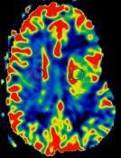
Imaging Acute Stroke and Cerebral Ischemia
|
|
|
- Kelley Singleton
- 6 years ago
- Views:
Transcription
1 Department of Radiology University of California San Diego Imaging Acute Stroke and Cerebral Ischemia John R. Hesselink, M.D.
2 Causes of Stroke Arterial stenosis Thrombosis Embolism Dissection Hypotension Anoxia / hypoxia Hypoglycemia
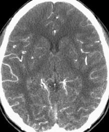
3 Imaging Acute Stroke Conventional Imaging Abnormal vascular density / signal Vascular enhancement Loss of gray / white contrast Cortical swelling Sulcal effacement Ventricular compression MRA \ CTA


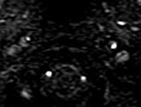
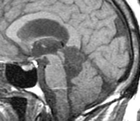
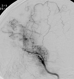
4 History: 85 y/o man with a right hemiparesis 291

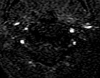
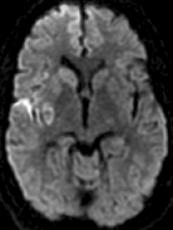
5 Dx: MCA embolus with cerebral infarction {Page 2}


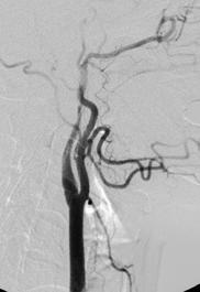
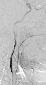
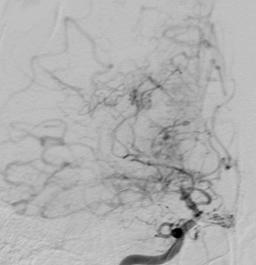
6 History: 62 y.o. male with transient weakness on the right side O2027


7 Dx: Left carotid occlusion {Page 2}


8 History: 58 y.o. man with altered mental status


9 History: 59 y/o woman with headache and left-sided weakness 187


10 1 Weakness {Page 2} 2 T1 - FATSAT T2W 3

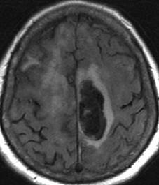
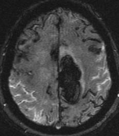
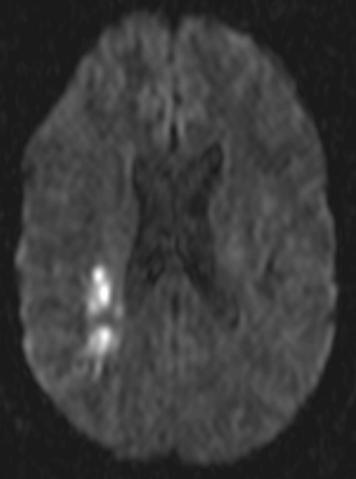
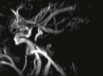
11 Dx: Right ICA dissection {Page 3}


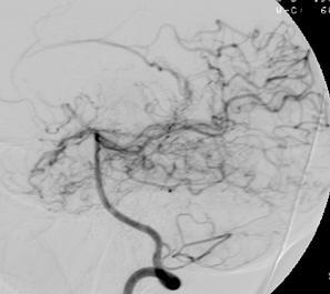
12 History: 37 y/o HIV + female with acute right sided weakness 172


13 Dx: Acute MCA infarct 18 hours later {Page 2}
14 History: 50 y/o male with acute right hemianopsia following coronary artery angioplasty & stenting 666

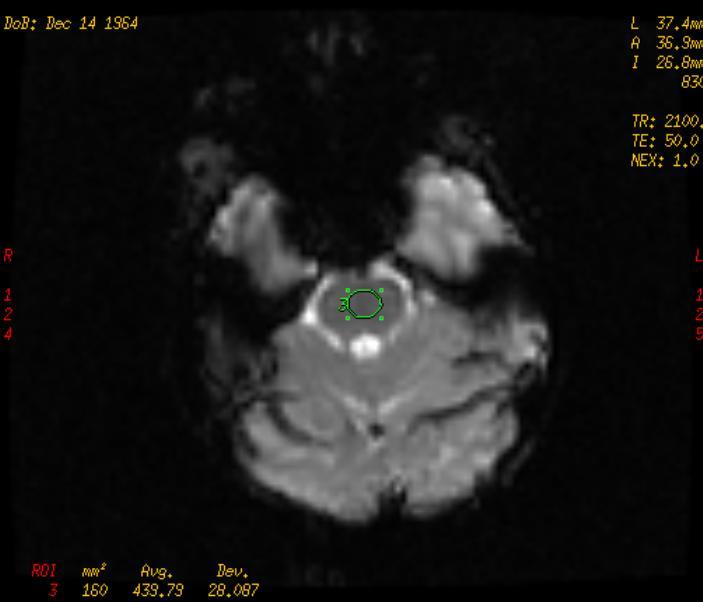
15 {Page 2} Treated with intra-arterial TPA 1 day later

16 {Page 3}

17 {Page 4} Dx: Hemorrhagic occipital infarct


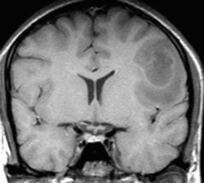
18 History: 77 y/o woman with right-sided weakness 733

19 {Page 2}

20 {Page 3} Dx: Amyloid angiopathy

21 Diffusion and Perfusion Imaging
22 Cerebral Ischemia / Infarction Physiology Brain requires glucose & oxygen Normal CBF ml/100gm/min If CBF < 18, electrical activity ceases If CBF < 10, neuronal metabolism stops CBF called the "ischemic penumbra" Pathologic effect depends on the degree & duration of ischemia
23 Diffusion Weighted Imaging Physical Principles Random molecular movement or "Brownian motion" Addition of a pair of strong gradient pulses 1st pulse - dephases the spins 2nd pulse - rephases spins if no net movement If net movement of spins occurs between gradient pulses, signal attenuation occurs Warach S: Diffusion & Perfusion MRI, in Clinical MRI, Edelman et al, Saunders, Chap. 26, pp
24 Acute Cerebral Ischemia Diffusion-Weighted Imaging CBF lowered to < 10 ml/100gm/min Cell membrane Na K pump fails Net movement of water from extracellular to intracellular compartment Diffusion restricted by cell membranes ADC & signal intensity on DWI


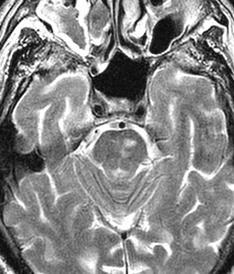
25 History: 47 y/o man with von-hippel-lindau disease & new left arm weakness 486


26 {Page 2} DWI ADC Dx: Infarct & MCA stenosis


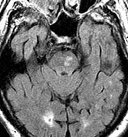
27 History: 80 y/o woman with a fluent aphasia 405

28 {Page 2} DWI


29 {Page 3} DWI Dx: Acute & chronic strokes


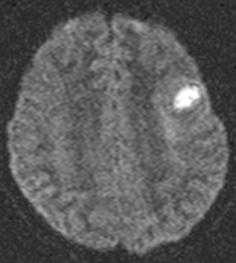
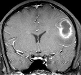
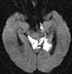
30 History: 73 y/o woman with weakness 427


31 {Page 2} {Video clip} Dx: Acute pontine infarct

32 History: 76 y.o. male with vertebral-basilar TIA's 119


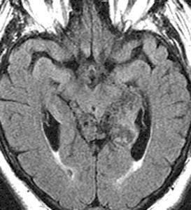
33 TIA's {Page 2} Dx: FLAIR & Diffusion - DWMI


34 History: 64 y/o man with bilateral leg weakness & ataxia 347

35 {Page 2}


36 Leg weakness {Page 3} Dx: Acute infarcts in pons & left corona radiata DWI


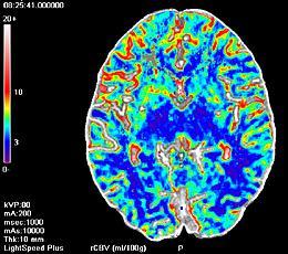
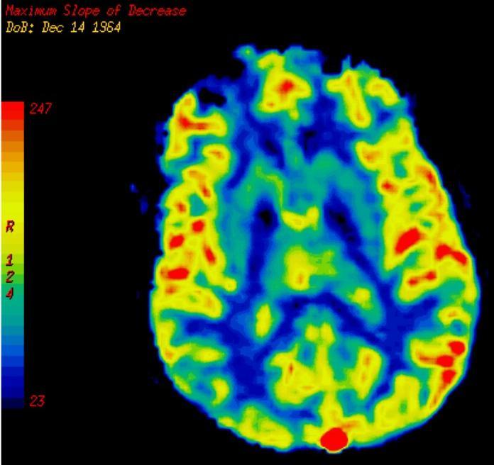
37 History: 16 y/o male with new onset of seizures 350


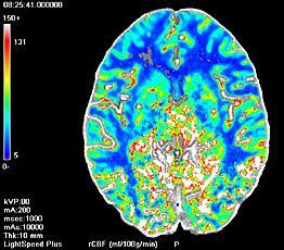
38 {Page 2} Diffusion

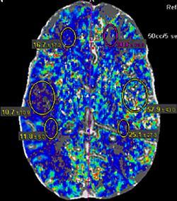
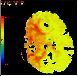
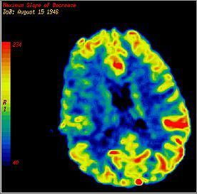
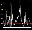
39 Dx: Brain abscess - Streptococcus milleri {Page 3}


40 History: 63 y/o man with seizures for 8 years 365
41 {Page 2}


42 {Page 3} Diffusion Dx: Epidermoid
43 Other Causes of Positive DWI Bacterial abscess, Epidermoid tumor Acute demyelination Acute encephalitis Tumors undergoing central necrosis Tumors with high nuclear:cytoplasmic ratios Creutzfeldt-Jakob disease Diffuse axonal injury T2 shine-through (High ADC)
44 Perfusion Techniques Cerebral blood flow PET Xenon CT CT and MRI Vascular transit time Cerebral blood volume
45 CT Perfusion Technique kvp, mas 2 8 sections / 5 10 mm thick Acquire 1 image set per second 40 second acquisition 40 ml of contrast ( mg I/ml) Inject 5-8 ml / sec
46 CT Perfusion Time Density Curve TTP CVA TTP rcbf = rcbv / MTT MTT Area = rcbv HU Baseline Injection Time (sec) Normal


47 History: 38 y/o male with a left hemiparesis DWI ADC 752
48 {Page 2}


49 MTT CBV CBF {Page 3} Dx: MCA embolus & stroke


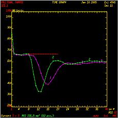
50 76 y/o woman developed a complete aphasia & right hemiplegia 35 minutes earlier Tomandl BF, et al: Radiographics 23:565-92, 2003

51 Symptoms resolved completely within 2 hours 24 hours later


52 History: 18 y/o male with confusion & 7 seizures 633

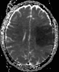
53 {Page 2}

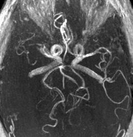
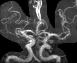
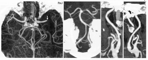
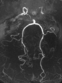
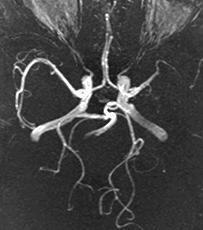
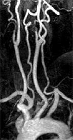
54 {Page 3} Dx: Moya Moya
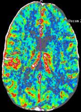
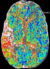
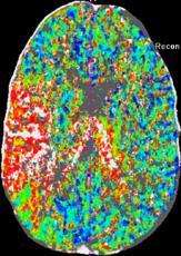
55 MR Perfusion Methods Intravascular magnetic susceptibility - Inject bolus of gadolinium - Obtain time-intensity curve - Measure area under curve EPISTAR (QUIPSS) - Tag in-flowing blood with 180 o inversion pulse - Presaturate slice of interest - 90 o readout pulse to slice - Repeat sequence without tag - Subtract 4 from 3 - Signal difference proportional to perfusion
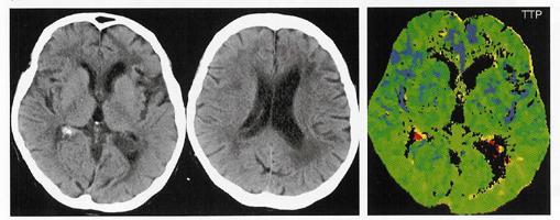
56 EPI Perfusion Sequence Gadolinium injection TR = 1000 msec; 90 o flip angle TE = 60 msec Fat saturation Matrix = 128 x 128 Acquisition time = 40 sec
57 EPI Perfusion Time Intensity Curve Baseline TTP CVA rcbf = rcbv / MTT Baseline Normal MTT Area = rcbv SI Injection TTP Time (sec)


58 History: 38 y/o man with altered mental status & a right hemiparesis ADC DWI 631


59 {Page 2} Dx: Embolic MCA infarct right ventricular cardiac thrombus rcbv CBV/MTT= rcbf Perfusion TTP rmtt


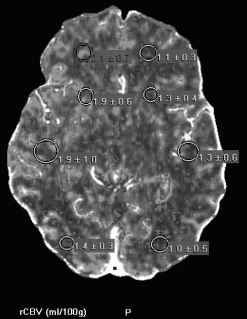
60 History: 59 y.o. woman with an upper GI bleed developed a left hemiparesis & slurred speech FLAIR DWI b=1000 ADC 623


61 {Page 2} 3D-TOF CE-MRA CTA

62 Perfusion TTP rmtt DWI rcbv CBV/MTT=rCBF


63 {Page 4} rmtt CT perfusion rcbv rcbf


64 {Page 5} CT perfusion Dx: DWI/PWI mismatch
65 Interpretation Right ACA-MCA watershed DWI abnormalities Perfusion imaging (4-5cc Gd/sec) N TTP MTT CBV (auto-regulation compensatory vasodilatation reduced functional reserve) CBF (CBV/MTT) Large DWI/PWI mismatch (penumbra) (large volume of tissue at risk = salvageable brain)
66 Ischemic Penumbra DWI / PWI Mismatch Diffusion Abnormality CBF < 10 ml/100g/min Cytotoxic edema Irreversible ischemia Perfusion Abnormality CBF = ml/100g/min Neuronal paralysis Penumbra Reversible ischemia


67 635 History: 56 y/o woman with hypertension & hyperlipidemia developed dysarthria & left facial droop


68 {Page 2} MTT rcbv TTP rcbf Dx: Acute infarct with matched DWI and perfusion


69 History: 40 y/o male metamphetamine abuser developed acute bilateral arm numbness, leg weakness & dysarthria 627
70 {Page 2}


71 {Page 3} L R Perfusion Imaging Pons

72 {Page 4} rcbv rmtt CBV/MTT=rCBF


73 {Page 5} Post tpa Day 3 Dx: Basilar thrombosis & posterior fossa ischemia
74 Acute Cerebral Ischemia Imaging Sensitivity CT scan MRI (T2/FLAIR) DWI Perfusion hours 6-12 hours < 1.5 hours Instantly
75 Acute Cerebral Ischemia The Integrated MR Exam T2 / FLAIR sequences Diffusion imaging (Diagnostic) Perfusion imaging (Prognostic) MR Angiography (Site for therapy)
76


77 UCSD Neuroradiology Teaching File Website URL -
Department of Radiology University of California San Diego. MR Angiography. Techniques & Applications. John R. Hesselink, M.D.
 Department of Radiology University of California San Diego MR Angiography Techniques & Applications John R. Hesselink, M.D. Vascular Imaging Arterial flow void Flow enhancement Gadolinium enhancement Vascular
Non-Traumatic Neuro Emergencies
 Department of Radiology University of California San Diego Non-Traumatic Neuro Emergencies John R. Hesselink, M.D. Nontraumatic Neuroemergencies 1. Acute focal neurological deficit 2. Worst headache of
Acute Ischemic Stroke Imaging. Ronald L. Wolf, MD, PhD Associate Professor of Radiology
 Acute Ischemic Stroke Imaging Ronald L. Wolf, MD, PhD Associate Professor of Radiology Title of First Slide of Substance An Illustrative Case 2 Disclosures No financial disclosures Off-label uses of some
Advanced Neuroimaging for Acute Stroke
 Advanced Neuroimaging for Acute Stroke E. Bradshaw Bunney, MD, FACEP Professor Department Of Emergency Medicine University of Illinois at Chicago Swedish American Belvidere Hospital Disclosures FERNE Board
Acute stroke imaging
 Acute stroke imaging Aims Imaging modalities and differences Why image acute stroke Clinical correlation to imaging appearance What is stroke Classic definition: acute focal injury to the central nervous
Complete Recovery of Perfusion Abnormalities in a Cardiac Arrest Patient Treated with Hypothermia: Results of Cerebral Perfusion MR Imaging
 pissn 2384-1095 eissn 2384-1109 imri 2018;22:56-60 https://doi.org/10.13104/imri.2018.22.1.56 Complete Recovery of Perfusion Abnormalities in a Cardiac Arrest Patient Treated with Hypothermia: Results
11/1/2018. Disclosure. Imaging in Acute Ischemic Stroke 2018 Neuro Symposium. Is NCCT good enough? Keystone Heart Consultant, Stock Options
 Disclosure Imaging in Acute Ischemic Stroke 2018 Neuro Symposium Keystone Heart Consultant, Stock Options Kevin Abrams, M.D. Chief of Radiology Medical Director of Neuroradiology Baptist Hospital, Miami,
STROKE - IMAGING. Dr RAJASEKHAR REDDY 2nd Yr P.G. RADIODIAGNOSIS KIMS,Narkatpalli.
 STROKE - IMAGING Dr RAJASEKHAR REDDY 2nd Yr P.G. RADIODIAGNOSIS KIMS,Narkatpalli. STROKE Describes a clinical event that consists of sudden onset of neurological symptoms Types Infarction - occlusion of
AMSER Case of the Month: March 2019
 AMSER Case of the Month: March 2019 62 year-old male with left-sided weakness Ashley Graziano OMS IV, Lake Erie College of Osteopathic Medicine Erik Yannone MD, Charles Q. Li MD, Warren Chang MD, Matthew
Place for Interventional Radiology in Acute Stroke
 Place for Interventional Radiology in Acute Stroke Dr Lakmalie Paranahewa MBBS, MD(Radiology), FRCR Consultant Interventional Radiologist Asiri Group of Hospitals Objectives Imaging in Stroke Neurovascular
Imaging in Stroke. D Nagaraja, N Karthik
 Imaging in Stroke D Nagaraja, N Karthik Cerebro-vascular disease (stroke) is the second leading cause of death. Prior to CT era, diagnosis was essentially clinical supported by angio and lumbar puncture.
Acute Ischemic Stroke Imaging Innovations
 Acute Ischemic Stroke Imaging Innovations Guilherme Dabus, MD, FAHA Director, Fellowship NeuroInterventional Surgery Miami Cardiac & Vascular Institute Baptist Neuroscience Center Baptist Neuroscience
: STROKE. other pertinent information such as recent trauma, illicit drug use, pertinent medical history or use of oral contraceptives.
 INTRODUCTION A cerebral vascular accident (CVA) or stroke is a lack of blood supply to the brain as a result of either ischemia or hemorrhage. 80% of CVAs are a result of ischemia (embolic or thrombotic)
UPSTATE Comprehensive Stroke Center. Neurosurgical Interventions Satish Krishnamurthy MD, MCh
 UPSTATE Comprehensive Stroke Center Neurosurgical Interventions Satish Krishnamurthy MD, MCh Regional cerebral blood flow is important Some essential facts Neurons are obligatory glucose users Under anerobic
Imaging ischemic strokes: Correlating radiological findings with the pathophysiological evolution of an infarct
 Imaging ischemic strokes: Correlating radiological findings with the pathophysiological evolution of an infarct Jay Chyung,, PhD, HMS III Patient A: history 91 y.o. woman Acute onset R sided weakness and
The Language of Stroke
 The Language of Stroke Examination / Imaging / Diagnosis / Treatment Dr Suzanne Busch A lot of letters! CBF CVA ICH CVD CBV DWI US MRI/MRA CAA CTA CTP ICA MCA SAH WMD TIA MCA Agnosia A lot of big words!
On Call Guide to CT Perfusion. Updated: March 2011
 On Call Guide to CT Perfusion Updated: March 2011 CT Stroke Protocol 1. Non contrast CT brain 2. CT perfusion: contrast 40cc bolus dynamic imaging at 8 slice levels ~ 60 sec creates perfusion color maps
Cerebrovascular Disorders. Blood, Brain, and Energy. Blood Supply to the Brain 2/14/11
 Cerebrovascular Disorders Blood, Brain, and Energy 20% of body s oxygen usage No oxygen/glucose reserves Hypoxia - reduced oxygen Anoxia - Absence of oxygen supply Cell death can occur in as little as
NEURO IMAGING OF ACUTE STROKE
 1 1 NEURO IMAGING OF ACUTE STROKE ALICIA RICHARDSON, MSN, RN, ACCNS-AG, ANVP-BC WENDY SMITH, MA, RN, MBA, SCRN, FAHA LYNN HUNDLEY, APRN, CNRN, CCNS, ANVP-BC 2 2 1 DISCLOSURES Alicia Richardson: Stryker
NEURORADIOLOGY Part I
 NEURORADIOLOGY Part I Vörös Erika University of Szeged Department of Radiology SZEGED BRAIN IMAGING METHODS Plain film radiography Ultrasonography (US) Computer tomography (CT) Magnetic resonance imaging
Stroke imaging. Why image stroke patients? Stroke. Treatment of infarct. Methods for infarct diagnosis. Treatment of infarct.
 Stroke imaging Stroke Infarct: -Arterial thrombosis/embolus -Hypoxic/ischemic -Venous thrombosis Non-traumatic hemorrhage: -Intracerebral -Subarachnoid Johan Wikström MD PhD Associate Professor of Radiology
Case Conference: Neuroradiology. Case 1: Tumor Case 1: 22yo F w/ HA and prior Seizures
 Case Conference: Neuroradiology Case 1: 22yo F w/ HA and prior Seizures David E. Rex, MD, PhD Stanford University Hospital Department of Radiology Case 1: Tumor Most likely gangiloglioma, oligodendroglioma,
Magnetic Resonance Imaging. Basics of MRI in practice. Generation of MR signal. Generation of MR signal. Spin echo imaging. Generation of MR signal
 Magnetic Resonance Imaging Protons aligned with B0 magnetic filed Longitudinal magnetization - T1 relaxation Transverse magnetization - T2 relaxation Signal measured in the transverse plane Basics of MRI
Imaging Modalities in Acute Stroke: Time is Brain
 April 2001 Imaging Modalities in Acute Stroke: Time is Brain Jeremiah Scharf, Harvard Medical School, MS IV Beth Israel-Deaconess Medical Center Department of Radiology Stroke - Definition and Statistics
Index. aneurysm, 92 carotid occlusion, 94 ICA stenosis, 95 intracranial, 92 MCA, 94
 A ADC. See Apparent diffusion coefficient (ADC) Aneurysm cerebral artery aneurysm, 93 CT scan, 93 gadolinium, 93 Angiography, 13 Anoxic brain injury, 25 Apparent diffusion coefficient (ADC), 7 Arachnoid
The Role of Neuroimaging in Acute Stroke. Bradley Molyneaux, HMS IV
 The Role of Neuroimaging in Acute Stroke Bradley Molyneaux, HMS IV Patient CR 62 yo F w/ 2 wk h/o altered mental status Presents to ED w/ confusion following a fall 1 day prior New onset left facial droop
Debbie Summers, MSN, RN, ACNS-BC, CNRN, SCRN. Debbie Summers, MSN, ACNS-BC Nothing
 Debbie Summers, MSN, RN, ACNS-BC, CNRN, SCRN Debbie Summers, MSN, ACNS-BC Nothing Identify anatomical location of stroke on CT, MRI, and perfusion scans and angiography. Relate underlying pathology to
Functional aspects of anatomical imaging techniques
 Functional aspects of anatomical imaging techniques Nilendu Purandare Associate Professor & Consultant Radiologist Tata Memorial Centre Functional/metabolic/molecular imaging (radioisotope scanning) PET
CEREBRO VASCULAR ACCIDENTS
 CEREBRO VASCULAR S MICHAEL OPONG-KUSI, DO MBA MORTON CLINIC, TULSA, OK, USA 8/9/2012 1 Cerebrovascular Accident Third Leading cause of deaths (USA) 750,000 strokes in USA per year. 150,000 deaths in USA
Outline. Neuroradiology. Diffusion Imaging in. Clinical Applications of. Basics of Diffusion Imaging. Basics of Diffusion Imaging
 Clinical Applications of Diffusion Imaging in Neuroradiology No disclosures Stephen F. Kralik Assistant Professor of Radiology Indiana University School of Medicine Department of Radiology and Imaging
Demyelinating Diseases of the Brain
 Department of Radiology University of California San Diego Demyelinating Diseases of the Brain John R. Hesselink, M.D. T1-Weighted Images Normal White Matter Contents Axons with envelope of myelin Neuroglia
Perfusion MRI. Youngkyoo Jung, PhD Associate Professor Radiology, Biomedical Engineering, and Clinical & Translational Science Institute
 Perfusion MRI Youngkyoo Jung, PhD Associate Professor Radiology, Biomedical Engineering, and Clinical & Translational Science Institute Perfusion The delivery of blood to a capillary bed in tissue Perfusion
Basilar artery stenosis with bilateral cerebellar strokes on coumadin
 Qaisar A. Shah, MD Patient Profile 68 years old female with a history of; Basilar artery stenosis with bilateral cerebellar strokes on coumadin Diabetes mellitus Hyperlipidemia Hypertension She developed
High Field MR of the Spine
 Department of Radiology University of California San Diego 3T for MR Applications Advantages High Field MR of the Spine Increased signal-to-noise Better fat suppression Increased enhancement with gadolinium
[(PHY-3a) Initials of MD reviewing films] [(PHY-3b) Initials of 2 nd opinion MD]
![[(PHY-3a) Initials of MD reviewing films] [(PHY-3b) Initials of 2 nd opinion MD]](/thumbs/89/98619893.jpg "[(PHY-3a) Initials of MD reviewing films] [(PHY-3b) Initials of 2 nd opinion MD]") 2015 PHYSICIAN SIGN-OFF (1) STUDY NO (PHY-1) CASE, PER PHYSICIAN REVIEW 1=yes 2=no [strictly meets case definition] (PHY-1a) CASE, IN PHYSICIAN S OPINION 1=yes 2=no (PHY-2) (PHY-3) [based on all available
2015 PHYSICIAN SIGN-OFF (1) STUDY NO (PHY-1) CASE, PER PHYSICIAN REVIEW 1=yes 2=no [strictly meets case definition] (PHY-1a) CASE, IN PHYSICIAN S OPINION 1=yes 2=no (PHY-2) (PHY-3) [based on all available
Endovascular Neurointervention in Cerebral Ischemia
 Endovascular Neurointervention in Cerebral Ischemia Beyond Thrombolytics Curtis A. Given II, MD Co-Director, Neurointerventional Services Baptist Physician Lexington 72 y/o female with a recent diagnosis
/ / / / / / Hospital Abstraction: Stroke/TIA. Participant ID: Hospital Code: Multi-Ethnic Study of Atherosclerosis
 Multi-Ethnic Study of Atherosclerosis Participant ID: Hospital Code: Hospital Abstraction: Stroke/TIA History and Hospital Record 1. Was the participant hospitalized as an immediate consequence of this
Nicolas Bianchi M.D. May 15th, 2012
 Nicolas Bianchi M.D. May 15th, 2012 New concepts in TIA Differential Diagnosis Stroke Syndromes To learn the new definitions and concepts on TIA as a condition of high risk for stroke. To recognize the
Vascular Disorders. Nervous System Disorders (Part B-1) Module 8 -Chapter 14. Cerebrovascular disease S/S 1/9/2013
 Module 8 -Chapter 14. Cerebrovascular disease S/S 1/9/2013") Nervous System Disorders (Part B-1) Module 8 -Chapter 14 Overview ACUTE NEUROLOGIC DISORDERS Vascular Disorders Infections/Inflammation/Toxins Metabolic, Endocrinologic, Nutritional, Toxic Neoplastic Traumatic
Nervous System Disorders (Part B-1) Module 8 -Chapter 14 Overview ACUTE NEUROLOGIC DISORDERS Vascular Disorders Infections/Inflammation/Toxins Metabolic, Endocrinologic, Nutritional, Toxic Neoplastic Traumatic
NEURORADIOLOGY DIL part 4
 NEURORADIOLOGY DIL part 4 Strokes and infarcts K. Agyem MD, G. Hall MD, D. Palathinkal MD, Alexandre Menard March/April 2015 OVERVIEW Introduction to Neuroimaging - DIL part 1 Basic Brain Anatomy - DIL
Background. Recommendations for Imaging of Acute Ischemic Stroke: A Scientific Statement From the American Heart Association
 for Imaging of Acute Ischemic Stroke: A Scientific Statement From the American Heart Association An Scientific Statement from the Stroke Council, American Heart Association and American Stroke Association
Speed, Comfort and Quality with NeuroDrive
 Speed, Comfort and Quality with NeuroDrive Echelon Oval provides a broad range of capabilities supporting fast, accurate diagnosis of brain conditions and injuries. From anatomical depiction to vascular
Interventions in the Management of Acute Stroke. Dr Md Shafiqul Islam Associate Professor Neurosurgery Dhaka Medical College Hospital
 Interventions in the Management of Acute Stroke Dr Md Shafiqul Islam Associate Professor Neurosurgery Dhaka Medical College Hospital Acute stroke intervention Number of stroke patients increasing day by
Neuroradiology: Imaging and Stroke
 Neuroradiology: Imaging and Stroke Stroke 2017 William Gallmann January 28, 2017 Stroke Arterial ischemia/infarct accounts for ~85% Cerebral venous occlusions - 0.5-1% Spontaneous intracranial hemorrhage
Stroke School for Internists Part 1
 Stroke School for Internists Part 1 November 4, 2017 Dr. Albert Jin Dr. Gurpreet Jaswal Disclosures I receive a stipend for my role as Medical Director of the Stroke Network of SEO I have no commercial
Acute Stroke Management LUKE BRADBURY, MD 10/8/14 FALL WAPA CONFERENCE
 Objectives Acute Stroke Management LUKE BRADBURY, MD 10/8/14 FALL WAPA CONFERENCE Recognize the clinical signs of acute stroke Differentiate between stroke and some of the more common stroke mimics Review
Title: Stability of Large Diffusion/Perfusion Mismatch in Anterior Circulation Strokes for 4 or More Hours
 Author's response to reviews Title: Stability of Large Diffusion/Perfusion Mismatch in Anterior Circulation Strokes for 4 or More Hours Authors: Ramon G. Gonzalez (rggonzalez@partners.org) Reza Hakimelahi
CT INTERPRETATION COURSE
 CT INTERPRETATION COURSE Refresher Course ASTRACAT October 2012 Stroke is a Clinical Diagnosis A clinical syndrome characterised by rapidly developing clinical symptoms and/or signs of focal loss of cerebral
PHYSICS OF MRI ACQUISITION. Alternatives to BOLD for fmri
 PHYSICS OF MRI ACQUISITION Quick Review for fmri HST-583, Fall 2002 HST.583: Functional Magnetic Resonance Imaging: Data Acquisition and Analysis Harvard-MIT Division of Health Sciences and Technology
Comparison of Five Major Recent Endovascular Treatment Trials
 Comparison of Five Major Recent Endovascular Treatment Trials Sample size 500 # sites 70 (100 planned) 316 (500 planned) 196 (833 estimated) 206 (690 planned) 16 10 22 39 4 Treatment contrasts Baseline
Disclosure. + Outline. What is a stroke? Role of imaging in stroke Ischemic stroke Venous infarct Current topics
 + Kathleen R. Fink, MD University of Washington 5 th Nordic Emergency Radiology Course May 21, 2015 + Disclosure My spouse receives research salary support from: Bracco BayerHealthcare Guerbet Thank you
Neuroradiology MR Protocols
 Neuroradiology MR Protocols Brain protocols N 1: Brain MRI without contrast N 2: Pre- and post-contrast brain MRI N 3 is deleted N 4: Brain MRI without or pre-/post-contrast (seizure protocol) N 5: Pre-
Diagnostic improvement from average image in acute ischemic stroke
 Diagnostic improvement from average image in acute ischemic stroke N. Magne (1), E.Tollard (1), O. Ozkul- Wermester (2), V. Macaigne (1), J.-N. Dacher (1), E. Gerardin (1) (1) Department of Radiology,
Essentials of Clinical MR, 2 nd edition. 14. Ischemia and Infarction II
 14. Ischemia and Infarction II Lacunar infarcts are small deep parenchymal lesions involving the basal ganglia, internal capsule, thalamus, and brainstem. The vascular supply of these areas includes the
57y WRH woman, controlled HTN only, presents with sudden LOC, fixed and dilated, quadraplegic Intubated on arrival and CT is negative CTA and CTP
 Case # 1 Hx 57y WRH woman, controlled HTN only, presents with sudden LOC, fixed and dilated, quadraplegic Intubated on arrival and CT is negative CTA and CTP show left PCA occlusion, some basilar stenosis,
CHAP 11 Contrast Enhanced MRI - Perfusion. CHAP 11 Contrast Enhanced MRI - Perfusion
 CHAP 11 Contrast Enhanced MRI - Perfusion Contrast agents Paramagnetic / superparamagnetic Relaxivity Water exchange Susceptibility effect Brain perfusion pathologies Perfusion measurement T2* (DSC-MRI)
Whole brain CT perfusion maps with paradoxical low mean transit time to predict infarct core
 Whole brain CT perfusion maps with paradoxical low mean transit time to predict infarct core Poster No.: B-292 Congress: ECR 2011 Type: Scientific Paper Topic: Neuro Authors: S. Chakraborty, M. E. Ahmad,
Disclosures. CREST Trial: Summary. Lecture Outline 4/16/2015. Cervical Atherosclerotic Disease
 Disclosures Your Patient Has Carotid Bulb Stenosis and a Tandem Intracranial Stenosis: How Do SAMMPRIS and Other Evidence Inform Your Treatment? UCSF Vascular Symposium 2015 Steven W. Hetts, MD Associate
CT Perfusion is Essential for Stroke Triage. Maarten Lansberg, MD PhD Associate Professor of Neurology Stanford University, Stanford Stroke Center
 CT Perfusion is Essential for Stroke Triage Maarten Lansberg, MD PhD Associate Professor of Neurology Stanford University, Stanford Stroke Center CT Perfusion is Essential for Stroke Triage Disclosures:
Neuroanatomy of a Stroke. Joni Clark, MD Professor of Neurology Barrow Neurologic Institute
 Neuroanatomy of a Stroke Joni Clark, MD Professor of Neurology Barrow Neurologic Institute No disclosures Stroke case presentations Review signs and symptoms Review pertinent exam findings Identify the
Endovascular Treatment for Acute Ischemic Stroke: Curtis A. Given II, MD Co-Director, Neurointerventional Services Baptist Physician Lexington
 Endovascular Treatment for Acute Ischemic Stroke: Curtis A. Given II, MD Co-Director, Neurointerventional Services Baptist Physician Lexington Disclosures: SWIFT PRIME site (Medtronic) Physician Proctor
9/18/16. Setting: Community ED, 30k admissions per year Time: Friday night, 11pm. CC: Syncope
 William A. Knight IV MD, FACEP Associate Professor Emergency Medicine & Neurosurgery University of Cincinnati September 21, 2016 (William.knight@uc.edu) ED as the Front Door Spectrum of care with Endovascular
Spontaneous Recanalization after Complete Occlusion of the Common Carotid Artery with Subsequent Embolic Ischemic Stroke
 Original Contribution Spontaneous Recanalization after Complete Occlusion of the Common Carotid Artery with Subsequent Embolic Ischemic Stroke Abstract Introduction: Acute carotid artery occlusion carries
Applicable Neuroradiology
 For the Clinical Neurology Clerkship LSU Medical School New Orleans Amy W Voigt, MD Clerkship Director Introduction The field of Radiology first developed following the discovery of X-Rays by Wilhelm Roentgen
Stroke 101. Maine Cardiovascular Health Summit. Eileen Hawkins, RN, MSN, CNRN Pen Bay Stroke Program Coordinator November 7, 2013
 Stroke 101 Maine Cardiovascular Health Summit Eileen Hawkins, RN, MSN, CNRN Pen Bay Stroke Program Coordinator November 7, 2013 Stroke Statistics Definition of stroke Risk factors Warning signs Treatment
NEURO IMAGING 2. Dr. Said Huwaijah Chairman of radiology Dep, Damascus Univercity
 NEURO IMAGING 2 Dr. Said Huwaijah Chairman of radiology Dep, Damascus Univercity I. EPIDURAL HEMATOMA (EDH) LOCATION Seventy to seventy-five percent occur in temporoparietal region. CAUSE Most likely caused
Management of Acute Ischemic Stroke. Learning Objec=ves. What is a Stroke? Jen Simpson Neurohospitalist
 Management of Acute Ischemic Stroke Jen Simpson Neurohospitalist Learning Objec=ves Iden=fy signs/symptoms of stroke Recognize pa=ents who may be eligible for treatment of acute stroke What is a Stroke?
Practical Considerations in the Early Treatment of Acute Stroke
 Practical Considerations in the Early Treatment of Acute Stroke Matthew E. Fink, MD Neurologist-in-Chief Weill Cornell Medical College New York-Presbyterian Hospital mfink@med.cornell.edu Disclosures Consultant
Carotid Embolectomy and Endarterectomy for Symptomatic Complete Occlusion of the Carotid Artery as a Rescue Therapy in Acute Ischemic Stroke
 This is an Open Access article licensed under the terms of the Creative Commons Attribution-NonCommercial-NoDerivs 3.0 License (www.karger.com/oa-license), applicable to the online version of the article
CT perfusion in Moyamoya disease
 CT perfusion in Moyamoya disease Poster No.: C-1726 Congress: ECR 2015 Type: Scientific Exhibit Authors: K. C. Lam, C. P. Tsang, K. K. Wong, R. LEE ; HK, Hong Kong/HK Keywords: Hemodynamics / Flow dynamics,
Neuroradiology. of Stroke and Headaches
 Neuroradiology of Stroke and Headaches Learning Objec:ves 1. Iden:fy T1 and T2 sequences 2. Recall the normal anatomy of the intracranial circula:on 3. Apply appropriate CT and MR imaging of the brain
Remission of diffusion lesions in acute stroke magnetic resonance imaging
 ORIGINAL RESEARCH Remission of diffusion lesions in acute stroke magnetic resonance imaging F. A. Fellner 1, M. R. Vosko 2, C. M. Fellner 1, D. Flöry 1 1. AKH Linz, Institute of Radiology, Austria. 2.
Overview of Stroke: Etiologies, Demographics, Syndromes, and Outcomes. Alex Abou-Chebl, MD, FSVIN Medical Director, Stroke Baptist Health Louisville
 Overview of Stroke: Etiologies, Demographics, Syndromes, and Outcomes Alex Abou-Chebl, MD, FSVIN Medical Director, Stroke Baptist Health Louisville Disclosure Statement of Financial Interest Within the
MR Advance Techniques. Vascular Imaging. Class II
 MR Advance Techniques Vascular Imaging Class II 1 Vascular Imaging There are several methods that can be used to evaluate the cardiovascular systems with the use of MRI. MRI will aloud to evaluate morphology
An Introduction to Imaging the Brain. Dr Amy Davis
 An Introduction to Imaging the Brain Dr Amy Davis Common reasons for imaging: Clinical scenarios: - Trauma (NICE guidelines) - Stroke - Tumours - Seizure - Neurological degeneration memory, motor dysfunction,
Carotid artery stenting for long CTO and pseudo occlusion of carotid artery -2 case reports-
 Carotid artery stenting for long CTO and pseudo occlusion of carotid artery -2 case reports- Katsutoshi Takayama, MD, Ph.D Department of Radiology and Interventional Neuroradiology Ishinkai Yao General
Perfusion-Based fmri. Thomas T. Liu Center for Functional MRI University of California San Diego May 7, Goal
 Perfusion-Based fmri Thomas T. Liu Center for Functional MRI University of California San Diego May 7, 2006 Goal To provide a basic understanding of the theory and application of arterial spin labeling
ACUTE ISCHEMIC STROKE. Current Treatment Approaches for Acute Ischemic Stroke
 ACUTE ISCHEMIC STROKE Current Treatment Approaches for Acute Ischemic Stroke EARLY MANAGEMENT OF ACUTE ISCHEMIC STROKE Rapid identification of a stroke Immediate EMS transport to nearest stroke center
CT Perfusion Parameter Values in Regions of Diffusion Abnormalities
 AJNR Am J Neuroradiol 25:1205 1210, August 2004 CT Perfusion Parameter Values in Regions of Diffusion Abnormalities Marcello Galvez, Gerald E. York, II, and James D. Eastwood BACKGROUND AND PURPOSE: Dynamic
Acute stroke. Ischaemic stroke. Characteristics. Temporal classification. Clinical features. Interpretation of Emergency Head CT
 Ischaemic stroke Characteristics Stroke is the third most common cause of death in the UK, and the leading cause of disability. 80% of strokes are ischaemic Large vessel occlusive atheromatous disease
Magnetic Resonance Angiography
 Magnetic Resonance Angiography 1 Magnetic Resonance Angiography exploits flow enhancement of GR sequences saturation of venous flow allows arterial visualization saturation of arterial flow allows venous
ISCHEMIC STROKE IMAGING
 ISCHEMIC STROKE IMAGING ผศ.พญ พญ.จ ร ร ตน ธรรมโรจน ภาคว ชาร งส ว ทยา คณะแพทยศาสตร มหาว ทยาล ยขอนแก น A case of acute hemiplegia Which side is the abnormality, right or left? Early Right MCA infarction
Code Stroke in real life. Disclosures. Parkland Memorial Hospital. I have no disclosures. Has 1 million patient visits annually. Level 1 Trauma Center
 Code Stroke in real life Alejandro Magadán, M.D. University of Texas Southwestern Medical Center Medical Director for Stroke Parkland Memorial Hospital Disclosures I have no disclosures Parkland Memorial
Imaging Stroke: Is There a Stroke Equivalent of the ECG? Albert J. Yoo, MD Director of Acute Stroke Intervention Massachusetts General Hospital
 Imaging Stroke: Is There a Stroke Equivalent of the ECG? Albert J. Yoo, MD Director of Acute Stroke Intervention Massachusetts General Hospital Disclosures Penumbra, Inc. research grant (significant) for
Cerebral Vascular Diseases. Nabila Hamdi MD, PhD
 Cerebral Vascular Diseases Nabila Hamdi MD, PhD Outline I. Stroke statistics II. Cerebral circulation III. Clinical symptoms of stroke IV. Pathogenesis of cerebral infarcts (Stroke) 1. Ischemic - Thrombotic
非對比劑與對比劑增強 MRA. 血管攝影與對比劑 A Course of MRI. 本週課程內容 -MR Angiography (MRA) Unenhanced MRA
 Unenhanced MRA") 本週課程內容 -MR Angiography (MRA) 血管攝影與對比劑 A Course of MRI 盧家鋒助理教授國立陽明大學物理治療暨輔助科技學系 alvin4016@ym.edu.tw 非對比劑增強 MRA(Unenhanced MRA) Time-of-flight (TOF) angiography Phase-contrast (PC) angiography 對比劑增強 MRA(Contrast-enhanced
本週課程內容 -MR Angiography (MRA) 血管攝影與對比劑 A Course of MRI 盧家鋒助理教授國立陽明大學物理治療暨輔助科技學系 alvin4016@ym.edu.tw 非對比劑增強 MRA(Unenhanced MRA) Time-of-flight (TOF) angiography Phase-contrast (PC) angiography 對比劑增強 MRA(Contrast-enhanced
occlusions. Cerebral perfusion is driven fundamentally by regional cerebral
 Appendix Figures Figure A1. Hemodynamic changes that may occur in major anterior circulation occlusions. Cerebral perfusion is driven fundamentally by regional cerebral perfusion pressure (CPP). In response
An Introduc+on to Stroke
 An Introduc+on to Stroke Elizabeth Huntoon MS, MD Assistant Professor Department of Physical Medicine and Rehabilita>on Vanderbilt University School of Medicine Defini+on Sudden focal neurologic deficit
False-negative and False-positive Diffusion-weighted MR Findings in Acute Ischemic Stroke and Stroke-like Episodes
 JRural Med 2005 ; 1 : 27 32 Original Article False-negative and False-positive Diffusion-weighted MR Findings in Acute Ischemic Stroke and Stroke-like Episodes Shuzo Shintani 1,HiroakiYokote 1,KaoruHanabusa
It s Always a Stroke; Except For When It s Not..
 It s Always a Stroke; Except For When It s Not.. TREVOR PHINNEY, D.O. Disclosures No Relevant Disclosures 1 Objectives Discuss variables of differential diagnosis for stroke Review when to TPA and when
Non-Contrast MRA. How and When 1996! Why Non-Contrast MRA? Angiography: What are our goals? Inflow Techniques Differences in excitation hx
 A major teaching hospital of Harvard Medical School Angiography: What are our goals? Non-Contrast MRA: How and When Neil M. Rofsky, M.D. Professor of Radiology, Harvard Medical School Director of MRI &
Pre-Hospital Stroke Care: Bringing It To The Street. by Bob Atkins, NREMT-Paramedic AEMD EMS Director Bedford Regional Medical Center
 Pre-Hospital Stroke Care: Bringing It To The Street by Bob Atkins, NREMT-Paramedic AEMD EMS Director Bedford Regional Medical Center Overview/Objectives Explain the reasons or rational behind the importance
The Egyptian Journal of Hospital Medicine (July 2018) Vol. 72 (10), Page
 Vol. 72 (10), Page") The Egyptian Journal of Hospital Medicine (July 2018) Vol. 72 (10), Page 5398-5402 The Role of Susceptibility Weighted Imaging (SWI) in Evaluation of Acute Stroke Maha Abdelhamed El Nouby*, Eman Ahmed
The Egyptian Journal of Hospital Medicine (July 2018) Vol. 72 (10), Page 5398-5402 The Role of Susceptibility Weighted Imaging (SWI) in Evaluation of Acute Stroke Maha Abdelhamed El Nouby*, Eman Ahmed
Noncontrast CT scan is currently the imaging modality
 Original Contributions MRI Features of Intracerebral Hemorrhage Within 2 Hours From Symptom Onset Italo Linfante, MD; Rafael H. Llinas, MD; Louis R. Caplan, MD; Steven Warach, MD, PhD Background and Purpose
Lecture Outline: 1/5/14
 John P. Karis, MD Lecture Outline: Provide a clinical overview of stroke: Risk Prevention Diagnosis Intervention Illustrate how MRI is used in the diagnosis and management of stroke. Illustrate how competing
Stroke/TIA. Tom Bedwell
 Stroke/TIA Tom Bedwell tab1g11@soton.ac.uk The Plan Definitions Anatomy Recap Aetiology Pathology Syndromes Brocas / Wernickes Investigations Management Prevention & Prognosis TIAs Key Definitions Transient
MRS and Perfusion of Brain Tumors
 Department of Radiology University of California San Diego MRS and Perfusion of Brain Tumors John R. Hesselink, M.D. MRS & Perfusion of Brain Tumors Tumor histology Degree of malignancy Delineate tumor
ORIGINAL CONTRIBUTION
 ORIGINAL CONTRIBUTION Magnetic Resonance Imaging in Basilar Artery Occlusion Richard du Mesnil de Rochemont, MD; Tobias Neumann-Haefelin, MD; Joachim Berkefeld, MD; Matthias Sitzer, MD; Heinrich Lanfermann,
IMAGING IN ACUTE ISCHEMIC STROKE
 IMAGING IN ACUTE ISCHEMIC STROKE Timo Krings MD, PhD, FRCP (C) Professor of Radiology & Surgery Braley Chair of Neuroradiology, Chief and Program Director of Diagnostic and Interventional Neuroradiology;
11/6/2013. Vanderbilt University Institute of Imaging Science (VUIIS) 7 Tesla Vascular Imaging. Evaluating stroke risk
 7 Tesla Vascular Imaging. Evaluating stroke risk") Vanderbilt University Institute of Imaging Science (VUIIS) New MI Techniques for Imaging Cerebrovascular Disease Manus J. Donahue Depts. of adiology, Physics, Neurology and Physics Vanderbilt University
Vanderbilt University Institute of Imaging Science (VUIIS) New MI Techniques for Imaging Cerebrovascular Disease Manus J. Donahue Depts. of adiology, Physics, Neurology and Physics Vanderbilt University