Thyroid and Parathyroid Ultrasound Protocol
|
|
|
- Clarissa Booker
- 6 years ago
- Views:
Transcription
1 Thyroid and Parathyroid Ultrasound Protocol Reviewed By: Anna Ellermeier, MD Last Reviewed: December 2017 Contact: (866) , Option 1 **NOTE for all examinations: 1. If documenting possible flow in a structure/mass, all color/doppler should be accompanied by a spectral gate for waveform tracing **EXCEPTION: Thyroid nodules; spectral tracing does not need to be provided 2. CINE clips to be labeled: -MIDLINE structures: right to left when longitudinal and superior to inferior when transverse -RIGHT/LEFT structures: lateral to medial when longitudinal and superior to inferior when transverse **each should be 1 sweep, NOT back and forth** Thyroid General -Longitudinal: lateral, mid, medial both lobes -Transverse: inferior, mid, superior both lobes and isthmus WHEN & WHAT to CINE: -If completely normal gland: no CINE is required -If any part of gland is abnormal:
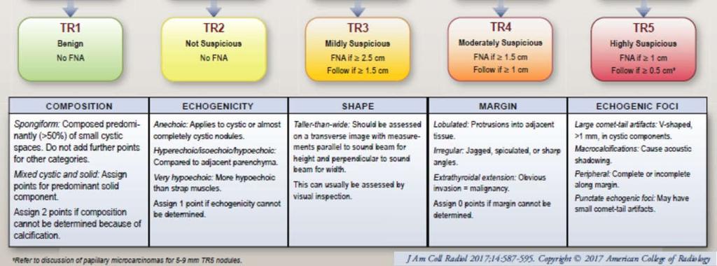
2 1. Both lobes (even if only 1 is abnormal): Transverse CINE from submandibular gland/hyoid bone to sternal notch through each lobe SEPARATELY If possible, include portion of isthmus on each side If not possible, separate transverse CINE through isthmus 2. ONLY abnormal lobe (s): Longitudinal CINE Size and Volume: -Measure size of each lobe and provide volume; provide prior volume if relevant prior is available (if no volume on prior, provide prior measurements) -Measure thickness (AP) of isthmus on transverse view Vascularity: -Representative color Doppler images in longitudinal of each lobe and transverse of isthmus Nodules: For reference, ACR TI-RADS Summary Table is attached at the end of this document. This is used by radiologists to describe and categorize nodules and highlights important features to document in nodule imaging. **If measuring a nodule that has been previously biopsied, please provide SIZE (at time of biopsy or immediately preceding formal study), DATE of biopsy and PATHOLOGY results (if available)** For organization: -Label each nodule on the images to match labels and description on worksheet: 1, 2, etc. --> Note, this may mean that the first nodule measured on the 2nd evaluated lobe is #4. Do not start numbers over from #1 on the 2nd lobe evaluated.
3 Document on worksheet: Location, Size, Basic features (composition, calcifications, margin) 1. Location: Which side: Right or left lobe Which third: Inferior pole, interpolar, or superior pole o Which area within this third: Anterior, posterior, medial, lateral, anteromedial, anterolateral, posteromedial, posterolateral 2. Size in 3 dimensions: measure in horizontal and vertical (rather than oblique) for consistency across exams NOTE: Make AP measurement for nodule in the transverse plane on the same image where you measure transverse dimension 3. DESCRIBE basic features: composition, calcifications, and margin (in Sonoreview: Select features on worksheet): 1. Composition: solid, cystic/partially cystic or spongiform -If possible show comet-tail artifact for colloid 2. Echogenic Foci/Calcification: absent or present -If possible, elaborate as to macrocalcification versus microcalcification 3. Margin: smooth or suspicious -Suspicious = jagged, irregular, portion of nodule extends beyond thyroid, etc. NOTE: For worksheets, please draw only an oval/circle for the nodule. It is not necessary to detail internal architecture on the diagrams. The legend has been removed for clarity. Provide adequate images that document nodule vascularity & echogenicity: Do not need to be described on worksheet by technologist. Radiologist will assess at time of imaging review. -Vascularity: Evaluate vascularity of each nodule in real-time. -Provide: Single color CINE (transverse) through gland.
4 -->Add more CINEs if gland size warrants to ensure that all nodules are visualized. -Dedicated color images of each nodule with color are not necessary, EXCEPT: NOTE EXCEPTIONS: (1) If there is truly dramatic vascularity of a nodule in real-time: provide short color Doppler of the nodule, CINE if helpful (2) Cyst: provide single image with color box over anechoic cyst (3) Vessel: if appears like a hypoechoic nodule or cyst on a still image, provide image with color box to show it is a vessel -Echogenicity: Provide images that clearly show nodule in relationship to remainder of thyroid and strap muscles -Technologist does not have to denote echogenicity on worksheet but it is important to tailor images to help radiologist document accurate echogenicity (CINEs can help here) For reference: o o Radiologists evaluate echogenicity as it relates to the thyroid gland and the strap muscles. Definitions used by radiologists (per TIRADS document, see table at end of protocol for reference) Echogenicity = hyper iso hypo - VERY hypo Hyperechoic is > thyroid Iso is = thyroid Hypoechoic is < than thyroid but = or > strap muscle VERY hypoechoic is < strap muscle Guidelines for what to measure: -Size: SOLID: Measure nodules that are 5 mm or more (in any dimension) CYSTIC/PARTIALLY CYSTIC: Measure nodules that are 1 cm or more (in any direction)
5 ** Smaller nodules (solid < 5mm; cystic/partially cystic < 1cm) do not need to be measured or formally recorded on worksheet o HOWEVER: Please note their presence and general description For example: multiple small additional solid/cystic nodules bilaterally -Multinodular gland: Measure the 3 most suspicious nodules per side and 2 most suspicious in the isthmus Neck Compartments: -Evaluate for abnormalities -Document enlarged lymph nodes: location and size (3 dimensions), comment on presence of calcification Measure 3 largest on each side, if enlarged or abnormal Provide specific images of the central hilum including color Doppler in abnormal lymph nodes -Accurately mark location of nodes on worksheet Examples of abnormal nodes: -Thick, irregular, and/or nodular cortex -Hypervascular cortex -Effaced echogenic hilum or non-visualized vascular pedicle -Microcalcifications (regardless of size or other morphology) -Short axis = or > 10 mm (submandibular: 15 mm), regardless of morphology -Anything else deemed worrisome by technologist
6 Partial or Complete Thyroidectomy -If available, provide date of surgery, side of malignancy/abnormality and pathology (from Epic, clinic order, etc.) **If partial, follow protocol above for Thyroid for the side still present** -Thyroid bed in longitudinal and transverse: Transverse CINE through thyroid bed Transverse CINE out laterally from submandibular gland/hyoid bone to sternal notch Longitudinal CINE through thyroidectomy bed Provide representative images: at least 3 in transverse and 3 in longitudinal -Any mass or cysts should be measured and documented Neck Compartments: -Evaluate for abnormalities -Document enlarged lymph nodes: location and size (3 dimensions), comment on presence of calcification Measure 3 largest on each side, if enlarged or abnormal (see above) Provide specific images of the central hilum including color Doppler in abnormal lymph nodes -Accurately mark location of nodes on worksheet PARATHYROID Note: Majority of parathyroid will be next to the thyroid gland, immediately posterior or inferior to the thyroid gland
7 Generally hypoechoic to normal thyroid gland with feeding vessel Representative still and CINE images in longitudinal and transverse images: -From carotid artery bifurcation superiorly to thoracic inlet inferiorly: scan through carotid arteries to midline bilaterally **As parathyroid glands may be hidden below the clavicles in the lower neck and upper mediastinum, it may be helpful to have the patient swallow during the examination with constant real-time observation. **Upper mediastinum may be imaged with an appropriate probe by angling under the sternum from the sternal notch.

8
Abdomen and Retroperitoneum Ultrasound Protocols
 Abdomen and Retroperitoneum Ultrasound Protocols Reviewed By: Anna Ellermeier, MD Last Reviewed: March 2018 Contact: (866) 761-4200, Option 1 **NOTE for all examinations: 1. If documenting possible flow
AACE/ACE Advanced Endocrine Neck Ultrasound Training Course 2016
 AACE/ACE Advanced Endocrine Neck Ultrasound Training Course 2016 This 9mm left inferior nodule should remind us all why we re here! There is no absolute number of images required for documentation
Ultrasonography of the Neck as an Adjunct to FNA. Nicole Massoll M.D.
 Ultrasonography of the Neck as an Adjunct to FNA Nicole Massoll M.D. Basic Features of Head and Neck Ultrasound and Anatomy Nicole Massoll M.D. University of Arkansas for Medical Sciences, Little Rock
Case-based discussion:
 Case-based discussion: Pailin Kongmebhol, M.D. Department of Radiology Faculty of Medicine Chiang Mai University There are many guidelines for managing thyroid nodules Two important guidelines: 2015 American
THYROID NODULES: THE ROLE OF ULTRASOUND
 THYROID NODULES: THE ROLE OF ULTRASOUND NOVEMBER 2017 DR. DEAN DURANT DEFINITION Thyroid nodule: Focal area within the thyroid gland with echogenicity different from surrounding parenchyma. THYROID NODULES
Thyroid Nodules: US Risk Stratification. Alex Tessnow, MD, FACE, ECNU University of Texas Southwestern Associate Professor of Medicine Dallas, Texas
 Thyroid Nodules: US Risk Stratification Alex Tessnow, MD, FACE, ECNU University of Texas Southwestern Associate Professor of Medicine Dallas, Texas Which of the following is true? A. All echogenic foci
FHS Appendicitis US Protocol
 FHS Appendicitis US Protocol Reviewed By: Shireen Khan, MD; Sarah Farley, MD; Anna Ellermeier, MD Last Reviewed: May 2018 Contact: (866) 761-4200 **NOTE for all examinations: 1. If documenting possible
Interpreting the Thyroid Ultrasound Report
 Interpreting the Thyroid Ultrasound Report Michael Neuman, MD Radiology Specialists of the Northwest February 2, 2018 Goals Review indications for thyroid ultrasound Review the role of ultrasound in evaluation
Endocrinology and Metabolic Disorder Unit Regina Apostolorum Hospital
 Enrico Papini Endocrinology and Metabolic Disorder Unit Regina Apostolorum Hospital Albano Laziale, Italy The Following Faculty have provide no information regarding significant relationship with commercial
Thyroid Nodules: US Risk Stratification and FNA Guidelines
 Thyroid Nodules: US Risk Stratification and FNA Guidelines Mark A. Lupo, MD, FACE, ECNU Thyroid & Endocrine Center of Florida Assistant Clinical Professor of Medicine Florida State University, College
Sonographic Features of Thyroid Nodules & Guidelines for Management
 Sonographic Features of Thyroid Nodules & Guidelines for Management Mark A. Lupo, MD, FACE, ECNU Thyroid & Endocrine Center of Florida Assistant Clinical Professor of Medicine Florida State University,
42 yr old male with h/o Graves disease and prior I 131 treatment presents with hyperthyroidism and undetectable TSH. 2 hr uptake 20%, 24 hr uptake 50%
 Pinhole images of the neck are acquired in multiple projections, 24hrs after the oral administration of approximately 200 µci of I123. Usually, 24hr uptake value if also calculated (normal 24 hr uptake
Neck Ultrasound. Faculty Info: Amy Kule, MD
 Neck Ultrasound Date: Friday, October 19, 2018 Time: 11:00 AM Location: SMALL GROUP LABORATORY SSOM L71 Watch: Ø Neck Ultrasound Scanning Protocol (4:00): https://www.youtube.com/watch?v=zozd2x2ll4q Faculty
Advanced Anatomy of the Neck
 AACE 2018 Advanced Anatomy of the Neck Alex Tessnow, MD, MBA, FACE, ECNU University of Texas Southwestern Dallas, TX Content contributed by: H. Jack Baskin, Daniel Duick, Diana Dean, Robert A. Levine,
Approach to Thyroid Nodules
 Approach to Thyroid Nodules Alice Y.Y. Cheng, MD, FRCPC Twitter: @AliceYYCheng Copyright 2017 by Sea Courses Inc. All rights reserved. No part of this document may be reproduced, copied, stored, or transmitted
Thyroid Nodules and Ultrasound. Patrick Vos Department of Radiology St. Paul s Hospital Vancouver, BC
 Thyroid Nodules and Ultrasound Patrick Vos Department of Radiology St. Paul s Hospital Vancouver, BC No Financial Disclosures Patrick Vos Department of Radiology St. Paul s Hospital Vancouver, BC Acknowledgements
Principal Site Investigator ENHANCE (Evaluation of Thyroid FNA Genomic Signature) study: An IRB approved study with funding to Rochester Regional
 study: An IRB approved study with funding to Rochester Regional") October 20 th 2018 Principal Site Investigator ENHANCE (Evaluation of Thyroid FNA Genomic Signature) study: An IRB approved study with funding to Rochester Regional Health from Veracyte Review ultrasound
October 20 th 2018 Principal Site Investigator ENHANCE (Evaluation of Thyroid FNA Genomic Signature) study: An IRB approved study with funding to Rochester Regional Health from Veracyte Review ultrasound
of Thyroid Lesions Comet Tail Crystals
 2 Ultrasound Features of Thyroid Lesions There are many different features indicating a certain benign or malignant tumor type, but many of these are overlapping signs. Combining several features is considered
Parathyroid Imaging: Current Concepts. Maria Gule-Monroe, M.D. Nancy Perrier, M.D.
 Parathyroid Imaging: Current Concepts Maria Gule-Monroe, M.D. Nancy Perrier, M.D. Disclosures None Objectives Ultrasound characteristics of parathyroid adenomas vs. lymph nodes 4D-CT evaluation of hyperparathyroidism
Abdominal Ultrasound
 Abdominal Ultrasound Imaging Control Buttons Depth The organ imaged should take up 3/4 of the screen Frequency = Penetration Use high frequencies (harmonics) for fluid filled and superficial structures
Contents. Basic Ultrasound Principles and Terminology. Ultrasound Nodule Characteristics
 Contents Basic Ultrasound Principles and Terminology Basic Ultrasound Principles... 1 Ultrasound System... 2 Linear Transducer for Superficial Images and Ultrasound-Guided FNA... 3 Scanning Planes... 4
Shadow because the air
 Thyroid Ultrasound Thyroid US examination needs: 1. high frequency transducer 2. extended patient's neck 3. check all the neck area because the swelling could be in areas other than the thyroid such as
Thyroid Nodule Risk Stratification and FNA Guidelines
 Thyroid Nodule Risk Stratification and FNA Guidelines Mark A. Lupo, MD, FACE, ECNU Thyroid & Endocrine Center of Florida Assistant Clinical Professor of Medicine Florida State University, College of Medicine
Thyroid in a Nutshell Dublin Catherine Kirkpatrick Consultant Sonographer ULHT
 Thyroid in a Nutshell Dublin 2017 Catherine Kirkpatrick Consultant Sonographer ULHT Acknowledgements Dr. Steve Colley Dr. Rhodri Evans Dr. Rhian Rhys Dr. Andrew McQueen Aims Anatomy & Physiology Incidence
Sonographic imaging of pediatric thyroid disorders in childhood. Experiences and report in 150 cases
 Sonographic imaging of pediatric thyroid disorders in childhood. Experiences and report in 150 cases M. Mearadji International Foundation for Pediatric Imaging Aid Sonographic technique. Use of high frequency
Carotid Abnormalities Coils, Kinks and Tortuosity David Lorelli M.D., RVT, FACS Michigan Vascular Association Conference Saturday, October 20, 2012
 Carotid Abnormalities Coils, Kinks and Tortuosity David Lorelli M.D., RVT, FACS Michigan Vascular Association Conference Saturday, October 20, 2012 Page 1 Table of Contents Carotid Anatomy Carotid Duplex
Ultrasound Physics & Doppler
 Ultrasound Physics & Doppler Endocrine University 2018 Mark Lupo, MD, FACE, ECNU Objectives Review the essential components of ultrasound physics in neck sonography Demonstrate the importance of ultrasound
ACRIN 6666 IM Additional Evaluation: Additional Views/Targeted US
 Additional Evaluation: Additional Views/Targeted US For revised or corrected form check box and fax to 215-717-0936. Instructions: The form is completed based on recommendations (from ID form) for additional
Scrotum Kacey Morrison Amanda Baxter Sabrina Tucker July 18, 2006 SCROTUM
 Scrotum Kacey Morrison Amanda Baxter Sabrina Tucker July 18, 2006 SCROTUM 1) Other Names: Scrotum None Testicles Testes (Curry Tempkin, p. 236, 2/3/2) Ductus deferens spermatic cord (Tempkin, p. 279, Anatomy
Ultrasound Interpretation of Non-Thyroid Neck Pathology
 Ultrasound Interpretation of Non-Thyroid Neck Pathology Kevin T. Brumund, M.D., F.A.C.S. Associate Professor of Surgery Head and Neck Surgery University of California, San Diego Health Sciences VA Medical
Management of Thyroid Nodules. February 2 nd, 2018 Sarah Hopkins
 Management of Thyroid Nodules February 2 nd, 2018 Sarah Hopkins No disclosures Goals: Review Initial Evaluation of Thyroid Nodules Review Indications for Biopsy Approach to Multinodular Goiter Review Management
Thyroidectomy. Siu Kwan Ng. Modified Radical Neck Dissection Type II 47
 06 Thyroidectomy Siu Kwan Ng Modified Radical Neck Dissection Type II 47 Thyroidectomy STEP 1. EXPOSING THE THYROID GLAND The collar incision Figure 1 (curvilinear skin crease incision) is made at 1.5-2
The radiological spectrum of thyroid malignancy
 The radiological spectrum of thyroid malignancy Poster No.: C-2575 Congress: ECR 2012 Type: Educational Exhibit Authors: K. Cortis, W. Scicluna, A. Mizzi ; Rabat/MT, Birkirkara/MT Keywords: Ultrasound-Colour
CLINICAL GUIDELINES. Introductory notes:
 CLINICAL GUIDELINES Thyroid Ultrasound Reporting Guideline Recommendations Thomas Gilbert, M.D., M.P.P., Robert Kanterman, M.D., Erik Rockswold, MHA Updated June, 2017 Introductory notes: Thyroid nodules
Parathyroid Glands: location, condition and value of imaging tests.
 Parathyroid Glands: location, condition and value of imaging tests. Poster No.: C-2283 Congress: ECR 2015 Type: Educational Exhibit Authors: E. Elías Cabot, P. Segui, G. D. Tobar Murgueitio; Cordoba/ES
Objectives. 1)To recall thyroid nodule ultrasound characteristics that increase the risk of malignancy
To recall thyroid nodule ultrasound characteristics that increase the risk of malignancy") Evaluation and Management of Thyroid Nodules in Primary Care Chris Sadler, MA, PA C, CDE, DFAAPA Medical Science Outcomes Liaison Intarcia Diabetes and Endocrine Associates La Jolla, CA Past President
Evaluation and Management of Thyroid Nodules in Primary Care Chris Sadler, MA, PA C, CDE, DFAAPA Medical Science Outcomes Liaison Intarcia Diabetes and Endocrine Associates La Jolla, CA Past President
Pre-operative Ultrasound of Lymph Nodes in Thyroid Cancer
 Pre-operative Ultrasound of Lymph Nodes in Thyroid Cancer AACE - Advances in Medical and Surgical Management of Thyroid Cancer - 2018 Robert A. Levine, MD, FACE, ECNU Thyroid Center of New Hampshire Geisel
Preoperative Evaluation
 Preoperative Evaluation Lateral compartment lymph nodes are easier to detect and are amenable to FNA Central compartment lymph nodes are much more difficult to detect and FNA (Tg washout testing is compromised)
The Thyroid Nodule: From the Ultrasound Image to the Anatomopathological Diagnosis
 The Thyroid Nodule: From the Ultrasound Image to the Anatomopathological Diagnosis Poster No.: C-2229 Congress: ECR 2014 Type: Educational Exhibit Authors: T. González de la Huebra Labrador, A. Herrero
Objectives. How to Investigate Thyroid Nodules like A Pro
 How to Investigate Thyroid Nodules like A Pro Chris Sadler, MA, PA C, CDE, DFAAPA Medical Science Outcomes Liaison Intarcia Diabetes and Endocrine Associates La Jolla, CA Past President ASEPA Disclosures
Alexander C Vlantis. Selective Neck Dissection 33
 05 Modified Radical Neck Dissection Type II Alexander C Vlantis Selective Neck Dissection 33 Modified Radical Neck Dissection Type II INCISION Various incisions can be used for a neck dissection. The incision
Ultrasound 5/1/2017. Ultrasound in the FNA Clinic. Uses of Ultrasound Outside of. Cardiology
 Ultrasound in the FNA Clinic Martha Bishop Pitman, M.D. Director, Cytopathology Massachusetts General Hospital Professor of Pathology Harvard Medical School Boston, MA Uses of Ultrasound Outside of Radiology
Background & Indications Probe Selection
 Teresa S. Wu, MD, FACEP Director, EM Ultrasound Program & Fellowship Co-Director, Simulation Based Training Program & Fellowship Associate Program Director, EM Residency Program Maricopa Medical Center
Lecture 01. The Thyroid & Parathyroid Glands. By: Dr Farooq Khan PMC Date: 12 th March. 2018
 Lecture 01 The Thyroid & Parathyroid Glands By: Dr Farooq Khan PMC Date: 12 th March. 2018 INTRODUCTION LAYERS OF THE NECK The neck has four major compartments or layer which are enclosed by an outer musculofascial
ULTRASOUND GUIDED FNA: WHEN, HOW, AND WHY
 ULTRASOUND GUIDED FNA: WHEN, HOW, AND WHY Marika Russell, MD, FACS Assistant Professor, UCSF OHNS Disclosures: none Overview Background Indications Technique Outcomes Survey Office-based ultrasound? USG-FNA?
Background. Structured Thyroid Ultrasound Reports 1/8/2018. Clear Communication Improves Management
 Thyroid Ultrasound Reports Clear Communication Improves Management Corey J. Hiti, MD Wendy Yang, MD 2 Michael Campbell, MD FACS 2 Thomas Loehfelm, MD PhD UC Davis Medical Center Department of Radiology,
Guidelines, Policies and Statements D5 Statement on Abdominal Scanning
 Guidelines, Policies and Statements D5 Statement on Abdominal Scanning Disclaimer and Copyright The ASUM Standards of Practice Board have made every effort to ensure that this Guideline/Policy/Statement
Neckmasses in infancy and childhood: Clinical and radiological classification and imaging approaches M. Mearadji
 Neckmasses in infancy and childhood: Clinical and radiological classification and imaging approaches M. Mearadji International Foundation for Pediatric Imaging Aid Introduction Neck masses are a frequent
10/14/2018 Dr. Shatarat
 2018 Objectives To discuss mediastina and its boundaries To discuss and explain the contents of the superior mediastinum To describe the great veins of the superior mediastinum To describe the Arch of
Recertification Process Manual
 Endocrine Certification in Neck Ultrasound ECNU - Recertification Process Manual For candidates who have successfully completed the Endocrine Certification in Neck Ultrasound process The Endocrine Certification
Complementary sestamibi scintigraphy and ultrasound for primary hyperparathyroidism
 Nuclear Medicine and Biomedical Imaging Research Article Complementary sestamibi scintigraphy and ultrasound for primary hyperparathyroidism Yang Z 1,3 *, Li AY 2, Alexander G 3 and Chadha M 3 1 Department
challenge is to distinguish the few clinically significant malignant nodules from many benign ones (Table 1).
.") Indian Journal of Mednodent and Allied Sciences Vol. 3, No. 2, June 2015, pp- 71-76 IndianJournals.com A product of Diva Enterprises Pvt. Ltd. DOI : 10.5958/2347-6206.2015.00018.7 Original Research Ultrasound
Indian Journal of Mednodent and Allied Sciences Vol. 3, No. 2, June 2015, pp- 71-76 IndianJournals.com A product of Diva Enterprises Pvt. Ltd. DOI : 10.5958/2347-6206.2015.00018.7 Original Research Ultrasound
Category Term Definition Comments 1 Major Categories 1a
 Working Lexicon Categories, Terms & Definitions Category Term Definition Comments 1 Major Categories 1a Physiologic Category (consistent with normal ovarian physiology) Follicle Simple 3 cm in premenopausal
PAPILLARY THYROID CARCINOMA PRESENTING AS A LATERAL NECK MASS MASS. Dr. Pamela Hanson DO PGY3
 PAPILLARY THYROID CARCINOMA PRESENTING AS A LATERAL NECK MASS MASS Dr. Pamela Hanson DO PGY3 MK CASE PRESENTATION 28 yo Female presented to the ENT Clinic in October 2016, with the complaint of chronic
Normal Sonographic Anatomy
 hapter 2:The Liver DUNSTAN ABRAHAM Normal Sonographic Anatomy Homogeneous, echogenic texture (Figure 2-1) Measures approximately 15 cm in length and 10 12.5 cm anterior to posterior; measurement taken
What is Ultrasound? What is Ultrasound? B A. Basic Principles of Ultrasound. Basic Principles of Ultrasound. Basic Principles of Ultrasound
 Introduction to Ultrasound Principles Mani Montazemi, RDMS Baylor College of Medicine Division of Maternal-Fetal Medicine Department of Obstetrics and Gynecology Manager, Maternal Fetal Center Imaging
Practical Approach to Thyroid Nodules:Ultrasound Criteria for Performing FNA Revisited
 Practical Approach to Thyroid Nodules:Ultrasound Criteria for Performing FNA Revisited Poster No.: C-0100 Congress: ECR 2013 Type: Educational Exhibit Authors: S. Kuzmich, S. Sritharan, S. MUKUNDHAN, M.
Surgical anatomy of thyroid and parathyroid glands
 Head & Neck Surgery Course Surgical anatomy of thyroid and parathyroid glands Dr Pierfrancesco PELLICCIA Pr Benjamin LALLEMANT Service ORL et CMF CHU de Nîmes CH de Arles Thyroid glands Dr Pierfrancesco
Thyroid Nodules: What to do next?
 Thyroid Nodules: What to do next? Ally P. H. Prebtani Professor of Medicine Internal Medicine, Endocrinology & Metabolism McMaster University Canada Copyright 2017 by Sea Courses Inc. All rights reserved.
Evaluation and Management of Thyroid Nodules. Nick Vernetti, MD, FACE Palm Medical Group Las Vegas, Nevada
 Evaluation and Management of Thyroid Nodules Nick Vernetti, MD, FACE Palm Medical Group Las Vegas, Nevada Disclosure Consulting Amgen Speaking Amgen Objectives Understand the significance of incidental
Cardiovascular system:
 Cardiovascular system: Mediastinum: The mediastinum: lies between the right and left pleura and lungs. It extends from the sternum in front to the vertebral column behind, and from the root of the neck
Risk of Thyroid Cancer Based on Thyroid Ultrasound Imaging Characteristics
 Risk of Thyroid Cancer Based on Thyroid Ultrasound Imaging Characteristics Diabetes Update and Advances in Endocrinology & Metabolism Vickie A Feldstein MD Rebecca Smith- Bindman MD Department of Radiology
Adina Alazraki, MD, FAAP Assistant Professor Radiology and Pediatrics Emory University and Children s Healthcare of Atlanta
 Adina Alazraki, MD, FAAP Assistant Professor Radiology and Pediatrics Emory University and Children s Healthcare of Atlanta Review recently published pediatric guidelines for management of thyroid nodules
Surface anatomy of Cardiovascular system
 Surface anatomy of Cardiovascular system Prof. Abdulameer Al-Nuaimi E-mail: a.al-nuaimi@sheffield.ac.uk E. mail: abdulameerh@yahoo.com The lines cover the front, side, and back of the thorax Midsternal
Study of validity of ultrasonographic diagnosis in relation to Fine Needle Aspiration Cytology (FNAC) diagnosis
 diagnosis") Original article: Study of validity of ultrasonographic diagnosis in relation to Fine Needle Aspiration Cytology (FNAC) diagnosis *Dr Rajvi Matalia, ** Dr Y.P.Sachdev, ***Dr D.S.Kulkarni *Junior Resident,
Original article: Study of validity of ultrasonographic diagnosis in relation to Fine Needle Aspiration Cytology (FNAC) diagnosis *Dr Rajvi Matalia, ** Dr Y.P.Sachdev, ***Dr D.S.Kulkarni *Junior Resident,
Ultrasound Evaluation of Thyroid Nodules. October 2016
 Ultrasound Evaluation of Thyroid Nodules October 2016 Thyroid Nodules Primary goal is to determine if a nodule is malignant and needs surgery, or is benign and does not need surgery. Concerning Clinical
Korean Thyroid Imaging Reporting and Data System features of follicular thyroid adenoma and carcinoma: a single-center study
 Korean Thyroid Imaging Reporting and Data System features of follicular thyroid adenoma and carcinoma: a single-center study Jung Won Park 1, Dong Wook Kim 1, Donghyun Kim 1, Jin Wook Baek 1, Yoo Jin Lee
Thyroid Ultrasound Physics and Doppler
 Thyroid Ultrasound Physics and Doppler Advanced AACE-ACE US training course 2017 Dev Abraham MD, MRCP(UK), ECNU, FACE Professor of Medicine, University of Utah No Disclosures Natural Ability to see with
Oh, I get it, the TSH goes up and down
 Evaluation and Management of the Thyroid Nodule Oh, I get it, the TSH goes up and down UCSF Head and Neck Conference October 24, 2008 Peter A. Singer, M.D. Professor and Chief Clinical Endocrinology University
Imaging in Pediatric Thyroid disorders: US and Radionuclide imaging. Deepa R Biyyam, MD Attending Pediatric Radiologist
 Imaging in Pediatric Thyroid disorders: US and Radionuclide imaging Deepa R Biyyam, MD Attending Pediatric Radiologist Imaging in Pediatric Thyroid disorders: Imaging modalities Outline ACR-SNM-SPR guidelines
Thyroid US. Background: Thyroid/Neck US. Use of Office Ultrasound in the Thyroid Surgery Practice
 2010 UCSF Head and Neck Endocrine Surgery Course Use of Office Ultrasound in the Thyroid Surgery Practice Lisa A. Orloff, MD FACS Dept of Otolaryngology-Head and Neck Surgery University of California,
THYROID & PARATHYROID. By Prof. Saeed Abuel Makarem & Dr. Sanaa Al-Sharawy
 THYROID & PARATHYROID By Prof. Saeed Abuel Makarem & Dr. Sanaa Al-Sharawy 1 OBJECTIVES By the end of the lecture, the student should be able to: Describe the shape, position, relations and structure of
The Neck the lower margin of the mandible above the suprasternal notch and the upper border of the clavicle
 The Neck is the region of the body that lies between the lower margin of the mandible above and the suprasternal notch and the upper border of the clavicle below Nerves of the neck Cervical Plexus Is formed
Endocrine University, 2016 AACE-ACE-MAYO CLINIC
 Endocrine University, 2016 AACE-ACE-MAYO CLINIC Dev Abraham MD, MRCP (UK), ECNU Professor of Medicine (clinical), Division of Endocrinology Adjunct Professor of Surgery and Pathology Medical Director,
DESCRIPTION: This is the part of the trunk, which is located between the root of the neck and the superior border of the abdominal region.
 1 THE THORACIC REGION DESCRIPTION: This is the part of the trunk, which is located between the root of the neck and the superior border of the abdominal region. SHAPE : T It has the shape of a truncated
GUNDERSEN/LUTHERAN ULTRASOUND DEPARTMENT POLICY AND PROCEDURE MANUAL
 GUNDERSEN/LUTHERAN ULTRASOUND DEPARTMENT POLICY AND PROCEDURE MANUAL SUBJECT: Carotid Duplex Ultrasound SECTION: Vascular Ultrasound ORIGINATOR: Deborah L. Richert, BSVT, RDMS, RVT DATE: October 15, 2015
Ultrasound for Pre-operative Evaluation of Well Differentiated Thyroid Cancer
 Ultrasound for Pre-operative Evaluation of Well Differentiated Thyroid Cancer Its Not Just About the Nodes AACE Advances in Medical and Surgical Management of Thyroid Cancer - 2017 Robert A. Levine, MD,
AACE/ACE Principles of Endocrine Neck Sonography Course
 AACE/ACE Principles of Endocrine Neck Sonography Course Primary objective of thyroid ultrasound: assess for malignant disease Nodular Disease Benign Malignant Goiter Iodine deficient Thyroiditis Organification
Tania Gallant MD, FRCPC Internal Medicine Update April
 Tania Gallant MD, FRCPC Internal Medicine Update April 28 2017 Disclosures Honoraria/Ad board: Sanofi-Aventis, Janssen, Merck Frosst, Eli-Lilly, Astra Zeneca, Boehringer-Ingelheim Objectives By the end
Ultrasound Physics & Terminology
 Ultrasound Physics & Terminology This module includes the following: Basic physics terms Basic principles of ultrasound Ultrasound terminology and terms Common artifacts seen Doppler principles Terms for
X-Rays. Kunal D Patel Research Fellow IMM
 X-Rays Kunal D Patel Research Fellow IMM The 12-Steps } 1: Name 2: Date 3: Old films 4: What type of view(s) 5: Penetration } Pre-read 6: Inspiration 7: Rotation Quality Control 8: Angulation 9: Soft tissues
Kidney Case 1 SURGICAL PATHOLOGY REPORT
 Kidney Case 1 Surgical Pathology Report February 9, 2007 Clinical History: This 45 year old woman was found to have a left renal mass. CT urography with reconstruction revealed a 2 cm medial mass which
AACE 2018 Advanced Endocrine Neck Ultrasound and UGFNA Course
 AACE 2018 Advanced Endocrine Neck Ultrasound and UGFNA Course Describe the sonographic appearance of diffuse thyroid diseases: autoimmune thyroid disease Review non thyroidal findings that can be encountered
Mediastinum and pericardium
 Mediastinum and pericardium Prof. Abdulameer Al-Nuaimi E-mail: a.al-nuaimi@sheffield.ac.uk E. mail: abdulameerh@yahoo.com The mediastinum: is the central compartment of the thoracic cavity surrounded by
The abdominal Esophagus, Stomach and the Duodenum. Prof. Oluwadiya KS
 The abdominal Esophagus, Stomach and the Duodenum Prof. Oluwadiya KS www.oluwadiya.com Viscera of the abdomen Abdominal esophagus: Terminal part of the esophagus The stomach Intestines: Small and Large
Thyroid & Parathyroid glands Ultrasound evaluation.
 Thyroid & Parathyroid glands Ultrasound evaluation. www.headandneckultrasound.co.uk Rhodri M Evans Incidence 70 Thyroid Nodules 30 Palpation 50 Age 100 Incidence 70 Thyroid Nodules US/Autopsy 30 Palpation
Validation of Competency Process Manual
 Endocrine Certification in Neck Ultrasound ECNU Part 2: Validation of Competency Process Manual For candidates who successfully completed the Comprehensive Certification Examination (CCE) The Endocrine
Copyright 2010 Pearson Education, Inc.
 E. VERTEBRAL COLUMN 1. The vertebral column extends from the skull to the pelvis and forms the vertical axis of the skeleton. 2. The vertebral column is composed of vertebrae that are separated by intervertebral
Learning Objectives (1&2)
") Learning Objectives (1&2) By the end of the session, students should be able to: 1) Identify anatomical position seated, standing, prone, supine. 2) Pronounce, define and be able to use directional and
Learning Objectives (1&2) By the end of the session, students should be able to: 1) Identify anatomical position seated, standing, prone, supine. 2) Pronounce, define and be able to use directional and
Chapter 5: Other mediastinal structures. The Large Arteries. The Aorta. Ascending aorta
 Chapter 5: Other mediastinal structures The Large Arteries The Aorta The aorta is the main arterial trunk of the systemic circulation and in the healthy state its wall contain a large amount of yellow
Evaluation of thyroid nodules: prediction and selection of malignant nodules for FNA (cytology)
") Evaluation of thyroid nodules: prediction and selection of malignant nodules for FNA (cytology) Poster No.: C-0221 Congress: ECR 2014 Type: Authors: Keywords: DOI: Scientific Exhibit E. Papadaki, I. Tritou,
Evaluation of thyroid nodules: prediction and selection of malignant nodules for FNA (cytology) Poster No.: C-0221 Congress: ECR 2014 Type: Authors: Keywords: DOI: Scientific Exhibit E. Papadaki, I. Tritou,
Repeat Ultrasound-Guided Fine-Needle Aspiration for Thyroid Nodules 10 mm or Larger Can Be Performed 10.7 Months After Initial Nondiagnostic Results
 Neuroradiology/Head and Neck Imaging Original Research Moon et al. Repeat US-Guided FNA of Thyroid Nodules After Nondiagnostic Results Neuroradiology/Head and Neck Imaging Original Research Hee Jung Moon
Guide to Small Animal Vascular Imaging using the Vevo 770 Micro-Ultrasound System
 Guide to Small Animal Vascular Imaging using the Vevo 770 Micro-Ultrasound System January 2007 Objectives: After completion of this module, the participant will be able to accomplish the following: Understand
Cervical Lymph Nodes
 Cervical Lymph Nodes Diana Gaitini, MD Unit of Ultrasound, Department of Medical Imaging Rambam Medical Center and Faculty of Medicine Technion, Israel Institute of Technology Haifa, Israel Learning Targets
Compliance of British Thyroid Ultrasound "U" Guidelines Are we all speaking the "Unified" Thyroid language?
 Compliance of British Thyroid Ultrasound "U" Guidelines Are we all speaking the "Unified" Thyroid language? Poster No.: C-1158 Congress: ECR 2016 Type: Scientific Exhibit Authors: P. Gopalan, S. Singh,
Anatomy of the Thyroid Gland
 Anatomy of the Thyroid Gland Introduction Nomenclature G, thyreos= shield, eidos= like Location Root of the neck ventrally (C5-T1) Function endocrine gland that secretes: Thyroxine (T4) T3 Calcitonin LWW,
PLEURAE and PLEURAL RECESSES
 PLEURAE and PLEURAL RECESSES By Dr Farooq Aman Ullah Khan PMC 26 th April 2018 Introduction When sectioned transversely, it is apparent that the thoracic cavity is kidney shaped: a transversely ovoid space
Yara saddam & Dana Qatawneh. Razi kittaneh. Maher hadidi
 1 Yara saddam & Dana Qatawneh Razi kittaneh Maher hadidi LECTURE 10 THORAX The thorax extends from the root of the neck to the abdomen. The thorax has a Thoracic wall Thoracic cavity and it is divided
Chest X-ray Interpretation
 Chest X-ray Interpretation Introduction Routinely obtained Pulmonary specialist consultation Inherent physical exam limitations Chest x-ray limitations Physical exam and chest x-ray provide compliment
Penis and Prostate. Holly White Jennifer Zang September 7, Penis and Prostate. 1) Other Names None
 Other Names None") Penis and Prostate Penis and Prostate Holly White Jennifer Zang September 7, 2006 1) Other Names None 2) Definition/ Location The prostate is a doughnut-like gland that lies inferior to the urinary bladder
Penis and Prostate Penis and Prostate Holly White Jennifer Zang September 7, 2006 1) Other Names None 2) Definition/ Location The prostate is a doughnut-like gland that lies inferior to the urinary bladder