Parathyroid Imaging: Current Concepts. Maria Gule-Monroe, M.D. Nancy Perrier, M.D.
|
|
|
- Bryan Dickerson
- 5 years ago
- Views:
Transcription

1 Parathyroid Imaging: Current Concepts Maria Gule-Monroe, M.D. Nancy Perrier, M.D.
2 Disclosures None
3 Objectives Ultrasound characteristics of parathyroid adenomas vs. lymph nodes 4D-CT evaluation of hyperparathyroidism Intraoperative support for minimally invasive parathyroidectomy and localization of transplanted parathyroid tissue
4 Embryology
5 Pathology Primary Hyperparathyroidism Single gland disease (89%) Hyperplasia of all four glands ( 6%) Double adenoma ( 4%) Parathyroid Carcinoma ( rare)
6 Preoperative imaging Goal: To accurately localize parathyroid adenoma To allow for minimally invasive parathyroidectomy Complimentary multimodality imaging is essential Comprehensive Pre-operative imaging at MD Anderson: - 4D-CT of the neck - Nuclear medicine Tc-99 Sestamibi SPECT CT - US of the neck
7 Preoperative Imaging Challenges Differentiation of parathyroid adenomas from lymph nodes Ectopic locations More than one abnormal parathyroid or parathyroid hyperplasia Intra-thyroid parathyroid adenoma Concurrent pathology in the neck

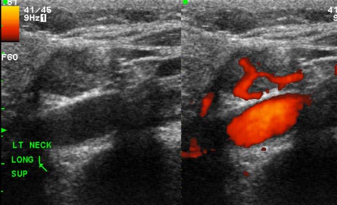
8 Ultrasound Technique High frequency (7-13 MHz) linear array transducers with gray- scale and color/power Doppler to evaluate the soft tissues of the neck Systematic evaluation of the thyroid and lateral neck compartments for the presence of co-existing thyroid disease or adenopathy
9 Parathyroid Imaging Ultrasound characteristics Hypoechoic with respect to adjacent thyroid parenchyma Oval or bean shaped, although larger adenomas may be multilobulated Can have cystic change Color of power Doppler may show a feeding vessel with peripheral internal vascularity - Polar feeding vessel


10 Thyroid Ultrasound Normal anatomy M M M Thy T Thy Thy CCA Transverse Longitudinal


11 Ultrasound Imaging of the Parathyroid Parathyroid adenoma Cyst Thy Cyst Thy Greyscale Color Doppler


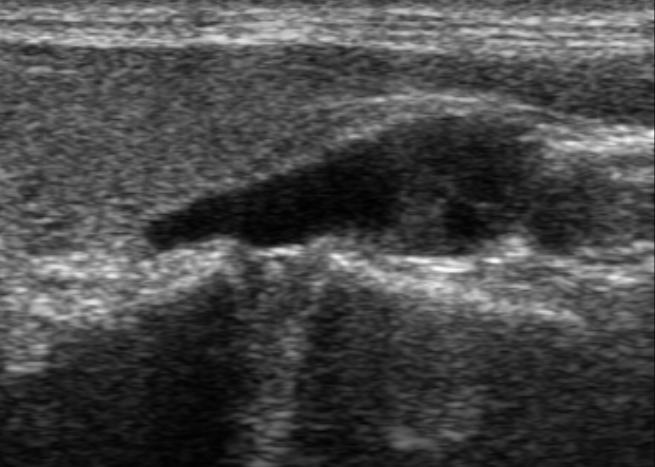
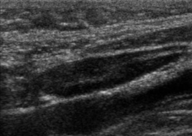
12 Parathyroid adenoma vs. lymph node Ultrasound characteristics Parathyroid adenoma Lymph node Thy JV Thy JV

13 Ultrasound Imaging Intra-thyroid parathyroid adenoma (ITPA) Ectopic Parathyroid gland completely surrounded on all sides by thyroid tissue Published incidence 0.5-4% 4D CT and Nuclear Medicine imaging cannot distinguish intra thyroid parathyroid adenoma from thyroid parenchyma
14 Intra-thyroid parathyroid adenoma Sonographic features: Variable- Overlap with imaging findings of thyroid nodules Hypoechoic echotexture with respect to thyroid parenchyma Presence of polar feeding vessel high sensitivity and specificity Caution! FNA findings can be indistinguishable from thyroid and sample must be sent for PTH assay


15 Imaging with Ultrasound Intra-thyroid parathyroid adenoma Thy Thy
16 Parathyroid Imaging Ultrasound Challenges Accuracy of US depends on the location and size of the lesion. Tumors may be obscured by the trachea or esophagus and may not be visualized if located in the mediastinum. Atypical sonographic appearance
17 Imaging with 4D-CT
18 4D-CT of the parathyroid Advantages Allows for detailed anatomic localization, superior to other imaging modalities, and helps aid surgery Accuracy of 87% as single modality in localizing adenoma
. Mortenson MM, et al.")
19 Imaging with 4D-CT Reoperative Neck and MDACC Experience Prospective study showed that 4DCT provides greater sensitivity than sestamibi imaging for accurate preoperative localization of parathyroid adenomas (88% vs 54%). Mortenson MM, et al. J Am Coll Surg 2008; 206.
20 Imaging with 4D-CT 4D CT parameters 18 G angiocatheter placed in the antecubital fossa 120 cc contrast at 4cc/sec Images obtained at 1.25 mm from the mandible to carina Images reconstructed at 4mm as multiplaner maximal intensity projection images: true axial true coronal true sagittal bilateral sagittal oblique planes
21 Imaging with 4D-CT 4D CT parameters (cont) Acquisitions: 1. Non- contrast 2. Arterial 3. Venous 4. Delayed- Venous

22 4D-CT Imaging Anatomy V v C T E THY S C M SCM C E LC VA

23 4D-CT Imaging Anatomy
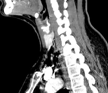
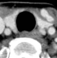
24 4D-CT Imaging Parathyroid adenoma : 4D CT findings Parathyroid adenomas are lower in attenuation relative to adjacent thyroid parenchyma on nonenhanced CT images Most parathyroid adenomas have rapid, arterial uptake with wash out of contrast during subsequent venous phase images Polar feeding vessel- secondary sign


25 T= 0 sec T= 55 sec T= 25 sec T= 85 sec
26 Imaging with 4D-CT Pitfalls on 4D CT False positive: - Lymph nodes - Thyroid tissue/exophytic thyroid nodules False Negative: - Ectopic glands - Small adenomas - Multiglandular disease/hyperplasia


27 4D-CT Parathyroid adenoma vs lymph node Parathyroid adenoma Rapid uptake with washout Lymph Node Slow continuous uptake with peak enhancement on delayed phase No contrast Arterial Venous
")
28 MDACC Nomenclature to organize and classify parathyroid adenomas Uniform and reliable description exact location of abnormal parathyroid glands Improves communication with surgeons Based on quadrants and the anterior-posterior depth relative to the recurrent laryngeal nerve and thyroid parenchyma ND Perrier World J Surgery (2009) 33:


29 Imaging with 4D-CT Case 1- Type A A- Superior Gland Adherent to the posterior surface of thyroid parenchyma Along the superior thyroid pole


30 Imaging with 4D-CT Case 1- Type A T

31 Imaging with 4D-CT Case 2- Type B T B- Ectopic Superior Gland Fallen posteriorly into tracheoesphageal groove. No contact with posterior surface of thyroid tissue. At the level of the superior thyroid pole


32 Imaging with 4D-CT Case 2- Type B T


33 Imaging with 4D-CT Case 3- Type C T C- Ectopic Superior Gland Fallen posteriorly into tracheoesphageal groove. No contact with posterior surface of thyroid tissue. At the level of or below the inferior thyroid pole
 In mid region of")


34 Imaging with 4D-CT Case 4- Type D D (Dangerous) In mid region of posterior surface of thyroid parenchyma. Could be superior or inferior gland Junction of recurrent laryngeal nerve, inferior thyroid artery and middle thyroid vein


35 Imaging with 4D-CT Case 5- Type E E- Inferior Gland Close to the inferior thyroid pole In the AP plane of thyroid and anterior trachea


36 Imaging with 4D-CT Case 5- Type E T


37 Imaging with 4D-CT Case 6- Type F T F Ectopic Inferior Gland Gland that has descended into Thyrothymic ligament. May appear to be ectopic or in mediastinum. AP view show it anterior/ near trachea


38 Imaging with 4D-CT Case 6- Type F T


39 Imaging with 4D-CT Double parathyroid adenoma Noncontrast Contrast Coronal MIP
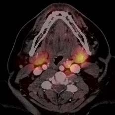
40 Imaging with Sestamibi Tc-99 x
41 Imaging with Sestamibi Tc-99 Technique: Early (30 min) and delayed (1.5 hour) planar imaging of the neck and chest Between the planar imaging we do SPECT/CT of the same area and 4DCT protocol CT done at 1.25 mm cuts SPECT images AC and non-ac processed with iterative reconstruction x
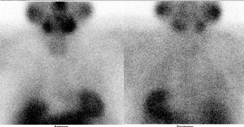
42 Imaging with Sestamibi Tc-99 Findings: Uptake in thyroid and salivary glands Parathyroid adenomas demonstrate avid uptake of tracer with retention of tracer on delayed imaging No uptake in normal parathyroid glands x


43 Imaging with Sestamibi Tc-99 Images: Thy Anterior Posterior Thy x


44 Imaging with Sestamibi Tc-99 Ectopic mediastinal parathyroid adenoma
45 Imaging with Sestamibi Tc-99 Pitfalls/limitations Cannot reliably differentiate between thyroid adenoma and parathyroid adenoma when tracer uptake is contiguous with thyroid parenchyma Small parathyroid adenomas may not accumulate enough tracer to be detectable Less sensitive in multigland hyperplasia Other malignancy can have tracer uptake and result in false positives- including thyroid cancer
46 Intraoperative Ultrasound localization
47 Intraoperative Ultrasound localization Used to help facilitate targeted, minimally invasive surgery, particularly in the reoperative neck Localize ectopic adenomas or transplanted parathyroid adenomas
48 Intraoperative Ultrasound localization Case 1 41 y/o female with recurrent primary hyperparathyroid disease Patient had undergone 2 prior neck explorations for parathyroid disease
49 Intraoperative Ultrasound localization Ectopic Parapharyngeal Parathyroid adenoma JV JV Anterior Posterior

50 Intraoperative Ultrasound localization Case 1: Ectopic parapharyngeal parathyroid adenoma IJV CCA Axial 4DCT with arterially enhancing parathyroid adenoma medial to the carotid space and posterior to the submandibular gland Noted pre-operatively is the depth of the lesion from skin surface and relationship to vessels


51 Intraoperative Ultrasound localization Case 1: Ectopic parapharyngeal parathyroid adenoma Pre-resection localization- skin marking CCA Transverse IOUS with Doppler demonstrates the adenoma and relationship to the carotid artery Skin marked overlying the ectopic parathyroid adenoma during IOUS


52 Intraoperative Ultrasound localization Case 1: Ectopic parapharyngeal parathyroid adenoma Intraoperative photographs The site of skin marking and planned incision 10 cm superior to the previous incision Offending parathyroid gland wedged between the facial vein and salivary gland


53 Intraoperative Ultrasound localization Case 1: Ectopic parapharyngeal parathyroid adenoma Photograph of the resected parathyroid adenoma placed on a template drawing Ruler documenting the size of the lesion
54 Intraoperative Ultrasound localization Case 2 82 year old female with parathyromatosis and recurrent primary parathyroid disease Patient had undergone 5 prior neck dissections
 Left pectoralis major parathyroid adenoma where")

55 Intraoperative Ultrasound localization Case 2: Multiple parathyroid adenomas (A) Left pectoralis major parathyroid adenoma where parathyroid tissue previously autotransplanted 0.9 cm A A IOUS was performed for localization and skin marking of multiple parathyroid adenomas
 Left")

56 Intraoperative Ultrasound localization Case 2: Multiple parathyroid adenomas (B) Left pretracheal/sternal notch implant B B 0.8 cm IOUS was performed for localization and skin marking of multiple parathyroid adenomas
 Anterior right")

57 Intraoperative Ultrasound localization Case 2: Multiple parathyroid adenomas (C) Anterior right suprasternal implant 1.1 cm C C IOUS was performed for localization and skin marking of multiple parathyroid adenomas
 Midline suprasternal")

58 Intraoperative Ultrasound localization Case 2: Multiple parathyroid adenomas (D) Midline suprasternal implant D D 0.7 cm 0.7 cm IOUS was performed for localization and skin marking of multiple parathyroid adenomas D D
59 Intraoperative Ultrasound localization Case 3 36 year old female with recurrent primary hyperparathyroidism Patient had undergone 2 prior parathyroidectomies Previous debulking of autotransplanted parathyroid tissue in the forearm with removal of surgical clip marking site of transplanted tissue 4D-CT, sestamibi and MR negative


60 Intraoperative Ultrasound localization Case 3: IOUS localization for re-operation debulking of left forearm Level of antecubital scar 0.3 cm 0.2 cm Three subtle areas of nodularity in the subcutenous soft tissues of the forearm were marked intraoperatively No surgical clips to identify site of autotransplanted tissue was seen


61 Intraoperative Ultrasound localization Case 3: IOUS localization for re-operation debulking of left forearm


62 Intraoperative Ultrasound localization Case 3: IOUS localization for re-operation debulking of left forearm


63 Intraoperative Ultrasound localization Case 3: IOUS localization for re-operation debulking of left forearm

64 Intraoperative Ultrasound localization Case 3: IOUS localization for re-operation debulking of left forearm
65 Summary Imaging is imperative for accurate localization of parathyroid adenomas facilitating minimally invasive parathyroidectomy Multimodality imaging complimentary Common nomenclature enables smooth communication between radiologist and surgeon Intraoperative US support can be key in challenging cases with ecopic locations or in the reoperative neck
66 Acknowledgements Dr. Nancy Perrier Dr. Beth Edeiken Dr. Thinh Vu

67
PARATHYROID IMAGING. James Lee, MD Chief, Endocrine Surgery Co-Director NY Thyroid-Parathyroid Center Columbia University Medical Center
 PARATHYROID IMAGING James Lee, MD Chief, Endocrine Surgery Co-Director NY Thyroid-Parathyroid Center Columbia University Medical Center NO DISCLOSURES Overview The hallmarks of the ideal test Benefits
Outline. Parathyroid Localization Studies. Mira Milas MD, FACS Associate Professor of Surgery Director, The Thyroid Center
 Parathyroid Localization Studies Mira Milas MD, FACS Associate Professor of Surgery Director, The Thyroid Center Outline Clinical Context of Primary Hyperparathyroidism Ultrasound, Sestamibi, and Other
PARATHYROID NUCLEAR MEDICINE IMAGING REVIEW DISCLOSURES
 PARATHYROID NUCLEAR MEDICINE IMAGING REVIEW Miguel Hernandez Pampaloni, M.D., Ph.D. Chief, Nuclear Medicine Assistant Professor of Radiology UCSF Department of Radiology and Biomedical Imaging DISCLOSURES
Surgical anatomy of thyroid and parathyroid glands
 Head & Neck Surgery Course Surgical anatomy of thyroid and parathyroid glands Dr Pierfrancesco PELLICCIA Pr Benjamin LALLEMANT Service ORL et CMF CHU de Nîmes CH de Arles Thyroid glands Dr Pierfrancesco
Parathyroid Glands: location, condition and value of imaging tests.
 Parathyroid Glands: location, condition and value of imaging tests. Poster No.: C-2283 Congress: ECR 2015 Type: Educational Exhibit Authors: E. Elías Cabot, P. Segui, G. D. Tobar Murgueitio; Cordoba/ES
AACE/ACE Advanced Endocrine Neck Ultrasound Training Course 2016
 AACE/ACE Advanced Endocrine Neck Ultrasound Training Course 2016 This 9mm left inferior nodule should remind us all why we re here! There is no absolute number of images required for documentation
Thyroid and Parathyroid Ultrasound Protocol
 Thyroid and Parathyroid Ultrasound Protocol Reviewed By: Anna Ellermeier, MD Last Reviewed: December 2017 Contact: (866) 761-4200, Option 1 **NOTE for all examinations: 1. If documenting possible flow
THE PARATHYROID GLAND THEORY AND NUCLEAR MEDICINE PRACTICE
 THE PARATHYROID GLAND THEORY AND NUCLEAR MEDICINE PRACTICE George N. Sfakianakis MD Professor of Radiology and Pediatrics Director, Division of Nuclear Medicine UM/JMMC Miami FL October 2009 ENDONCRINE
Chapter 2 Preoperative Parathyroid Imaging for the Endocrine Surgeon
 Chapter 2 Preoperative Parathyroid Imaging for the Endocrine Surgeon Elizabeth G. Grubbs, Beth S. Edeiken, Maria K. Gule, Brett J. Monroe, Edmund Kim, Thinh Vu, and Nancy D. Perrier Keywords Parathyroid
42 yr old male with h/o Graves disease and prior I 131 treatment presents with hyperthyroidism and undetectable TSH. 2 hr uptake 20%, 24 hr uptake 50%
 Pinhole images of the neck are acquired in multiple projections, 24hrs after the oral administration of approximately 200 µci of I123. Usually, 24hr uptake value if also calculated (normal 24 hr uptake
Case 2: 30 yr-old woman with 7 yr history of recurrent kidney stones
 Case 2: 30 yr-old woman with 7 yr history of recurrent kidney stones Giuliano Mariani Regional Center of Nuclear Medicine, University of Pisa Medical School, Pisa (Italy) 30 yr-old woman with 7 yr history
Outline. Primary Hyperparathyriodism. SPECT/CT in Parathyroid Localisation. Ann-Marie Quigley Nuclear Medicine Royal Free Hospital London
 SPECT/CT in Parathyroid Localisation Ann-Marie Quigley Nuclear Medicine Royal Free Hospital London Outline Pathophysiology Current guidelines SPECT/CT the evidence SPECT/CT in clinical scenarios MGD, Nodular
Outline. SPECT/CT in Parathyroid Disease. Pathophysiology. Current guidelines. SPECT/CT the evidence. SPECT/CT in clinical scenarios
 SPECT/CT in Parathyroid Disease Ann-Marie Quigley Nuclear Medicine Royal Free Hospital London Outline Pathophysiology Current guidelines SPECT/CT the evidence SPECT/CT in clinical scenarios MGD, Nodular
Minimally invasive parathyroidectomy
 Minimally invasive parathyroidectomy Jessica E. Gosnell MD Assistant Professor of Surgery March 22, 2011 1 Minimally invasive parathyroidectomy 1. What? 2. When? 3. How? 4. Convert? 5. What adjuncts? Primary
Complementary sestamibi scintigraphy and ultrasound for primary hyperparathyroidism
 Nuclear Medicine and Biomedical Imaging Research Article Complementary sestamibi scintigraphy and ultrasound for primary hyperparathyroidism Yang Z 1,3 *, Li AY 2, Alexander G 3 and Chadha M 3 1 Department
Thyroid Ultrasound for the Endocrine Surgeon: A Valuable Clinical Tool that Enhances Diagnostic and Therapeutic Outcomes
 Thyroid Ultrasound for the Endocrine Surgeon: A Valuable Clinical Tool that Enhances Diagnostic and Therapeutic Outcomes Allan Siperstein MD The Cleveland Clinic Audience Quiz Taken ultrasound course Perform
HYPERPARATHYROIDIS M FAISAL GHANI SIDDIQUI MBBS; FCPS; PGDIP-BIOMEDICAL ETHICS; MCPS-HPE
 HYPERPARATHYROIDIS M FAISAL GHANI SIDDIQUI MBBS; FCPS; PGDIP-BIOMEDICAL ETHICS; MCPS-HPE PROFESSOR OF SURGERY J I N N A H S I N D H M E D I C A L U N I V E R S I T Y PREAMBLE Anatomy & physiology of the
Advanced Anatomy of the Neck
 AACE 2018 Advanced Anatomy of the Neck Alex Tessnow, MD, MBA, FACE, ECNU University of Texas Southwestern Dallas, TX Content contributed by: H. Jack Baskin, Daniel Duick, Diana Dean, Robert A. Levine,
ORIGINAL ARTICLE. Appearance of Ectopic Undescended Inferior Parathyroid Adenomas on Technetium Tc 99m Sestamibi Scintigraphy
 ORIGINAL ARTICLE Appearance of Ectopic Undescended Inferior Parathyroid Adenomas on Technetium Tc 99m Sestamibi Scintigraphy A Lesson From Reoperative Parathyroidectomy David Axelrod, MD; James C. Sisson,
Shadow because the air
 Thyroid Ultrasound Thyroid US examination needs: 1. high frequency transducer 2. extended patient's neck 3. check all the neck area because the swelling could be in areas other than the thyroid such as
Primary Hyperparathyroidism
 November 2002 Primary Hyperparathyroidism Lori Coburn, Harvard Medical School Year III Hyperparathyroidism An increase in parathyroid hormone (PTH) production Divided into Primary, Secondary and Tertiary
SPECT/CT in Endocrine Diseases and Dosimetry
 SPECT/CT in Endocrine Diseases and Dosimetry Heather A. Jacene, MD Division of Nuclear Medicine Russell H. Morgan Dept. of Radiology and Radiological Science Johns Hopkins University Baltimore, MD Disclosures
Index. radiologic.theclinics.com. Note: Page numbers of article titles are in boldface type.
 Index Note: Page numbers of article titles are in boldface type. A ACC. See Adrenal cortical carcinoma. Acromegaly and the pituitary gland, 551 Acute suppurative thyroiditis, 405, 406 Addison, Thomas and
Preoperative Evaluation
 Preoperative Evaluation Lateral compartment lymph nodes are easier to detect and are amenable to FNA Central compartment lymph nodes are much more difficult to detect and FNA (Tg washout testing is compromised)
Marcin Barczynski, 1 Aleksander Konturek, 2 Alicja Hubalewska-Dydejczyk, 2. Filip Gołkowski, 1 Stanislaw Cichon, 1 Piotr Richter, 1 Wojciech Nowak
 3 rd Chair and Department of General Surgery 1 and Chair and Department of Endocrinology 2 Jagiellonian University, Medical College Head: Prof. Wojciech Nowak, MD, PhD INTRAOPERATIVE BILATERAL INTERNAL
A CASE OF A Huge Submandibular Pleomorphic Adenoma
 ISPUB.COM The Internet Journal of Head and Neck Surgery Volume 4 Number 2 S VERMA Citation S VERMA.. The Internet Journal of Head and Neck Surgery. 2009 Volume 4 Number 2. Abstract Pleomorphic adenoma
The mystery of the hidden parathyroid adenoma. Case #1. Imaging. Case of missed adenoma. Ultrasound false nega9ves. Case of missed adenoma 5/16/16
 The mystery of the hidden parathyroid adenoma AACE Annual Mee+ng 2016 Orlando, FL Case #1 Moderator: Panel: Jennifer L Mar+ MD FACS Endocrine Surgery NYC Azeez Farooki MD Endocrinology MSKCC Peter Sadow
Contents. Basic Ultrasound Principles and Terminology. Ultrasound Nodule Characteristics
 Contents Basic Ultrasound Principles and Terminology Basic Ultrasound Principles... 1 Ultrasound System... 2 Linear Transducer for Superficial Images and Ultrasound-Guided FNA... 3 Scanning Planes... 4
PTH > 60pg/ml PRIMARY HYPERPARATHYROIDISM. Introduction Biochemical Diagnosis. Normal Parathyroid. Parathyroid Glands
 next speaker: Declan Neeson Belfast/UK SPECT/CT scanning and parathyroid surgery in Southern Trust, N. Ireland D Neeson M Korda, G Gray, C Leonard, M Fawzy, R Lambon Parathyroid Glands PRIMARY HYPERPARATHYROIDISM
HPI joint pain/arthritis serum calcium 11.5 PTH 147pg/ml
 HPI 45 yo female Increased calcium level during evaluation for joint pain/arthritis W/U showed serum calcium 11.5 and PTH 147pg/ml (Normal 11-67pg/ml) Otherwise asymptomatic PMH/PSH Arthritis Tonsillectomy
Ultrasonography of the Neck as an Adjunct to FNA. Nicole Massoll M.D.
 Ultrasonography of the Neck as an Adjunct to FNA Nicole Massoll M.D. Basic Features of Head and Neck Ultrasound and Anatomy Nicole Massoll M.D. University of Arkansas for Medical Sciences, Little Rock
Head & Neck Clinical Sub Group. Network Agreed Imaging Guidelines for UAT and Thyroid Cancer. Measure Nos: 11-1C-105i & 11-1C-106i
 Greater Manchester, Lancashire & South Cumbria Strategic Clinical Network & Senate Head & Neck Clinical Sub Group Network Agreed Imaging Guidelines for UAT and Thyroid Cancer Measure Nos: 11-1C-105i &
Thyroid & Parathyroid glands Ultrasound evaluation.
 Thyroid & Parathyroid glands Ultrasound evaluation. www.headandneckultrasound.co.uk Rhodri M Evans Incidence 70 Thyroid Nodules 30 Palpation 50 Age 100 Incidence 70 Thyroid Nodules US/Autopsy 30 Palpation
hypercalcemia of malignancy hyperparathyroidism PHPT the most common cause of hypercalcemia in the outpatient setting the second most common cause
 hyperparathyroidism A 68-year-old woman with documented osteoporosis has blood tests showing elevated serum calcium and parathyroid hormone (PTH) levels: 11.2 mg/dl (8.8 10.1 mg/dl) and 88 pg/ml (10-60),
Neck Ultrasound. Faculty Info: Amy Kule, MD
 Neck Ultrasound Date: Friday, October 19, 2018 Time: 11:00 AM Location: SMALL GROUP LABORATORY SSOM L71 Watch: Ø Neck Ultrasound Scanning Protocol (4:00): https://www.youtube.com/watch?v=zozd2x2ll4q Faculty
Parathyroid Imaging. A Guide to Parathyroid Surgery
 Parathyroid Imaging A Guide to Parathyroid Surgery Primary Hyperparathyroidism (PHPT) 3 rd most common endocrine disorder after diabetes and hyperthyroidism Prevalence in women 2% Often discovered in asymptomatic
Case 4: 27 yr-old woman with history of kidney stones and hyperparathyroidism.
 Case 4: 27 yr-old woman with history of kidney stones and hyperparathyroidism. Giuliano Mariani Regional Center of Nuclear Medicine, University of Pisa Medical School, Pisa (Italy) Hyperparathyroidism
Karoline Nowillo, MD. February 1, 2008
 Case Presentation Karoline Nowillo, MD SUNY Downstate t February 1, 2008 Case Presentation Chief complaint enlarging goiter x 8 months History of present illness shortness of breath, heaviness in chest
Alexander C Vlantis. Selective Neck Dissection 33
 05 Modified Radical Neck Dissection Type II Alexander C Vlantis Selective Neck Dissection 33 Modified Radical Neck Dissection Type II INCISION Various incisions can be used for a neck dissection. The incision
Parathyroids, Small but Mighty Current Pathways to Early Diagnosis and Cure of Parathyroid Disease
 Parathyroids, Small but Mighty Current Pathways to Early Diagnosis and Cure of Parathyroid Disease Mira Milas MD, FACS Professor of Surgery Director of Endocrine Surgery No conflicts of interest or financial
Endoscopic Parathyroidectomy: Why and When?
 World J Surg (2008) 32:2509 2515 DOI 10.1007/s00268-008-9709-3 Endoscopic Parathyroidectomy: Why and When? Jean-François Henry Æ Frédéric Sebag Æ Mariya Cherenko Æ Giuseppe Ippolito Æ David Taieb Æ Josiane
Ultrasound Interpretation of Non-Thyroid Neck Pathology
 Ultrasound Interpretation of Non-Thyroid Neck Pathology Kevin T. Brumund, M.D., F.A.C.S. Associate Professor of Surgery Head and Neck Surgery University of California, San Diego Health Sciences VA Medical
General Surgery Curriculum Royal Australasian College of Surgeons, General Surgeons Australia & New Zealand Association of General Surgeons
 General Surgery Curriculum Royal Australasian College of Surgeons, General Surgeons Australia & New Zealand Association of General Surgeons MODULE TITLE: ENDOCRINE 5-May-2013 DEVELOPED BY: Jonathan Serpell
Lecture 01. The Thyroid & Parathyroid Glands. By: Dr Farooq Khan PMC Date: 12 th March. 2018
 Lecture 01 The Thyroid & Parathyroid Glands By: Dr Farooq Khan PMC Date: 12 th March. 2018 INTRODUCTION LAYERS OF THE NECK The neck has four major compartments or layer which are enclosed by an outer musculofascial
The Neck the lower margin of the mandible above the suprasternal notch and the upper border of the clavicle
 The Neck is the region of the body that lies between the lower margin of the mandible above and the suprasternal notch and the upper border of the clavicle below Nerves of the neck Cervical Plexus Is formed
Role of imaging in RCC. Ultrasonography. Solid lesion. Cystic RCC. Solid RCC 31/08/60. From Diagnosis to Treatment: the Radiologist Perspective
 Role of imaging in RCC From Diagnosis to Treatment: the Radiologist Perspective Diagnosis Staging Follow up Imaging modalities Limitations and pitfalls Duangkamon Prapruttam, MD Department of Therapeutic
Thyroid INTRODUCTION ANATOMY SUMMARY OF CHANGES
 AJC 7/14/06 1:19 PM Page 67 Thyroid C73.9 Thyroid gland SUMMARY OF CHANGES Tumor staging (T) has been revised and the categories redefined. T4 is now divided into T4a and T4b. Nodal staging (N) has been
Endocrine University 2016 AACE-ACE-MAYO CLINIC
 Endocrine University 2016 AACE-ACE-MAYO CLINIC Dev Abraham MD, MRCP (UK), ECNU Professor of Medicine (clinical), Division of Endocrinology Adjunct Professor of Surgery and Pathology Medical Director, Utah
Ovid: Oxford Textbook of Endocrinology & Diabetes
 Página 1 de 6 Copyright 2002 Oxford University Press Wass, John A.H., Shalet, Stephen M., Gale, Edwin, Amiel, Stephanie A. Oxford Textbook of Endocrinology & Diabetes, 1st Edition Surgical procedure Part
Sonoanatomy Of The Brachial Plexus With Single Broad Band-High Frequency (L17-5 Mhz) Linear Transducer
 Linear Transducer") ISPUB.COM The Internet Journal of Anesthesiology Volume 11 Number 2 Sonoanatomy Of The Brachial Plexus With Single Broad Band-High Frequency (L17-5 Mhz) Linear A Thallaj Citation A Thallaj.. The Internet
ISPUB.COM The Internet Journal of Anesthesiology Volume 11 Number 2 Sonoanatomy Of The Brachial Plexus With Single Broad Band-High Frequency (L17-5 Mhz) Linear A Thallaj Citation A Thallaj.. The Internet
Ultrasound Evaluation of Hyperparathyroidism
 Ultrasound Evaluation of Hyperparathyroidism OBJECTIVES: 1. Why use US for localization? Advanced US course, Orlando, Florida September, 2018 2. Review the US findings and features of parathyroid adenomas
Nuclear Medicine Head and Neck Region. Bán Zsuzsanna, MD University of Pécs, Department of Nuclear Medicine
 Nuclear Medicine Head and Neck Region Bán Zsuzsanna, MD University of Pécs, Department of Nuclear Medicine Thyroid scintigraphy Parathyroid scintigraphy F18-FDG PET examinations in head and neck cancer
Sectional Anatomy Quiz - III
 Sectional Anatomy - III Rashid Hashmi * Rural Clinical School, University of New South Wales (UNSW), Wagga Wagga, NSW, Australia A R T I C L E I N F O Article type: Article history: Received: 30 Jun 2018
Salivary ultrasound. Dr T J Beale Royal National Throat Nose & Ear and UCLH Hospitals London UK
 Salivary ultrasound Dr T J Beale Royal National Throat Nose & Ear and UCLH Hospitals London UK Two main groups of patients with presenting symptoms of: Obstructive or chronic inflammatory symptoms (salivary
THYROID & PARATHYROID. By Prof. Saeed Abuel Makarem & Dr. Sanaa Al-Sharawy
 THYROID & PARATHYROID By Prof. Saeed Abuel Makarem & Dr. Sanaa Al-Sharawy 1 OBJECTIVES By the end of the lecture, the student should be able to: Describe the shape, position, relations and structure of
Sectional Anatomy Quiz II
 Sectional Anatomy II Rashid Hashmi Rural Clinical School, University of New South Wales, Wagga Wagga, New South Wales, Australia A R T I C L E I N F O Article type: Article history: Received: 3 Aug 2017
PAPILLARY THYROID CARCINOMA PRESENTING AS A LATERAL NECK MASS MASS. Dr. Pamela Hanson DO PGY3
 PAPILLARY THYROID CARCINOMA PRESENTING AS A LATERAL NECK MASS MASS Dr. Pamela Hanson DO PGY3 MK CASE PRESENTATION 28 yo Female presented to the ENT Clinic in October 2016, with the complaint of chronic
Background & Indications Probe Selection
 Teresa S. Wu, MD, FACEP Director, EM Ultrasound Program & Fellowship Co-Director, Simulation Based Training Program & Fellowship Associate Program Director, EM Residency Program Maricopa Medical Center
Minimally invasive parathyroid surgery
 Review Article Minimally invasive parathyroid surgery Salem I. Noureldine, Zhen Gooi, Ralph P. Tufano Division of Head and Neck Endocrine Surgery, Department of Otolaryngology, Head and Neck Surgery, Johns
Re-explorative Parathyroid Surgery for Persistent and Recurrent Primary Hyperparathyroidism
 10.5005/jp-journals-10002-1070 ORIGINAL ARTICLE WJOES Re-explorative Parathyroid Surgery for Persistent and Recurrent Primary Hyperparathyroidism Rachel L O Connell, Karolina Afors, Martin H Thomas Ashford
Preoperative Tc-99m-sestamibi (MIBI) scintigraphy and
 scintigraphy and") Otolaryngology Head and Neck Surgery (2006) 134, 316-320 ORIGINAL RESEARCH In Vivo Characterisation of Parathyroid Lesions by Use of Gamma Probe: Comparison With Ex Vivo Count Method and Frozen Section
Otolaryngology Head and Neck Surgery (2006) 134, 316-320 ORIGINAL RESEARCH In Vivo Characterisation of Parathyroid Lesions by Use of Gamma Probe: Comparison With Ex Vivo Count Method and Frozen Section
Imaging in gastric cancer
 Imaging in gastric cancer Gastric cancer remains a deadly disease because of late diagnosis. Adenocarcinoma represents 90% of malignant tumors. Diagnosis is based on endoscopic examination with biopsies.
ANTERIOR CERVICAL TRIANGLE (Fig. 2.1 )
") 2 Neck Anatomy ANTERIOR CERVICAL TRIANGLE (Fig. 2.1 ) The boundaries are: Lateral: sternocleidomastoid muscle Superior: inferior border of the mandible Medial: anterior midline of the neck This large triangle
2 Neck Anatomy ANTERIOR CERVICAL TRIANGLE (Fig. 2.1 ) The boundaries are: Lateral: sternocleidomastoid muscle Superior: inferior border of the mandible Medial: anterior midline of the neck This large triangle
27 Reoperative Parathyroid Surgery
 327 27 Reoperative Parathyroid Surgery Contents 27.1 Definition and Introduction... 327 27.2 Anatomy and Embryology of the Parathyroid Gland... 328 27.3 Causes of Persistent and Recurrent Hyperparathyroidism...
objectives Pitfalls and Pearls in PET/CT imaging Kevin Robinson, DO Assistant Professor Department of Radiology Michigan State University
 objectives Pitfalls and Pearls in PET/CT imaging Kevin Robinson, DO Assistant Professor Department of Radiology Michigan State University To determine the regions of physiologic activity To understand
Ultrasound for Pre-operative Evaluation of Well Differentiated Thyroid Cancer
 Ultrasound for Pre-operative Evaluation of Well Differentiated Thyroid Cancer Its Not Just About the Nodes AACE Advances in Medical and Surgical Management of Thyroid Cancer - 2017 Robert A. Levine, MD,
THYROID NODULES: THE ROLE OF ULTRASOUND
 THYROID NODULES: THE ROLE OF ULTRASOUND NOVEMBER 2017 DR. DEAN DURANT DEFINITION Thyroid nodule: Focal area within the thyroid gland with echogenicity different from surrounding parenchyma. THYROID NODULES
Abdomen Sonography Examination Content Outline
 Abdomen Sonography Examination Content Outline (Outline Summary) # Domain Subdomain Percentage 1 2 3 Anatomy, Perfusion, and Function Pathology, Vascular Abnormalities, Trauma, and Postoperative Anatomy
Anatomy of the Thyroid Gland
 Anatomy of the Thyroid Gland Introduction Nomenclature G, thyreos= shield, eidos= like Location Root of the neck ventrally (C5-T1) Function endocrine gland that secretes: Thyroxine (T4) T3 Calcitonin LWW,
Thyroid Nodules: US Risk Stratification. Alex Tessnow, MD, FACE, ECNU University of Texas Southwestern Associate Professor of Medicine Dallas, Texas
 Thyroid Nodules: US Risk Stratification Alex Tessnow, MD, FACE, ECNU University of Texas Southwestern Associate Professor of Medicine Dallas, Texas Which of the following is true? A. All echogenic foci
Mediastinum and pericardium
 Mediastinum and pericardium Prof. Abdulameer Al-Nuaimi E-mail: a.al-nuaimi@sheffield.ac.uk E. mail: abdulameerh@yahoo.com The mediastinum: is the central compartment of the thoracic cavity surrounded by
CURRENTLY THERE is considerable discussion about
 0013-7227/02/$15.00/0 The Journal of Clinical Endocrinology & Metabolism 87(3):1024 1029 Printed in U.S.A. Copyright 2002 by The Endocrine Society Parathyroid Surgery: Separating Promise from Reality NANCY
Comparison Of Sestamibi Scintigraphy And Ultrasonography In Preoperative Localization Of Primary Hyperparathyroidism
 ISPUB.COM The Internet Journal of Surgery Volume 16 Number 1 Comparison Of Sestamibi Scintigraphy And Ultrasonography In Preoperative Localization Of Primary S Nasiri, A Sorush, A Hashemi, F Mehrkhani,
DESCRIPTION: This is the part of the trunk, which is located between the root of the neck and the superior border of the abdominal region.
 1 THE THORACIC REGION DESCRIPTION: This is the part of the trunk, which is located between the root of the neck and the superior border of the abdominal region. SHAPE : T It has the shape of a truncated
The Concept of GOSTT
 IAEA Regional Training Course on Sentinel Lymph Node Mapping and Radioguided Surgery The Concept of GOSTT Giuliano Mariani Regional Center of Nuclear Medicine, University of Pisa Medical School, Pisa,
Imaging Technique. Ultrasound Imaging of the Salivary Glands. Parotid Gland. The Major Salivary Glands. Parotid Gland: Stenson s Duct.
 Ultrasound Imaging of the Salivary Glands Edward G. Grant MD Professor & Chairman, Dept of Radiology USC Keck School of Medicine edgrant@usc.edu edgrant@usc.edu Imaging Technique Linear array transducer
Learning Objectives (1&2)
") Learning Objectives (1&2) By the end of the session, students should be able to: 1) Identify anatomical position seated, standing, prone, supine. 2) Pronounce, define and be able to use directional and
Learning Objectives (1&2) By the end of the session, students should be able to: 1) Identify anatomical position seated, standing, prone, supine. 2) Pronounce, define and be able to use directional and
Hyperparathyroidism is caused by overproduction of parathyroid
 Published July 18, 2013 as 10.3174/ajnr.A3615 ORIGINAL RESEARCH HEAD & NECK 4D-CT for Preoperative Localization of Abnormal Parathyroid Glands in Patients with Hyperparathyroidism: Accuracy and Ability
Health Sciences Centre, Team A, Dr. L. Bohacek (Endocrine Surgery) Medical Expert
 Medical Expert") Health Sciences Centre, Team A, Dr. L. Bohacek (Endocrine Surgery) Introduction Medical Expert This is a three month PGY 1-5 rotation in which residents gain exposure in the care and management of patients
Health Sciences Centre, Team A, Dr. L. Bohacek (Endocrine Surgery) Introduction Medical Expert This is a three month PGY 1-5 rotation in which residents gain exposure in the care and management of patients
Anterior triangle of neck
 Anterior triangle of neck Dept. of Anatomy Zhou Hong Ying Outline boundary and subdivisions of ant. triangle contents of the triangle Muscles: suprahyoid muscles, infrahyoid muscles Nerves: CNⅩ, CNⅪ, CNⅫ,
10/14/2018 Dr. Shatarat
 2018 Objectives To discuss mediastina and its boundaries To discuss and explain the contents of the superior mediastinum To describe the great veins of the superior mediastinum To describe the Arch of
Case Papillary thyroid carcinoma(ptc):local recurrence post thyroidectomy
:local recurrence post thyroidectomy") Case 16070 Papillary thyroid carcinoma(ptc):local recurrence post thyroidectomy Dr Alka Ashmita Singhal, Dr Deepak Sarin 1, Dr Monika Aggarwal, Dr Haimanti Sarin2 Medanta The Medicity Hospital, Medanta
Case 16070 Papillary thyroid carcinoma(ptc):local recurrence post thyroidectomy Dr Alka Ashmita Singhal, Dr Deepak Sarin 1, Dr Monika Aggarwal, Dr Haimanti Sarin2 Medanta The Medicity Hospital, Medanta
Multimodality imaging of the parathyroid adenoma
 Multimodality imaging of the parathyroid adenoma Poster No.: C-0256 Congress: ECR 2015 Type: Educational Exhibit Authors: S. Liddy, J. Feeney; Dublin/IE Keywords: Head and neck, Nuclear medicine, Thyroid
Thyroid and Parathyroid Glands
 Thyroid and Parathyroid Glands Please view our Editing File before studying this lecture to check for any changes. Color Code Important Doctors Notes Notes/ explanation Objectives: By the end of the lecture,
Scrotum Kacey Morrison Amanda Baxter Sabrina Tucker July 18, 2006 SCROTUM
 Scrotum Kacey Morrison Amanda Baxter Sabrina Tucker July 18, 2006 SCROTUM 1) Other Names: Scrotum None Testicles Testes (Curry Tempkin, p. 236, 2/3/2) Ductus deferens spermatic cord (Tempkin, p. 279, Anatomy
Neck-2. Dr. Heba Kalbouneh Associate Professor of Anatomy and Histology
 Neck-2 ` Dr. Heba Kalbouneh Associate Professor of Anatomy and Histology Triangles of the neck Side of the neck Midline Lower border of mandible Line between angle of mandible and mastoid Superior nuchal
Kidney Case 1 SURGICAL PATHOLOGY REPORT
 Kidney Case 1 Surgical Pathology Report February 9, 2007 Clinical History: This 45 year old woman was found to have a left renal mass. CT urography with reconstruction revealed a 2 cm medial mass which
Women s Health in General Practice Symposium 2015 Thyroid & Parathyroid Cases
 Women s Health in General Practice Symposium 2015 Thyroid & Parathyroid Cases Bill Fleming Epworth Freemasons Hospital 1 Common Endocrine Presentations anatomical problems thyroid nodule / goitre embryological
Reoperative central neck surgery
 Reoperative central neck surgery R. Pandev, I. Tersiev, M. Belitova, A. Kouizi, D. Damyanov University Clinic of Surgery, Section Endocrine Surgery University Hospital Queen Johanna ISUL Medical University
Head & Neck Contouring
 Head & Neck Contouring Presented by James Wheeler, MD Center for Cancer Care Goshen, IN 46526 September 12, 2014 Special Thanks to: Spencer Boulter, Director of Operations (AAMD) Adam Moore, RT(T), CMD
Thyroid Ultrasonography: clinical and radiological correlations
 Thyroid Ultrasonography: clinical and radiological correlations Dr.M.Thijs Radiology Anatomy Inflammatory Thyroid Disease Benign lesions Thyroid tumors Thyroglossal duct cyst Anatomy Transverse Longitudinal
O~iginalArtrc!~'" MINIMALLY INVASIVE RADIO-GUIDED PARATHYROIDECTOMY IN 152 CONSECUTIVE PATIENTS WITH PRIMARY HYPERPARATHYROIDISM
 O~iginalArtrc!~'",,_.~.~_.,_,,~_......_. ~.o:-'';:...:/-.~. ~'.:::.., MINIMALLY INVASIVE RADIO-GUIDED PARATHYROIDECTOMY IN 152 CONSECUTIVE PATIENTS WITH PRIMARY HYPERPARATHYROIDISM Douglas Politz, MD,
THYROID CANCER IN CHILDREN. Humberto Lugo-Vicente MD FACS FAAP Professor Pediatric Surgery UPR School of Medicine
 THYROID CANCER IN CHILDREN Humberto Lugo-Vicente MD FACS FAAP Professor Pediatric Surgery UPR School of Medicine Thyroid nodules Rare Female predominance 4-fold as likely to be malignant Hx Radiation exposure?
Polymorphous Low-Grade. December 5 th, 2008
 Polymorphous Low-Grade Adenocarcinoma December 5 th, 2008 Epidemiology Represents 2 nd or 3 rd most common minor salivary gland malignancy (17-26%) 1 st mucoepidermoid carcinoma Rare in reported Asian
Parathyroid glands: so small but yet so big
 Parathyroid glands: so small but yet so big Poster No.: C-1520 Congress: ECR 2016 Type: Educational Exhibit Authors: M. Pérez Dávila, C. Ferreiro Arguelles, J. A. Camacho Oviedo, P. BARÓN RODIZ, P. Núñez
The GOSTT concept. (radio)guided intraoperative Scintigraphic Tumor Targeting. Emmanuel Deshayes. GOSTT = Radioguided Surgery
guided intraoperative Scintigraphic Tumor Targeting. Emmanuel Deshayes. GOSTT = Radioguided Surgery") IAEA WorkShop, November 2017 Emmanuel Deshayes With the kind help of Pr Francesco Giammarile The GOSTT concept GOSTT = Radioguided Surgery (radio)guided intraoperative Scintigraphic Tumor Targeting 1 Radioguided
IAEA WorkShop, November 2017 Emmanuel Deshayes With the kind help of Pr Francesco Giammarile The GOSTT concept GOSTT = Radioguided Surgery (radio)guided intraoperative Scintigraphic Tumor Targeting 1 Radioguided
Evaluation of Neck Mass. Disclosure. Learning Objectives 3/24/2014. Karen T. Pitman MD, FACS Banner MDACC, Gilbert AZ. Nothing to disclose
 Evaluation of Neck Mass Karen T. Pitman MD, FACS Banner MDACC, Gilbert AZ Nothing to disclose Disclosure Learning Objectives 1. Describe a systematic method to evaluate a patient with a neck mass 2. Select
Dr. Weyrich G07: Superior and Posterior Mediastina. Reading: 1. Gray s Anatomy for Students, chapter 3
 Dr. Weyrich G07: Superior and Posterior Mediastina Reading: 1. Gray s Anatomy for Students, chapter 3 Objectives: 1. Subdivisions of mediastinum 2. Structures in Superior mediastinum 3. Structures in Posterior
PEDIATRICS WK 3 HEAD AND NECK ALISON WALLACE MD, PHD
 PEDIATRICS WK 3 HEAD AND NECK ALISON WALLACE MD, PHD Topics 1. Cervical lymphadenopathy 2. Lymphatic malformation 3. Thyroglossal duct cysts 4. Branchial cleft cysts 5. Thyroid masses CASE 1 Case 1 A 2