Advanced Anatomy of the Neck
|
|
|
- Kerry Greer
- 5 years ago
- Views:
Transcription

1 AACE 2018 Advanced Anatomy of the Neck Alex Tessnow, MD, MBA, FACE, ECNU University of Texas Southwestern Dallas, TX Content contributed by: H. Jack Baskin, Daniel Duick, Diana Dean, Robert A. Levine, Mark Lupo, Dev Abraham, John Abele,

2 Thyroid Term derived from Greek word, Thureos, an ancient oblong shield German = Shilddruesse Shield Gland

3 Neck Anatomy Wharton 1656: purpose is to beautify the neck particularly in females to whom for this reason a larger gland.. Deposition Van der Weyden c. 1435
 C-cells scattered through posterior/superior")
4 Neck Anatomy Derived from endodermal tissue at base of tongue 1 st gland to develop day 24 Embryonal remnants form Thyroglossal duct; pyramidal lobe; lingual thyroid Fuse with C-cells (neural crest origin) C-cells scattered through posterior/superior lobes

5

6 Thyroid Anatomy Largest endocrine gland (~20 g) Fills the tracheoesophageal space Overlies RLN bilaterally

7

8
9
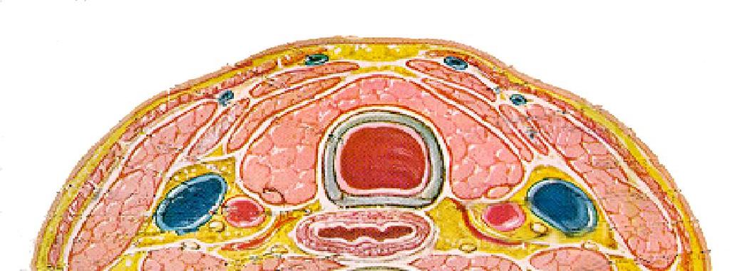
10 Sternocleidomastoid muscle trachea Isthmus Sternohyoid muscle Sternothyroid muscle Omohyoid muscle Internal jugular vein Carotid artery esophagus Parathyroid gland

11 Normal Thyroid Composite View Strap SCM IJ CA R Lobe Trach L Lobe LCM Esoph

12

13 Transverse View High Neck

14 Transverse View Mid Neck


15 Transverse View Low Neck STM SHM SCM I RL T LL IJV CA CA
16 Diagnostic Ultrasound Images (13 minimum): Right Trans Inferior Right Trans Mid* Right Trans Superior Right Sag Lateral Right Sag Mid* Right Sag Medial Left Trans Inferior Left Trans Mid* Left Trans Superior Left Sag Lateral Left Sag Mid* Left Sag Medial Isthmus* * = measurements made here
17


18 Normal Thyroid R Transverse Superior Mid Inferior


19 Normal Thyroid L Transverse Superior Mid Inferior

20 Left Medial Sagittal View Note tangential image of esophagus


21 Normal Thyroid L Sagittal Lateral Mid Medial


22 Appearance of the Esophagus (post thyroidectomy)


23 ISTHMUS

24 Pyramidal Lobe

25 Measurement of Thyroid Width and Depth
 Volume =")
26 Measurement of Thyroid Length (sagittal / longitudinal) Volume = p/6(lxdxw)
27 Thyroid Echogenicity Normal thyroid: High intensity homogeneous echo pattern with little identifiable internal architecture Muscles located anteriorly and anterolaterally are less echogenic
 Note bright")

28 Thyroid Echogenicity Normal thyroid (longitudinal view) Note bright homogeneous echotexture Hashimoto s thyroiditis Note heterogeneous (hypoechoic) echotexture


29

30

31 Surgical Levels of the Neck
32 Central Neck (levels I and VI) Level I: from mandible to hyoid, to lateral edge of posterior belly of digastrics/submandibular gland Level VI: Hyoid to Sternum between the carotids Most common location for thyroid cancer recurrence Contains thyroid/bed, cricoid and thyroid cartilages
33 Lateral Neck: II,III, IV, V Level II Skull base (superior) to Hyoid (inferior) Posterior digastric (anterior), lateral edge of SCM (posterior) Can use carotid bifurcation to approximate level of hyoid
34 Level III Medial edge of carotid (medial), Lateral edge of SCM (Lateral) Hyoid (superior) to cricoid (inferior) Includes mid-portion SCM Can use omohyoid crossing the carotid to approximate cricoid cartilage
35 Level IV Medial edge of carotid (medial), Lateral edge of SCM (Lateral) Cricoid (superior) to clavicle (inferior) Includes inferior portion of SCM Level V Lateral edge of SCM to Trapezius Posterior triangle Inferior border is clavicle
36

37


38
39 Characteristics of Benign Nodes Flattened or oval shape (AP/T <0.5) Echogenic (hilar) line Hilar vascular flow on Doppler Size varies with compartment and is less important than morphology.


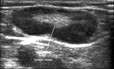

40 Normal Lymph Nodes

41 Typical location of Parathyroid Glands
42 Summary Ultrasound is an anatomic imaging tool, whose utility requires a thorough understanding of neck anatomy Think of structures in relationship to one another to help you understand the three dimensional picture Diagnostic ultrasound of the thyroid requires 13 images at a minimum Memorize the borders of the surgical levels of the neck. Your surgeon will thank you!
Lecture 01. The Thyroid & Parathyroid Glands. By: Dr Farooq Khan PMC Date: 12 th March. 2018
 Lecture 01 The Thyroid & Parathyroid Glands By: Dr Farooq Khan PMC Date: 12 th March. 2018 INTRODUCTION LAYERS OF THE NECK The neck has four major compartments or layer which are enclosed by an outer musculofascial
THYROID & PARATHYROID. By Prof. Saeed Abuel Makarem & Dr. Sanaa Al-Sharawy
 THYROID & PARATHYROID By Prof. Saeed Abuel Makarem & Dr. Sanaa Al-Sharawy 1 OBJECTIVES By the end of the lecture, the student should be able to: Describe the shape, position, relations and structure of
Thyroid and Parathyroid Glands
 Thyroid and Parathyroid Glands Please view our Editing File before studying this lecture to check for any changes. Color Code Important Doctors Notes Notes/ explanation Objectives: By the end of the lecture,
Anatomy of the Thyroid Gland
 Anatomy of the Thyroid Gland Introduction Nomenclature G, thyreos= shield, eidos= like Location Root of the neck ventrally (C5-T1) Function endocrine gland that secretes: Thyroxine (T4) T3 Calcitonin LWW,
AACE/ACE Advanced Endocrine Neck Ultrasound Training Course 2016
 AACE/ACE Advanced Endocrine Neck Ultrasound Training Course 2016 This 9mm left inferior nodule should remind us all why we re here! There is no absolute number of images required for documentation
Ultrasonography of the Neck as an Adjunct to FNA. Nicole Massoll M.D.
 Ultrasonography of the Neck as an Adjunct to FNA Nicole Massoll M.D. Basic Features of Head and Neck Ultrasound and Anatomy Nicole Massoll M.D. University of Arkansas for Medical Sciences, Little Rock
Neck-2. Dr. Heba Kalbouneh Associate Professor of Anatomy and Histology
 Neck-2 ` Dr. Heba Kalbouneh Associate Professor of Anatomy and Histology Triangles of the neck Side of the neck Midline Lower border of mandible Line between angle of mandible and mastoid Superior nuchal
Neck Ultrasound. Faculty Info: Amy Kule, MD
 Neck Ultrasound Date: Friday, October 19, 2018 Time: 11:00 AM Location: SMALL GROUP LABORATORY SSOM L71 Watch: Ø Neck Ultrasound Scanning Protocol (4:00): https://www.youtube.com/watch?v=zozd2x2ll4q Faculty
Thyroid and Parathyroid Ultrasound Protocol
 Thyroid and Parathyroid Ultrasound Protocol Reviewed By: Anna Ellermeier, MD Last Reviewed: December 2017 Contact: (866) 761-4200, Option 1 **NOTE for all examinations: 1. If documenting possible flow
Anterior triangle of neck
 Anterior triangle of neck Dept. of Anatomy Zhou Hong Ying Outline boundary and subdivisions of ant. triangle contents of the triangle Muscles: suprahyoid muscles, infrahyoid muscles Nerves: CNⅩ, CNⅪ, CNⅫ,
The Neck the lower margin of the mandible above the suprasternal notch and the upper border of the clavicle
 The Neck is the region of the body that lies between the lower margin of the mandible above and the suprasternal notch and the upper border of the clavicle below Nerves of the neck Cervical Plexus Is formed
Pre-operative Ultrasound of Lymph Nodes in Thyroid Cancer
 Pre-operative Ultrasound of Lymph Nodes in Thyroid Cancer AACE - Advances in Medical and Surgical Management of Thyroid Cancer - 2018 Robert A. Levine, MD, FACE, ECNU Thyroid Center of New Hampshire Geisel
OBJECTIVE: To obtain a fundamental knowledge of the root of the neck with respect to structure and function
 The root of the neck Jeff Dupree, Ph.D. e mail: jldupree@vcu.edu OBJECTIVE: To obtain a fundamental knowledge of the root of the neck with respect to structure and function READING ASSIGNMENT: Moore and
The Neck. BY: Lina Abdullah & Rahaf Jreisat
 The Neck BY: Lina Abdullah & Rahaf Jreisat Boundaries of the Neck: generally from base of the skull to root of the neck Superior margin :From superior nuchal line of occipital bone up to mastoid process
1. Thyroxine (inactive form) also called T4 (90% of the secretion). 2. Triiodothyronine (active form) also called T3 (10% of the secretion).
 also called T4 (90% of the secretion). 2. Triiodothyronine (active form) also called T3 (10% of the secretion).") A Introduction The nomenclature of the thyroid gland comes from its close relation to the thyroid cartilage (the thyroid cartilage was named like this because thyroid means shield and it is shielding the
A Introduction The nomenclature of the thyroid gland comes from its close relation to the thyroid cartilage (the thyroid cartilage was named like this because thyroid means shield and it is shielding the
Head & Neck Contouring
 Head & Neck Contouring Presented by James Wheeler, MD Center for Cancer Care Goshen, IN 46526 September 12, 2014 Special Thanks to: Spencer Boulter, Director of Operations (AAMD) Adam Moore, RT(T), CMD
Histology. Dr.shatarat
 Histology Dr.shatarat Dr.shatarat Dr.shatarat Cells of the adenohypophysis adenohypophysis Dr.shatarat Dr.shatarat Adenohypophysis high power acidopill basophill Chromophobes Dr.shatarat 1-Chromophils
Clinical Anatomy of the Thyroid and Adrenal Glands
 Clinical Anatomy of the Thyroid and Adrenal Glands Handout download: http://www.oucom.ohiou.edu/dbms-witmer/gs-rpac.htm 28 October 2003 Lawrence M. Witmer, PhD Department of Biomedical Sciences College
Anatomy: head and Neck (6 questions) 1. Prevertebral Flexor Musculature (lying in front of the vertebrae) include all, EXCEPT: Longus Colli.
 1. Prevertebral Flexor Musculature (lying in front of the vertebrae) include all, EXCEPT: Longus Colli.") Anatomy: head and Neck (6 questions) 1. Prevertebral Flexor Musculature (lying in front of the vertebrae) include all, EXCEPT: Longus Colli. Rectus Capitis Anterior. Rectus Capitis Lateralis. Rectus Capitis
Anatomy: head and Neck (6 questions) 1. Prevertebral Flexor Musculature (lying in front of the vertebrae) include all, EXCEPT: Longus Colli. Rectus Capitis Anterior. Rectus Capitis Lateralis. Rectus Capitis
Part Ten: Thyroid / Parathyroid. Chapter 133: Anatomy. Daniel O. Graney, Ronald C. Hamaker. Development of the Thyroid Gland
 Part Ten: Thyroid / Parathyroid Chapter 133: Anatomy Daniel O. Graney, Ronald C. Hamaker Development of the Thyroid Gland The thyroid gland begins as an endodermal bud from the floor of the pharynx between
Thyroid gland. importance. relations and connections. external laryngeal nerves. malformations.
 Thyroid gland 1. Recognize and understand the coverings of the thyroid gland and their clinical importance. 2. Recognize and understand the main parts of the thyroid gland and their locations, relations
Thyroid INTRODUCTION ANATOMY SUMMARY OF CHANGES
 AJC 7/14/06 1:19 PM Page 67 Thyroid C73.9 Thyroid gland SUMMARY OF CHANGES Tumor staging (T) has been revised and the categories redefined. T4 is now divided into T4a and T4b. Nodal staging (N) has been
Ultrasound for Pre-operative Evaluation of Well Differentiated Thyroid Cancer
 Ultrasound for Pre-operative Evaluation of Well Differentiated Thyroid Cancer Its Not Just About the Nodes AACE Advances in Medical and Surgical Management of Thyroid Cancer - 2017 Robert A. Levine, MD,
PCM1 Physical Exam Skills Session: Head and Neck FACILITATOR & STUDENT COPY
 PATIENT CENTERED MEDICINE - 1 GOALS & OUTCOMES: PCM1 Physical Exam Skills Session: Head and Neck FACILITATOR & STUDENT COPY 1. To introduce the applied anatomy relevant for the examination of the head
Thyroidectomy. Siu Kwan Ng. Modified Radical Neck Dissection Type II 47
 06 Thyroidectomy Siu Kwan Ng Modified Radical Neck Dissection Type II 47 Thyroidectomy STEP 1. EXPOSING THE THYROID GLAND The collar incision Figure 1 (curvilinear skin crease incision) is made at 1.5-2
Alexander C Vlantis. Selective Neck Dissection 33
 05 Modified Radical Neck Dissection Type II Alexander C Vlantis Selective Neck Dissection 33 Modified Radical Neck Dissection Type II INCISION Various incisions can be used for a neck dissection. The incision
Ultrasound Physics & Doppler
 Ultrasound Physics & Doppler Endocrine University 2018 Mark Lupo, MD, FACE, ECNU Objectives Review the essential components of ultrasound physics in neck sonography Demonstrate the importance of ultrasound
CERVICAL LYMPH NODES
 CERVICAL LYMPH NODES (ANATOMY & EXAMINATION) Hemant (DTCD 1 st YEAR) 1. Lymphatic Tissues: A Type of connective tissue that contains large numbers of lymphocytes. 2. Lymphatic Vessels: Are Tubes that assist
Shadow because the air
 Thyroid Ultrasound Thyroid US examination needs: 1. high frequency transducer 2. extended patient's neck 3. check all the neck area because the swelling could be in areas other than the thyroid such as
Parathyroid Imaging: Current Concepts. Maria Gule-Monroe, M.D. Nancy Perrier, M.D.
 Parathyroid Imaging: Current Concepts Maria Gule-Monroe, M.D. Nancy Perrier, M.D. Disclosures None Objectives Ultrasound characteristics of parathyroid adenomas vs. lymph nodes 4D-CT evaluation of hyperparathyroidism
Surgical anatomy of thyroid and parathyroid glands
 Head & Neck Surgery Course Surgical anatomy of thyroid and parathyroid glands Dr Pierfrancesco PELLICCIA Pr Benjamin LALLEMANT Service ORL et CMF CHU de Nîmes CH de Arles Thyroid glands Dr Pierfrancesco
Preoperative Evaluation
 Preoperative Evaluation Lateral compartment lymph nodes are easier to detect and are amenable to FNA Central compartment lymph nodes are much more difficult to detect and FNA (Tg washout testing is compromised)
Cervical Lymph Nodes
 Cervical Lymph Nodes Diana Gaitini, MD Unit of Ultrasound, Department of Medical Imaging Rambam Medical Center and Faculty of Medicine Technion, Israel Institute of Technology Haifa, Israel Learning Targets
Thyroid Nodules: US Risk Stratification. Alex Tessnow, MD, FACE, ECNU University of Texas Southwestern Associate Professor of Medicine Dallas, Texas
 Thyroid Nodules: US Risk Stratification Alex Tessnow, MD, FACE, ECNU University of Texas Southwestern Associate Professor of Medicine Dallas, Texas Which of the following is true? A. All echogenic foci
ANTERIOR CERVICAL TRIANGLE (Fig. 2.1 )
") 2 Neck Anatomy ANTERIOR CERVICAL TRIANGLE (Fig. 2.1 ) The boundaries are: Lateral: sternocleidomastoid muscle Superior: inferior border of the mandible Medial: anterior midline of the neck This large triangle
2 Neck Anatomy ANTERIOR CERVICAL TRIANGLE (Fig. 2.1 ) The boundaries are: Lateral: sternocleidomastoid muscle Superior: inferior border of the mandible Medial: anterior midline of the neck This large triangle
For the following questions, indicate the letter that corresponds to the SINGLE MOST APPROPRIATE ANSWER
 GROSS ANATOMY EXAMINATION May 15, 2000 For the following questions, indicate the letter that corresponds to the SINGLE MOST APPROPRIATE ANSWER 1. Pain associated with an infection limited to the middle
University of Palestine. Midterm Exam 2013/2014 Total Grade:
 Course No: DNTS2208 Course Title: Head and Neck Anatomy Date: 09/11/2013 No. of Questions: (50) Time: 1hour Using Calculator (No) University of Palestine Midterm Exam 2013/2014 Total Grade: Instructor
Thyroid Ultrasound for the Endocrine Surgeon: A Valuable Clinical Tool that Enhances Diagnostic and Therapeutic Outcomes
 Thyroid Ultrasound for the Endocrine Surgeon: A Valuable Clinical Tool that Enhances Diagnostic and Therapeutic Outcomes Allan Siperstein MD The Cleveland Clinic Audience Quiz Taken ultrasound course Perform
SURGERY. This article is one of a. of the Head and Neck
 SURGERY of the Head and Neck Mary Sutton, CST, CFA This article is one of a series that will discuss head and neck surgeries from an otolaryngology perspective. Most of these surgeries involve cancer,
Alexander C Vlantis. Total Laryngectomy 57
 07 Total Laryngectomy Alexander C Vlantis Total Laryngectomy 57 Total Laryngectomy STEP 1 INCISION AND POSITION OF STOMA A superiorly based apron flap incision is marked with the horizontal limb placed
Tympanic Bulla Temporal Bone. Digastric Muscle. Masseter Muscle
 Superior view Hyoid Bone The hyoid bone does not articulate with any other bones. It is held in place by ligaments to the styloid process of the temporal bone and the thyroid cartilage of the larynx. It
Larynx. Rudimentary. Behind the posterior surface : -stylopharyngeus - salpingopharyngeus -platopharyngeus
 Larynx The larynx is an organ that provides a protective sphincter at the inlet of the air passages and is responsible for voice production. It extends from C3-C6: *Posterior: the pharynx *Lateral: the
Ultrasound Interpretation of Non-Thyroid Neck Pathology
 Ultrasound Interpretation of Non-Thyroid Neck Pathology Kevin T. Brumund, M.D., F.A.C.S. Associate Professor of Surgery Head and Neck Surgery University of California, San Diego Health Sciences VA Medical
AACE/ACE Principles of Endocrine Neck Sonography Course
 AACE/ACE Principles of Endocrine Neck Sonography Course Primary objective of thyroid ultrasound: assess for malignant disease Nodular Disease Benign Malignant Goiter Iodine deficient Thyroiditis Organification
Posterior Triangle of the Neck By Prof. Dr. Muhammad Imran Qureshi
 Posterior Triangle of the Neck By Prof. Dr. Muhammad Imran Qureshi For the purpose of anatomical description the neck is sub divided into two major triangles, the Anterior and the Posterior by muscle bellies
REVIEW/PREVIEW OF HEAD AND NECK ANATOMY FOR ENT EXAM
 REVIEW/PREVIEW OF HEAD AND NECK ANATOMY FOR ENT EXAM - 2017 PALPATE CAROTID ARTERY: AT LEVEL OF CAROTID BIFURCATION VERTEBRAL LEVEL C4 Sternocleidomastoid Muscle INTERNAL CAROTID EXTERNAL CAROTID COMMON
Lies in front and sides of the neck. Consists of two lobe connected anterior to the trachea by an isthmus.
 THYROID GLAND 1 Lies in front and sides of the neck. Consists of two lobe connected anterior to the trachea by an isthmus. A small pyramidal lobe projects upwards from the left lobe in 40% of cases. The
Veins of the Face and the Neck
 Veins of the Face and the Neck Facial Vein The facial vein is formed at the medial angle of the eye by the union of the supraorbital and supratrochlear veins. connected through the ophthalmic veins with
Lecture 07. Lymphatic's of Head & Neck. By: Dr Farooq Amanullah Khan PMC
 Lecture 07 Lymphatic's of Head & Neck By: Dr Farooq Amanullah Khan PMC Dated: 28.11.2017 Lymphatic Vessels Of the 800 lymph nodes in the human body, 300 are in the Head & neck region. The lymphatic vessels
Title. CitationInternational Cancer Conference Journal, 4(1): Issue Date Doc URL. Rights. Type. File Information
: Issue Date Doc URL. Rights. Type. File Information") Title Lymph node metastasis in the suprasternal space from Homma, Akihiro; Hatakeyama, Hiromitsu; Mizumachi, Ta Author(s) Tomohiro; Fukuda, Satoshi CitationInternational Cancer Conference Journal, 4(1):
Title Lymph node metastasis in the suprasternal space from Homma, Akihiro; Hatakeyama, Hiromitsu; Mizumachi, Ta Author(s) Tomohiro; Fukuda, Satoshi CitationInternational Cancer Conference Journal, 4(1):
DEVELOPMENT & STRUCTURE OF THYROID GLAND DR TATHEER ZAHRA ASSISTANT PROFESSOR ANATOMY
 DEVELOPMENT & STRUCTURE OF THYROID GLAND DR TATHEER ZAHRA ASSISTANT PROFESSOR ANATOMY DEVELOPMENT OF THYROID Concept of pharyngeal arch 3 rd week 4 th week Adults 7 th week HISTOGENESIS OF THYROID GLAND
Anatomical Complications in General Surgery. John E. Skandalakis, Stephen W. Gray, Joseph S. Rowe. Chapter 1: The Neck
 Anatomical Complications in General Surgery John E. Skandalakis, Stephen W. Gray, Joseph S. Rowe Chapter 1: The Neck The human neck is so designed that the swelling of a normal structure or the presence
AACE 2018 Advanced Endocrine Neck Ultrasound and UGFNA Course
 AACE 2018 Advanced Endocrine Neck Ultrasound and UGFNA Course Describe the sonographic appearance of diffuse thyroid diseases: autoimmune thyroid disease Review non thyroidal findings that can be encountered
Over the past 18 years, numerous classifications have been proposed to distinguish
 ORGNAL ARTCLE An maging-based Classification for the Cervical Nodes Designed as an Adjunct to Recent Clinically Based Nodal Classifications Peter M. Som, MD; Hugh D. Curtin, MD; Anthony A. Mancuso, MD
Anatomy. Contents Brain (Questions)
") Anatomy 12 Contents 12.1 Brain (Questions).................................................... 683 12.2 Head and Neck (Questions)............................................. 685 12.3 Thorax (Questions)...................................................
Anatomy 12 Contents 12.1 Brain (Questions).................................................... 683 12.2 Head and Neck (Questions)............................................. 685 12.3 Thorax (Questions)...................................................
THE ANGULAR TRACT: AN ANATOMICAL
 British Journal of Oral Surgery (1981) 19, 116-120 0 The British Association of Oral Surgeons 0007-117X/81/00170116$02.00 THE ANGULAR TRACT: AN ANATOMICAL OF SURGICAL SIGNIFICANCE STRUCTURE HAITHEM A.
Since the first description of the radical neck dissection by George Crile almost a century
 ORIGINAL ARTICLE Neck Dissection Classification Update Revisions Proposed by the American Head and Neck Society and the American Academy of Otolaryngology Head and Neck Surgery K. Thomas Robbins, MD; Garry
Chapter 28: The neck. Fascia of the neck
 Chapter 28: The neck Fascia of the neck The superficial fascia is a fatty areolar layer between the skin and the more obvious deep fascia. It contains the platysma muscles and the external jugular veins
SCHOOL OF ANATOMICAL SCIENCES Mock Run Questions. 4 May 2012
 SCHOOL OF ANATOMICAL SCIENCES Mock Run Questions 4 May 2012 1. With regard to the muscles of the neck: a. the platysma muscle is supplied by the accessory nerve. b. the stylohyoid muscle is supplied by
42 yr old male with h/o Graves disease and prior I 131 treatment presents with hyperthyroidism and undetectable TSH. 2 hr uptake 20%, 24 hr uptake 50%
 Pinhole images of the neck are acquired in multiple projections, 24hrs after the oral administration of approximately 200 µci of I123. Usually, 24hr uptake value if also calculated (normal 24 hr uptake
PEDIATRICS WK 3 HEAD AND NECK ALISON WALLACE MD, PHD
 PEDIATRICS WK 3 HEAD AND NECK ALISON WALLACE MD, PHD Topics 1. Cervical lymphadenopathy 2. Lymphatic malformation 3. Thyroglossal duct cysts 4. Branchial cleft cysts 5. Thyroid masses CASE 1 Case 1 A 2
10/14/2018 Dr. Shatarat
 2018 Objectives To discuss mediastina and its boundaries To discuss and explain the contents of the superior mediastinum To describe the great veins of the superior mediastinum To describe the Arch of
Ovid: Oxford Textbook of Endocrinology & Diabetes
 Página 1 de 6 Copyright 2002 Oxford University Press Wass, John A.H., Shalet, Stephen M., Gale, Edwin, Amiel, Stephanie A. Oxford Textbook of Endocrinology & Diabetes, 1st Edition Surgical procedure Part
Anatomy and Physiology II. Spine
 Anatomy and Physiology II Spine Bones and Other Structures Vertibrae Contains Cervical, Thoracic, Lumbar, Sacral and Coccygeal regions We use Capital letters to refer to these (C, T, L, S, and Co) and
Clinical Anatomy of the Endocrine System HYPOPTHALAMUS; HYPOPHYSIS; PINEAL GLAND
 STUDY COMPONENT Clinical Anatomy of the Endocrine System UNIT THEME 1: UNIT THEME 2: UNIT THEME 3: UNIT THEME 4: HYPOPTHALAMUS; HYPOPHYSIS; PINEAL GLAND THYROID AND PARATHYROID PANCREAS; ADRENAL GLANDS
University of Palestine. Midterm Exam 2013/2014 Total Grade:
 [ Course No: DNTS2208 Course Title: Head and Neck Anatomy Date: 17/11/1024 No. of Questions: (52) Time: 2hours Using Calculator (No) University of Palestine Midterm Exam 2013/2014 Total Grade: Instructor
Compartmentalization of the larynx Sites and subsites Supraglottis Glottis subglottis Spaces Pre-epiglottic epiglottic space Para-glottic space
 Stroboscopy Rounds February 8, 2008 C. Matt Stewart, M.D.,Ph.D. Compartmentalization of the larynx Sites and subsites Supraglottis Glottis subglottis Spaces Pre-epiglottic epiglottic space Para-glottic
Surgical Anatomy of the Neck. M. J. Jurkiewicz, John Bostwick. Surgical Clinics of North America, Vol 54, No 6, December 1974.
 Surgical Anatomy of the Neck M. J. Jurkiewicz, John Bostwick Surgical Clinics of North America, Vol 54, No 6, December 1974. The radical neck dissection is a safe, effective therapeutic procedure for eradication
Tikrit University collage of dentistry Dr.Ban I.S. head & neck anatomy 2 nd y. Lec [5] / Temporal fossa :
![Tikrit University collage of dentistry Dr.Ban I.S. head & neck anatomy 2 nd y. Lec [5] / Temporal fossa :](/thumbs/88/115294566.jpg "Tikrit University collage of dentistry Dr.Ban I.S. head & neck anatomy 2 nd y. Lec [5] / Temporal fossa :") Lec [5] / Temporal fossa : Borders of the Temporal Fossa: Superior: Superior temporal line. Inferior: gap between zygomatic arch and infratemporal crest of sphenoid bone. Anterior: Frontal process of the
Lec [5] / Temporal fossa : Borders of the Temporal Fossa: Superior: Superior temporal line. Inferior: gap between zygomatic arch and infratemporal crest of sphenoid bone. Anterior: Frontal process of the
Ultrasonographic identification of the anatomical landmarks that define cervical lymph nodes spaces
 Review Med Ultrason 2013, Vol. 15, no. 1, 29-34 DOI: 10.11152/mu.2013.2066.151.lml1uia2 Ultrasonographic identification of the anatomical landmarks that define cervical lymph nodes spaces Lavinia Manuela
General Anatomic Layout
 General Anatomic Layout 2 Core Messages At the start of the dissection exercise, we must take a panoramic look for orientation. We then establish the limits of the area of operation and the main landmarks.
Dr. Weyrich G07: Superior and Posterior Mediastina. Reading: 1. Gray s Anatomy for Students, chapter 3
 Dr. Weyrich G07: Superior and Posterior Mediastina Reading: 1. Gray s Anatomy for Students, chapter 3 Objectives: 1. Subdivisions of mediastinum 2. Structures in Superior mediastinum 3. Structures in Posterior
Bones of the Skull Lateral View
 Bones of the Skull Lateral View Frontal Bone Parietal Bone Occipital Bone Temporal Bone Sphenoid Bone Pterion Sutures of the Skull Lateral View Coronal Suture Lambdoid Suture Squamous Suture Sutures of
CHAPTER 9. The Neck THE "CERVICAL CAVITY" TRACHEA ESOPHAGUS THYROID GLAND PARATHYROID GLANDS
 281 CHAPTER 9 The Neck CERVICAL VERTEBRAE POSTERIOR TRIANGLE OF NECK Trapezius and Sternocleidomastoid Trapezius CLINICAL CONSIDERATIONS REGARDING TRAPEZIUS Sternocleidomastoid CLINICAL CONSIDERATIONS
Uncommon ultrasound findings in the cervical region
 Uncommon ultrasound findings in the cervical region Poster No.: C-1132 Congress: ECR 2014 Type: Educational Exhibit Authors: V. Donciu, S. M. Petrescu, E. NEGUT, C. ZAHARIA ; 1 1 1 2 1 2 Bucharest/RO,
Management of unknown primary with neck node metastasis: Current evidence
 Management of unknown primary with neck node metastasis: Current evidence Dr. Pooja Nandwani Patel Associate Professor Dept. of Radiation Oncology GCRI, Ahmedabad Introduction- Approach to Topic What is
Contents. Basic Ultrasound Principles and Terminology. Ultrasound Nodule Characteristics
 Contents Basic Ultrasound Principles and Terminology Basic Ultrasound Principles... 1 Ultrasound System... 2 Linear Transducer for Superficial Images and Ultrasound-Guided FNA... 3 Scanning Planes... 4
Salivary ultrasound. Dr T J Beale Royal National Throat Nose & Ear and UCLH Hospitals London UK
 Salivary ultrasound Dr T J Beale Royal National Throat Nose & Ear and UCLH Hospitals London UK Two main groups of patients with presenting symptoms of: Obstructive or chronic inflammatory symptoms (salivary
DESCRIPTION: This is the part of the trunk, which is located between the root of the neck and the superior border of the abdominal region.
 1 THE THORACIC REGION DESCRIPTION: This is the part of the trunk, which is located between the root of the neck and the superior border of the abdominal region. SHAPE : T It has the shape of a truncated
Spinal nerves and cervical plexus Prof. Abdulameer Al Nuaimi. E mail: a.al E. mail:
 Spinal nerves and cervical plexus Prof. Abdulameer Al Nuaimi E mail: a.al nuaimi@sheffield.ac.uk E. mail: abdulameerh@yahoo.com Branches of ophthalmic artery Muscles of face A spinal nerve Spinal
REVIEW OF CLINICAL EMBRYOLOGY OF HEAD AND NECK
 REVIEW OF CLINICAL EMBRYOLOGY OF HEAD AND NECK OUTLINE - EMBRYOLOGY UNDERLYING CLINICAL CONDITIONS I. EARLY DEVELOPMENT OF FACE: CLEFT LIP, CLEFT PALATE, OBSTRUCTED NASOLACRIMAL DUCT II. BRANCHIAL ARCHES
Mediastinum and pericardium
 Mediastinum and pericardium Prof. Abdulameer Al-Nuaimi E-mail: a.al-nuaimi@sheffield.ac.uk E. mail: abdulameerh@yahoo.com The mediastinum: is the central compartment of the thoracic cavity surrounded by
The Pharynx. Dr. Nabil Khouri MD. MSc, Ph.D
 The Pharynx Dr. Nabil Khouri MD. MSc, Ph.D Introduction The pharynx is the Musculo-fascial halfcylinder that links the oral and nasal cavities in the head to the larynx and esophagus in the neck Common
HEAD & NECK ANATOMY - MCQ HEAD & NECK ANATOMY
 . ' HEAD & NECK ANATOMY I. Deep investing layer of cervical fascia splits to enclose: A. Sternocleidomastoid B. Trapezius C. Parotid gland D. Omohyoid 2. Regarding the prevertebral fascia, the following
The following images were all acquired using a CTI Biograph
 Positron Emission Tomography/ Computed Tomography Imaging of Head and Neck Tumors: An Atlas Michael M. Graham, MD, PhD, and Yusuf Menda, MD Department of Radiology, University of Iowa, Iowa City, IA. Address
Neck of Condylar. Process. Anterior Border of Ramus. Mandibular. Foramen. Posterior Border of Ramus Incisive Fossa.
 Learning Outcomes The Mandible Surface Anatomy Muscle Attachments The (FOM) Muscles of the FOM The Tongue Muscles of the Tongue The Submandibular Region Submandibular Gland Sublingual Gland Lingual The
Difficult Problems in Thyroid Surgery
 Difficult Problems in Thyroid Surgery T he successful diagnosis and treatment of thyroid disease is based on a thorough understanding of the normal anatomic and physiologic features of the thyroid gland.
Ultrasonographic anatomy of head and neck a pictorial for the ENT specialist
 Continuing education Med Ultrason 2015, Vol. 17, no. 1, 104-108 DOI: 10.11152/mu.2013.2066.171.aco Ultrasonographic anatomy of head and neck a pictorial for the ENT specialist Adrian Costache 1, Mihai
Small Incision Thyroid Surgery -Vascular Compartment Surgery
 IOSR Journal of Dental and Medical Sciences (IOSR-JDMS) e-issn: 2279-0853, p-issn: 2279-0861.Volume 17, Issue 1 Ver. 13 January. (2018), PP 60-64 www.iosrjournals.org Small Incision Thyroid Surgery -Vascular
A Pathologist s Guide to Neck Dissection. Neck Dissections. Lymphatics of head and neck. Neck Dissections
 A Pathologist s Guide to Neck Dissection North American Society for Head and Neck Pathology Companion Meeting 2006 Sigrid Wayne, M.D. Department of Pathology University of Iowa The presence of cervical
THE NECK PART I BONES AND JOINTS OF THE NECK MUSCLES OF THE NECK SCM CAROTID TRIANGLE ENDOCRINE LAYER OF THE CERVICAL VISCERA
 THE NECK PART I BONES AND JOINTS OF THE NECK MUSCLES OF THE NECK SCM CAROTID TRIANGLE ENDOCRINE LAYER OF THE CERVICAL VISCERA THE NECK THE NECK The neck has four major compartments, which are enclosed
A Rare Case of Bilateral Jugular Venous Malformation
 JOURNAL OF CASE REPORTS 2013;3(2):326-330 A Rare Case of Bilateral Jugular Venous Malformation Prasanna LC, Alva R, D Souza AS, Bhat KMR Department of Anatomy, Kasturba Medical College, Manipal University,
A220: Larynx cancer tissues. (formalin fixed)
") A220: Larynx cancer tissues (formalin fixed) For research use only Specifications: No. of cases: 45 Tissue type: Larynx cancer tissues No. of spots: 2 spots from each cancer case (90 spots) 4 non-neoplastic
A220: Larynx cancer tissues (formalin fixed) For research use only Specifications: No. of cases: 45 Tissue type: Larynx cancer tissues No. of spots: 2 spots from each cancer case (90 spots) 4 non-neoplastic
12 Larynx. I - Cartilages. Learning Objectives
 12 Larynx Learning Objectives By the end of this topic you should be able to: Identify the cartilages, membranes, muscles and nerves of the larynx. Describe the attachments of the larynx to other structures
2017 ATA Victoria Advanced Thyroid US
 2017 ATA Victoria Advanced Thyroid US DIFFUSE THYROID CONDITIONS Stephanie L. Lee, M.D., Ph.D. Director of the BMC Thyroid Nodule and Cancer Center Section of Endocrinology, Diabetes and Nutrition Boston
Prevertebral Region, Pharynx and Soft Palate
 Unit 20: Prevertebral Region, Pharynx and Soft Palate Dissection Instructions: Step1 Step 2 Step 1: Insert your fingers posterior to the sternocleidomastoid muscle, vagus nerve, internal jugular vein,
Thyroid Nodules: US Risk Stratification and FNA Guidelines
 Thyroid Nodules: US Risk Stratification and FNA Guidelines Mark A. Lupo, MD, FACE, ECNU Thyroid & Endocrine Center of Florida Assistant Clinical Professor of Medicine Florida State University, College
Scrotum Kacey Morrison Amanda Baxter Sabrina Tucker July 18, 2006 SCROTUM
 Scrotum Kacey Morrison Amanda Baxter Sabrina Tucker July 18, 2006 SCROTUM 1) Other Names: Scrotum None Testicles Testes (Curry Tempkin, p. 236, 2/3/2) Ductus deferens spermatic cord (Tempkin, p. 279, Anatomy
Anatomy and Physiology. Bones, Sutures, Teeth, Processes and Foramina of the Human Skull
 Anatomy and Physiology Chapter 6 DRO Bones, Sutures, Teeth, Processes and Foramina of the Human Skull Name: Period: Bones of the Human Skull Bones of the Cranium: Frontal bone: forms the forehead and the