What is Ultrasound? What is Ultrasound? B A. Basic Principles of Ultrasound. Basic Principles of Ultrasound. Basic Principles of Ultrasound
|
|
|
- Milton Lee
- 6 years ago
- Views:
Transcription


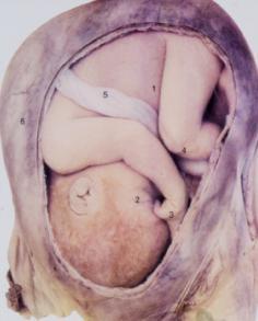
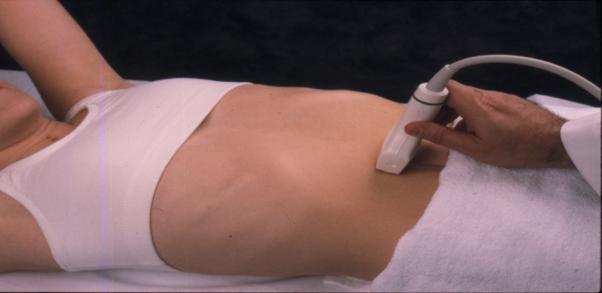
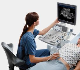
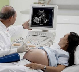
1 Introduction to Ultrasound Principles Mani Montazemi, RDMS Baylor College of Medicine Division of Maternal-Fetal Medicine Department of Obstetrics and Gynecology Manager, Maternal Fetal Center Imaging Texas Children s Hospital, Pavilion for Women Houston Texas & Clinical Instructor Thomas Jefferson University Hospital Philadelphia, Pennsylvania B A D Basic Principles of Ultrasound Ultrasound diagnostically is used in 2 ways Basic Principles of Ultrasound Ultrasound diagnostically is used in 2 ways Anatomic information Basic Principles of Ultrasound Ultrasound diagnostically is used in 2 ways Anatomic information Blood flow information What is Ultrasound? What is Ultrasound? 1


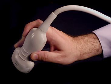
2 Basic Principles of Ultrasound What is sound Sound is a form of energy Basic Principles of Ultrasound What is sound Sound is a form of energy What is energy Energy is the capacity to do work Moving an object, heating the room, lighting electricity Basic Principles of Ultrasound Sound is mechanical energy transmitted by pressure waves in a material medium Sound is not electromagnetic Matter must be present for sound to travel How Does an Ultrasound Instrument Work? Pulsed-echo Principle The energy within a pulsed-echo system is electrical, but the energy in the patient s body is sound, which is mechanical What is a Transducer? Converts one form of energy into another form 2


3 Video Clip - Transducer Transducer Frequency MHz 3.5 MHz 5-6 MHz MHz 9.0 MHz MHz Transducer Configurations Curved linear Straight linear Conventional sector Microsector Endocavity Intraoperative Image Configurations Linear Curved Sector Transducer Configurations Mechanical Phased array Transducer Configurations Mechanical moving parts Wobbler or rotating Single or multi-element Mechanical beam steering & focusing Motor driven/fluid filled scan head FOV is sector Single or multi-frequency 3


 Resolution: What is it?")
 One Million cycles / second")
4 Transducer Configurations Phased array no moving parts Flat linear Curved linear Sector Curved Linear Array Transducer Multi-element Elements arranged in an arc and are sequentially pulsed FOV is trapezoidal-sector Produce larger FOV Benefits: High quality imaging Faster frame rate Electronic focusing Video Clip - Transducer Categories of Sound Infra Sound Audible Sound below 20 Hz 20-20,000 Hz Ultrasound Above 20,000 Hz Sound is categorized according to the frequency of vibration per seconds Frequencies Used For Medical Diagnostic Ultrasound Are Normally Above 1 MHz One cycle / second = one Hertz (Hz) Resolution: What is it? Clarity & sharpness of an ultrasound image The capability of clearly distinguishing two points located close to each other on the sonogram One thousand cycles / second = one Kilohertz (khz) One Million cycles / second = one Megahertz 4


5 Video Clip Safety Safety - Prudent Use Minimize risk Minimize exposure Medical indication only Minimize exposure time Minimize exposure output Ultrasound Safety Prudent use dictates that the principle of ALARA be observed and the ultrasound power levels be maintained As Low As Reasonably Achievable, in order to provide the maximum benefit / risk ratio Output Display Values Power Thermal Index heat Ratio of the in-situ power to the acoustic power required to raise tissue temprature by 1 o C Mechanical Index cavitation Acoustic output in terms of the likelihood of tissue cavitation 5


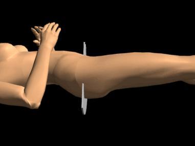
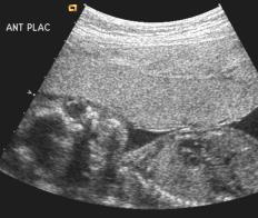
6 Caution Image Orientation & Scanning Planes Longitudinal Transverse Oblique Scanning Planes Sagittal Scan Longitudinal, vertical planes Divide the body into left & right Sagittal Scan Sagittal Scan H A P F 6


7 Sagittal Scan Sagittal Scan Sagittal Scan Sagittal Scan Anterior Maternal Head Maternal Feed Posterior Sagittal Scan Sagittal Scan 7


8 Anterior Sagittal Scan H F Posterior Anterior H F Posterior Transverse Scan Horizontal, transaxial planes Divide the body into superior & inferior 8
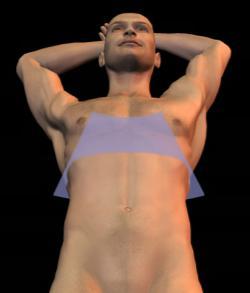
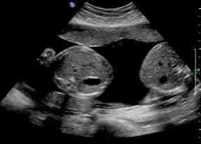
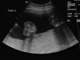
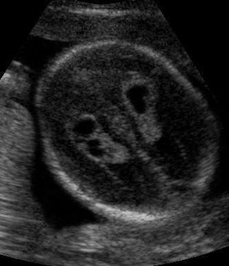
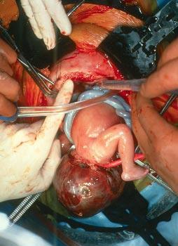
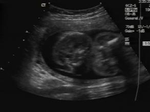
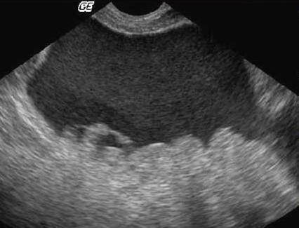
9 Transverse Scan Transverse Scan R A P L Transverse Scan Transabdominal Transverse Transverse Scan Transverse Scan Twin Pregnancy 9


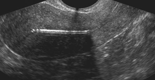
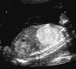
10 Oblique Scan Oblique Scan Oblique planes Inclined from the standard planes In any direction Artifacts Posterior Enhancement Acoustical Shadowing Attenuation Reverberations Refraction Posterior Enhancement Increased intensity in echoes from reflectors behind a structure that weakly attenuates sound Classical feature of fluid Posterior Enhancement Posterior Enhancement UT OV 10


11 Acoustical Shadowing Acoustical Shadowing Marked decrease in intensity of echoes from reflectors that lie behind a structure that is strongly reflecting or attenuating sound Complete lack of acoustical & anatomical information in the area of shadowing Acoustical Shadowing Acoustical Shadowing Head Acoustical Shadowing Acoustical Shadowing 11


12 Acoustical Shadowing Attenuation Reduction of the sound beam s amplitude and intensity as it travels through a medium Attenuation Attenuation Normal Attenuation Reverberations When 2 or more reflectors in the sound path cause multiple, repetitive artifactual echoes 12


13 Reverberations Reverberations Video Clip Artifacts Cervix US Appearance Artifacts Posterior Enhancement Acoustical Shadowing Attenuation Reverberations Refraction Echogenicity Intensity of echoes reflected by tissues or structures from inside the body 13


14 Echogenicity Liquids Solids Vessels & Ducts Soft Tissue Bone Gallbladder Fibrous Cysts Gall Stone Urinary Bladder Tumors Kidney Stone Cysts Polyps & Lesions Calcific Plaque Echogenicity of a structure is described relative to surrounding or adjacent tissue Echogenicity Anechoic Anechoic Hypoechoic Hyperechoic Sonolucent No internal echoes Anechoic Anechoic 14
15 Anechoic Anechoic Hyperechoic Hyperechoic High intensity echoes Increased echogenicity Echogenic Hyperechoic Hyperechoic 15


16 Hypoechoic Hypoechoic Low intensity echoes Decreased echogenicity Clotted Blood Hemoperitoneum Homogeneous Heterogeneous Echotexture Homogeneous Homogeneous Uniform echoes Fine, smooth texture 16


17 Homogeneous Heterogeneous Non-uniform echoes Irregular texture Heterogeneous Heterogeneous Normal Normal Cystic Solid Complex Types of Masses Cystic Masses Anechoic Fluid filled Smooth, well defined margins Posterior enhancement 17


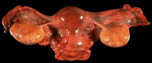
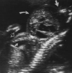
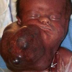
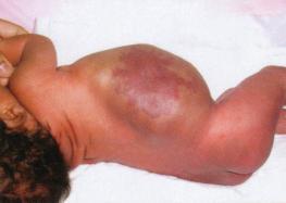
18 Cystic Masses Cystic Masses Cephalocele Solid Masses Solid Masses Contains varying amounts of internal echoes Regular or irregular margins Poorly defined back wall Posterior attenuation of sound Distal acoustic shadow Uterine Leiomyoma US Findings Discrete solid mass single or multiple Uterine Leiomyoma US Findings Variable echogenecity hypoechoic, hyperechoic 18


19 Uterine Leiomyoma US Findings Diagnostic Challenge Solid Masses Complex Masses Predominantly Cystic Mostly cystic Posterior enhancement Internal echoes Thin or thick septations Thin or thick walls Hemorrhagic Cyst Mural Nodule vs. Clot Lace-like pattern of internal echoes 19


20 Mural Nodule vs. Clot Complex Masses Normal Sacrococcygeal Teratoma Complex Masses Complex Masses Cervical Teratoma Complex Masses Complex Masses Predominantly Solid Mostly echogenic Internal cystic changes Posterior attenuation Cystic Lymphangioma 20

 Posterior")


21 Complex Masses Describing A Mass Size Shape, borders, wall Acoustic properties (cystic, solid, complex) Posterior enhancement or attenuation Location Normal Hepatoblastoma How Would You Describe This Mass? How Would You Describe This Mass? In the right adnexa TRV Pelvis How Would You Describe This Mass? How Would You Describe This Mass? In the right adnexa there is a round, well marginated thin walled mass with a sharply defined back wall, In the right adnexa there is a round, well marginated thin walled mass with a sharply defined back wall, thin internal septations, 21


22 How Would You Describe This Mass? How Would You Describe This Mass? In the right adnexa there is a round, well marginated thin walled mass with a sharply defined back wall, thin internal septations, low intensity echoes, In the right adnexa there is a round, well marginated thin walled mass with a sharply defined back wall, thin internal septations, low intensity echoes, and excellent posterior enhancement. How Would You Describe This Mass? Basic System Overview In the right adnexa there is a round, well marginated thin walled mass with a sharply defined back wall, thin internal septations, low intensity echoes, and excellent posterior enhancement. The features are those of a complex, predominantly cystic mass. Patient Data Entry Patient Data Entry 22


23 Transducer / Exam Selection Transducer / Exam Selection Transducer Selection Probe Handling Resolution vs. Penetration Right frequency for penetration Highest frequency for resolution? Probe Handling Sagittal Probe Handling Sagittal Incorrect Incorrect 23


24 Probe Handling Sagittal Probe Handling Sagittal Incorrect Correct Probe Handling Sagittal Probe Handling Transverse Incorrect Probe Handling Transverse Probe Handling Transverse Incorrect Correct 24


25 Probe Handling Don t Probe Handling Basic Controls Freeze Overall Gain TGC Depth Focus Overall Gain Overall Gain - Increase An increase or decrease of the gain will change the brightness of the image Affects entire field of view 25


26 Overall Gain - Increase Overall Gain - Increase Overall Gain - Decrease Overall Gain - Decrease Overall Gain - Decrease Overall Gain 26


27 Time Gain Compensation These slide pots controls amplification of returning echo signals at specific depths The TGC compensates for loss in signal strength as the ultrasound beam passes through an organ Affects specific portions of the field of view Time Gain Compensation Near Field Mid Field Far Field Time Gain Compensation Time Gain Compensation Brighter Darker Time Gain Compensation Time Gain Compensation Brighter 27


28 Time Gain Compensation Time Gain Compensation Darker The diagnosis is only As good as the information your image provides Time Gain Compensation What Constitute a Good Image? What Constitute a Good Image? 28


29 Controls the depth of field of view Depth Depth When the image depth is changed, the field of view increases or decreases Too Small Depth Just Right Too Big Focus Transmit Zone Highlights area of interest Place at or slightly below area of interest Focus Region of minimum beam width in a focused ultrasound beam 29


30 Focus Focus Don t Get Fooled By Poor Technique Pay Attention to Focal Zone Ancillary Functions Cine Loop Stores and displays images with no loss of quality M-Mode Useful in evaluating motion and velocity of moving structures Fetal Heart Motion 30


![[Single/A/B] soft key](/docs-images/77/75954318/images/31-2.jpg "if needed OB")


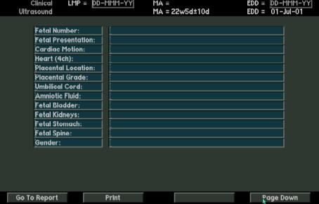
31 OB Calculations Sac Diameter Highlight and Select OB Calc OB Calculations Select [Single/A/B] soft key if needed OB Calculations Highlight and select Biometry or AFI Measurements Calipers, Add Caliper Individual measurements Trace, Ellipse Measures area and circumference OB Worksheet Edit or Delete Measurements Enter Previous Exam Data Enter Comments 31


32 OB Worksheet OB Worksheet OB Worksheet Thank You 32
ULTRASOUND. OB/Gyn (Core) Ultrasound PIEZOELECTRIC EFFECT. Principles of Ultrasound Physics and Instrumentation. Nathan Pinkney, BS, CDOS
 Ultrasound PIEZOELECTRIC EFFECT. Principles of Ultrasound Physics and Instrumentation. Nathan Pinkney, BS, CDOS") 1 OB/Gyn (Core) Ultrasound Principles of Ultrasound Physics and Instrumentation Nathan Pinkney, BS, CDOS Philadelphia College of Osteopathic Medicine 2016 ULTRASOUND CATEGORIES OF SOUND INFRASOUND = below
1 OB/Gyn (Core) Ultrasound Principles of Ultrasound Physics and Instrumentation Nathan Pinkney, BS, CDOS Philadelphia College of Osteopathic Medicine 2016 ULTRASOUND CATEGORIES OF SOUND INFRASOUND = below
Physical Principles of Ultrasound
 Physical Principles of Ultrasound Grateful appreciation to Richard A. Lopchinsky, MD, FACS and Nancy H. Van Name, RDMS, RTR, and MarleneKattaron, RDMS 2000 UIC All Rights Reserved. Course Objectives Identify
ULTRASOUND NOMENCLATURE
 Chapter 1: Ultrasound Nomenclature, Image Orientation, and Basic Instrumentation CYNTHIA SIKOWSKI Ultrasound waves are sound waves that have a frequency exceeding 20,000 Hz. When sound waves are transmitted
Principles of Ultrasound. Cara C. Prideaux, M.D. University of Utah PM&R Sports Medicine Fellow March 14, 2012
 Principles of Ultrasound Cara C. Prideaux, M.D. University of Utah PM&R Sports Medicine Fellow March 14, 2012 None Disclosures Outline Introduction Benefits and Limitations of US Ultrasound (US) Physics
Preamble (disclaimer)
") Preamble (disclaimer) PHYSICS AND PRINCIPLES OF HEAD/NECK ULTRASOUND Joseph C. Sniezek, MD FACS LTC, MC, USA Otolaryngology/H&N Surgery Tripler Army Medical Center 1. I am not a physicist 2. ACS has recommended
Preamble (disclaimer) PHYSICS AND PRINCIPLES OF HEAD/NECK ULTRASOUND Joseph C. Sniezek, MD FACS LTC, MC, USA Otolaryngology/H&N Surgery Tripler Army Medical Center 1. I am not a physicist 2. ACS has recommended
The Physics of Ultrasound. The Physics of Ultrasound. Claus G. Roehrborn. Professor and Chairman. Ultrasound Physics
 The Physics of Ultrasound Pipe Organ 10-8000 Emission Dog 452-1080 Man 85-1100 Spectrum Bat 10,000-120,000 Porpoise 7000-120,000 Claus G. Roehrborn Professor and Chairman 10 20 Cycles per second Reception
Ultrasound Physics & Terminology
 Ultrasound Physics & Terminology This module includes the following: Basic physics terms Basic principles of ultrasound Ultrasound terminology and terms Common artifacts seen Doppler principles Terms for
Ultrasound Principles cycle Frequency Wavelength Period Velocity
 ! Teresa S. Wu, MD, FACEP Director, EM Ultrasound Program & Fellowship Co-Director, Simulation Based Training Program & Fellowship Associate Program Director, EM Residency Program Maricopa Medical Center
Basic of Ultrasound Physics E FAST & Renal Examination. Dr Muhammad Umer Ihsan MBBS,MD, DCH CCPU,DDU1,FACEM
 Basic of Ultrasound Physics E FAST & Renal Examination Dr Muhammad Umer Ihsan MBBS,MD, DCH CCPU,DDU1,FACEM What is Sound? Sound is Mechanical pressure waves What is Ultrasound? Ultrasounds are sound waves
Chapter 3. Sonographic Image Interpretation
 Chapter 3 Sonographic Image Interpretation Sonograms are two-dimensional gray-scale images that allow assessment and diagnosis of many anatomic and pathologic changes that can occur in the human body.
Basic Physics of Ultrasound and Knobology
 WELCOME TO UTMB Basic Physics of Ultrasound and Knobology By Daneshvari Solanki, FRCA Laura B. McDaniel Distinguished Professor Anesthesiology and Pain Medicine University of Texas Medical Branch Galveston,
Terminology Tissue Appearance
 By Marc Nielsen, MD Advantages/Disadvantages Generation of Image Ultrasound Machine/Transducer selection Modes of Ultrasound Terminology Tissue Appearance Scanning Technique Real-time Portable No ionizing
Lesson 07: Ultrasound Transducers. This lesson contains 73 slides plus 16 multiple-choice questions.
 Lesson 07: Ultrasound Transducers This lesson contains 73 slides plus 16 multiple-choice questions. This lesson was derived from pages 33 through 42 in the textbook: Ultrasound Transducers Ultrasound Transducers
CONTENTS. Test Number cpd Tanya Reynolds (Nat. Dip. Diag. Rad., B. Tech. Diag. Rad., B. Tech. Ultrasound)
") CONTENTS page 1-15 page 16 BASIC 2-DIMENSIONAL ULTRASOUND PRINCIPLES Multiple Choice Test Test Number cpd 41640 Tanya Reynolds (Nat. Dip. Diag. Rad., B. Tech. Diag. Rad., B. Tech. Ultrasound) Tanya is
CONTENTS page 1-15 page 16 BASIC 2-DIMENSIONAL ULTRASOUND PRINCIPLES Multiple Choice Test Test Number cpd 41640 Tanya Reynolds (Nat. Dip. Diag. Rad., B. Tech. Diag. Rad., B. Tech. Ultrasound) Tanya is
1 Fundamentals. Basic Definitions and Physics Principles. Fundamentals
 1 To become versed in the language of ultrasonography, it is necessary to review some of the basic principles of physics. The wave physics principles of ordinary (i.e., audible) sound apply to ultrasound
Basic Training Programme. 16 Februrary 2018, ROTTERDAM. Pre and Post-Course Test Answers
 Basic Training Programme 16 Februrary 2018, ROTTERDAM Pre and Post-Course Test Answers Your details: Name: Conference registration number/ BT delegate number: Email address: Are you already performing
Introduction to Ultrasound Guided Region Anesthesia
 Introduction to Ultrasound Guided Region Anesthesia Brian D. Sites, MD Dept of Anesthesiology Dartmouth-Hitchcock Medical Center INTRODUCTION Welcome to Introduction to Ultrasound Guided Regional Anesthesia.
Category Term Definition Comments 1 Major Categories 1a
 Working Lexicon Categories, Terms & Definitions Category Term Definition Comments 1 Major Categories 1a Physiologic Category (consistent with normal ovarian physiology) Follicle Simple 3 cm in premenopausal
Diagnostic Ultrasound. Sutiporn Khampunnip, M.D.
 Diagnostic Ultrasound Sutiporn Khampunnip, M.D. Definition of Ultrasound Ultrasound is simply sound waves, like audible sound. High-frequency sound and refers to mechanical vibrations above 20 khz. Human
Abdominal ultrasound:
 Abdominal ultrasound: Non-traumatic acute abdomen Wittanee Na-ChiangMai, MD Department of Radiology ChiangMai University 26/04/2017 Contents Technique of examination Normal anatomy Emergency conditions
Introduction & Physics of ED Ultrasound. Objectives. What? - Limited Studies. Who? - ED Docs
 Introduction & Physics of ED Ultrasound Martine Sargent, MD Ultrasound Director, Assistant Professor UCSF Department of Emergency Medicine San Francisco General Hospital & Trauma Center Objectives Who?
Ultrasound Physics & Doppler
 Ultrasound Physics & Doppler Endocrine University 2018 Mark Lupo, MD, FACE, ECNU Objectives Review the essential components of ultrasound physics in neck sonography Demonstrate the importance of ultrasound
Chapter 14. Imaging Artifacts
 Chapter 14 Image Artifacts The complex physical interactions that occur between an ultrasound beam and human anatomy and the intricate and sophisticated technological components of a sonographic imaging
Ultrasound. Principles of Medical Imaging. Contents. Prof. Dr. Philippe Cattin. MIAC, University of Basel. Oct 17th, 2016
 Ultrasound Principles of Medical Imaging Prof. Dr. Philippe Cattin MIAC, University of Basel Contents Abstract 1 Image Generation Echography A-Mode B-Mode M-Mode 2.5D Ultrasound 3D Ultrasound 4D Ultrasound
Normal Sonographic Anatomy
 hapter 2:The Liver DUNSTAN ABRAHAM Normal Sonographic Anatomy Homogeneous, echogenic texture (Figure 2-1) Measures approximately 15 cm in length and 10 12.5 cm anterior to posterior; measurement taken
WELCOME! Introduction to Bedside Ultrasound
 WELCOME! Introduction to Bedside Ultrasound TEACHERS University of California-Irvine School of Medicine Nathan Molina nathan.d.molina@gmail.com Trevor Plescia taplescia90@gmail.com Jack Silva jpsilva42@gmail.com
The 2 nd Cambridge Advanced Emergency Ultrasound Course
 The 2 nd Cambridge Advanced Emergency Ultrasound Course Addenbrooke s Hospital Cambridge Sept 2008 1 2 Faculty! UK! USA! Australia! Toshiba! Emergency Medicine! Radiology 3 Programme! Day 1 Introduction
Ultrasound Knobology
 Ultrasound Knobology Raj Dasgupta MD, FACP, FCCP, FASSM Assistant Professor of Clinical Medicine Pulmonary / Critical Care / Sleep Medicine University of Southern California (USC) Objectives Physics of
Thyroid and Parathyroid Ultrasound Protocol
 Thyroid and Parathyroid Ultrasound Protocol Reviewed By: Anna Ellermeier, MD Last Reviewed: December 2017 Contact: (866) 761-4200, Option 1 **NOTE for all examinations: 1. If documenting possible flow
Guidelines, Policies and Statements D5 Statement on Abdominal Scanning
 Guidelines, Policies and Statements D5 Statement on Abdominal Scanning Disclaimer and Copyright The ASUM Standards of Practice Board have made every effort to ensure that this Guideline/Policy/Statement
Ultrasound Physics and Knobology Alan Macfarlane. Consultant Anaesthetist Glasgow Royal Infirmary
 Ultrasound Physics and Knobology Alan Macfarlane Consultant Anaesthetist Glasgow Royal Infirmary RAPM 2009; 34: 40-46 Ultrasound Proficiency Understanding US image generation and device operation Image
The Essentials Tissue Characterization and Knobology
 The Essentials Tissue Characterization and Knobology Randy E. Moore, DC, RDMS RMSK No relevant financial relationships Ultrasound The New Standard of Care Musculoskeletal sonography has become the standard
AACE/ACE Advanced Endocrine Neck Ultrasound Training Course 2016
 AACE/ACE Advanced Endocrine Neck Ultrasound Training Course 2016 This 9mm left inferior nodule should remind us all why we re here! There is no absolute number of images required for documentation
FAST Focused Assessment with Sonography in Trauma
 FAST Focused Assessment with Sonography in Trauma Wilma Rodriguez Mojica,MD,FACR Professor of Radiology UPR School of Medicine Ultrasound Section - Radiological Sciences Department OBJECTIVES Understand
Dr Emma Chung. Safety first - Physical principles for excellent imaging
 Safety first - Physical principles for excellent imaging Dr Emma Chung Lecturer in Medical Physics, University of Leicester Clinical Scientist, University Hospitals of Leicester NHS Trust Thanks to Caroline
APPLICATION AND DEPLOYMENT OF ADVANCED NDE TECHNIQUES IN HIGH PRESSURE VESSELS
 APPLICATION AND DEPLOYMENT OF ADVANCED NDE TECHNIQUES IN HIGH PRESSURE VESSELS Jeffrey P. Milligan, Daniel T. Peters, Structural Integrity Associates, Inc., USA Many advances in Non-Destructive Examination
Breast Imaging Essentials
 Breast Imaging Essentials Module 9 Transcript 2016 ASRT. All rights reserved. Breast Imaging Essentials Module 9 Breast Ultrasound 1. ASRT Animation 2. Welcome Welcome to Module 9 of Breast Imaging Essentials
Point-of-Care Ultrasound: An Introduction
 Point-of-Care Ultrasound: An Introduction Delegation Teaching Package for Registered Respiratory Therapists and Anesthesia Assistants Developed by: Rob Bryan RRT, AA Edited by: Kelly Hassall RRT, FCSRT,
Ultrasonography of the Neck as an Adjunct to FNA. Nicole Massoll M.D.
 Ultrasonography of the Neck as an Adjunct to FNA Nicole Massoll M.D. Basic Features of Head and Neck Ultrasound and Anatomy Nicole Massoll M.D. University of Arkansas for Medical Sciences, Little Rock
Contents. Basic Ultrasound Principles and Terminology. Ultrasound Nodule Characteristics
 Contents Basic Ultrasound Principles and Terminology Basic Ultrasound Principles... 1 Ultrasound System... 2 Linear Transducer for Superficial Images and Ultrasound-Guided FNA... 3 Scanning Planes... 4
ULTRASOUND IMAGING EE 472 F2018. Prof. Yasser Mostafa Kadah
 ULTRASOUND IMAGING EE 472 F2018 Prof. Yasser Mostafa Kadah www.k-space.org Recommended Textbook Diagnostic Ultrasound: Physics and Equipment, 2nd ed., by Peter R. Hoskins (Editor), Kevin Martin (Editor),
Supplement (videos)
") Supplement (videos) Ruben s tube (sound): http://www.youtube.com/watch?v=gpcquuwqayw Doppler US (diagnostic use): http://www.youtube.com/watch?v=fgxzg-j_hfw http://www.youtube.com/watch?v=upsmenyoju8 High
Supplement (videos) Ruben s tube (sound): http://www.youtube.com/watch?v=gpcquuwqayw Doppler US (diagnostic use): http://www.youtube.com/watch?v=fgxzg-j_hfw http://www.youtube.com/watch?v=upsmenyoju8 High
Background & Indications Probe Selection
 Teresa S. Wu, MD, FACEP Director, EM Ultrasound Program & Fellowship Co-Director, Simulation Based Training Program & Fellowship Associate Program Director, EM Residency Program Maricopa Medical Center
Thyroid Ultrasound Physics and Doppler
 Thyroid Ultrasound Physics and Doppler Advanced AACE-ACE US training course 2017 Dev Abraham MD, MRCP(UK), ECNU, FACE Professor of Medicine, University of Utah No Disclosures Natural Ability to see with
Thyroid Nodules: US Risk Stratification. Alex Tessnow, MD, FACE, ECNU University of Texas Southwestern Associate Professor of Medicine Dallas, Texas
 Thyroid Nodules: US Risk Stratification Alex Tessnow, MD, FACE, ECNU University of Texas Southwestern Associate Professor of Medicine Dallas, Texas Which of the following is true? A. All echogenic foci
Policies, Standards, and Guidelines. Guidelines for Abdominal Ultrasound Examination
 Policies, Standards, and Guidelines Guidelines for Abdominal Ultrasound Examination Approved by Council Feb 2018 Disclaimer and Copyright The ASUM Standards of Practice Board have made every effort to
Penis and Prostate. Holly White Jennifer Zang September 7, Penis and Prostate. 1) Other Names None
 Other Names None") Penis and Prostate Penis and Prostate Holly White Jennifer Zang September 7, 2006 1) Other Names None 2) Definition/ Location The prostate is a doughnut-like gland that lies inferior to the urinary bladder
Penis and Prostate Penis and Prostate Holly White Jennifer Zang September 7, 2006 1) Other Names None 2) Definition/ Location The prostate is a doughnut-like gland that lies inferior to the urinary bladder
US in non-traumatic acute abdomen. Lalita, M.D. Radiologist Department of radiology Faculty of Medicine ChiangMai university
 US in non-traumatic acute abdomen Lalita, M.D. Radiologist Department of radiology Faculty of Medicine ChiangMai university Sagittal Orientation Transverse (Axial) Orientation Coronal Orientation Intercostal
Guide to Small Animal Reproductive Imaging using the Vevo 770
 Guide to Small Animal Reproductive Imaging using the Vevo 770 Course Objectives: After completion of this module, the participant will be able to accomplish the following: Recognize reproductive female
Scrotum Kacey Morrison Amanda Baxter Sabrina Tucker July 18, 2006 SCROTUM
 Scrotum Kacey Morrison Amanda Baxter Sabrina Tucker July 18, 2006 SCROTUM 1) Other Names: Scrotum None Testicles Testes (Curry Tempkin, p. 236, 2/3/2) Ductus deferens spermatic cord (Tempkin, p. 279, Anatomy
of Thyroid Lesions Comet Tail Crystals
 2 Ultrasound Features of Thyroid Lesions There are many different features indicating a certain benign or malignant tumor type, but many of these are overlapping signs. Combining several features is considered
Concepts of Imaging and Knobology
 Concepts of Imaging and Knobology Pravin Patil, MD FACC FASE Associate Professor of Medicine Director, Cardiovascular Disease Training Program Lewis Katz School of Medicine at Temple University Disclosures
My Patient Has Abdominal Pain PoCUS of the Biliary Tract and the Urinary Tract
 My Patient Has Abdominal Pain PoCUS of the Biliary Tract and the Urinary Tract Objectives PoCUS for Biliary Disease PoCUS for Renal Colic PoCUS for Urinary Retention Biliary Disease A patient presents
Abdominal Ultrasound
 Abdominal Ultrasound Imaging Control Buttons Depth The organ imaged should take up 3/4 of the screen Frequency = Penetration Use high frequencies (harmonics) for fluid filled and superficial structures
DIGITAL IMAGE PROCESSING IN ULTRASOUND IMAGES
 DIGITAL IMAGE PROCESSING IN ULTRASOUND IMAGES Kamaljeet Kaur Computer Science & Engineering Department Guru Nanak Dev Engg. College, Ludhiana. Punjab-India meetk.89@gmail.com ABSTRACT-- Image processing
Table of contents. Foreword. Preface. 1 Introduction Historical Perspective 00
 Table of contents Foreword Preface 1 Introduction 00 1.1 Historical Perspective 00 2 Fundamentals of musculoskeletal ultrasound 00 2.1 Frequency and wavelength 00 2.2 Generating ultrasound waves 00 2.3
Diploma of Medical Ultrasonography (DMU) Physical Principles of Ultrasound and Instrumentation Syllabus
 Physical Principles of Ultrasound and Instrumentation Syllabus") Diploma of Medical Ultrasonography (DMU) Physical Principles of Ultrasound and Instrumentation Syllabus Page 1 of 7 11/18 Candidates are expected to cover all of the content of this syllabus when preparing
Diploma of Medical Ultrasonography (DMU) Physical Principles of Ultrasound and Instrumentation Syllabus Page 1 of 7 11/18 Candidates are expected to cover all of the content of this syllabus when preparing
Sound in medicine. CH.12. Dr.Rajaa أ.م.د. رجاء سهيل جنم جامعة تكريت كلية طب االسنان. General Properties of Sound
 CH.12. Dr.Rajaa Sound in medicine أ.م.د. رجاء سهيل جنم جامعة تكريت كلية Sound : It is the audible waves of frequency between 20 Hz and 20 khz. Infrasound : refers to the sound of frequency below the normal
What is Ultrasound? Resolution Image production Attenuation Imaging modes Ultrasound artifacts... 7
 What is Ultrasound?... 1 Resolution... 3 Image production... 3 Attenuation... 4 Imaging modes... 5 Ultrasound artifacts... 7 0 What is Ultrasound? High frequency sound of frequencies 2-50 MHz is used in
Focused Assessment Sonography of Trauma (FAST) Scanning Protocol
 Scanning Protocol") Focused Assessment Sonography of Trauma (FAST) Scanning Protocol Romolo Gaspari CHAPTER 3 GOAL OF THE FAST EXAM Demonstrate free fluid in abdomen, pleural space, or pericardial space. EMERGENCY ULTRASOUND
Focused Assessment Sonography of Trauma (FAST) Scanning Protocol Romolo Gaspari CHAPTER 3 GOAL OF THE FAST EXAM Demonstrate free fluid in abdomen, pleural space, or pericardial space. EMERGENCY ULTRASOUND
Routine Quality Assurance Cookbook
 This Cookbook is a companion guide to the AIUM Routine Quality Assurance (QA) for Diagnostic Ultrasound Equipment document, which outlines the basic QA requirements for AIUM-accredited practices. The Guide
Abdomen and Retroperitoneum Ultrasound Protocols
 Abdomen and Retroperitoneum Ultrasound Protocols Reviewed By: Anna Ellermeier, MD Last Reviewed: March 2018 Contact: (866) 761-4200, Option 1 **NOTE for all examinations: 1. If documenting possible flow
Carotid Abnormalities Coils, Kinks and Tortuosity David Lorelli M.D., RVT, FACS Michigan Vascular Association Conference Saturday, October 20, 2012
 Carotid Abnormalities Coils, Kinks and Tortuosity David Lorelli M.D., RVT, FACS Michigan Vascular Association Conference Saturday, October 20, 2012 Page 1 Table of Contents Carotid Anatomy Carotid Duplex
Lesson 03: Sound Wave Propagation and Reflection. This lesson contains 15 slides plus 14 multiple-choice questions.
 Lesson 03: Sound Wave Propagation and Reflection This lesson contains 15 slides plus 14 multiple-choice questions. Accompanying text for the slides in this lesson can be found on pages 8 through 14 in
3/20/2017. Disclosures. Ultrasound Fundamentals. Ultrasound Fundamentals. Bone Anatomy. Tissue Characteristics
 Disclosures Images of ultrasound equipment in this presentation are not an endorsement Fundamentals of Musculoskeletal Ultrasound Physics and Knobology Shane A. Shapiro, M.D. Assistant Professor Orthopedic
Ultrasonic Testing Level I:
 Ultrasonic Testing Level I: 1- Sound Wave - Introduction - ASNT Level I - Sound Wave Propagation - Velocity / Frequency / Wave Length - Acoustic Impedance - Energy / Intensity 2- Ultrasound Wave Modes
Application of Phased Array Radar Theory to Ultrasonic Linear Array Medical Imaging System
 Application of Phased Array Radar Theory to Ultrasonic Linear Array Medical Imaging System R. K. Saha, S. Karmakar, S. Saha, M. Roy, S. Sarkar and S.K. Sen Microelectronics Division, Saha Institute of
Ultrasound in Anesthesia: Applying Scientific Principles to Clinical Practice
 AANA Journal Course Update for Nurse Anesthetists 3 6 CE Credits* Ultrasound in Anesthesia: Applying Scientific Principles to Clinical Practice Christian R. Falyar, CRNA, DNAP The use of ultrasound as
Introduction to Biomedical Imaging
 Alejandro Frangi, PhD Computational Imaging Lab Department of Information & Communication Technology Pompeu Fabra University www.cilab.upf.edu Basic principles. Comparison to X-rays Ultrasound > 20kHz
ISUOG Basic Training. Examining Fetal Anatomy from Longitudinal Sections Titia Cohen-Overbeek, The Netherlands
 ISUOG Basic Training Examining Fetal Anatomy from Longitudinal Sections Titia Cohen-Overbeek, The Netherlands Learning objectives 2 & 3 At the end of the lecture you will be able to: describe how to obtain
Emergency Medicine Interest Group (EMIG) 2016
 2016") Emergency Medicine Interest Group (EMIG) 2016 Welcome to the flipped classroom (learning objectives summary) for the 2016 Emergency Medicine Interest Group (EMIG) Procedures Workshop. Overview - Tuesday
Emergency Medicine Interest Group (EMIG) 2016 Welcome to the flipped classroom (learning objectives summary) for the 2016 Emergency Medicine Interest Group (EMIG) Procedures Workshop. Overview - Tuesday
CSB 046 Complementary Imaging Techniques
 CSB 046 Complementary Imaging Techniques - Quizzes are only ultrasound, final includes nuc med and ultrasound Week 1 Intro to Ultrasound Physics - Uses 1 to 20 MHz frequencies, which is way above the sound
High resolution ultrasound scanner for skin imaging
 High resolution ultrasound scanner for skin imaging Christine Turlat Sales Director Atys medical 17 Parc d Arbora 69510 SOUCIEU EN JARREST Atys company Principle of ultrasound imaging DERMCUP Normal image
IN THE NAME OF GOD POV: CYSTIC OVARIAN LESION
 IN THE NAME OF GOD POV: CYSTIC OVARIAN LESION CASE 1 20 years old girl with AUB and pelvic pain from 2 weeks ago Impression :Simple unilocular 6 cm ovarian cyst Next step? Almost certainly benign so FU
ADVANCED PHASED ARRAY TECHNOLOGIES
 IRNDT 2016 3rd Iranian International NDT Conference ADVANCED PHASED ARRAY TECHNOLOGIES Wolfram A. Karl Deutsch Karl Deutsch Pruef- und Messgeraetebau GmbH + Co KG, Wuppertal, Germany, E-Mail: info@karldeutsch.de
Breast Ultrasound: Improving Your Skills & Patient Care
 Breast Ultrasound: Improving Your Skills & Patient Care Objectives Discuss US techniques available for image optimization. Review & compare the US appearances of benign & malignant masses. Cherie M. Kuzmiak,
FHS Appendicitis US Protocol
 FHS Appendicitis US Protocol Reviewed By: Shireen Khan, MD; Sarah Farley, MD; Anna Ellermeier, MD Last Reviewed: May 2018 Contact: (866) 761-4200 **NOTE for all examinations: 1. If documenting possible
for the Veterinary Technician
 An Overview of Abdominal Ultrasound for the Veterinary Technician Valerie Gates, CVT, VTS (ECC) Learning Objective: The reader should gain a basic understanding of ultrasound, including physics, terminology,
Image optimization for critical care US
 Image optimization for critical care US 1 Although we assume you are already familiar with focused US in the ED, it might not hurt to revise the basics: Machines & transducers US appearance of normal tissues
Transducer Selection. Renal Artery Duplex Exam. Renal Scan. Renal Scan Echogenicity. How to Perform an Optimal Renal Artery Doppler Examination
 How to Perform an Optimal Renal Artery Doppler Examination Director of Ultrasound Education & Quality Assurance Baylor College of Medicine Division of Maternal-Fetal Medicine Maternal Fetal Center Imaging
An Overview of Ultrasound Testing For Lesion Detection in Human Kidney
 Journal of Tomography System & Sensors Application Vol.1, Issue 1, June 2018 An Overview of Ultrasound Testing For Lesion Detection in Human Kidney Aina Fadhilah Abd Rahim 1, Zawin Najah Abd Halim 1, Jaysuman
(sound with frequency) above hertz / 20 khz. frequencies above (human) audible range. (sound) cannot be heard by humans 2
 above hertz / 20 khz. frequencies above (human) audible range. (sound) cannot be heard by humans 2") M. (a) any two from: (sound with frequency) above 20 000 hertz / 20 khz frequencies above (human) audible range (sound) cannot be heard by humans 2 either two appropriate points gain mark each either both
M. (a) any two from: (sound with frequency) above 20 000 hertz / 20 khz frequencies above (human) audible range (sound) cannot be heard by humans 2 either two appropriate points gain mark each either both
The Evolution and Benefits of Phased Array Technology for the Every Day Inspector
 ECNDT 2006 - Poster 198 The Evolution and Benefits of Phased Array Technology for the Every Day Inspector Dan KASS, Tom NELLIGAN, and Erich HENJES Olympus NDT, Waltham, USA Abstract. Phased arrays were
Case-based discussion:
 Case-based discussion: Pailin Kongmebhol, M.D. Department of Radiology Faculty of Medicine Chiang Mai University There are many guidelines for managing thyroid nodules Two important guidelines: 2015 American
Underwater Acoustic Measurements in Megahertz Frequency Range.
 Underwater Acoustic Measurements in Megahertz Frequency Range. Current State and Prospects of Development in Russia Alexander M. Enyakov,, Many medical applications of underwater acoustic measurements
Real Time Spatial Compound Imaging in breast ultrasound: technology and early clinical experience
 R. Entrekin 1, P. Jackson 1, J.R. Jago 1 and B.A. Porter 2 Real Time Spatial Compound Imaging in breast ultrasound: technology and early clinical experience In current clinical practice, high-resolution
Basic Physics of Ultrasound in Transesophageal Echocardiography
 SPECIAL ARTICLE IJUTPC Basic Physics of Ultrasound in Transesophageal Echocardiography Basic Physics of Ultrasound in Transesophageal Echocardiography 1 Mary Korula, 2 Ravi Hebballi 1 Senior Consultant,
Ultrasound Applied Physics
 Ultrasound Applied Physics University of Toronto Department of Medical Imaging Applied Physics Mini-Course #3 2016 Ultrasound Laboratory Manual and Examination Booklet 1/21/2016 Ultrasound Applied Physics
ISUOG Basic Training. Distinguishing between Normal & Abnormal Appearances of the Urinary Tract. Seshadri Suresh, India
 ISUOG Basic Training Distinguishing between Normal & Abnormal Appearances of the Urinary Tract Seshadri Suresh, India Learning objectives 13 & 14 At the end of the lecture you will be able to: describe
Ultrasound in Medicine
 Ultrasound in Medicine Experimental Equipment for Medical Education Universities Colleges Medical Schools Medical and Med-Technical Training Education can befun! WELCOME TO GAMPT Devices and accessories
Roaa M.Hussein Professor, Department of Physics, College of Science, Ramadi, Iraq. Abstract:
 Abstract: Ultrasound Waves Employment in the Medical Diagnostic for Lıver and Gallbladder Faik H. Antar Professor, Department of Physics, College of Science, AL- Anbar University, Ramadi, Iraq. Ultrasound
ACRIN 6666 IM Additional Evaluation: Additional Views/Targeted US
 Additional Evaluation: Additional Views/Targeted US For revised or corrected form check box and fax to 215-717-0936. Instructions: The form is completed based on recommendations (from ID form) for additional
Abdominal Ultrasound. Diane Hallinen, MD. Bloodroot
 Abdominal Ultrasound Diane Hallinen, MD Bloodroot Abdominal Ultrasound Vasculature Hepatobiliary Spleen Kidney Bladder Bowel Where to put the probe? Vasculature We are going to talk about Celiac Trunk
INTRODUCTION. Getting the best scan. Choosing a probe. Choosing the frequency
 Getting the best scan Choosing a probe Select the most appropriate probe for the particular scan required. s vary in their: operating frequency range higher ultrasound frequencies provide better discrimination
Shadow because the air
 Thyroid Ultrasound Thyroid US examination needs: 1. high frequency transducer 2. extended patient's neck 3. check all the neck area because the swelling could be in areas other than the thyroid such as
Chapter Overview. Chapter 1. Anatomy. Physiology
 Chapter Overview Chapter 1 An Introduction to the Human Body Define Anatomy and Physiology Levels of Organization Characteristics of Living Things Homeostasis Anatomical Terminology 1 2 Anatomy Describes
Thyroid Nodules: US Risk Stratification and FNA Guidelines
 Thyroid Nodules: US Risk Stratification and FNA Guidelines Mark A. Lupo, MD, FACE, ECNU Thyroid & Endocrine Center of Florida Assistant Clinical Professor of Medicine Florida State University, College
Developments in Ultrasonic Inspection II
 Developments in Ultrasonic Inspection II An Ultrasonic Technique for the Testing of Plates Embedded in Concrete with Synthesis of Signals from a Multi-element Probe H. Ishida, Y. Kurozumi, Institute of
Tissue Strain Analytics Virtual Touch Tissue Imaging and Quantification
 Whitepaper Tissue Strain Analytics Virtual Touch Tissue Imaging and Quantification ACUSON S2000 Ultrasound System Answers for life. Page 1 Tissue Strain Analytics: Virtual Touch Tissue Imaging and Quantification
Abdominal Ultrasonography
 Abdominal Ultrasonography David A. Masneri, DO, FACEP, FAAEM Assistant Professor of Emergency Medicine Assistant Director, Emergency Medicine Residency Medical Director, Operational Medicine Division Center