Evalua&on)of)Le-)Ventricular)Diastolic) Dysfunc&on)by)Echocardiography:) Role)of)Ejec&on)Frac&on)
|
|
|
- Jasmin Phelps
- 6 years ago
- Views:
Transcription
by)Echocardiography:)")
 Department)of)Cardiology)")
1 Evalua&on)of)Le-)Ventricular)Diastolic) Dysfunc&on)by)Echocardiography:) Role)of)Ejec&on)Frac&on) N.Koutsogiannis) Department)of)Cardiology) University)Hospital)of)Patras)!
2 I have no conflicts of interest to declare
3 For)normal)cardiac)performance) the)le-)ventricle)must)be)able) to):) Eject)an)adequate)stroke)volume)at)arterial) pressure)(systolic)func&on)) Fill)without)requiring)an)elevated)le-)atrial) pressure)(diastolic)func&on)) ) These))func&ons)must)be)adequate)to)meet)the) needs)of)the)body)both)at)rest)and)during)stress)
4 Mechanisms)of)Diastolic)Dysfunc&on) Resistance to filling by : Impaired)relaxa&on)(early!diastole)!!!!!! energy!dependent!mechanism!of!ac5ve!filling!and!first! func5on!to!slow!down! ) Reduced)compliance)(mid:!to!late!diastole)! passive!lv!proper5es!(myocardial!s5ffness,!chamber! geometry,!wall!thickness)! Pericardial!restraint,!ventricular!interac5on!
5 The!main!physiologic!consequence!of!!diastolic!dysfunc5on! is!elevated!lv!filling!pressures,!which!represent!the!common! feature!for!heart!failure!regardless!underlying!ae5ology! Assessment of LV filling pressures provides important information for: Diagnosis Prognosis Monitoring therapy Mean LA pressure (PCWP) >12 mmhg LVEDP > 16mmHg
6 Determinants)of)diastolic)func&on) Active elements Actin-myosine crossbrige inactivation Passive elements Elasticity (diastolic recoil) Lengthening load (Filling pressures,geometry) Pressure-volume relation intrinsic factors: myocardial stiffness, wall thikness and chamber geometry extrinsic factors : pericardial restraint, ventriular interaction Early diastole Relaxation Restoring forces Filling load Mid-late diastole Compliance
7 Early!diastole! Diastolic recoil (Restoring forces) (+) Residual crossbridge interaction (impaired relaxation) (-) The relative importance of these forces depends on the remodeling process and systolic function Lengthening load (filling pressures and geometry) (+)
8 Laplace law T = P x r / 2h Concentric remodeling (small LV cavity size and thick LV walls) Eccentrc remodeling (large LV cavity size and thin LV walls) Lengthening load is low Lenghening load is high
9 Early!diastole!!Concrentric!Remodeling! Diastolic recoil (Restoring forces) (+) Residual crossbridge interaction (impaired relaxation) (-) Doppler findings of early diastole are mainly determined from relaxation kinetics Lengthening load (filling pressures and geometry) (+)
10 Early!diastole!!Eccentric!Remodeling! Diastolic recoil (Restoring forces) (+) Residual crossbridge interaction (impaired relaxation) (-) Doppler findings of early diastole are mainly determined from filling pressures Lengthening load (filling pressures and geometry) (+)
11 Late diastole Pressure Reduced compliance Hypertension Hypertrophy Diadetes Obesity Fibrosis Myocardial stiffness c b Compliance is load depended a Diuretics Volume

12 Systolic!func5on! EF = SV /EDV Simple evidence based prognostic
13 How!to!assess!Diastolic!func5on!by!echo!!Step!by!step!Approach! Are!there!morphologic!and!func5onal!correlates! for!diastolic!dysfunc5on!?! Does!mitral!inflow!velocity!paNern!indicates! diastolic!dysfunc5on!/elevated!filling!pressures!?! How!abnormal!is!myocardial!relaxa5on! (especially!when!ef!is!preserved)!?! Is!filling!pressures!elevated!at!rest!and/or!with! exer5on!?!


14 2-D findings and diastolic dysfunction LVEF <50% slowed relaxation interpretation of Doppler findings LA volume Cumulative effects of diastolic dysfunction and filling pressures over time (especially when EF>50%) LA volume > 34ml/m2 (diagnostic and prognostic implications) LV Hypertrophy slowed relaxation Exclude : Atrial fibrillation Mitral valve disease Volume overload Athletes heart
² peak TR + RA pressure PA")


15 Pulmonary artery pressures and diastolic dysfunction PA systolic pressure 4 (V)² peak TR + RA pressure PA diastolic pressure 4 (V)² end diast PR + RA pressure Significant correlation between PAS and LV filling pressures in the absence of pulmonary disease Correlates well with invasively measured PCWP in patients without mean PA pressure > 40mmHg

 LA contractility")

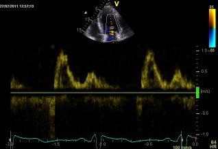
16 Mitral inflow and Hemodynamics E LA pressure LV impaired relaxation DT LV stiffness (LVDP) LV impaired relaxation A LV stiffness (LVEDP) LA contractility Age Heart rate and rhythm P-R interval Cardiac output Mitral annular size LA function
17 E / A ratio Filling pressure Compliance Relaxation EF < 50% (dilated cardiomyopathies, after MI) Mitral inflow pattern correlate with filling pressures, functional class and prognosis better than EF. As E/A ratio increases and DT shortens filling normal pressures increases, functional class is worse and prognosis is poor Therapeutic goal EF > 50% Gray zone EF > 50% restrictive and morfologic correlates of diastolic dysfunction pseudonormal (LA enlargment, severe hypertrorhy, HCM,amyolidosis) Restrictive pattern indicates elevated Impaired relaxation filling pressures and poor prognosis Disease severity
18 Biphasic)response)of)tradi&onal) indices)to)diastolic)dysfunc&on) Diastolic normal mild severe Parameter function dysfunction dysfunction Systolic IVTR dysfuction E/A ratio EF < 50% DT S/D pulm veins + age dependence


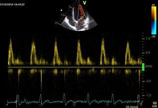
19 Pulmonary venous flow pattern and filling pressures Mitral flow velocity LV pressure mm Hg Ma=100 ms PVa=165 ms Pulmonary vein EF <50% S/D <1 Systolic fraction < 40% Increased PCWP Any EF Ar duration > 30msec from A duration Increased LVEDP

20 Asymptomatic NYHA III Recurrent Hospitalizations NYHA II

21 ?


22 The Valsalva Maneuver Baseline Strain Pseudonormalization impaired relaxation Normal normal A decrease of >50% in the E/A ratio is highly specfic for increased fillinig pressures A decrease of 20cm/sec in mitral peak E velocity is considered an adequate effort
23 Tissue Doppler Annular velocities Color M-mode flow propagation velocity How abnormal is myocardial relaxation? Time interval between E and e Global longitudinal SR IVRT
24 Tissue Doppler longitudinal Annular Velocities and Hemodynamics Recoil (systolic force) (+) Residual crossbridge interaction (Impaired Relaxation) (-) LA contraction(+) e is reduced and delayed LVEDP (-) Lengthening load (+) (filling pressures and geometry) When relaxation is impaired especially with normal EF


 Nagueh et al: JACC 37(1):")
25 e vs Maximal Instantaneous Transmitral Pressure Gradient Divided According to Tau e cm/ sec Mitral flow Mitral annulus 6 velocity Tau < Tau > Maximal transmitral pressure gradient (mmhg) Nagueh et al: JACC 37(1): , 2001

26 Clinical Application in patients with cardiac disease Most patients with e (lateral) < 10cm/sec or e (septal) < 8cm/sec have impaired myocardial relaxation Transmitral E velocity depends on LA pressure, relaxation kinetics and age Mitral annular e velocity depends on relaxation kinetics and age. The E/e ratio eliminates the effect of relaxation and age e as a surrogate of LV relaxation E/e ratio as predictor of LV filling pressures

 should de used for increased")
27 E/e ratio for the prediction of LV filling pressures E/e < 8 associated with normal filling pressures E/e > 15 associated with increased filling pressures Omen et al Circ 2000 E/e between 8 15 other echo indices should be used When using lateral or average values lower cutoff (12 for lateral and 13 for average) should de used for increased filling pressures
28

29 E/e = 7 E/e = 20 E/e = 16?
30 E/e!and!diastolic!func5on! E/e E/e is unreliable in Normal health people (e preload dependent) MItral stenosis/regurg/calcification/prosthetic valves Hypertrophic cardiomyopathy Severe LV systolic dysfunction ( e load dependent) Constictive pericarditis (annulus paradoxus) E/e works were we need it most, in patients with Heart Failure and normal EF
31 After Acute MI Hillis JACC 2004 Cardiomyopathies (Ishaemic, Dilated) Troughton AJC 2005, Yamamoto JASE 2003 Wang JACC 2005, Dokainish JACC 2005 Atrial fibrillation Okura Heart 2006 E /e and prognosis Secondary MR Bruch AJC 2007 Hypertensive heart disease Wang J Hypertens 2005 Sharp AS Eur Heart J 2010 End stage Renal disease Sharma JASE 2006
 E / Vp 2,5")
32 Color M-Mode flow propagation Velocity (Vp) Slope of the first aliasing velocity during early filling Normaly Vp > 50cm/sec Index of relaxation (inverse relation to τ) and load independent but only in patients with depressed EF Patients with normal LV volumes and EF but elevated filling pressures can have misleadingly normal Vp (dependency of load, geometry,contractile function) E / Vp 2,5 Predicts elevated filling pressures in patients with depressed EF when other indices appear inconclusive
33 Time interval between onset of the E and onset of the e When myocardial relaxation in normal both E and e coincided with the crossover of the LA-LV pressure. When relaxation is abnormal the e is reduced and delayed and occurs after LA-LV pressure crossover (asymmetrical longitudinal LV expansion) T E-e is an index of impaired relaxation (directly related to τ) IVRT / T E-e ratio < 2 Rivas-Gotz JACC 2003 Predicts increased filling pressures Useful when: - E/e is inconclusive - In mitral valve diseases
34 Global longitudinal SR IVRT by 2D speckle tracking Index strongly dependent on LV relaxation E/ SR IVRT > 236 Predicts increased filling pressures Useful when : -E/e is inconclusive, -EF is normal -In patients with regional dysfunction Wang J Circulation 2007
35
36


37 Heart failure symptoms with normal EF restrictive filling pattern, IVC dilatation Abnormal Pericardium Constrictive Restrictive 1.Increased thickness yes no 2.Septal fluttening yes no 3.Mitral inflow respiratory yes no variation 4.Hepatic vein expiratory yes no diastolic flow reversal Normal Myocardium 1.Ventricular septal Strain normal reduced 2. Septal annular e > 7cm/sec yes no 3. E/E normal Increased
38 Special populations
39 Conclusions) Tradi5onal!doppler!indices!are!usually! adequate!for!the!evalua5on!of!filling! pressures!in!pa5ents!with!low!ef.! A!relaxa5on!index!(usually!e )!mast!be!added! in!pa5ents!with!normal!ef! Diastolic!func5on!assessment!should!always! consider!all!available!echo!parameters!and! clinical!informa5on.!
An Integrated Approach to Study LV Diastolic Function
 An Integrated Approach to Study LV Diastolic Function Assoc. Prof. Adriana Ilieşiu, FESC University of Medicine Carol Davila Bucharest, Romania LV Diastolic Dysfunction impaired relaxation (early diastole)
How to Assess Diastolic Dysfunction?
 How to Assess Diastolic Dysfunction? Fausto J Pinto, MD, PhD, FESC, FACC, FASE Lisbon University Dyastolic Dysfunction Impaired relaxation Elevated filling pressures Ischemic heart disease Cardiomyopathies
The Patient with Atrial Fibrilation
 Assessment of Diastolic Function The Patient with Atrial Fibrilation Assoc. Prof. Adriana Ilieşiu, FESC University of Medicine Carol Davila Bucharest, Romania Associated Conditions with Atrial Fibrillation
Left ventricular diastolic function and filling pressure in patients with dilated cardiomyopathy
 Left ventricular diastolic function and filling pressure in patients with dilated cardiomyopathy Bogdan A. Popescu University of Medicine and Pharmacy Bucharest, Romania My conflicts of interest: I have
Diastolic Function Assessment Practical Ways to Incorporate into Every Echo
 Diastolic Function Assessment Practical Ways to Incorporate into Every Echo Jae K. Oh, MD Echo Hawaii 2018 2018 MFMER 3712003-1 Learning Objectives My presentation will help you to Appreciate the importance
Diastolic Function: What the Sonographer Needs to Know. Echocardiographic Assessment of Diastolic Function: Basic Concepts 2/8/2012
 Diastolic Function: What the Sonographer Needs to Know Pat Bailey, RDCS, FASE Technical Director Beaumont Health System Echocardiographic Assessment of Diastolic Function: Basic Concepts Practical Hints
Diastology State of The Art Assessment
 Diastology State of The Art Assessment Dr. Mohammad AlGhamdi Assistant professor, KSAU-HS Consultant Cardiologist King AbdulAziz Cardiac Center Ministry of National Guard Health Affairs Diagnostic Clinical
The importance of left atrium in LV diastolic function
 II Baltic Heart Failure Meeting and Congress of Latvian Society of Cardiology The importance of left atrium in LV diastolic function Dr. Artem Kalinin Eastern Clinical University Hospital Riga 30.09.2010.
Imaging in Heart Failure: A Multimodality Approach. Thomas Ryan, MD
 Imaging in Heart Failure: A Multimodality Approach Thomas Ryan, MD Heart Failure HFrEF HFpEF EF50% Lifetime risk 20% Prevalence 6M Americans Societal costs - $30B 50% 5-year survival 1 Systolic
LV geometric and functional changes in VHD: How to assess? Mi-Seung Shin M.D., Ph.D. Gachon University Gil Hospital
 LV geometric and functional changes in VHD: How to assess? Mi-Seung Shin M.D., Ph.D. Gachon University Gil Hospital LV inflow across MV LV LV outflow across AV LV LV geometric changes Pressure overload
Diastology Disclosures: None. Dias2011:1
 Diastology 2011 James D. Thomas, M.D., F.A.C.C. Cardiovascular Imaging Center Department of Cardiology Cleveland Clinic Foundation Cleveland, Ohio, USA Disclosures: None Dias2011:1 Is EVERYBODY a member!?!
LV FUNCTION ASSESSMENT: WHAT IS BEYOND EJECTION FRACTION
 LV FUNCTION ASSESSMENT: WHAT IS BEYOND EJECTION FRACTION Jamilah S AlRahimi Assistant Professor, KSU-HS Consultant Noninvasive Cardiology KFCC, MNGHA-WR Introduction LV function assessment in Heart Failure:
Diastolic Function Assessment New Guideline Update Practical Approach
 Mayo Clinic Department of Cardiovascular Diseases Mayo Clinic Echocardiography Review Course for Boards and Recertification Diastolic Function Assessment New Guideline Update Practical Approach Jae K.
Basic Approach to the Echocardiographic Evaluation of Ventricular Diastolic Function
 Basic Approach to the Echocardiographic Evaluation of Ventricular Diastolic Function J A F E R A L I, M D U N I V E R S I T Y H O S P I T A L S C A S E M E D I C A L C E N T E R S T A F F C A R D I O T
Μαρία Μπόνου Διευθύντρια ΕΣΥ, ΓΝΑ Λαϊκό
 Μαρία Μπόνου Διευθύντρια ΕΣΥ, ΓΝΑ Λαϊκό Diastolic HF DD: Diastolic Dysfunction DHF: Diastolic HF HFpEF: HF with preserved EF DD Pathophysiologic condition: impaired relaxation, LV compliance, LV filling
Diastole is Not a Single Entity Four Components of Diastolic Dysfunction
 Physiology of Diastolic Function Made Easy James D. Thomas, MD, FACC, FASE Director, Center for Heart Valve Disease Bluhm Cardiovascular Institute Professor of Medicine, Feinberg School of Medicine, Northwestern
Diastolic Heart Failure
 Chronic Heart Failure Prevalence overall = 2-3 % Diastolic Heart Failure Patrick Wouters University Hospital Ghent Belgium (Heart Failure + Asymptomatic Ventricular Dysfunction) Prevalence > 70 y = 10-20
Diastolic Heart Function: Applying the New Guidelines Case Studies
 Diastolic Heart Function: Applying the New Guidelines Case Studies Mitral Regurgitation The New ASE William Guidelines: A. Zoghbi Role MD, of FASE, 2D/3D MACCand CMR Professor and Chairman, Department
Choose the grading of diastolic function in 82 yo woman
 Question #1 Choose the grading of diastolic function in 82 yo woman E= 80 cm/s A= 70 cm/s LAVI < 34 ml/m 2 1= Grade 1 2= Grade 2 3= Grade 3 4= Normal 5= Indeterminate 2018 MFMER 3712003-1 Choose the grading
DOPPLER HEMODYNAMICS (1) QUANTIFICATION OF PRESSURE GRADIENTS and INTRACARDIAC PRESSURES
 QUANTIFICATION OF PRESSURE GRADIENTS and INTRACARDIAC PRESSURES") THORAXCENTRE DOPPLER HEMODYNAMICS (1) QUANTIFICATION OF PRESSURE GRADIENTS and INTRACARDIAC PRESSURES J. Roelandt DOPPLER HEMODYNAMICS Intracardiac pressures and pressure gradients Volumetric measurement
THORAXCENTRE DOPPLER HEMODYNAMICS (1) QUANTIFICATION OF PRESSURE GRADIENTS and INTRACARDIAC PRESSURES J. Roelandt DOPPLER HEMODYNAMICS Intracardiac pressures and pressure gradients Volumetric measurement
Adel Hasanin Ahmed 1
 Adel Hasanin Ahmed 1 PERICARDIAL DISEASE The pericardial effusion ends anteriorly to the descending aorta and is best visualised in the PLAX. PSAX is actually very useful sometimes for looking at posterior
E/Ea is NOT an essential estimator of LV filling pressures
 Euroecho Kopenhagen Echo in Resynchronization in 2010 E/Ea is NOT an essential estimator of LV filling pressures Wilfried Mullens, MD, PhD December 10, 2010 Ziekenhuis Oost Limburg Genk University Hasselt
Evaluation of Left Ventricular Diastolic Dysfunction by Doppler and 2D Speckle-tracking Imaging in Patients with Primary Pulmonary Hypertension
 ESC Congress 2011.No 85975 Evaluation of Left Ventricular Diastolic Dysfunction by Doppler and 2D Speckle-tracking Imaging in Patients with Primary Pulmonary Hypertension Second Department of Internal
LA Function analysis Marcia Barbosa Vice Presidente - Brazilian Soc of Cardiology President-elect - Interamerican Soc of Cardiology
 LA Function analysis Marcia Barbosa Vice Presidente - Brazilian Soc of Cardiology President-elect - Interamerican Soc of Cardiology Belo Horizonte Brazil DECLARATION OF CONFLICT OF INTEREST Nothing to
Heart Failure in Women: Dr Goh Ping Ping Cardiologist Asian Heart & Vascular Centre
 Heart Failure in Women: More than EF? Dr Goh Ping Ping Cardiologist Asian Heart & Vascular Centre Overview Review pathophysiology as it relates to diagnosis and management Rational approach to workup:
Advanced imaging of the left atrium - strain, CT, 3D, MRI -
 Advanced imaging of the left atrium - strain, CT, 3D, MRI - Monica Rosca, MD Carol Davila University of Medicine and Pharmacy, Bucharest, Romania Declaration of interest: I have nothing to declare Case
P = 4V 2. IVC Dimensions 10/20/2014. Comprehensive Hemodynamic Evaluation by Doppler Echocardiography. The Simplified Bernoulli Equation
 Comprehensive Hemodynamic Evaluation by Doppler Echocardiography Itzhak Kronzon, MD North Shore LIJ/ Lenox Hill Hospital New York, NY Disclosure: Philips Healthcare St. Jude Medical The Simplified Bernoulli
Diastolic Function Overview
 Diastolic Function Overview Richard Palma BS, RDCS, RCS, APS, FASE Director and Clinical Coordinator The Hoffman Heart and Vascular Institute School of Cardiac Ultrasound None Disclosures Learning Objectives
22 nd Annual Conference of the Saudi Heart Association Riyadh, Saudi Arabia
 22 nd Annual Conference of the Saudi Heart Association Riyadh, Saudi Arabia New Echocardiographic Modalities to Evaluate Ventricular Function in Congenital Heart Disease: Tissue Doppler & Strain Rate Imaging
Echo-Doppler evaluation of left ventricular diastolic function. Michel Slama Amiens France
 Echo-Doppler evaluation of left ventricular diastolic function Michel Slama Amiens France Left ventricular pressure Pressure A wave [ LVEDP LVEDP préa Congestive cardiac failure with preserved systolic
OPTIMIZING ECHO ACQUISTION FOR STRAIN AND DIASTOLOGY
 OPTIMIZING ECHO ACQUISTION FOR STRAIN AND DIASTOLOGY October 8, 2017 Deborah Agler, ACS, RDCS, FASE Coordinator of Education and Training Cleveland Clinic General Principles Diastology Clinical Data Heart
Echo Doppler Assessment of Right and Left Ventricular Hemodynamics.
 Echo Doppler Assessment of Right and Left Ventricular Hemodynamics. Itzhak Kronzon, MD, FASE, FACC, FESC, FAHA, FACP, FCCP Northwell, Lenox Hill Hospital, New York Professor of Cardiology Hofstra University
Prognostic Value of Left Atrial Size and Function
 Prognostic Value of Left Atrial Size and Function James D. Thomas, M.D., F.A.C.C. Cardiovascular Imaging Center Department of Cardiology Cleveland Clinic Foundation Cleveland, Ohio, USA Conflicts: None
GENERAL PRINCIPLES FOR ECHO ASSESSMENT OF DIASTOLIC FUNCTION (For full recommendation refer to the Left Ventricular Diastolic Function Guideline)
") 1 THE AMERICAN SOCIETY OF ECHOCARDIOGRAPHY RECOMMENDATIONS FOR THE EVALUATION OF LEFT VENTRICULAR DIASTOLIC FUNCTION BY ECHOCARDIOGRAPHY: A QUICK REFERENCE GUIDE FROM THE ASE WORKFLOW AND LAB MANAGEMENT
1 THE AMERICAN SOCIETY OF ECHOCARDIOGRAPHY RECOMMENDATIONS FOR THE EVALUATION OF LEFT VENTRICULAR DIASTOLIC FUNCTION BY ECHOCARDIOGRAPHY: A QUICK REFERENCE GUIDE FROM THE ASE WORKFLOW AND LAB MANAGEMENT
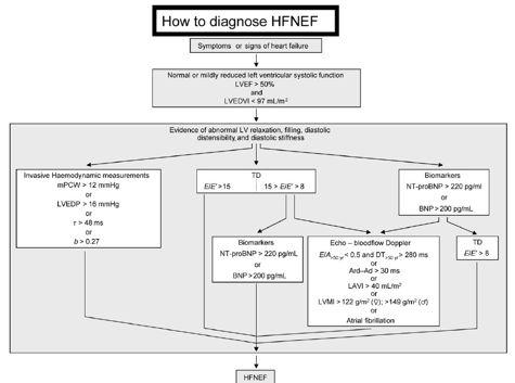
HFNEF. Heart Failure is
 HFNEF Bijoy K. Khandheria, MD. FASE, FACP, FACC FESC Professor of Medicine University of Wisconsin Director. Echocardiography Services Aurora Health Care No conflicts or off label use CP1173868-1 Heart
Diastolic Dysfunction: Hypertension to Hypertrophy to Heart Failure
 Diastolic Dysfunction: Hypertension to Hypertrophy to Heart Failure Dr. Shelley Zieroth FRCPC Assistant Professor, Cardiology, University of Manitoba Director of Cardiac Transplant and Heart Failure Clinics
Diastolic Functions: Evaluation & Clinical Applications
 Special Articles Diastolic Functions: Evaluation & Clinical Applications Senior Consultant Cardiologist, Metro Heart Institute, Delhi Immediate Past President, Cardiological Society of India (Cardiovasc.
HFpEF. April 26, 2018
 HFpEF April 26, 2018 (J Am Coll Cardiol 2017;70:2476 86) HFpEF 50% or more (40-71%) of patients with CHF have preserved LV systolic function. HFpEF is an increasingly frequent hospital discharge. Outcomes
ΚΑΡΔΙΟΛΟΓΟΣ EUROPEAN ACCREDITATION IN TRANSTHORACIC AND TRANSESOPHAGEAL ECHOCARDIOGRAPHY
 1 ΚΑΡΔΙΟΛΟΓΟΣ EUROPEAN ACCREDITATION IN TRANSTHORACIC AND TRANSESOPHAGEAL ECHOCARDIOGRAPHY 2 Constrictive pericarditis (CP) is characterized by impaired ventricular filling due to a stiffened or noncompliant
The Doppler Examination. Katie Twomley, MD Wake Forest Baptist Health - Lexington
 The Doppler Examination Katie Twomley, MD Wake Forest Baptist Health - Lexington OUTLINE Principles/Physics Use in valvular assessment Aortic stenosis (continuity equation) Aortic regurgitation (pressure
European Heart Journal - Cardiovascular Imaging Advance Access published July 18, 2016
 European Heart Journal - Cardiovascular Imaging Advance Access published July 18, 2016 European Heart Journal Cardiovascular Imaging doi:10.1093/ehjci/jew082 Recommendations for the Evaluation of Left
Mechanisms of heart failure with normal EF Arterial stiffness and ventricular-arterial coupling. What is the pathophysiology at presentation?
 Mechanisms of heart failure with normal EF Arterial stiffness and ventricular-arterial coupling What is the pathophysiology at presentation? Ventricular-arterial coupling elastance Central arterial pressure
Echo assessment of the failing heart
 Echo assessment of the failing heart Mark K. Friedberg, MD The Labatt Family Heart Center The Hospital for Sick Children Toronto, Ontario, Canada Cardiac function- definitions Cardiovascular function:
Nancy Goldman Cutler, MD Beaumont Children s Hospital Royal Oak, Mi
 Nancy Goldman Cutler, MD Beaumont Children s Hospital Royal Oak, Mi Identify increased LV wall thickness (WT) Understand increased WT in athletes Understand hypertrophic cardiomyopathy (HCM) Enhance understanding
RIGHT VENTRICULAR SIZE AND FUNCTION
 RIGHT VENTRICULAR SIZE AND FUNCTION Edwin S. Tucay, MD, FPCC, FPCC, FPSE Philippine Society of Echocardiography Quezon City, Philippines Echo Mission, BRTTH, Legaspi City, July 1-2, 2016 NO DISCLOSURE
Aortic valve Stenosis: Insights in the evaluation of LV function. Erwan DONAL Cardiologie CHU Rennes
 Aortic valve Stenosis: Insights in the evaluation of LV function Erwan DONAL Cardiologie CHU Rennes erwan.donal@chu-rennes.fr Preload Afterload Myocardial Fiber Shortening Circumferential Longitudinal
Pericardial Diseases. Smonporn Boonyaratavej, MD. Division of Cardiology, Department of Medicine Chulalongkorn University
 Pericardial Diseases Smonporn Boonyaratavej, MD Division of Cardiology, Department of Medicine Chulalongkorn University Cardiac Center, King Chulalongkorn Memorial Hospital 21 AUGUST 2016 Pericardial
Echocardiographic Evaluation of the Cardiomyopathies. Stephanie Coulter, MD, FACC, FASE April, 2016
 Echocardiographic Evaluation of the Cardiomyopathies Stephanie Coulter, MD, FACC, FASE April, 2016 Cardiomyopathies (CMP) primary disease intrinsic to cardiac muscle Dilated CMP Hypertrophic CMP Infiltrative
Objectives. Diastology: What the Radiologist Needs to Know. LV Diastolic Function: Introduction. LV Diastolic Function: Introduction
 Objectives Diastology: What the Radiologist Needs to Know. Jacobo Kirsch, MD Cardiopulmonary Imaging, Section Head Division of Radiology Cleveland Clinic Florida Weston, FL To review the physiology and
Constriction vs Restriction Case-based Discussion
 Mayo Clinic Department of Cardiovascular Diseases Mayo Clinic Echocardiography Review Course for Boards and Recertification Constriction vs Restriction Case-based Discussion Jae K. Oh, MD Samsung Professor
Comprehensive Hemodynamics By Doppler Echocardiography. The Echocardiographic Swan-Ganz Catheter.
 Comprehensive Hemodynamics By Doppler Echocardiography. The Echocardiographic Swan-Ganz Catheter. Itzhak Kronzon, MD, FASE, FACC, FESC, FAHA, FACP, FCCP North Shore HS, LIJ/Lenox Hill Hospital, New York
Value of echocardiography in chronic dyspnea
 Value of echocardiography in chronic dyspnea Jahrestagung Schweizerische Gesellschaft für /Schweizerische Gesellschaft für Pneumologie B. Kaufmann 16.06.2016 Chronic dyspnea Shortness of breath lasting
How does the heart pump? From sarcomere to ejection volume
 How does the heart pump? From sarcomere to ejection volume Piet Claus Cardiovascular Imaging and Dynamics Department of Cardiovascular Diseases University Leuven, Leuven, Belgium Course on deformation
Diastolic Function. Rick Nishimura Leighton Professor of CV Diseases Mayo Clinic No Disclosures
 Diastolic Function Rick Nishimura Leighton Professor of CV Diseases Mayo Clinic No Disclosures Heart = Pump Heart Failure Systolic Dysfunction Diastolic Dysfunction Diastole is a complex sequence of multiple
Strain/Untwisting/Diastolic Suction
 What Is Diastole and How to Assess It? Strain/Untwisting/Diastolic Suction James D. Thomas, M.D., F.A.C.C. Cardiovascular Imaging Center Department of Cardiology Cleveland Clinic Foundation Cleveland,
We are IntechOpen, the world s leading publisher of Open Access books Built by scientists, for scientists. International authors and editors
 We are IntechOpen, the world s leading publisher of Open Access books Built by scientists, for scientists 4,100 116,000 120M Open access books available International authors and editors Downloads Our
Review of Cardiac Imaging Modalities in the Renal Patient. George Youssef
 Review of Cardiac Imaging Modalities in the Renal Patient George Youssef ECHO Left ventricular hypertrophy (LVH) assessment Diastolic dysfunction Stress ECHO Cardiac CT angiography Echocardiography - positives
Diastolic Heart Failure
 58 Diastolic Heart Failure Muhamed Saric and Itzhak Kronzon PRACTICAL POINTS Diastole starts with the closure of the aortic valve and ends with the closure of the mitral valve. It consists of four phases:
THE DIASTOLIC STRESS TEST: A NEW CLINICAL TOOL? THE CONCEPT OF DIASTOLIC RESERVE
 Thierry C. Gillebert University of Ghent ESC Education Committee THE DIASTOLIC STRESS TEST: A NEW CLINICAL TOOL? THE CONCEPT OF DIASTOLIC RESERVE 1 Case: Ann, 63 years Suffered from metabolic syndrome
Restrictive Cardiomyopathy
 ESC Congress 2011, Paris Imaging Unusual Causes of Cardiomyopathy Restrictive Cardiomyopathy Kazuaki Tanabe, MD, PhD Professor of Medicine Chair, Division of Cardiology Izumo, Japan I Have No Disclosures
What is controversial in diagnostic imaging?
 Controversies in the management of pulmonary hypertension What is controversial in diagnostic imaging? G. Derumeaux Lyon University Hospices Civils de Lyon France Déclaration de Relations Professionnelles
Constrictive/Restrictive Cardiomyopathies: Diagnosis and Management Update; Radiation Induced Heart Disease. Alexander (Sandy) Dick, MD
 Dick, MD") Constrictive/Restrictive Cardiomyopathies: Diagnosis and Management Update; Radiation Induced Heart Disease Alexander (Sandy) Dick, MD Outline Pericardial Constriction Diagnosis: Imaging, Hemodynamics
Constrictive/Restrictive Cardiomyopathies: Diagnosis and Management Update; Radiation Induced Heart Disease Alexander (Sandy) Dick, MD Outline Pericardial Constriction Diagnosis: Imaging, Hemodynamics
Diagnosis is it really Heart Failure?
 ESC Congress Munich - 25-29 August 2012 Heart Failure with Preserved Ejection Fraction From Bench to Bedside Diagnosis is it really Heart Failure? Prof. Burkert Pieske Department of Cardiology Med.University
Dilated Cardiomyopathy
 EAE EDUCATIONAL COURSE, Bucharest 2010 Dilated Cardiomyopathy Diagnosis, Prognosis, F/U: The Role of Echo? Aleksandar N. Neskovic Clinical Hospital Zemun Belgrade University School of Medicine Echo in
Constrictive Pericarditis
 Constrictive Pericarditis Never Confused with Anything Else Jae K. Oh, MD 2018 MFMER 3712003-1 ARS #1 CP Which of following patients has constrictive pericarditis? 1 2 3 Medial e 13 cm/s Medial e 3 cm/s
Ejection across stenotic aortic valve requires a systolic pressure gradient between the LV and aorta. This places a pressure load on the LV.
 Valvular Heart Disease Etiology General Principles Cellular and molecular mechanism of valve damage Structural pathology Functional pathology - stenosis/regurgitation Loading conditions - pressure/volume
Noninvasive assessment of left ventricular (LV)
") Comparative Value of Tissue Doppler Imaging and M-Mode Color Doppler Mitral Flow Propagation Velocity for the Evaluation of Left Ventricular Filling Pressure* Michal Kidawa, MD; Lisa Coignard, MD; Gérard
Comparative Value of Tissue Doppler Imaging and M-Mode Color Doppler Mitral Flow Propagation Velocity for the Evaluation of Left Ventricular Filling Pressure* Michal Kidawa, MD; Lisa Coignard, MD; Gérard
Pericardial Disease: Case Examples. Echo Fiesta 2017
 Pericardial Disease: Case Examples Echo Fiesta 2017 2014 2014 MFMER MFMER 3346252-1 slide-1 Objectives Have a systematic approach to evaluation of constriction 2014 MFMER 3346252-2 CASE 1 2013 MFMER 3248567-3
Highlights from EuroEcho 2009 Echo in cardiomyopathies
 Highlights from EuroEcho 2009 Echo in cardiomyopathies Bogdan A. Popescu University of Medicine and Pharmacy, Bucharest, Romania ESC Congress 2010 Hypertrophic cardiomyopathy To determine the differences
Dr.Fayez EL Shaer Consultant cardiologist Assistant professor of cardiology KKUH
 Pulmonary Hypertension in patients with Heart Failure with Preserved Ejection Fraction Dr.Fayez EL Shaer Consultant cardiologist Assistant professor of cardiology KKUH Recent evaluation of available data
Tissue Doppler Imaging
 Cronicon OPEN ACCESS Hesham Rashid* Tissue Doppler Imaging CARDIOLOGY Editorial Department of Cardiology, Benha University, Egypt *Corresponding Author: Hesham Rashid, Department of Cardiology, Benha University,
Imaging in dilated cardiomyopathy : factors associated with a poor outcome
 Imaging in dilated cardiomyopathy : factors associated with a poor outcome Johan De Sutter, MD, PhD, FESC AZ Maria Middelares Gent and University Gent - Belgium Dilated cardiomyopathy Cardiomyopathy with
Echocardiographic and Doppler Assessment of Cardiac Functions in Patients of Non-Insulin Dependent Diabetes Mellitus
 ORIGINAL ARTICLE JIACM 2002; 3(2): 164-8 Echocardiographic and Doppler Assessment of Cardiac Functions in Patients of Non-Insulin Dependent Diabetes Mellitus Rajesh Rajput*, Jagdish**, SB Siwach***, A
The study of left ventricular diastolic function by Doppler echocardiography: the essential for the clinician
 Heart International / Vol. 3 no. 1-2, 2007 / pp. 42-50 Wichtig Editore, 2007 Review The study of left ventricular diastolic function by Doppler echocardiography: the essential for the clinician POMPILIO
Elevated LV filling pressure is a major determinant of cardiac symptoms and
 LEFT VENTRICULAR FILLING PRESSURE, DIASTOLIC FUNCTION, AND HEART RATE PATRIZIO LANCELLOTTI, MD, PhD, FESC PERSPECTIVES Author affiliations: University of Liège hospital, GIGA Cardiovascular Science, Heart
Ejection across stenotic aortic valve requires a systolic pressure gradient between the LV and aorta. This places a pressure load on the LV.
 Valvular Heart Disease General Principles Etiology Cellular and molecular mechanism of valve damage Structural pathology Functional pathology - stenosis/regurgitation Loading conditions - pressure/volume
Load and Function - Valvular Heart Disease. Tom Marwick, Cardiovascular Imaging Cleveland Clinic
 Load and Function - Valvular Heart Disease Tom Marwick, Cardiovascular Imaging Cleveland Clinic Indications for surgery in common valve lesions Risks Operative mortality Failed repair - to MVR Operative
Index. K Knobology, TTE artifact, image resolution, ultrasound, 14
 A Acute aortic regurgitation (AR), 124 128 Acute aortic syndrome (AAS) classic aortic dissection diagnosis, 251 263 evolutive patterns, 253 255 pathology, 250 251 classifications, 247 248 incomplete aortic
THE RIGHT VENTRICLE IN PULMONARY HYPERTENSION R. DRAGU
 THE RIGHT VENTRICLE IN PULMONARY HYPERTENSION R. DRAGU Cardiology Dept. Rambam Health Care Campus Rappaport Faculty of Medicine Technion, Israel Why the Right Ventricle? Pulmonary hypertension (PH) Right
Jong-Won Ha*, Jeong-Ah Ahn, Jae-Yun Moon, Hye-Sun Suh, Seok-Min Kang, Se-Joong Rim, Yangsoo Jang, Namsik Chung, Won-Heum Shim, Seung-Yun Cho
 Eur J Echocardiography (2006) 7, 16e21 CLINICAL/ORIGINAL PAPERS Triphasic mitral inflow velocity with mid-diastolic flow: The presence of mid-diastolic mitral annular velocity indicates advanced diastolic
Atrial dysfunction and chronotropic incompetence
 Pathophysiology of heart failure with preserved ejection fraction Atrial dysfunction and chronotropic incompetence Vojtech Melenovsky IKEM, Prague, Czech Republic DECLARATION OF CONFLICT OF INTEREST :
Hypertensive heart disease and failure
 Hypertensive heart disease and failure Prof. Dr. Alan Fraser Cardiff University The heart in hypertension Pathophysiology of LV adaptation Regional development of hypertrophy Stress testing - inducible
HEMODYNAMIC ASSESSMENT
 HEMODYNAMIC ASSESSMENT INTRODUCTION Conventionally hemodynamics were obtained by cardiac catheterization. It is possible to determine the same by echocardiography. Methods M-mode & 2D echo alone can provide
Clinical Outcome of Tricuspid Regurgitation. David Messika-Zeitoun
 Clinical Outcome of Tricuspid Regurgitation David Messika-Zeitoun I have financial relationships to disclose Consultant for: Edwards, Symetis and Valtech Tricuspid Regurgitation is a Common Finding Tricuspid
Echocardiography for the Electrophysiologist: Day-to-day practice. Emmanuel Fares, MD
 Echocardiography for the Electrophysiologist: Day-to-day practice Emmanuel Fares, MD EP and pacing service, Department of Cardiovascular Medicine, Cairo University Agenda Role of echo in arrhythmia management:
Left atrial Function In Echocardiography
 Left atrial Function In Echocardiography Pr Erwan DONAL erwan.donal@chu-rennes.fr Inserm Diastole LA Pressure (mmhg) -t/ 17 LA function 15 A-Loop 13 V-Loop 11 9 7 5 1 2 3 4 20 30 40 50 60 70 LA Volume
HYPERTROPHY: Behind the curtain. V. Yotova St. Radboud Medical University Center, Nijmegen
 HYPERTROPHY: Behind the curtain V. Yotova St. Radboud Medical University Center, Nijmegen Disclosure of interest: none Relative wall thickness (cm) M 0.22 0.42 0.43 0.47 0.48 0.52 0.53 F 0.24 0.42 0.43
Managing Hypertrophic Cardiomyopathy with Imaging. Gisela C. Mueller University of Michigan Department of Radiology
 Managing Hypertrophic Cardiomyopathy with Imaging Gisela C. Mueller University of Michigan Department of Radiology Disclosures Gadolinium contrast material for cardiac MRI Acronyms Afib CAD Atrial fibrillation
The Pathophysiology of Cardiogenic Shock Knowledge Gaps & Opportunities
 The Pathophysiology of Cardiogenic Shock Knowledge Gaps & Opportunities Navin K. Kapur, MD, FACC, FSCAI, FAHA Associate Professor, Department of Medicine Interventional Cardiology & Advanced Heart Failure
Global left ventricular circumferential strain is a marker for both systolic and diastolic myocardial function
 Global left ventricular circumferential strain is a marker for both systolic and diastolic myocardial function Toshinari Onishi 1, Samir K. Saha 2, Daniel Ludwig 1, Erik B. Schelbert 1, David Schwartzman
Pathophysiology: Heart Failure
 Pathophysiology: Heart Failure Mat Maurer, MD Irving Assistant Professor of Medicine Outline Definitions and Classifications Epidemiology Muscle and Chamber Function Pathophysiology Heart Failure: Definitions
Valvular Regurgitation: Can We Do Better Than Colour Doppler?
 Valvular Regurgitation: Can We Do Better Than Colour Doppler? A/Prof David Prior St Vincent s Hospital Melbourne Sports Cardiology Valvular Regurgitation Valve regurgitation volume loads the ventricles
Prof. JL Zamorano Hospital Universitario Ramón y Cajal
 Prof. JL Zamorano Hospital Universitario Ramón y Cajal Should we forget TR? Nath J et al. Impact of tricuspid regurgitation on long-term survival. J Am Coll Cardiol. 2004; 43:405-409 Why is it difficult
Velocity, strain and strain rate: Doppler and Non-Doppler methods. Thoraxcentre, Erasmus MC,Rotterdam
 Velocity, strain and strain rate: Doppler and Non-Doppler methods J Roelandt J. Roelandt Thoraxcentre, Erasmus MC,Rotterdam Basics of tissue Doppler imaging Instantaneous annular velocity profiles IVCT
2/2/2011. Strain and Strain Rate Imaging How, Why and When? Movement vs Deformation. Doppler Myocardial Velocities. Movement. Deformation.
 Strain and Strain Rate Imaging How, Why and When? João L. Cavalcante, MD Advanced Cardiac Imaging Fellow Cleveland Clinic Foundation Disclosures: No conflicts of interest Movement vs Deformation Movement
SUPPLEMENTAL MATERIAL
 SUPPLEMENTAL MATERIAL Supplemental methods Pericardium In several studies, it has been shown that the pericardium significantly modulates ventricular interaction. 1-4 Since ventricular interaction has
Left atrial function. Aliakbar Arvandi MD
 In the clinic Left atrial function Abstract The left atrium (LA) is a left posterior cardiac chamber which is located adjacent to the esophagus. It is separated from the right atrium by the inter-atrial
Stephen Glen ISCHAEMIC HEART DISEASE AND LEFT VENTRICULAR FUNCTION
 Stephen Glen ISCHAEMIC HEART DISEASE AND LEFT VENTRICULAR FUNCTION Overview Coronary arteries Terminology to describe contractility Measuring ventricular function Systolic dysfunction Practice cases- LV
Advanced Applica,on of Point- of- Care Echocardiography in Cri,cal Care. Dr. Mark Tutschka Dr. Rob ArnAield
 Advanced Applica,on of Point- of- Care Echocardiography in Cri,cal Care Dr. Mark Tutschka Dr. Rob ArnAield OBJECTIVES Provide an overview of common advanced echocardiographic techniques suitable for use
Outline. Pathophysiology: Heart Failure. Heart Failure. Heart Failure: Definitions. Etiologies. Etiologies
 Outline Pathophysiology: Mat Maurer, MD Irving Assistant Professor of Medicine Definitions and Classifications Epidemiology Muscle and Chamber Function Pathophysiology : Definitions An inability of the