Introduction. Key terms. Key terms. Key terms 10/12/2016 EQUINE UPPER DIGESTIVE SYSTEM. Learning Objectives for My Lectures. What are the functions?
|
|
|
- Brittney Mason
- 5 years ago
- Views:
Transcription


 = gastroenteric Alimentary canal = gut Oris ---- oral ---- mouth Labial --- lip Buccal --- cheek Dento = donto ----- teeth Lingual/glossal = tongue Gingiva = gum")
1 Learning Objectives for My Lectures Recognize the importance of the head. List the functions of the digestive system. EQUINE UPPER DIGESTIVE SYSTEM Dr. Fawzy Elnady Prof. of Anatomy and Embryology Cairo University Correlate the anatomy with the clinical study. Understand the parts and boundaries of oral cavity and its anatomical features. List and give description to the salivary glands. Describe the anatomy of the tongue. Describe the anatomy of the pharynx. Introduction Key terms Why the head is important? What are the functions? Prehension Mastication Chemical digestion Absorption Elimination of wastes Herbivores Carnivores Omnivores Vegetarian Vegan Key terms Key terms Digestive or gastrointestinal tube/tract (GIT) = gastroenteric Alimentary canal = gut Oris ---- oral ---- mouth Labial --- lip Buccal --- cheek Dento = donto teeth Lingual/glossal = tongue Gingiva = gum (mucosa over the jaws, it is less sensible, no glands) Entery ---- intestine Peristalsis Stomatitis mouth Gingivitis gingiva Glossitis tongue Cheilitis lips Pharyngitis pharynx Esophagus esophagitis 1


2 Boundaries of the maxillary sinus: Approach to RMS Approach to CMS Maxillary Sinus Width of mandible and maxilla Oral mucosa 1 st line of defense Maxilla is wider than mandible Outside of upper cheek teeth and inside of lower teeth ecome sharp Smooth, shiny, pink Stratified squamous epithelium 2
 It is bounded by soft palate --- dorsally root of tongue ---")

3 Capillary refill time (CRT) Oral cavity Boundaries DOG Rostrally --- lips Laterally --- cheeks Dorsally --- Hard palate Ventrally --- tongue and mucous membrane Caudally --- isthmus faucium Isthmus faucium Divisions of the oral cavity The oral cavity communicates with the oro-pharynx through the isthmus faucium (aditus pharyngis) It is bounded by soft palate --- dorsally root of tongue --- ventrally palato-glossal fold --- laterally Oral vestibule Oral cavity Oral cavity proper Labial v. between incisors and lips Buccal v. between cheek teeth and cheek The space within the teeth Labial Vestibule Structures within oral vestibule Labial vestibule Labial glands Frenula labii --- fold of m.m. from lip to gum Buccal vestibule Buccal glands --- dorsal and ventral Parotid papillae (papilla salivalis) --- opening of the parotid duct at the level of the 3 rd upper cheek teeth. Vomeronasal organ Flehmen reaction 3


4 Oral cavity Oral cavity proper within the dental arcades Vestibule -- between the teeth and cheeks Sublingual floor of oral cavity Pre-frenulum part Frenulum linguae 3 Compartments 2 sublingual caruncles - Rostrolateral to the frenulum linguae - Surrounded by lymphoid tissue (Sublingual tonsil) - The mandibular ducts open here Pre-frenulum part 2 Sublingual recesses Orobasal organ: rudiment of rostral sublingual salivary glands in reptiles. 2 Sublingual recesses Hard palate Two sublingual folds on either side of frenulum linguae (openings of sublingual s.gl). They sublingual caruncles. Pterygomandibular fold: vertical fold caudal to last molar (extends between upper and lower jaws) Extends from incisors to last molars (of equal width) Boundries: Rostrally and Laterally by the upper dental arch Caudally soft palate 4

: 18 curved")

 Palatocheiloschisis")


.")


5 Bony support of hard p. Features of hard palate Palatine raphe: median line joining the 2 halves of the palate Palatine ridges (Rugae): 18 curved ridges on either side of p. raphe Clinical: Alpaca Incisive papilla: Palatoschisis = cleft palate Cheiloschisis = cleft lip (hare lip) Palatocheiloschisis = both Aspiration Pneumonia Oval elevation of mucous membrane at the rostral end of the p. raphe (just cd to central incisors). It conceals the orifices of the incisive ducts (obliterated in equine and represented by 2 small depressions). Incisive (nasopalatine) duct: Incisive duct or nasopalatine duct: Small tube of m.m. extends oblique in the palatine fissure. Its palatine end is blind and the nasal end communicates with the vomeronasal organ. 5


 2- Palatine glands (about 1 cm.")
 of the soft palate Palato-glossal arch (anterior pillar): m.m pass from soft p.")

6 Palatine venous plexus Soft palate Def. musculo-mucosal curtain, extends from the caudal end of the hard palate, separating the nasopharynx from the oropharynx. Soft palate Features of the soft palate 15 cm (very long, so no mouth breathing). Vomit occurs through nostril. Structure: 1- epithelial covering: i. Ventral surface: Stratified squamous epithelium as hard palate. ii. Dorsal surface: respiratory epithelium as nasal cavity (PSCC) 2- Palatine glands (about 1 cm. thickness) 3- Tendinous aponeurosis --- replaces bony support. Def. musculo-mucosal curtain, extends from the caudal end of the hard palate, separating the nasopharynx from the oropharynx. Folds (arches) of the soft palate Palato-glossal arch (anterior pillar): m.m pass from soft p. to base of tongue. Palato-pharyngeal arch (posterior pillar): from s.p. to sides of pharynx (the 2 folds fuse at entrance of the oesophagus, forming intrapharyngeal opening Tonsillar fossa ---- the space between ant. and post. Pillars and contain the tonsil 6


7 DDSP 1- things go wrong!!! 2- epiglottis 3- dynamic endoscope 4- examination 5- softpalate The bookmark at the time of intermittent DDSP Notice the nasopharynx and how the rostral portion of the soft palate billows dorsally and ventrally into the airway during inhalation and exhalation repectively.. 7


8 Musculature of the soft palate 1. Tensor veli palatini 2. Levator veli palatini 3. Palatinus 4. Palatopharyngeus soft palate Muscles 1. Palatinus 2. Levator veli palatini 3. Tensor veli palatini Origin Insertion Action Nerve Palatine bone Muscular process of petrous temporal bone + lateral surface of auditory tube 4. Palatopharypalatine aponeurosis and Free edge of s.p s.p. Pass around hamulus of pterygoid bone into aponeurosis of s.p. pharyngeal Shorten s.p. Elevates the s.p. during swallowing to close the choanae Tenses s.p. مرجيحة shortens the s.p. and draws the larynx and Pharynge al branch of vagus (X) Mandibul ar of Trigeminal As Normal DDSP 8


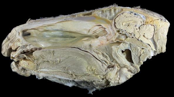
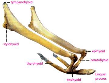
9 Hyoid apparatus 9
 soft palate")


10 Pharynx pharynx Pharyngeal muscles Def. Structure: Muscles (constricors 5 and dilator 1) soft palate Regions 3 Constrictors: Rostral Group Middle group Ca. group Dilators Nasopharynx Oropharynx Laryngeopharynx Openings of pharynx Single Isthmus faucium (aditus pharynges) Aditus laryngis Aditus oesophageus Paired Openings of auditory tube (Eustachian tubes) Tongue Choanae 10


11 Regions Apex Body Root fixation Mucous membrane Mandible & myoglosus M Hyoid bone Frenulum linguae Glosso-epiglotic fold Palato-glossal fold Extrinsic muscles Blood vessels and nerves Median dorsal cartilage Frenulum linguae lingual glands --- in submucosa of the root and lateral surface Lingual papillae Lingual muscles Mechanical --- filiform Gustatory: fungiform --- apex Intrinsic Longitudinal, vertical and transverse Extrinsic Styloglosseus Hyoglosseus genioglosseus Vallate no. between body and root folliate cm just rostral to palatoglossal arch Origin Insertion action 11


12 Innervation of tongue Filiform papillae Sensation (pain, temperature) Taste Motor Rostral 2/3 Lingual of Mandibular of Trigeminal (V) chorda tympani of facial n. (VII) Hypoglossal n (XII) Caudal 1/3 glossopharyngeal n. (IX) 12
Oral cavity : consist of two parts: the oral vestibule and the oral cavity proper. Oral vestibule : is slit like space between.
 Oral cavity Oral cavity : consist of two parts: the oral vestibule and the oral cavity proper Oral vestibule : is slit like space between the teeth, buccal gingiva, lips, and cheeks 1 Oral cavity Oral
Subdivided into Vestibule & Oral cavity proper
 Extends from the lips to the oropharyngeal isthmus The oropharyngeal isthmus: Is the junction of mouth and pharynx. Is bounded: Above by the soft palate and the palatoglossal folds Below by the dorsum
-Ibrahim Al-Naser. -Dr Al- Muhtaseb. 1 P a g e
 -1 -Ibrahim Al-Naser - -Dr Al- Muhtaseb 1 P a g e The Digestive System The doctor started the lecture by talking about the class rules. The GI system is an organ system, it is divided into: The Alimentary
Nose & Mouth OUTLINE. Nose. - Nasal Cavity & Its Walls. - Paranasal Sinuses. - Neurovascular Structures. Mouth. - Oral Cavity & Its Contents
 Dept. of Human Anatomy, Si Chuan University Zhou hongying eaglezhyxzy@163.com Nose & Mouth OUTLINE Nose - Nasal Cavity & Its Walls - Paranasal Sinuses - Neurovascular Structures Mouth - Oral Cavity & Its
Basic Anatomy and Physiology of the Lips and Oral Cavity. Dr. Faghih
 Basic Anatomy and Physiology of the Lips and Oral Cavity Dr. Faghih It is divided into seven specific subsites : 1. Lips 2. dentoalveolar ridges 3. oral tongue 4. retromolar trigone 5. floor of mouth 6.
Anatomy of Oral Cavity DR. MAAN AL-ABBASI
 Anatomy of Oral Cavity DR. MAAN AL-ABBASI By the end of this lecture you should be able to: 1. Differentiate different parts of the oral cavity 2. Describe the blood and nerve supply of mucosa and muscles
The Digestive System in the Head and Neck
 The Digestive System in the Head and Neck The Mouth The Lips The lips are two fleshy folds that surround the oral orifice They are covered on the outside by skin and are lined on the inside by mucous membrane
Lips and labial mucosa
 Lips and labial mucosa External portion of the lips: the vermilion border and the skin Vermilion border : the exposed red portion of the lip, covered by mucous membrane, no mucous glands Boundary: the
ORAL CAVITY, ESOPHAGUS AND STOMACH
 ORAL CAVITY, ESOPHAGUS AND STOMACH 1 OBJECTIVES By the end of the lecture you should be able to: Describe the anatomy the oral cavity, (boundaries, parts, nerve supply). Describe the anatomy of the palate,
Anatomy Sheet: Oral cavity Done by: rasha Rakan edited by: khansaa Mahmoud
 Anatomy Sheet: Oral cavity Done by: rasha Rakan edited by: khansaa Mahmoud The oral cavity has 2 parts: 1. Oral vestibule: outer part that consists of outside the teeth, between the teeth, the cheeks and
Prevertebral Region, Pharynx and Soft Palate
 Unit 20: Prevertebral Region, Pharynx and Soft Palate Dissection Instructions: Step1 Step 2 Step 1: Insert your fingers posterior to the sternocleidomastoid muscle, vagus nerve, internal jugular vein,
Dr.Ban I.S. head & neck anatomy 2 nd y. جامعة تكريت كلية طب االسنان املرحلة الثانية
 جامعة تكريت كلية طب االسنان التشريح مادة املرحلة الثانية أ.م.د. بان امساعيل صديق 6102-6102 1 The Palate The palate forms the roof of the mouth and the floor of the nasal cavity. It is divided into two
The Pharynx. Dr. Nabil Khouri MD. MSc, Ph.D
 The Pharynx Dr. Nabil Khouri MD. MSc, Ph.D Introduction The pharynx is the Musculo-fascial halfcylinder that links the oral and nasal cavities in the head to the larynx and esophagus in the neck Common
THE INTERIOR OF THE PHARYNX. By Dr. Muhammad Imran Qureshi
 THE INTERIOR OF THE PHARYNX By Dr. Muhammad Imran Qureshi The Cavity The cavity of the pharynx is divided into: 1. The Nasal part (called Nasopharynx) 2. The Oral part (called the Oropharynx), 3. And the
Oral Cavity and Pharynx. The Oral Cavity. The oral cavity is divided into two major portions: the vestibule and the cavum oris.
 11 Oral Cavity and Pharynx Persons who specialize in the care and treatment of the oral cavity have a great responsibility. The oral cavity participates actively in respiration, nutrition, and excretion
Today's lecture discuss : 1- the mouth. 5-the salivary glands
 Today's lecture discuss : 1- the mouth 3-the tongue 2-the teeth 4-the palates 5-the salivary glands ( u dnt have to refer to the slides, I've included everything in slides ( 1-27 ) except some figures.
بسم اهلل الرحمن الرحيم
 بسم اهلل الرحمن الرحيم Today we will talk about digestive system in the head & neck We have the mouth, teeth, tongue, palate & salivary glands all of these are included in this lecture *First we will start
Lec [8]: Mandibular nerve:
![Lec [8]: Mandibular nerve:](/thumbs/94/121295776.jpg "Lec [8]: Mandibular nerve:") Lec [8]: Mandibular nerve: The mandibular branch from the trigeminal ganglion lies in the middle cranial fossa lateral to the cavernous sinus. With the motor root of the trigeminal nerve [motor roots lies
Lec [8]: Mandibular nerve: The mandibular branch from the trigeminal ganglion lies in the middle cranial fossa lateral to the cavernous sinus. With the motor root of the trigeminal nerve [motor roots lies
Temporal fossa Infratemporal fossa Pterygopalatine fossa Terminal branches of external carotid artery Pterygoid venous plexus
 Outline of content Temporal fossa Infratemporal fossa Pterygopalatine fossa Terminal branches of external carotid artery Pterygoid venous plexus Boundary Content Communication Mandibular division of trigeminal
Bisection of Head & Nasal Cavity 頭部對切以及鼻腔. 解剖學科馮琮涵副教授 分機
 Bisection of Head & Nasal Cavity 頭部對切以及鼻腔 解剖學科馮琮涵副教授 分機 3250 E-mail: thfong@tmu.edu.tw Outline: The structure of nose The concha and meatus in nasal cavity The openings of paranasal sinuses Canals, foramens
The PHARYNX. Dr. Nabil Khouri MD Ph.D
 The PHARYNX Dr. Nabil Khouri MD Ph.D PHARYNX Fibromuscular tube lined with mucous membrane extends from base of skull to lower border of cricoid cartilage (C-6). 12-14 cm long At the lower border of cricoid
Trigeminal Nerve Anatomy. Dr. Mohamed Rahil Ali
 Trigeminal Nerve Anatomy Dr. Mohamed Rahil Ali Trigeminal nerve Largest cranial nerve Mixed nerve Small motor root and large sensory root Motor root Nucleus of motor root present in the pons and medulla
Temporal region. temporal & infratemporal fossae. Zhou Hong Ying Dept. of Anatomy
 Temporal region temporal & infratemporal fossae Zhou Hong Ying Dept. of Anatomy Temporal region is divided by zygomatic arch into temporal & infratemporal fossae. Temporal Fossa Infratemporal fossa Temporal
ORAL & PHARYNGEAL STRUCTURES
 ORAL & PHARYNGEAL STRUCTURES Pedro Amarante Andrade, PhD LCSC06 BIOSCIENCES FOR SPEECH AND LANGUAGE THERAPY 09/10/2017 1 Oral & pharyngeal structures Dentition THIS SESSION Tongue Taste & Sensation Tonsillar
abstract Key words: Hard palate, Tongue, Buccal floor, Sambar deer, Gross anatomy
 Gross anatomical studies on the hard palate, tongue and buccal floor in sambar deer (Cervus unicolor) a.r sreeranjini 1, c.v rajani 2 and n. ashok 3 Department of Veterinary Anatomy & Histology College
Structure and Nerve Supply of The Larynx
 Kingdom of Bahrain Arabian Gulf University College of Medicine and Medical sciences Structure and Nerve Supply of The Larynx This presentation was originally prepared by: Dr. Kumar Notes were added by:
Cranial nerves.
 Cranial nerves eaglezhyxzy@163.com Key Points of Learning Name Components Passing through Peripheral distribution Central connection Function Cranial nerves Ⅰ olfactory Ⅱ optic Ⅲ occulomotor Ⅳ trochlear
Pharyngeal Apparatus. Pouches Endoderm Grooves Ectoderm Arch Neural Crest Somitomeres Aortic Arch - Vessel
 Pharyngeal Apparatus Pouches Endoderm Grooves Ectoderm Arch Neural Crest Somitomeres Aortic Arch - Vessel Segmental Organization Humans: Arch 1-4 prominent Arch 5 absent Arch 6 - transient First Arch Face
SCHOOL OF ANATOMICAL SCIENCES Mock Run Questions. 4 May 2012
 SCHOOL OF ANATOMICAL SCIENCES Mock Run Questions 4 May 2012 1. With regard to the muscles of the neck: a. the platysma muscle is supplied by the accessory nerve. b. the stylohyoid muscle is supplied by
Respiratory System. Cambridge University Press Concise Anatomy for Anaesthesia Andreas G. Erdmann Excerpt More information
 Respiratory System 1 The mouth DESCRIPTION The mouth extends from the lips (anterior) to the isthmus of the fauces (posterior). There are two sections: Vestibule slit-like cavity between the cheeks/lips
Gross Anatomy of the. TEMPORAL BONE, EXTERNAL EAR, and MIDDLE EAR
 Gross Anatomy of the TEMPORAL BONE, EXTERNAL EAR, and MIDDLE EAR M1 Gross and Developmental Anatomy 9:00 AM, December 11, 2008 Dr. Milton M. Sholley Professor of Anatomy and Neurobiology Assignment: Head
Upper arch. 1Prosthodontics. Dr.Bassam Ali Al-Turaihi. Basic anatomy & & landmark of denture & mouth
 1Prosthodontics Lecture 2 Dr.Bassam Ali Al-Turaihi Basic anatomy & & landmark of denture & mouth Upper arch Palatine process of maxilla: it form the anterior three quarter of the hard palate. Horizontal
Lungs a. d. b. c. e.
 Lungs d. e. Lungs Right superior lobe Right middle lobe Right inferior lobe d. Left superior lobe e. Left inferior lobe Sinuses d. Nasal Cavity & Sinuses g. g. i. Nasal Cavity & Sinuses g. h. d. f. e.
Infratemporal fossa: Tikrit University college of Dentistry Dr.Ban I.S. head & neck Anatomy 2 nd y.
 Infratemporal fossa: This is a space lying beneath the base of the skull between the lateral wall of the pharynx and the ramus of the mandible. It is also referred to as the parapharyngeal or lateral pharyngeal
INTRODUCTION: ANATOMY UNDERLYING CLINICAL TESTS OF CRANIAL NERVES
 INTRODUCTION: ANATOMY UNDERLYING CLINICAL TESTS OF CRANIAL NERVES CRANIAL NERVE I - OLFACTORY I - OLFACTORY NERVE - SMELL TEST: SMELL ODORS (note: not ammonia; pain in nasal cavity CN5 DAMAGE: LOSS OF
Cranial Nerve VII - Facial Nerve. The facial nerve has 3 main components with distinct functions
 Cranial Nerve VII - Facial Nerve The facial nerve has 3 main components with distinct functions Somatic motor efferent Supplies the muscles of facial expression; posterior belly of digastric muscle; stylohyoid,
Brain and spinal nerve. By: shirin Kashfi
 Brain and spinal nerve By: shirin Kashfi Nervous system: central nervous system (CNS) peripheral nervous system (PNS) Brain (cranial) nerves Spinal nerves Ganglions (dorsal root ganglions, sympathetic
APRIL
 APRIL - 2003 OCTOBER - 2003 February 2009 [KU 652] Sub. Code : 4131 FIRST B.D.S DEGREE EXAMINATION (Modified Regulations III) Paper I HUMAN ANATOMY, HISTOLOGY AND EMBRYOLOGY Time : Three hours
04 Development of the Face and Neck. Development of the Face Development of the neck
 04 Development of the Face and Neck Development of the Face Development of the neck Development of the face Overview of facial development The fourth week ~ the twelfth week of prenatal development Between
Gross Anatomy of the. TEMPORAL BONE, EXTERNAL EAR, and MIDDLE EAR. Assignment: Head to Toe Temporomandibular Joint (TMJ)
") Gross Anatomy the TEMPORAL BONE, EXTERNAL EAR, and MIDDLE EAR M1 Gross and Developmental Anatomy 9:00 AM, December 11, 2008 Dr. Milton M. Sholley Pressor Anatomy and Neurobiology Assignment: Head to Toe
Gross Anatomy the TEMPORAL BONE, EXTERNAL EAR, and MIDDLE EAR M1 Gross and Developmental Anatomy 9:00 AM, December 11, 2008 Dr. Milton M. Sholley Pressor Anatomy and Neurobiology Assignment: Head to Toe
Anatomic Relations Summary. Done by: Sohayyla Yasin Dababseh
 Anatomic Relations Summary Done by: Sohayyla Yasin Dababseh Anatomic Relations Lecture 1 Part-1 - The medial wall of the nose is the septum. - The vestibule lies directly inside the nostrils (Nares). -
REVIEW OF CLINICAL EMBRYOLOGY OF HEAD AND NECK
 REVIEW OF CLINICAL EMBRYOLOGY OF HEAD AND NECK OUTLINE - EMBRYOLOGY UNDERLYING CLINICAL CONDITIONS I. EARLY DEVELOPMENT OF FACE: CLEFT LIP, CLEFT PALATE, OBSTRUCTED NASOLACRIMAL DUCT II. BRANCHIAL ARCHES
Oral cavity landmarks
 By: Dr. Ahmed Rabah Oral cavity landmarks The knowledge of oral anatomy and physiology will help the operator and provides enough landmarks to act as positive guide during denture construction. This subject
Anatomy 2. Parotid bed (V.imp): meaning that gland is sleeping on structures and they are:
: meaning that gland is sleeping on structures and they are:") Anatomy 2 Parotid Gland: "refer to previous sheet for extra details." Its pyramidal in shape, apex is toward pharynx. Its Medial surface is divided into Anterio-medial and posterio-medial and its posterio-medial
Anatomy 2 Parotid Gland: "refer to previous sheet for extra details." Its pyramidal in shape, apex is toward pharynx. Its Medial surface is divided into Anterio-medial and posterio-medial and its posterio-medial
Anatomy of the Trigeminal Nerve
 19 Anatomy of the Trigeminal Nerve.1 Introduction 0. The Central Part of the Trigeminal Nerve 1..1 Origin 1.. Trigeminal Nuclei.3 The Peripheral Part of the Trigeminal Nerve 4.3.1 Ophthalmic Nerve 4.3.
Digestive Anatomy Lab
 Digestive Anatomy Lab In-Lab Exercises I have included the word list in this document. Any descrepencies between this document and the wordlist, you should default to this document. There is a lot of repetition
PTERYGOPALATINE FOSSA
 PTERYGOPALATINE FOSSA Outline Anatomical Structure and Boundaries Foramina and Communications with other spaces and cavities Contents Pterygopalatine Ganglion Especial emphasis on certain arteries and
Trigeminal Nerve Worksheets, Distributions Page 1
 Trigeminal Nerve Worksheet #1 Distribution by Nerve Dr. Darren Hoffmann Dental Gross Anatomy, Spring 2013 We have drawn out each of the branches of CN V in lecture and you have an idea now for their basic
Bio 104 Digestive System
 13 Lecture Outline: Digestive System Hole s HAP [Chapters 17 & 18] General Characteristics of the Alimentary Canal A. Functions 1. Ingestion 2. Mechanical digestion 3. Chemical digestion 4. Propulsion
Dental Anatomy and Physiology for Clinical Dental Technicians. with Marnie Hayward
 Dental Anatomy and Physiology for Clinical Dental Technicians with Marnie Hayward Salivary glands Parotid Submandibular Sublingual Salivary glands position Parotid glands Lie below ear and behind angle
Anatomy and Physiology. Bones, Sutures, Teeth, Processes and Foramina of the Human Skull
 Anatomy and Physiology Chapter 6 DRO Bones, Sutures, Teeth, Processes and Foramina of the Human Skull Name: Period: Bones of the Human Skull Bones of the Cranium: Frontal bone: forms the forehead and the
RESPIRATORY SYSTEM. described: pp. 744,746 fig. 25.1, described: p. 746 fig described: p. 776 fig. 26.3
 ACTIVITY 11: RESPIRATORY AND DIGESTIVE SYSTEMS OBJECTIVES: 1) How to get ready: Read Chapters 25 and 26, McKinley et al., Human Anatomy, 5e. All text references are for this textbook. 2) Identify structures
Anatomy of the Airway
 Anatomy of the Airway Nagelhout, 5 th edition, Chapter 26 Morgan & Mikhail, 5 th edition, Chapter 23 Mary Karlet, CRNA, PhD Airway Anatomy The airway consists of the nose, pharynx, larynx, trachea, and
The Ear The ear consists of : 1-THE EXTERNAL EAR 2-THE MIDDLE EAR, OR TYMPANIC CAVITY 3-THE INTERNAL EAR, OR LABYRINTH 1-THE EXTERNAL EAR.
 The Ear The ear consists of : 1-THE EXTERNAL EAR 2-THE MIDDLE EAR, OR TYMPANIC CAVITY 3-THE INTERNAL EAR, OR LABYRINTH 1-THE EXTERNAL EAR Made of A-AURICLE B-EXTERNAL AUDITORY MEATUS A-AURICLE It consists
Figure (2-6): Labial frenum and labial notch.
: Labial frenum and labial notch.") The anatomy of the edentulous ridge in the maxilla and mandible is very important for the design of a complete denture. The consistency of the mucosa and architecture of the underlying bone is different
The anatomy of the edentulous ridge in the maxilla and mandible is very important for the design of a complete denture. The consistency of the mucosa and architecture of the underlying bone is different
Anatomical Considerations for Lab Practical II
 Anatomical Considerations for Lab Practical II For each of the following please be prepared to provide: Identification System Organ(s) or ducts to Function(s) location which it is attached Use your lecture
ACTIVITY 11: RESPIRATORY AND DIGESTIVE SYSTEMS RESPIRATORY SYSTEM
 ACTIVITY 11: RESPIRATORY AND DIGESTIVE SYSTEMS OBJECTIVES: 1) How to get ready: Read Chapters 25 and 26, McKinley et al., Human Anatomy, 4e. All text references are for this textbook. 2) Identify structures
Mohammad Hisham Al-Mohtaseb. Lina Mansour. Reyad Jabiri. 0 P a g e
 2 Mohammad Hisham Al-Mohtaseb Lina Mansour Reyad Jabiri 0 P a g e This is only correction for the last year sheet according to our record. If you already studied this sheet just read the yellow notes which
Mohammad Mohtaseb. Nour Hussein. Faisal Nimri
 2 Mohammad Mohtaseb Nour Hussein Faisal Nimri Muscles of the tongue The tongue is a muscular organ and contains intrinsic and extrinsic muscles. The intrinsic muscle contains vertical, oblique, and transverse
Parotid Gland, Temporomandibular Joint and Infratemporal Fossa
 M1 - Anatomy Parotid Gland, Temporomandibular Joint and Infratemporal Fossa Jeff Dupree Sanger 9-057 jldupree@vcu.edu Parotid gland: wraps around the mandible positioned between the mandible and the sphenoid
(A) Diarrhea. (B) Stomach cramps. (C) Dehydration due to excess fluid loss. (D) A, B, and C are correct. (E) Only answer B is correct.
 Diarrhea. (B) Stomach cramps. (C) Dehydration due to excess fluid loss. (D) A, B, and C are correct. (E) Only answer B is correct.") Human Anatomy - Problem Drill 21: The Digestive System Question No. 1 of 10 1. A 26-year-old male is treated in the emergency department for severe gastrointestinal disturbance. Which of the following
Human Anatomy - Problem Drill 21: The Digestive System Question No. 1 of 10 1. A 26-year-old male is treated in the emergency department for severe gastrointestinal disturbance. Which of the following
Tongue In the buccal cavity of the digestive system
 Tongue In the buccal cavity of the digestive system same layers as those of tubular organs Mucosa, submucosa, and muscularis muscularis = the muscularis externa no muscularis mucosa 1 Tongue ling = tongue
Lab 5 Digestion and Hormones of Digestion. 7/16/2015 MDufilho 1
 Lab 5 Digestion and Hormones of Digestion 1 Figure 23.1 Alimentary canal and related accessory digestive organs. Mouth (oral cavity) Tongue* Parotid gland Sublingual gland Submandibular gland Salivary
Trigeminal Nerve (V)
") Trigeminal Nerve (V) Lecture Objectives Discuss briefly how the face is developed. Follow up the course of trigeminal nerve from its point of central connections, exit and down to its target areas. Describe
Trigeminal Nerve (V) Lecture Objectives Discuss briefly how the face is developed. Follow up the course of trigeminal nerve from its point of central connections, exit and down to its target areas. Describe
Upper Respiratory Tract
 Upper Respiratory Tract Lectures Objectives Describe the structure of nasal cavity including nasal septum. Describe the structure of lateral wall of nasal cavity including conchae and meatuses. Locate
Introduction to Head and Neck Anatomy
 Introduction to Head and Neck Anatomy Nervous Tissue Controls and integrates all body activities within limits that maintain life Three basic functions 1. sensing changes with sensory receptors 2. interpreting
HUMAN ANATOMY II STUDY NOTES. At the end of this chapter the student should be able to answer the following questions:
 HUMAN ANATOMY II STUDY NOTES CHAPTER ONE The Special Senses Learning objectives At the end of this chapter the student should be able to answer the following questions: 1. What is the gross and histological
Anatomy #9. Rashed AL-Jomared. The Cranial Nerves IX. Amneh Hazaimeh & Alanood Bostanji
 Anatomy #9 The Cranial Nerves IX Rashed AL-Jomared Amneh Hazaimeh & Alanood Bostanji السالم عليكم This lecture talks about the cranial nerves IX & X:: *Glossopharyngeal nerve : The nerve gets out of the
Dental Morphology and Vocabulary
 Dental Morphology and Vocabulary Palate Palate Palate 1 2 Hard Palate Rugae Hard Palate Palate Palate Soft Palate Palate Palate Soft Palate 4 Palate Hard Palate Soft Palate Maxillary Arch (Maxilla) (Uppers)
Anatomy #1; Respiratory Nose and the Nasal Cavity December 1st, 2013
 Note #1: the doctor skipped some slides in the lecture. Those slides are not included in this sheet and so you will have to review the slides to study them. The reason they were not included is because
Chapter 9. The digestive system. Glossary. Louise McErlean
 Chapter 9 The digestive system Louise McErlean Glossary Absorption Process whereby the products of digestion move into the blood or lymph fluid. Acini glands Produce pancreatic juice. Amylase Carbohydrate
Biology 323 Human Anatomy for Biology Majors Week 10; Lecture 1; Tuesday Dr. Stuart S. Sumida. Cranial Nerves and Soft Tissues of the Skull
 Biology 323 Human Anatomy for Biology Majors Week 10; Lecture 1; Tuesday Dr. Stuart S. Sumida Cranial Nerves and Soft Tissues of the Skull FOREBRAIN MIDBRAIN HINDBRAIN Forebrain: Cerebrum Perception,
A deep groove encircles the body of the circumvallate papilla. Serous (von Ebner s) glands (serous) drain into the base of this groove.
 glands (serous) drain into the base of this groove.") By Dr. Raja Ali A deep groove encircles the body of the circumvallate papilla. Serous (von Ebner s) glands (serous) drain into the base of this groove. The flow of fluid from these glands serves to wash
By Dr. Raja Ali A deep groove encircles the body of the circumvallate papilla. Serous (von Ebner s) glands (serous) drain into the base of this groove. The flow of fluid from these glands serves to wash
SESSION 2: THE MOUTH AND PHARYNX
 SESSION 2: THE MOUTH AND PHARYNX 9 In the pig s digestive tract, food flows in only one direction from mouth to anus.this allows for greatly specialized sections that can act independently of each other.
Parotid Gland. Parotid Gland. Largest of 3 paired salivary glands (submandibular; sublingual) Ramus of Mandible. Medial pterygoid.
 Ramus of Mandible. Medial pterygoid.") Parotid region Parotid Gland Largest of 3 paired salivary glands (submandibular; sublingual) Ramus of Mandible Medial pterygoid Cross section of mandible Masseter D S SCM Parotid Gland Mastoid Process
Parotid region Parotid Gland Largest of 3 paired salivary glands (submandibular; sublingual) Ramus of Mandible Medial pterygoid Cross section of mandible Masseter D S SCM Parotid Gland Mastoid Process
The Digestive System. Chapter 23 Anatomy of the Digestive System Part 1
 The Digestive System Chapter 23 Anatomy of the Digestive System Part 1 Overview Organs: Mouth, pharynx, esophagus, stomach, small intestine, and large intestine Overview Accessory Organs Teeth, tongue,
Respiratory System. Functional Anatomy of the Respiratory System
 Respiratory System Overview of the Respiratory System s Job Major Duty Respiration Other important aspects ph control Vocalization Processing incoming air Protection Metabolism (ACE) What structures allow
Structure Location Function
 Frontal Bone Cranium forms the forehead and roof of the orbits Occipital Bone Cranium forms posterior and inferior portions of the cranium Temporal Bone Cranium inferior to the parietal bone forms the
By : Prof Saeed Abuel Makarem & Dr.Sanaa Alshaarawi
 By : Prof Saeed Abuel Makarem & Dr.Sanaa Alshaarawi OBJECTIVES By the end of the lecture, students shouldbe able to: List the nuclei of the deep origin of the trigeminal and facial nerves in the brain
Oral Cavity, Soft Palate, Pharynx, and Larynx; Development of the Face and Palate
 Oral Cavity, Soft Palate, Pharynx, and Larynx; Development of the Face and Palate Think on this. The ability to eat and drink safely and efficiently is fundamental to our quality of life. The wide variety
Pharyngeal apparatus. - At the third week, it is a 3 layered structure: ectoderm, mesoderm and endoderm. This is called trilaminar disc
 Pharyngeal apparatus Remember from the first year embryology - The embryo was disc shaped in the second week of development (this is called embryonic disc) and it is a 2 layered disc (composed of two layers)---bilaminar
Face. Definition: The area between the two ears and from the chin to the eye brows. The muscles of the face
 Face Definition: The area between the two ears and from the chin to the eye brows. The muscles of the face The muscle of facial expression (include the muscle of the face and the scalp). All are derived
Veins of the Face and the Neck
 Veins of the Face and the Neck Facial Vein The facial vein is formed at the medial angle of the eye by the union of the supraorbital and supratrochlear veins. connected through the ophthalmic veins with
Objectives. Module A2: Upper Airway Anatomy & Physiology. Function of the Lungs/Heart. The lung is for gas exchange. Failure of the Lungs/Heart
 Module A2: Upper Airway Anatomy & Physiology Objectives Classify epithelial tissue based on cell type and tissue layers. Identify location of tissue epithelium in the respiratory system. Describe the major
OPEN ACCESS ATLAS OF OTOLARYNGOLOGY, HEAD & NECK OPERATIVE SURGERY
 OPEN ACCESS ATLAS OF OTOLARYNGOLOGY, HEAD & NECK OPERATIVE SURGERY BUCCINATOR MYOMUCOSAL FLAP The Buccinator Myomucosal Flap is an axial flap, based on the facial and/or buccal arteries. It is a flexible
Dr.Ban I.S. head & neck anatomy 2 nd y. جامعة تكريت كلية طب االسنان املرحلة الثانية أ.م.د. بان امساعيل صديق 6102/6102
 جامعة تكريت كلية طب االسنان التشريح مادة املرحلة الثانية أ.م.د. بان امساعيل صديق 6102/6102 Parotid region The part of the face in front of the ear and below the zygomatic arch is the parotid region. The
CRANIAL NERVES. Dr. Amani A. Elfaki Associate Professor Department of Anatomy
 CRANIAL NERVES Dr. Amani A. Elfaki Associate Professor Department of Anatomy LEARNING OBJECTIVES Named the cranial nerves Identify the funcunal component of each cranial nerve Identify the effect of each
Digestive System Lecture Notes Read Ch 14; review questions start on page 477 S/A # 4, 5, 7, 8, 10, 11, 12, 13, 16, 17, 19, 20, 21, 22, 26, 35
 The PRINCIPLE structure of the digestive system is the Alimentary Canal (Gastrointestinal Tract) = hollow tube much like a hallway in a home. I. Functions of the Digestive System a. Ingestion: Putting
The ORAL CAVITY I. Dr. Nabil Khouri MD, PhD
 The ORAL CAVITY I Dr. Nabil Khouri MD, PhD LIPS These are two fleshy folds that circumscribe the mouth and closes the cavity. At the sides they unite to form the oral commisures 5 LAYERS 1. Skin made of
- Reem Akiely. -Wardeh Al-Swalmeh. - Mohammad Al-Muhtaseb. 1 P a g e
 -2 - Reem Akiely -Wardeh Al-Swalmeh - Mohammad Al-Muhtaseb 1 P a g e The palate: * Hard palate * Soft palate the Uvula: is a muscular structure present In the midline of the soft palate (اللهاة) The Hard
Digestive System. In one end and out the other.
 Digestive System In one end and out the other. Overview Every cell in the body needs nourishment, yet most cells cannot leave their position in the body and travel to a food source, so the food must be
The Digestive System and Body Metabolism Premedical Biology
 The Digestive System and Body Metabolism Premedical Biology Copyright 2003 Pearson Education, Inc. publishing as Benjamin Cummings The Digestive System and Body Digestion Metabolism Breakdown of ingested
Lecture 07. Lymphatic's of Head & Neck. By: Dr Farooq Amanullah Khan PMC
 Lecture 07 Lymphatic's of Head & Neck By: Dr Farooq Amanullah Khan PMC Dated: 28.11.2017 Lymphatic Vessels Of the 800 lymph nodes in the human body, 300 are in the Head & neck region. The lymphatic vessels
Brainstem and Cranial Nerves II. Nerves covered in other lectures. A reminder about embryology. Prof. Stuart Bunt
 Brainstem and Cranial Nerves II Prof. Stuart Bunt Nerves covered in other lectures 1 Olfactory 2 Optic 3,4,6 Extraocular eye muscles 8 Vestibulo-cochlear 5 Motor and Sensory to the face and muscles of
Dr.Noor Hashem Mohammad Lecture (5)
") Dr.Noor Hashem Mohammad Lecture (5) 2016-2017 If the mandible is discarded, the anterior part of this aspect of the skull is seen to be formed by the hard palate. The palatal processes of the maxillae
Dr.Noor Hashem Mohammad Lecture (5) 2016-2017 If the mandible is discarded, the anterior part of this aspect of the skull is seen to be formed by the hard palate. The palatal processes of the maxillae
Oral Surgery. Basic Techniques of Dental Local Anesthesia. A variety of techniques used in administration and deposition of local anesthesia:
 Oral Surgery Lecture: 9 Dr. Saif Saadedeen Basic Techniques of Dental Local Anesthesia A variety of techniques used in administration and deposition of local anesthesia: 1. Topical anesthesia 2. Infiltration
C h a p t e r PowerPoint Lecture Slides prepared by Jason LaPres North Harris College Houston, Texas
 C h a p t e r 15 The Nervous System: The Brain and Cranial Nerves PowerPoint Lecture Slides prepared by Jason LaPres North Harris College Houston, Texas Copyright 2009 Pearson Education, Inc., publishing