Upper Respiratory Tract
|
|
|
- Gabriella Baker
- 5 years ago
- Views:
Transcription
1 Upper Respiratory Tract
2 Lectures Objectives Describe the structure of nasal cavity including nasal septum. Describe the structure of lateral wall of nasal cavity including conchae and meatuses. Locate the openings of the paranasal air sinuses and naso lacrimal duct in the meatuses. Describe nasal innervations, blood supply, and its relation to epistaxis. Study the structure of nasopharynx and associated openings with their clinical importance. Describe the structure of various cartilages and membranes of the larynx. Describe muscles of the larynx including their action, nerve and blood supply. Describe the structure of vocal cords and the mechanism of voice production and control of air passageway.
3 Nose
4 Nose External nose portion visible on face Internal nose large cavity beyond nasal vestibule Internal nares or choanae Ducts from paranasal sinuses and nasolacrimal ducts open into internal nose Nasal cavity divided by nasal septum Nasal conchae subdivide cavity into meatuses Increase surface area and prevents dehydration Olfactory receptors in olfactory epithelium
 Ala External")
5 Parts Root Dorsum Apex Naris (nostrils, anterior nasal apertures) Ala External Nose

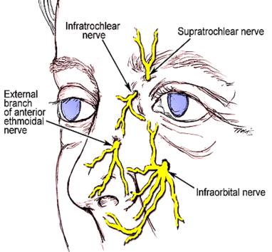
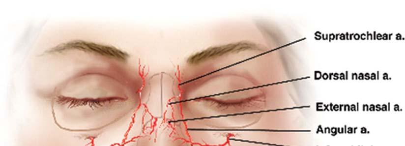
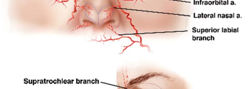
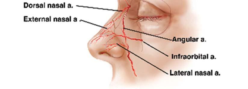
6 External Nose Skeleton of the nose Bony part Frontal, nasal, maxillary bones Cartilaginous part Lateral, septal, alar cartilages Blood supply: branches of ophthalmic, maxillary & facial aa. Nerve supply Infratrochlear (V1) External nasal (V1) Infraorbital (V2)
7

8 Nasal septum Bony part Perpendicular plate of ethmoid Vomer Cartilaginous part Septal cartilage

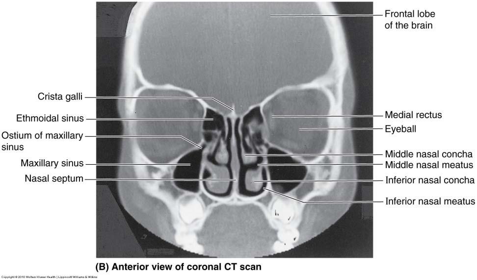
9 Parts Nasal vestibule Nasal septum Choanae (posterior nasal apertures) Walls of the nasal cavity Floor Roof Lateral wall Sphenoethmoidal recess Nasal conchae Superior, middle, & inferior Meatuses Superior, middle, & inferior Nasal Cavity

10 Sphenoethmoidal recess Sphenoid sinus Meatuses Superior Posterior ethmoid sinus Middle Bulla ethmoidalis Middle ethmoid sinus Hiatus semilunaris Maxillary sinus Infundibulum Frontal sinus Anterior ethmoid sinus Inferior Nasolacrimal duct Nasal Meatuses
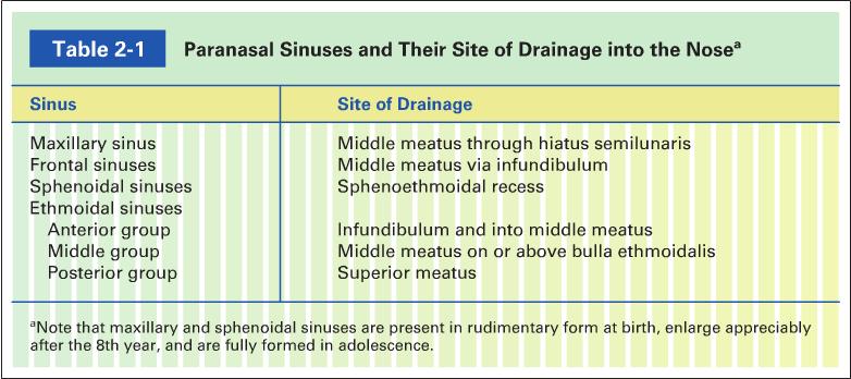
11 Paranasal Sinuses Paired cavities in ethmoid, sphenoid, frontal and maxillary bones Lined with mucous membranes and open into nasal cavity Resonating chambers for voice, lighten the skull Sinusitis is inflammation of the membrane
12 Paranasal Sinuses Maxillary sinus Between floor of orbit and roots of upper molars and premolars Superior alveolar nn. (V2) Frontal sinus Supraorbital nn. (V1) Sphenoid sinus Body of sphenoid Posterior ethmoidal nn. (V1) Ethmoid sinus Anterior, middle & posterior Between nasal cavity and orbit Anterior & posterior ethmoidal nn. From nasociliary n. (V1)
13

14 Paranasal Sinuses: X ray


15 Nasal Cavity: Innervation Olfactory nerve Trigeminal nerve Ophthalmic Maxillary


16 Nasal Cavity: Blood Supply Anterior & posterior ethmoidal aa. From ophthalmic a. Sphenopalatine a. From maxillary a. Septal branch from facial a. Kiesselbach s area & plexus Epistaxis Lymph drainage Deep cervical lymph nodes Vestibule submandibular lymph nodes
17 Pharynx
18 Pharynx Muscular tube (5 inch long) hanging from skull Skeletal muscle & mucous membrane Completed posteriorly & deficient anteriorly (openings into nose, mouth & larynx) Extends from internal nares to cricoid cartilage (C6) Funnel shape wide superiorly & narrow inferiorly (1.5 cm) Functions Passageway for food and air Resonating chamber for speech production Tonsil (lymphatic tissue) in the walls protects entryway into body

19 Regions of the Pharynx Distinct regions nasopharynx, oropharynx and laryngopharynx
 Tubal elevation Tubal tonsils Salpingopharyngeal fold Salpingopharyngeus m.")
20 Nasopharynx Above soft palate Openings Internal nares Pharyngeal isthmus Auditory (Eustachian, pharyngotympanic) tube Structures Pharyngeal tonsil (adenoids) Tubal elevation Tubal tonsils Salpingopharyngeal fold Salpingopharyngeus m. Pharyngeal recess

21 Oropharynx From soft palate to epiglottis Structures At floor Posterior ⅓ of tongue Lingual tonsils Median glossoepiglotic fold Lateral glossoepiglotic folds Valleculae

22 Oropharynx At lateral wall Palatoglossal fold Palatoglossus m. Oropharyngeal isthmus Palatopharyngeal fold Palatopharyngeus m. Palatine tonsil
23 Laryngopharynx Extends from epiglottis to cricoid cartilage Posterior to laryngeal inlet Piriform fossa Between aryepiglotic fold and thyroid cartilage


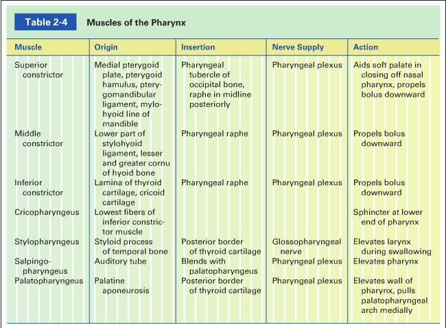
24 Pharyngeal Muscles External circular muscles Inserts into the pharyngeal raphe Superior, middle, & inferior constrictor mm. Overlap each other (inferior is more superficial) Cricopharyngeus m. Lower end Sphincter Internal longitudinal muscles Stylopharyngeus m. Palatopharengeus m. Salpingopharyngeus m. Nerve supply: pharyngeal plexus except stylopharyngeus muscle (IX)
25
26 Pharynx Sensory innervation Nasopharynx (V2) Oropharynx (IX) Laryngopharynx (X) internal laryngeal nerve Blood supply Ascending pharyngeal aa. Tonsillar branch of facial aa. Branches of maxillary & lingual aa. Lymph drainage Deep cervical nodes Retropharyngeal or paratracheal nodes deep cervical


27 Cartilage & connective tissue tube Below hyoid bone Anterior to C4 to C6 Short passageway connecting laryngopharynx with trachea Constructed of 3 single & 3 paired cartilages Functions Passageway for air Voice production Prevent entrance of food Larynx


28 Larynx Relations Infrahyoid mm. Thyroid gland Major blood vessels
29 Larynx: Cartilages
 Oblique line Superior & inferior cornua Cricothyroid joints Larynx:")
30 Thyroid cartilage The largest Incomplete ring Laminae Laryngeal prominence (Adam s apple) Oblique line Superior & inferior cornua Cricothyroid joints Larynx: Cartilages
")

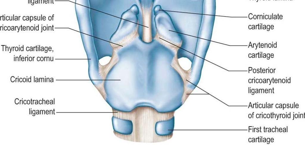
31 Larynx: Cartilages Cricoid cartilage Complete ring Below thyroid cartilage Lamina (posteriorly) Cricoarytenoid joints Arch (anteriorly) Arytenoid cartilages Apex, base, vocal process, & muscular process
 Stalk attached to thyroid")

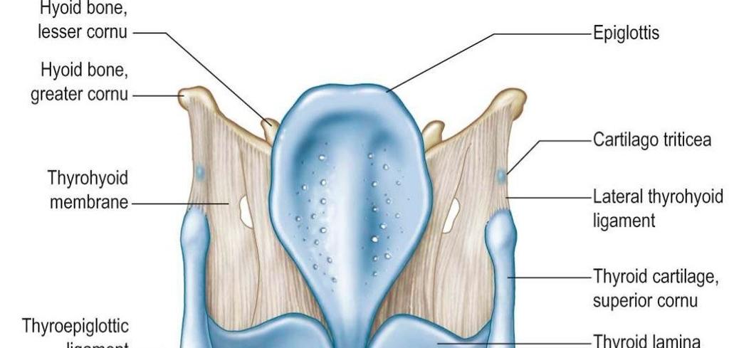
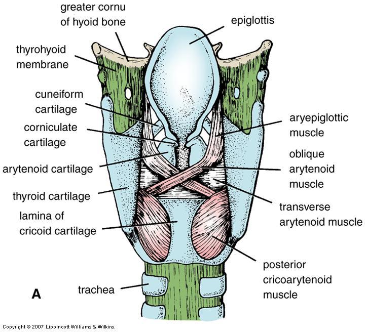
32 Epiglottis Leaf shape cartilage (elastic cartilage) Stalk attached to thyroid cartilage Aryepiglottic fold Median and lateral glossoepiglottic folds valleculae Larynx: Cartilages
 Cuneiform cartilages In the aryepiglottic fold")
33 Larynx: Cartilages Corniculate cartilages Above arytenoids (attachment of aryepiglottic fold) Cuneiform cartilages In the aryepiglottic fold (support)

 Vocal fold Mobile, avascular (whitish) Rima")
34 Larynx: Membranes & Ligaments Thyrohyoid membrane Median thyrohyoid ligament Cricotracheal ligament Quadrangular membrane Between epiglottis & arytenoid Vestibular ligament (inferior margin) Vestibular fold Immovable, vascular (pinkish) Cricothyroid ligament Vocal ligament (superior margin) Vocal fold Mobile, avascular (whitish) Rima glottidis (glottis)

 Between vocal folds & lower border of")
35 Larynx: Cavity Inlet of larynx Orientation Boundaries Vestibule Between inlet & vestibular folds Middle region Between laryngeal folds Laryngeal sinus (ventricle) Laryngeal saccule Lower region (infraglottic cavity) Between vocal folds & lower border of cricoid
36 Extrinsic muscles Larynx: Muscles Elevators Suprahyoids (Digastric, stylohyoid, mylohyoid, & geniohyoid) Longitudinal pharyngeal (stylopharyngeus, salpingopharyngeus, & palatopharyngeus) Depressors (infrahyoid) Sternothyroid, sternohyoid, & omohyoid


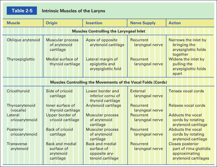
37 Larynx: Muscles Intrinsic muscles Modification of laryngeal inlet Narrowing Oblique arytenoid m. Widening Thyroepiglottic m. Movement of vocal cords Tensing Cricothyroid m. Relaxing Thyroarytenoids (vocalis) m. Adducting Lateral cricoarytenoid m. Abducting Posterior cricoarytenoid m. Approximating aretyneoids Transverse arytenoid m.

38 Larynx: Muscles
39

40 Voice Production Vocal folds are adducted Muscle contraction pulls elastic ligaments which stretch vocal folds out into airway Vibrate and produce sound with released air (frequency or pitch) Folds can move apart or together, elongate or shorten, tighter or looser Androgens make folds thicker and longer slower vibration and lower pitch Quality of voice determined by other structures (mouth, lips, tongue, pharynx, soft palate, & teeth)
 Blood supply Upper half: superior laryngeal branch of superior thyroid a.")
41 Nerve supply Sensory innervation (X) Above vocal cords: internal laryngeal n. Below vocal cords: recurrent laryngeal. Motor innervation All intrinsic muscles innervated by recurrent laryngeal except cricothyroid muscle (external laryngeal n.) Blood supply Upper half: superior laryngeal branch of superior thyroid a. Lower half: inferior laryngeal branch of the inferior thyroid a. Lymph drainage Deep cervical nodes Larynx
Structure and Nerve Supply of The Larynx
 Kingdom of Bahrain Arabian Gulf University College of Medicine and Medical sciences Structure and Nerve Supply of The Larynx This presentation was originally prepared by: Dr. Kumar Notes were added by:
Larynx. Rudimentary. Behind the posterior surface : -stylopharyngeus - salpingopharyngeus -platopharyngeus
 Larynx The larynx is an organ that provides a protective sphincter at the inlet of the air passages and is responsible for voice production. It extends from C3-C6: *Posterior: the pharynx *Lateral: the
Dr. Sami Zaqout Faculty of Medicine IUG
 The Nose External Nose Nasal Cavity External Nose Blood and Nerve Supplies of the External Nose Blood Supply of the External Nose The skin of the external nose Branches of the ophthalmic and the maxillary
Respiratory System. Cambridge University Press Concise Anatomy for Anaesthesia Andreas G. Erdmann Excerpt More information
 Respiratory System 1 The mouth DESCRIPTION The mouth extends from the lips (anterior) to the isthmus of the fauces (posterior). There are two sections: Vestibule slit-like cavity between the cheeks/lips
The Pharynx. Dr. Nabil Khouri MD. MSc, Ph.D
 The Pharynx Dr. Nabil Khouri MD. MSc, Ph.D Introduction The pharynx is the Musculo-fascial halfcylinder that links the oral and nasal cavities in the head to the larynx and esophagus in the neck Common
NURSE-UP RESPIRATORY SYSTEM
 NURSE-UP RESPIRATORY SYSTEM FUNCTIONS OF THE RESPIRATORY SYSTEM Pulmonary Ventilation - Breathing Gas exchanger External Respiration between lungs and bloodstream Internal Respiration between bloodstream
The Respiratory System
 The Respiratory System Respiration Includes Pulmonary ventilation Air moves in and out of lungs Continuous replacement of gases in alveoli (air sacs) External respiration Gas exchange between blood and
Larynx - cartilaginous structure holding the vocal folds which protrude into airstream
 1! Larynx - cartilaginous structure holding the vocal folds which protrude into airstream 2! Flow increase - like thumb over garden hose Pressure drop - narrower space forces pressure drop due to speed
Anatomy #1; Respiratory Nose and the Nasal Cavity December 1st, 2013
 Note #1: the doctor skipped some slides in the lecture. Those slides are not included in this sheet and so you will have to review the slides to study them. The reason they were not included is because
THE INTERIOR OF THE PHARYNX. By Dr. Muhammad Imran Qureshi
 THE INTERIOR OF THE PHARYNX By Dr. Muhammad Imran Qureshi The Cavity The cavity of the pharynx is divided into: 1. The Nasal part (called Nasopharynx) 2. The Oral part (called the Oropharynx), 3. And the
Nose & Mouth OUTLINE. Nose. - Nasal Cavity & Its Walls. - Paranasal Sinuses. - Neurovascular Structures. Mouth. - Oral Cavity & Its Contents
 Dept. of Human Anatomy, Si Chuan University Zhou hongying eaglezhyxzy@163.com Nose & Mouth OUTLINE Nose - Nasal Cavity & Its Walls - Paranasal Sinuses - Neurovascular Structures Mouth - Oral Cavity & Its
Anatomy of the Airway
 Anatomy of the Airway Nagelhout, 5 th edition, Chapter 26 Morgan & Mikhail, 5 th edition, Chapter 23 Mary Karlet, CRNA, PhD Airway Anatomy The airway consists of the nose, pharynx, larynx, trachea, and
Nose, Nasal cavity, Paranasal Sinuses & Pharynx
 Nose, Nasal cavity, Paranasal Sinuses & Pharynx Respiratory block-anatomy-lecture 2 Editing file Objectives At the end of the lecture, the students should be able to: Describe the boundaries of the nasal
Nasal region. cartilages: septal cartilage (l); lateral nasal cartilage (2); greater alar cartilages (2); lesser alar cartilages (?
; lateral nasal cartilage (2); greater alar cartilages (2); lesser alar cartilages (?") Nasal region skull bones: nasal and frontal processes of maxilla cartilages: septal cartilage (l); lateral nasal cartilage (2); greater alar cartilages (2); lesser alar cartilages (?) 1 Nasal cavity Roof
Nasal region skull bones: nasal and frontal processes of maxilla cartilages: septal cartilage (l); lateral nasal cartilage (2); greater alar cartilages (2); lesser alar cartilages (?) 1 Nasal cavity Roof
The PHARYNX. Dr. Nabil Khouri MD Ph.D
 The PHARYNX Dr. Nabil Khouri MD Ph.D PHARYNX Fibromuscular tube lined with mucous membrane extends from base of skull to lower border of cricoid cartilage (C-6). 12-14 cm long At the lower border of cricoid
Bisection of Head & Nasal Cavity 頭部對切以及鼻腔. 解剖學科馮琮涵副教授 分機
 Bisection of Head & Nasal Cavity 頭部對切以及鼻腔 解剖學科馮琮涵副教授 分機 3250 E-mail: thfong@tmu.edu.tw Outline: The structure of nose The concha and meatus in nasal cavity The openings of paranasal sinuses Canals, foramens
The Larynx. Prof. Dr.Mohammed Hisham Al-Muhtaseb
 The Larynx Prof. Dr.Mohammed Hisham Al-Muhtaseb The Larynx Extends from the middle of C3 vertebra till the level of the lower border of C6 Continue as Trachea Above it opens into the laryngo-pharynx Suspended
Prevertebral Region, Pharynx and Soft Palate
 Unit 20: Prevertebral Region, Pharynx and Soft Palate Dissection Instructions: Step1 Step 2 Step 1: Insert your fingers posterior to the sternocleidomastoid muscle, vagus nerve, internal jugular vein,
Please refer back to the slides as these are extra notes only. Slide 2 -The Larynx is a Box of cartilage.
 [ANATOMY #3] 1 بسم رلاهللا Please refer back to the slides as these are extra notes only. Slide 2 -The Larynx is a Box of cartilage. -The lower border of c6 is the lower border of cricoid cartilage. -The
SCHOOL OF ANATOMICAL SCIENCES Mock Run Questions. 4 May 2012
 SCHOOL OF ANATOMICAL SCIENCES Mock Run Questions 4 May 2012 1. With regard to the muscles of the neck: a. the platysma muscle is supplied by the accessory nerve. b. the stylohyoid muscle is supplied by
CHAPTER 22 RESPIRATORY
 pulmonary ventilation move air external respiration exchange gases transportation of gases internal respiration exchange gases CHAPTER 22 RESPIRATORY in / out lungs air - blood blood - cells cell respiration
I. Anatomy of the Respiratory System A. Upper Respiratory System Structures 1. Nose a. External Nares (Nostrils) 1) Vestibule Stratified Squamous
 1) Vestibule Stratified Squamous") I. Anatomy of the Respiratory System A. Upper Respiratory System Structures 1. Nose a. External Nares (Nostrils) 1) Vestibule Stratified Squamous Epithelium b. Nasal Cartilages 1) Nasal Cavity Pseudostratified
I. Anatomy of the Respiratory System A. Upper Respiratory System Structures 1. Nose a. External Nares (Nostrils) 1) Vestibule Stratified Squamous Epithelium b. Nasal Cartilages 1) Nasal Cavity Pseudostratified
The Respiratory System
 The Respiratory System Cells continually use O2 & release CO2 Respiratory system designed for gas exchange Cardiovascular system transports gases in blood Failure of either system rapid cell death from
Read Me. We are the Learning Lab. to look
 Respiratory Tract Anatomy Lab In-Lab Exercises Read Me We are going to look at models and slides. Much of this can be done in the Learning Lab on your own time. The steps do not have to be done in order,
Respiratory System. Functional Anatomy of the Respiratory System
 Respiratory System Overview of the Respiratory System s Job Major Duty Respiration Other important aspects ph control Vocalization Processing incoming air Protection Metabolism (ACE) What structures allow
Respiratory System. Ling Shucai
 Respiratory System Ling Shucai General Description Ⅰ. Constituents: Respiratory tract Lungs Pleura and plural cavity Ⅱ. Function: exchange O 2 and CO 2 mainly Mediastinum Respiratory tract Upper respiratory
12 Larynx. I - Cartilages. Learning Objectives
 12 Larynx Learning Objectives By the end of this topic you should be able to: Identify the cartilages, membranes, muscles and nerves of the larynx. Describe the attachments of the larynx to other structures
THE ANATOMY AND PHYSIOLOGY OF THE RESPIRATORY SYSTEM
 42790_01_ch01_001-052.qxd 7/6/07 12:23 PM Page 1 CHAPTER ONE THE ANATOMY AND PHYSIOLOGY OF THE RESPIRATORY SYSTEM O B J E C T I V E S By the end of this chapter, the student should be able to: 1. List
Anatomy 2. Parotid bed (V.imp): meaning that gland is sleeping on structures and they are:
: meaning that gland is sleeping on structures and they are:") Anatomy 2 Parotid Gland: "refer to previous sheet for extra details." Its pyramidal in shape, apex is toward pharynx. Its Medial surface is divided into Anterio-medial and posterio-medial and its posterio-medial
Anatomy 2 Parotid Gland: "refer to previous sheet for extra details." Its pyramidal in shape, apex is toward pharynx. Its Medial surface is divided into Anterio-medial and posterio-medial and its posterio-medial
Oral cavity : consist of two parts: the oral vestibule and the oral cavity proper. Oral vestibule : is slit like space between.
 Oral cavity Oral cavity : consist of two parts: the oral vestibule and the oral cavity proper Oral vestibule : is slit like space between the teeth, buccal gingiva, lips, and cheeks 1 Oral cavity Oral
The Respiratory System. Supplies body with oxygen Disposes of carbon dioxide Four processes in respiration
 C H A P T E R 22 The Respiratory System The Respiratory System Supplies body with oxygen Disposes of carbon dioxide Four processes in respiration Pulmonary ventilation External respiration Transport of
The Respiratory System
 13 PART A The Respiratory System PowerPoint Lecture Slide Presentation by Jerry L. Cook, Sam Houston University ESSENTIALS OF HUMAN ANATOMY & PHYSIOLOGY EIGHTH EDITION ELAINE N. MARIEB Organs of the Respiratory
The Digestive System in the Head and Neck
 The Digestive System in the Head and Neck The Mouth The Lips The lips are two fleshy folds that surround the oral orifice They are covered on the outside by skin and are lined on the inside by mucous membrane
The Respiratory System:
 The Respiratory System: Respiration Involves both the respiratory and the circulatory systems Four processes that supply the body with O 2 and dispose of CO 2 Respiration Pulmonary ventilation (breathing):
The Respiratory System
 PowerPoint Lecture Slide Presentation by Vince Austin Human Anatomy & Physiology FIFTH EDITION Elaine N. Marieb The Respiratory System Dr Nabil Khouri. MD, Ph.D Respiratory System Consists of a conducting
LARYNX ANATOMY. Elena Rizzo Riera R1 ORL HUSE
 LARYNX ANATOMY Elena Rizzo Riera R1 ORL HUSE INTRODUCTION v Odd and median organ v Infrahyoid region v Phonation, swallowing and breathing v Triangular pyramid v Postero- superior base à pharynx and hyoid
Dr.Ban I.S. head & neck anatomy 2 nd y. جامعة تكريت كلية طب االسنان املرحلة الثانية
 جامعة تكريت كلية طب االسنان التشريح مادة املرحلة الثانية أ.م.د. بان امساعيل صديق 6102-6102 1 The Palate The palate forms the roof of the mouth and the floor of the nasal cavity. It is divided into two
Subdivided into Vestibule & Oral cavity proper
 Extends from the lips to the oropharyngeal isthmus The oropharyngeal isthmus: Is the junction of mouth and pharynx. Is bounded: Above by the soft palate and the palatoglossal folds Below by the dorsum
Anterior triangle of neck
 Anterior triangle of neck Dept. of Anatomy Zhou Hong Ying Outline boundary and subdivisions of ant. triangle contents of the triangle Muscles: suprahyoid muscles, infrahyoid muscles Nerves: CNⅩ, CNⅪ, CNⅫ,
Lungs a. d. b. c. e.
 Lungs d. e. Lungs Right superior lobe Right middle lobe Right inferior lobe d. Left superior lobe e. Left inferior lobe Sinuses d. Nasal Cavity & Sinuses g. g. i. Nasal Cavity & Sinuses g. h. d. f. e.
Omran Saeed. Luma Taweel. Mohammad Almohtaseb. 1 P a g e
 2 Omran Saeed Luma Taweel Mohammad Almohtaseb 1 P a g e I didn t include all the photos in this sheet in order to keep it as small as possible so if you need more clarification please refer to slides In
Anatomical Considerations for Lab Practical II
 Anatomical Considerations for Lab Practical II For each of the following please be prepared to provide: Identification System Organ(s) or ducts to Function(s) location which it is attached Use your lecture
Raneen Hamdan. Raghad Abu Jebbeh. Mohammad almuhtaseb
 1 Raneen Hamdan Raghad Abu Jebbeh Mohammad almuhtaseb Introduction Respiratory System Organs: 1) Starting from the Nose (nasal cavity). 2) Pharynx (Nasopharynx, oropharynx and laryngopharynx, previously
University of Palestine. Midterm Exam 2013/2014 Total Grade:
 Course No: DNTS2208 Course Title: Head and Neck Anatomy Date: 09/11/2013 No. of Questions: (50) Time: 1hour Using Calculator (No) University of Palestine Midterm Exam 2013/2014 Total Grade: Instructor
THE RESPIRATORY SYSTEM
 THE RESPIRATORY SYSTEM Functions of the Respiratory System Provides extensive gas exchange surface area between air and circulating blood Moves air to and from exchange surfaces of lungs Protects respiratory
Anatomic Relations Summary. Done by: Sohayyla Yasin Dababseh
 Anatomic Relations Summary Done by: Sohayyla Yasin Dababseh Anatomic Relations Lecture 1 Part-1 - The medial wall of the nose is the septum. - The vestibule lies directly inside the nostrils (Nares). -
B. Correct! As air travels through the nasal cavities, it is warmed and humidified.
 Human Anatomy - Problem Drill 20: The Respiratory System Question No. 1 of 10 1. Which of the following statements about the portion of the respiratory system labeled in the image below is correct? Question
The Respiratory System
 The Respiratory System If you have not done so already, please print and bring to class the Laboratory Practical II Preparation Guide. We will begin using this shortly in preparation of your second laboratory
Larynx, Trachea & Bronchi
 Larynx, Trachea & Bronchi Respiratory block-anatomy-lecture 3 Editing file Objectives By the end of the lecture, you should be able to: Describe the Extent, structure and functions of the larynx. Describe
Basic Anatomy and Physiology of the Lips and Oral Cavity. Dr. Faghih
 Basic Anatomy and Physiology of the Lips and Oral Cavity Dr. Faghih It is divided into seven specific subsites : 1. Lips 2. dentoalveolar ridges 3. oral tongue 4. retromolar trigone 5. floor of mouth 6.
*in general the blood supply of the nose comes from branches of the internal and external carotid arteries.
 In the previous lecture we talked about the anatomy of the nasal cavity, today we will talk about its blood supply, venous drainage, innervations, and finally about the paranasal sinuses. When we describe
CHAPTER 24. Respiratory System
 CHAPTER 24 Respiratory System RESPIRATION INCLUDES Air moves in and out of lungs Continuous replacement of gases in alveoli (air sacs) Gas exchange between blood and air at alveoli Transport of respiratory
Oral Cavity, Soft Palate, Pharynx, and Larynx; Development of the Face and Palate
 Oral Cavity, Soft Palate, Pharynx, and Larynx; Development of the Face and Palate Think on this. The ability to eat and drink safely and efficiently is fundamental to our quality of life. The wide variety
Anatomy of Oral Cavity DR. MAAN AL-ABBASI
 Anatomy of Oral Cavity DR. MAAN AL-ABBASI By the end of this lecture you should be able to: 1. Differentiate different parts of the oral cavity 2. Describe the blood and nerve supply of mucosa and muscles
A. The supraclavicular nerves supply sensory fibers to the skin of the clavicular area
 YR 1 GROSS ANATOMY WRITTEN EXAM 2 -- October 10, 1997. CHOOSE THE SINGLE BEST ANSWER FOR QUESTIONS 1-42. 1. Each of the following statements is CORRECT EXCEPT: A. The supraclavicular nerves supply sensory
Respiratory System Structures and Gas Exchange
 A. Respiratory medium the oxygen source 1. Air 2. Water Respiratory Medium Organism Cellular Respiration O 2 CO 2 B. Respiratory surface the structure where exchange of gases with the surrounding environment
Chapter 26: The temporomandibular joint, pharynx and larynx. The Temporomandibular Joint. Ligaments. (a) Capsular
 Capsular") Chapter 26: The temporomandibular joint, pharynx and larynx The Temporomandibular Joint This is a synovial joint of a condyloid (modified hinge) variety between the condyle of the mandible and the mandibular
Chapter 26: The temporomandibular joint, pharynx and larynx The Temporomandibular Joint This is a synovial joint of a condyloid (modified hinge) variety between the condyle of the mandible and the mandibular
Ch16: Respiratory System
 Ch16: Respiratory System Function: - O2 in and CO2 out of the blood vessels in the lungs - O2 out and CO2 into the blood vessels around the cells - Gas exchange happens in - Other organs purify, humidify,
PTERYGOPALATINE FOSSA
 PTERYGOPALATINE FOSSA Outline Anatomical Structure and Boundaries Foramina and Communications with other spaces and cavities Contents Pterygopalatine Ganglion Especial emphasis on certain arteries and
AIRWAY MANAGEMENT SUZANNE BROWN, CRNA
 AIRWAY MANAGEMENT SUZANNE BROWN, CRNA OBJECTIVE OF LECTURE Non Anesthesia Sedation Providers Review for CRNA s Informal Questions encouraged 2 AIRWAY MANAGEMENT AWARENESS BASICS OF ANATOMY EQUIPMENT 3
Lecture Overview. Respiratory System. Martini s Visual Anatomy and Physiology First Edition. Chapter 20 - Respiratory System Lecture 11
 Martini s Visual Anatomy and Physiology First Edition Martini Ober Chapter 20 - Respiratory System Lecture 11 1 Lecture Overview Overview of respiration Functions of breathing Organs of the respiratory
The Respiratory System
 PowerPoint Lecture Slides prepared by Leslie Hendon University of Alabama, Birmingham C H A P T E R 22 Part 1 The Respiratory System The Respiratory System Basic functions of the respiratory system Supplies
LECTURE 2 THE RESPIRATORY SYSTEM
 LECTURE 2 THE RESPIRATORY SYSTEM Respiratory system - a complex of organs and anatomical structures exercising function of external respiration. Functions of the respiratory system: - Provides the organism
University of Palestine. Midterm Exam 2013/2014 Total Grade:
 [ Course No: DNTS2208 Course Title: Head and Neck Anatomy Date: 17/11/1024 No. of Questions: (52) Time: 2hours Using Calculator (No) University of Palestine Midterm Exam 2013/2014 Total Grade: Instructor
Oral Cavity and Pharynx. The Oral Cavity. The oral cavity is divided into two major portions: the vestibule and the cavum oris.
 11 Oral Cavity and Pharynx Persons who specialize in the care and treatment of the oral cavity have a great responsibility. The oral cavity participates actively in respiration, nutrition, and excretion
RESPIRATORY SYSTEM. described: pp. 744,746 fig. 25.1, described: p. 746 fig described: p. 776 fig. 26.3
 ACTIVITY 11: RESPIRATORY AND DIGESTIVE SYSTEMS OBJECTIVES: 1) How to get ready: Read Chapters 25 and 26, McKinley et al., Human Anatomy, 5e. All text references are for this textbook. 2) Identify structures
Vocal Systems * Marcos Gridi-Papp. 1 The Human Larynx
 OpenStax-CNX module: m66861 1 Vocal Systems * Marcos Gridi-Papp This work is produced by OpenStax-CNX and licensed under the Creative Commons Attribution License 4.0 Abstract The human larynx is protects
Respiratory System. Clinical notes. Published on Second Faculty of Medicine, Charles University ( https://www.lf2.cuni.cz)
") Published on Second Faculty of Medicine, Charles University ( https://www.lf2.cuni.cz) Respiratory System The test of the respiratory system follows the general rules for written tests (see Continuous
Published on Second Faculty of Medicine, Charles University ( https://www.lf2.cuni.cz) Respiratory System The test of the respiratory system follows the general rules for written tests (see Continuous
ACTIVITY 11: RESPIRATORY AND DIGESTIVE SYSTEMS RESPIRATORY SYSTEM
 ACTIVITY 11: RESPIRATORY AND DIGESTIVE SYSTEMS OBJECTIVES: 1) How to get ready: Read Chapters 25 and 26, McKinley et al., Human Anatomy, 4e. All text references are for this textbook. 2) Identify structures
Maxilla, ORBIT and infratemporal fossa. Neophytos C Demetriades MD, DDS, MSc Associate professor European University of Cyprus School of Medicine
 Maxilla, ORBIT and infratemporal fossa Neophytos C Demetriades MD, DDS, MSc Associate professor European University of Cyprus School of Medicine MAXILLA Superior, middle, and inferior meatus Frontal sinus
Anatomy & Regional Anaesthesia
 THE MOUTH the vestibule is formed from the lips and cheeks, bounded within by the teeth & gums the opening to the parotid duct is adjacent the 2 nd upper molar the potential space is minimised by facial
Laryngeal Anatomy. Dr.Hani Abdulsattar Shaker
 1/14 Laryngeal Anatomy The larynx ("organ of voice") is a valve separating the trachea from the upper aerodigestive tract. It is placed at the upper part of the air passage. It is situated between the
Organs and Structures of the Respiratory System
 Organs and Structures of the Respiratory System Bởi: OpenStaxCollege The major organs of the respiratory system function primarily to provide oxygen to body tissues for cellular respiration, remove the
Bones Ethmoid bone Inferior nasal concha Lacrimal bone Maxilla Nasal bone Palatine bone Vomer Zygomatic bone Mandible
 splanchnocranium - Consists of part of skull that is derived from branchial arches - The facial bones are the bones of the anterior and lower human skull Bones Ethmoid bone Inferior nasal concha Lacrimal
Tympanic Bulla Temporal Bone. Digastric Muscle. Masseter Muscle
 Superior view Hyoid Bone The hyoid bone does not articulate with any other bones. It is held in place by ligaments to the styloid process of the temporal bone and the thyroid cartilage of the larynx. It
BELLWORK DAY 1 RESEARCH THE DIFFERENCE BETWEEN INTERNAL AND EXTERNAL RESPIRATION. COPY BOTH OF THE STATE STANDARDS ENTIRELY ON THE NEXT SLIDE.
 BELLWORK DAY 1 RESEARCH THE DIFFERENCE BETWEEN INTERNAL AND EXTERNAL RESPIRATION. COPY BOTH OF THE STATE STANDARDS ENTIRELY ON THE NEXT SLIDE. STANDARDS 42) Review case studies that involve persons with
RESPIRATORY SYSTEM. A. Upper respiratory tract (Fig. 23.1) Use the half-head models.
 Use the half-head models.") RESPIRATORY SYSTEM I. OVERVIEW OF THE RESPIRATORY SYSTEM AND THORAX A. Upper respiratory tract (Fig. 23.1) Use the half-head models. Nasal cavity Pharynx (fare-rinks) B. Lower respiratory tract (Fig. 23.1)
RESPIRATORY SYSTEM I. OVERVIEW OF THE RESPIRATORY SYSTEM AND THORAX A. Upper respiratory tract (Fig. 23.1) Use the half-head models. Nasal cavity Pharynx (fare-rinks) B. Lower respiratory tract (Fig. 23.1)
Pharynx. Muscles of Pharynx
 Pharynx A funnel shaped fibromuscular tube that extends from the base of the skull & continues below with the esophagus at the level of C6 in the neck. It is divided into 3 parts: (1) Nasal: nasopharynx;
Objectives. Module A2: Upper Airway Anatomy & Physiology. Function of the Lungs/Heart. The lung is for gas exchange. Failure of the Lungs/Heart
 Module A2: Upper Airway Anatomy & Physiology Objectives Classify epithelial tissue based on cell type and tissue layers. Identify location of tissue epithelium in the respiratory system. Describe the major
Organs of the Respiratory System Laboratory Exercise 52
 Organs of the Respiratory System Laboratory Exercise 52 Background The organs of the respiratory system include the nose, nasal cavity, sinuses, pharynx, larynx, trachea, bronchial tree, and lungs. They
HEAD & NECK ANATOMY - MCQ HEAD & NECK ANATOMY
 . ' HEAD & NECK ANATOMY I. Deep investing layer of cervical fascia splits to enclose: A. Sternocleidomastoid B. Trapezius C. Parotid gland D. Omohyoid 2. Regarding the prevertebral fascia, the following
Mohammad Mohtaseb. Nour Hussein. Faisal Nimri
 2 Mohammad Mohtaseb Nour Hussein Faisal Nimri Muscles of the tongue The tongue is a muscular organ and contains intrinsic and extrinsic muscles. The intrinsic muscle contains vertical, oblique, and transverse
-Ibrahim Al-Naser. -Dr Al- Muhtaseb. 1 P a g e
 -1 -Ibrahim Al-Naser - -Dr Al- Muhtaseb 1 P a g e The Digestive System The doctor started the lecture by talking about the class rules. The GI system is an organ system, it is divided into: The Alimentary
The Neck. BY: Lina Abdullah & Rahaf Jreisat
 The Neck BY: Lina Abdullah & Rahaf Jreisat Boundaries of the Neck: generally from base of the skull to root of the neck Superior margin :From superior nuchal line of occipital bone up to mastoid process
04 Development of the Face and Neck. Development of the Face Development of the neck
 04 Development of the Face and Neck Development of the Face Development of the neck Development of the face Overview of facial development The fourth week ~ the twelfth week of prenatal development Between
The Neck the lower margin of the mandible above the suprasternal notch and the upper border of the clavicle
 The Neck is the region of the body that lies between the lower margin of the mandible above and the suprasternal notch and the upper border of the clavicle below Nerves of the neck Cervical Plexus Is formed
Dr.Ban I.S. head & neck anatomy 2 nd y جامعة تكريت كلية طب االسنان مادة التشريح املرحلة الثانية أ.م.د. بان امساعيل صديق 6102/6102
 جامعة تكريت كلية طب االسنان مادة التشريح املرحلة الثانية أ.م.د. بان امساعيل صديق 6102/6102 Pterygopalatine fossa: The pterygopalatine fossa is a cone-shaped depression, It is located between the maxilla,
- Reem Akiely. -Wardeh Al-Swalmeh. - Mohammad Al-Muhtaseb. 1 P a g e
 -2 - Reem Akiely -Wardeh Al-Swalmeh - Mohammad Al-Muhtaseb 1 P a g e The palate: * Hard palate * Soft palate the Uvula: is a muscular structure present In the midline of the soft palate (اللهاة) The Hard
Karachi King s College of Nursing
 Karachi King s College of Nursing Badil Dass Lecturer Respiratory system Respiratory System Respiratory system consist of: Nose Pharynx (Throat) Larynx (Voice Box) Trachea (Wind Pipe) Bronchi Bronchioles
Infratemporal fossa: Tikrit University college of Dentistry Dr.Ban I.S. head & neck Anatomy 2 nd y.
 Infratemporal fossa: This is a space lying beneath the base of the skull between the lateral wall of the pharynx and the ramus of the mandible. It is also referred to as the parapharyngeal or lateral pharyngeal
Chapter 23 The Respiratory System
 Chapter 23 The Respiratory System Cells continually use O 2 & release CO 2 Respiratory System designed for gas exchange Cardiovascular system transports gases in blood Failure of either system rapid cell
Organs and Structures of the Respiratory System
 OpenStax-CNX module: m46548 1 Organs and Structures of the Respiratory System OpenStax College This work is produced by OpenStax-CNX and licensed under the Creative Commons Attribution License 4.0 By the
APRIL
 APRIL - 2003 OCTOBER - 2003 February 2009 [KU 652] Sub. Code : 4131 FIRST B.D.S DEGREE EXAMINATION (Modified Regulations III) Paper I HUMAN ANATOMY, HISTOLOGY AND EMBRYOLOGY Time : Three hours
Lecture 07. Lymphatic's of Head & Neck. By: Dr Farooq Amanullah Khan PMC
 Lecture 07 Lymphatic's of Head & Neck By: Dr Farooq Amanullah Khan PMC Dated: 28.11.2017 Lymphatic Vessels Of the 800 lymph nodes in the human body, 300 are in the Head & neck region. The lymphatic vessels
Anatomy of the Respiratory System
 Anatomy of the Respiratory System Respiration is a term used to refer to ventilation of the lungs (breathing) In other contexts it can be used to refer to part of cellular metabolism Functions of respiration
Cranial nerves.
 Cranial nerves eaglezhyxzy@163.com Key Points of Learning Name Components Passing through Peripheral distribution Central connection Function Cranial nerves Ⅰ olfactory Ⅱ optic Ⅲ occulomotor Ⅳ trochlear
The RESPIRATORY System
 The RESPIRATORY System Respira5on The exchange of gases between the atmosphere, blood, and cells Pulmonary Ven5la5on - the exchange of air between the atmosphere and lungs External (Pulmonary) Respira5on
Trigeminal Nerve (V)
") Trigeminal Nerve (V) Lecture Objectives Discuss briefly how the face is developed. Follow up the course of trigeminal nerve from its point of central connections, exit and down to its target areas. Describe
Trigeminal Nerve (V) Lecture Objectives Discuss briefly how the face is developed. Follow up the course of trigeminal nerve from its point of central connections, exit and down to its target areas. Describe
ORAL CAVITY, ESOPHAGUS AND STOMACH
 ORAL CAVITY, ESOPHAGUS AND STOMACH 1 OBJECTIVES By the end of the lecture you should be able to: Describe the anatomy the oral cavity, (boundaries, parts, nerve supply). Describe the anatomy of the palate,