Parotid Gland. Parotid Gland. Largest of 3 paired salivary glands (submandibular; sublingual) Ramus of Mandible. Medial pterygoid.
|
|
|
- Cecilia Park
- 5 years ago
- Views:
Transcription

1 Parotid region
2
3
 Ramus of Mandible Medial")

4 Parotid Gland Largest of 3 paired salivary glands (submandibular; sublingual) Ramus of Mandible Medial pterygoid Cross section of mandible Masseter D S SCM Parotid Gland Mastoid Process Facial nerve and its branches


5 Parotid Duct Parotid Duct Parotid Gland Buccal Branch of the Facial Nerve Masseter Buccinator Parotid Duct

6 Parotid Duct 3 parallel structures!! Transverse Facial Artery Parotid Duct Buccinator Buccal Branch of the Facial Nerve Retromandibular v. Parotid Gland Masseter 2nd Molar

7 Intraoral opening of the parotid duct (next to the upper second molar)
8

9 Innervation of the parotid gland
10

11 O S T E O L O G Y Temporal fossa Frontal Zygomatic Maxilla Grtr wing sphenoid Mand ramus Parietal Temporal Zygomatic arch (zygoma)


12 Boundaries of the Infratemporal Fossa: Frontal Parietal Zyg Grtr wing Sphenoid Roof Temporal Ext. aud meatus Maxilla Styloid P. Infratemporal fossa Lat pterygoid plate
13 Channels communicating with the infratemporal fossa: Inferior orbital fissure Frontal Grtr wing Sphenoid Parietal Temporal Zyg Pterygomaxillary fissure Maxilla Foramen ovale + Foramen spinosum
14
 Maxillary artery + branches Pterygoid plexus of")
15 Contents of the infratemporal fossa: Three (of four) muscles of mastication Mandibular nerve (V 3 ) + branches Otic ganglion Chorda tympani nerve (between facial and lingual nerve) Maxillary artery + branches Pterygoid plexus of veins
16 Muscles of Mastication: Temporalis Temporalis fascia Masseter Parotid duct


17 Muscles of Mastication: Lateral pterygoid Articular disk Head of mandible Medial pterygoid

18 Temporalis and Masseter muscles

19 Jaw opening muscles

20 Anterior belly of digastric muscle
:")
 Elevation (close mouth, occlusion): masseter (#2),")
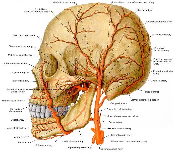
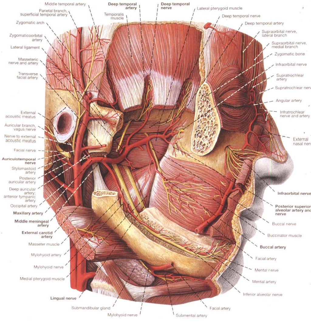
 + masseter (#2) Retrusion")
21 Lateral (side to side) motion: lateral and medial pterygoids (#3,4) Action of muscles of mastication on the mandible: Depression (open mouth): anterior belly of digastric, geniohyoid, lateral pterygoids (#3), mylohyoid (minor role) Elevation (close mouth, occlusion): masseter (#2), temporalis (#1), medial pterygoids (#4) Protrusion (protraction): mostly medial and lateral pterygoids (#3,4) + masseter (#2) Retrusion (retraction): temporalis (#1)
 fossa Mandibular head (condyle)")
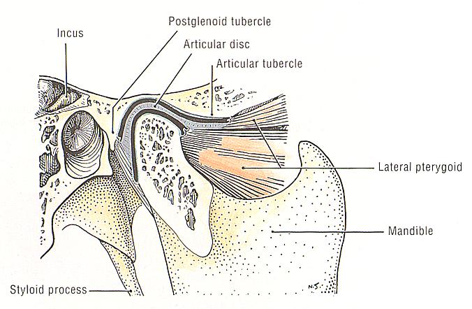
22 Temporomandibular Joint: Articulation of condyle of mandible with mandibular fossa plus articular eminence of temporal bone Articular eminence Mandibular (glenoid) fossa Mandibular head (condyle)
23 TMJ: details of articulation Gliding Action Upper synovial space Mandibular fossa Articular disc Lateral pterygoid tendon Condyle Hinge Action Lower synovial space

 requires translation of the condyle+articular disc on the articular")
24 TMJ movement: Initial opening of mouth involves rotation of the condyle in the lower compartment of the TMJ. This is the HINGE motion. Further opening (beyond 20 mm.) requires translation of the condyle+articular disc on the articular eminence, which occurs in the upper compartment. This is the GLIDING motion.
25

26 Capsular ligament of the TMJ (enclosing the TMJ)

27 Ligaments of the temporomandibular joint


28 Review: Trigeminal nerve in middle cranial fossa: V V 1 V 2 V 3 V 3 Exits through foramen ovale to enter infratemporal fossa.
 SS Lingual")
29 SS+parapost from otic ganglia (IX) Auriculotemporal MANDIBULAR NERVE (V 3 ) V 3 Somatosensory and somatomotor Chorda tympani* n (VII) Para pre & Taste SS Inferior alveolar n Mylohyoid n SM to mylohyoid & anterior digastric mm * Taste & parasympathetic pre-ganglionic Mental n SS SS Buccal n To go in and innervate cheek (don t get confused with buccal branch of CNVII) SS Lingual n

30 Foramen ovale Eye Greater wing of sphenoid Ear rior V 3 Maxilla Posterio Mandible

31 Chorda tympani B Posterior Anterior L I Ramus fractured M
32 Maxillary artery Maxillary artery
33

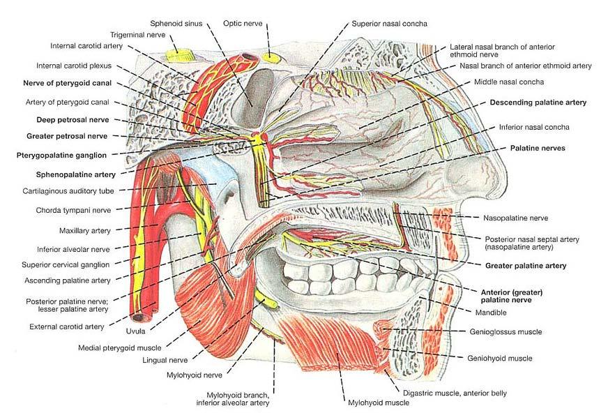
34 The pterygopalatine fossa is somewhat cone-shaped, located between the infratemporal fossa laterally and nasal cavity medially. It has six openings. Sphenoid bone posterior view Nasal cavity Infratemporal fossa
35 Six openings in to or out of the pterygopalatine fossa: Infraorbital (V 2 ) & artery 4. Inferior orbital fissure Nasal nerves Sphenopalatine artery 6. Sphenopalatine foramen Nasal Cavity Right side 1. Foramen rotundum Maxillary (V 2 ) Infratemporal Fossa 2. Pterygoid canal Greater petrosal nerve nerve of pterygoid canal (Vidian s) 5. Pterygomaxillary fissure Maxillary artery 3. Greater palatine foramen Greater & lesser palatine n.

36 Maxillary Artery entering pterygopalatine fossa ECA Maxillary artery arising from external carotid artery

37 Superficial temporal a Maxillary artery I Maxillary a Ext carotid a Inferior alveolar artery and nerve

38 Maxillary Artery II Maxillary a Auriculotemporal n Middle meningeal a

39 Auriculotemporal nerve Middle meningeal artery V 3 Maxillary artery ECA
40

41 Maxillary artery entering the pterygopalatine fossa
42

43 Veins in the facial region
Temporal region. temporal & infratemporal fossae. Zhou Hong Ying Dept. of Anatomy
 Temporal region temporal & infratemporal fossae Zhou Hong Ying Dept. of Anatomy Temporal region is divided by zygomatic arch into temporal & infratemporal fossae. Temporal Fossa Infratemporal fossa Temporal
Parotid Gland, Temporomandibular Joint and Infratemporal Fossa
 M1 - Anatomy Parotid Gland, Temporomandibular Joint and Infratemporal Fossa Jeff Dupree Sanger 9-057 jldupree@vcu.edu Parotid gland: wraps around the mandible positioned between the mandible and the sphenoid
Temporal fossa Infratemporal fossa Pterygopalatine fossa Terminal branches of external carotid artery Pterygoid venous plexus
 Outline of content Temporal fossa Infratemporal fossa Pterygopalatine fossa Terminal branches of external carotid artery Pterygoid venous plexus Boundary Content Communication Mandibular division of trigeminal
The Skull and Temporomandibular joint II Prof. Abdulameer Al-Nuaimi. E. mail:
 The Skull and Temporomandibular joint II Prof. Abdulameer Al-Nuaimi E-mail: a.al-nuaimi@sheffield.ac.uk E. mail: abdulameerh@yahoo.com Temporal fossa The temporal fossa is a depression on the temporal
Infratemporal fossa: Tikrit University college of Dentistry Dr.Ban I.S. head & neck Anatomy 2 nd y.
 Infratemporal fossa: This is a space lying beneath the base of the skull between the lateral wall of the pharynx and the ramus of the mandible. It is also referred to as the parapharyngeal or lateral pharyngeal
Lec [8]: Mandibular nerve:
![Lec [8]: Mandibular nerve:](/thumbs/94/121295776.jpg "Lec [8]: Mandibular nerve:") Lec [8]: Mandibular nerve: The mandibular branch from the trigeminal ganglion lies in the middle cranial fossa lateral to the cavernous sinus. With the motor root of the trigeminal nerve [motor roots lies
Lec [8]: Mandibular nerve: The mandibular branch from the trigeminal ganglion lies in the middle cranial fossa lateral to the cavernous sinus. With the motor root of the trigeminal nerve [motor roots lies
PTERYGOPALATINE FOSSA
 PTERYGOPALATINE FOSSA Outline Anatomical Structure and Boundaries Foramina and Communications with other spaces and cavities Contents Pterygopalatine Ganglion Especial emphasis on certain arteries and
Trigeminal Nerve Worksheets, Distributions Page 1
 Trigeminal Nerve Worksheet #1 Distribution by Nerve Dr. Darren Hoffmann Dental Gross Anatomy, Spring 2013 We have drawn out each of the branches of CN V in lecture and you have an idea now for their basic
Anatomy and Physiology. Bones, Sutures, Teeth, Processes and Foramina of the Human Skull
 Anatomy and Physiology Chapter 6 DRO Bones, Sutures, Teeth, Processes and Foramina of the Human Skull Name: Period: Bones of the Human Skull Bones of the Cranium: Frontal bone: forms the forehead and the
Tikrit University collage of dentistry Dr.Ban I.S. head & neck anatomy 2 nd y. Lec [5] / Temporal fossa :
![Tikrit University collage of dentistry Dr.Ban I.S. head & neck anatomy 2 nd y. Lec [5] / Temporal fossa :](/thumbs/88/115294566.jpg "Tikrit University collage of dentistry Dr.Ban I.S. head & neck anatomy 2 nd y. Lec [5] / Temporal fossa :") Lec [5] / Temporal fossa : Borders of the Temporal Fossa: Superior: Superior temporal line. Inferior: gap between zygomatic arch and infratemporal crest of sphenoid bone. Anterior: Frontal process of the
Lec [5] / Temporal fossa : Borders of the Temporal Fossa: Superior: Superior temporal line. Inferior: gap between zygomatic arch and infratemporal crest of sphenoid bone. Anterior: Frontal process of the
Mohammad Hisham Al-Mohtaseb. Lina Mansour. Reyad Jabiri. 0 P a g e
 2 Mohammad Hisham Al-Mohtaseb Lina Mansour Reyad Jabiri 0 P a g e This is only correction for the last year sheet according to our record. If you already studied this sheet just read the yellow notes which
Dr.Ban I.S. head & neck anatomy 2 nd y. جامعة تكريت كلية طب االسنان املرحلة الثانية أ.م.د. بان امساعيل صديق 6102/6102
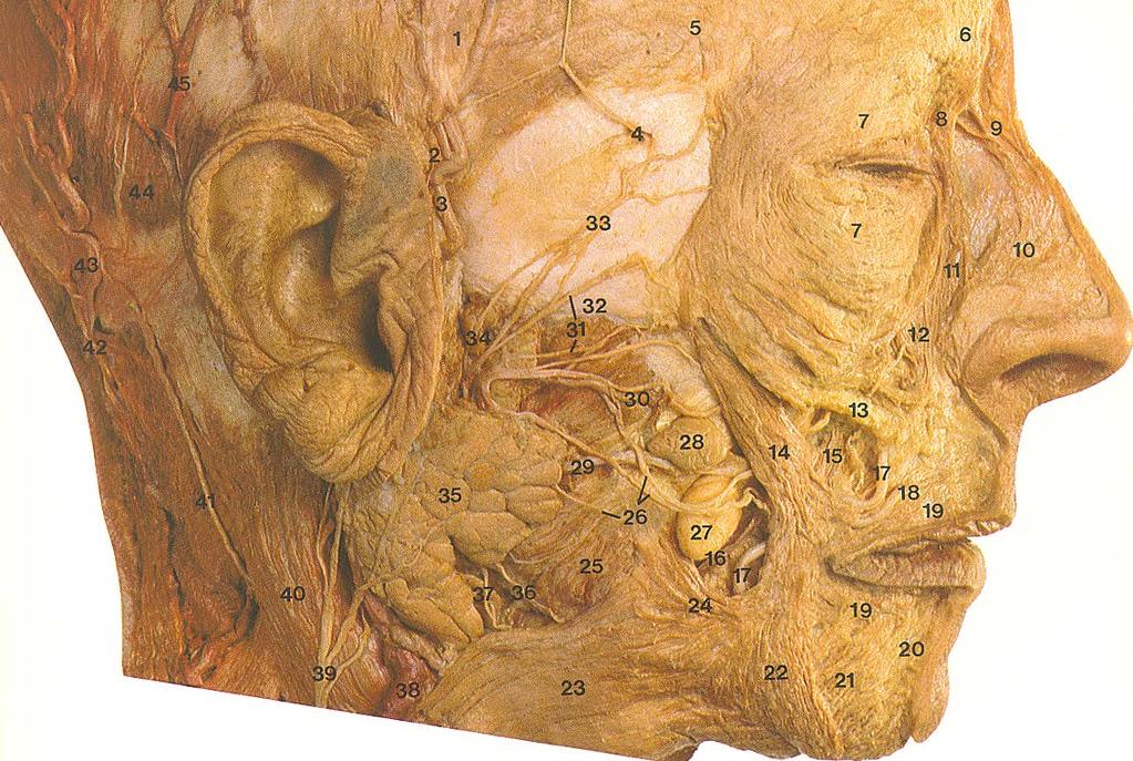
 جامعة تكريت كلية طب االسنان التشريح مادة املرحلة الثانية أ.م.د. بان امساعيل صديق 6102/6102 Parotid region The part of the face in front of the ear and below the zygomatic arch is the parotid region. The
Omran Saeed. Luma Taweel. Mohammad Almohtaseb. 1 P a g e
 2 Omran Saeed Luma Taweel Mohammad Almohtaseb 1 P a g e I didn t include all the photos in this sheet in order to keep it as small as possible so if you need more clarification please refer to slides In
SCHOOL OF ANATOMICAL SCIENCES Mock Run Questions. 4 May 2012
 SCHOOL OF ANATOMICAL SCIENCES Mock Run Questions 4 May 2012 1. With regard to the muscles of the neck: a. the platysma muscle is supplied by the accessory nerve. b. the stylohyoid muscle is supplied by
Anatomy of the Trigeminal Nerve
 19 Anatomy of the Trigeminal Nerve.1 Introduction 0. The Central Part of the Trigeminal Nerve 1..1 Origin 1.. Trigeminal Nuclei.3 The Peripheral Part of the Trigeminal Nerve 4.3.1 Ophthalmic Nerve 4.3.
Trigeminal Nerve Anatomy. Dr. Mohamed Rahil Ali
 Trigeminal Nerve Anatomy Dr. Mohamed Rahil Ali Trigeminal nerve Largest cranial nerve Mixed nerve Small motor root and large sensory root Motor root Nucleus of motor root present in the pons and medulla
Dr.Ban I.S. head & neck anatomy 2 nd y جامعة تكريت كلية طب االسنان مادة التشريح املرحلة الثانية أ.م.د. بان امساعيل صديق 6102/6102
 جامعة تكريت كلية طب االسنان مادة التشريح املرحلة الثانية أ.م.د. بان امساعيل صديق 6102/6102 Pterygopalatine fossa: The pterygopalatine fossa is a cone-shaped depression, It is located between the maxilla,
Introduction to Local Anesthesia and Review of Anatomy
 5-Sep Introduction and Anatomy Review 12-Sep Neurophysiology and Pain 19-Sep Physiology and Pharmacology part 1 26-Sep Physiology and Pharmacology part 2 Introduction to Local Anesthesia and Review of
Maxilla, ORBIT and infratemporal fossa. Neophytos C Demetriades MD, DDS, MSc Associate professor European University of Cyprus School of Medicine
 Maxilla, ORBIT and infratemporal fossa Neophytos C Demetriades MD, DDS, MSc Associate professor European University of Cyprus School of Medicine MAXILLA Superior, middle, and inferior meatus Frontal sinus
Structure Location Function
 Frontal Bone Cranium forms the forehead and roof of the orbits Occipital Bone Cranium forms posterior and inferior portions of the cranium Temporal Bone Cranium inferior to the parietal bone forms the
University of Palestine. Final Exam 1 st Semester 2014/2015 Total Grade: 60
 Question One: MCQ: 1- The coronal suture joins the a) frontal and parietal bones. b) left and right parietal bones. c) parietal and occipital bones. d) parietal, squamous temporal and greater wing of the
Bones of the skull & face
 Bones of the skull & face Cranium= brain case or helmet Copyright The McGraw-Hill Companies, Inc. Permission required for reproduction or display. The cranium is composed of eight bones : frontal Occipital
Trigeminal Nerve (V)
") Trigeminal Nerve (V) Lecture Objectives Discuss briefly how the face is developed. Follow up the course of trigeminal nerve from its point of central connections, exit and down to its target areas. Describe
Trigeminal Nerve (V) Lecture Objectives Discuss briefly how the face is developed. Follow up the course of trigeminal nerve from its point of central connections, exit and down to its target areas. Describe
Anatomic Relations Summary. Done by: Sohayyla Yasin Dababseh
 Anatomic Relations Summary Done by: Sohayyla Yasin Dababseh Anatomic Relations Lecture 1 Part-1 - The medial wall of the nose is the septum. - The vestibule lies directly inside the nostrils (Nares). -
Dr. Sami Zaqout Faculty of Medicine IUG
 Auricle External Ear External auditory meatus The Ear Middle Ear (Tympanic Cavity) Auditory ossicles Internal Ear (Labyrinth) Bony labyrinth Membranous labyrinth External Ear Auricle External auditory
Skull-2. Norma Basalis Interna Norma Basalis Externa. Dr. Heba Kalbouneh Associate Professor of Anatomy and Histology
 Skull-2 Norma Basalis Interna Norma Basalis Externa Dr. Heba Kalbouneh Associate Professor of Anatomy and Histology Norma basalis interna Base of the skull- superior view The interior of the base of the
University of Palestine. Midterm Exam 2013/2014 Total Grade:
 [ Course No: DNTS2208 Course Title: Head and Neck Anatomy Date: 17/11/1024 No. of Questions: (52) Time: 2hours Using Calculator (No) University of Palestine Midterm Exam 2013/2014 Total Grade: Instructor
By : Prof Saeed Abuel Makarem & Dr.Sanaa Alshaarawi
 By : Prof Saeed Abuel Makarem & Dr.Sanaa Alshaarawi OBJECTIVES By the end of the lecture, students shouldbe able to: List the nuclei of the deep origin of the trigeminal and facial nerves in the brain
3. The Jaw and Related Structures
 Overview and objectives of this dissection 3. The Jaw and Related Structures The goal of this dissection is to observe the muscles of jaw raising. You will also have the opportunity to observe several
Muscles of mastication [part 1]
![Muscles of mastication [part 1]](/thumbs/76/73586850.jpg "Muscles of mastication [part 1]") Muscles of mastication [part 1] In this lecture well have the muscles of mastication, neuromuscular function, and its relationship to the occlusion morphology. The fourth determinant of occlusion is the
Muscles of mastication [part 1] In this lecture well have the muscles of mastication, neuromuscular function, and its relationship to the occlusion morphology. The fourth determinant of occlusion is the
MAXILLA, ORBIT & PTERYGOPALATINE FOSSA. Neophytos C Demetriades MD, DDS, MSc Associate professor European University of Cyprus School of Medicine
 MAXILLA, ORBIT & PTERYGOPALATINE FOSSA Neophytos C Demetriades MD, DDS, MSc Associate professor European University of Cyprus School of Medicine Maxilla MAXILLA Superior, middle, and inferior meatus Frontal
Dr. Sami Zaqout, IUG Medical School
 The skull The skull is composed of several separate bones united at immobile joints called sutures. Exceptions? Frontal bone Occipital bone Vault Cranium Sphenoid bone Zygomatic bones Base Ethmoid bone
Trigeminal nerve. Slide in bold and please go back to see the pictures, if I skipped any part of record that because it wasn t clear to me
 Trigeminal nerve Slide in bold and please go back to see the pictures, if I skipped any part of record that because it wasn t clear to me Hala nsour 2/26/2018 P a g e 1 this lecture contain two topics
For the following questions, indicate the letter that corresponds to the SINGLE MOST APPROPRIATE ANSWER
 GROSS ANATOMY EXAMINATION May 15, 2000 For the following questions, indicate the letter that corresponds to the SINGLE MOST APPROPRIATE ANSWER 1. Pain associated with an infection limited to the middle
Skeletal System: Skull.
 Skeletal System: Skull www.fisiokinesiterapia.biz Bones of the Skull SPLANCHNOCRANIUM Nasal (2) Maxilla (2) Lacrimal (2) Zygomatic (2) Palatine (2) Inferior concha (2) Vomer Mandible NEUROCRANIUM Frontal
Tracing the Cranial Nerves Osteologically
 CN I II III IV V 1 Supra-orbital ethmoidal nn. Ext. nasal V 2 Tracing the Cranial Nerves Osteologically Nucleus of Origin Olfactory tracts of frontal lobe of cerebrum Optic tracts from optic chiasma and
Temporomandibular Joint. Dr Noman ullah wazir
 Temporomandibular Joint Dr Noman ullah wazir Type of Joint TMJ is a Synovial joint between : The condylar head of the mandible. The mandibular fossa of squamous part of temporal bone. The joint cavity
SKULL / CRANIUM BONES OF THE NEUROCRANIUM (7) Occipital bone (1) Sphenoid bone (1) Temporal bone (2) Frontal bone (1) Parietal bone (2)
 Occipital bone (1) Sphenoid bone (1) Temporal bone (2) Frontal bone (1) Parietal bone (2)") Important! 1. Memorizing these pages only does not guarantee the succesfull passing of the midterm test or the semifinal exam. 2. The handout has not been supervised, and I can not guarantee, that these
Important! 1. Memorizing these pages only does not guarantee the succesfull passing of the midterm test or the semifinal exam. 2. The handout has not been supervised, and I can not guarantee, that these
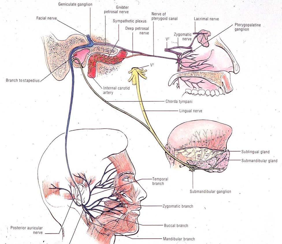
Cranial Nerve VII - Facial Nerve. The facial nerve has 3 main components with distinct functions
 Cranial Nerve VII - Facial Nerve The facial nerve has 3 main components with distinct functions Somatic motor efferent Supplies the muscles of facial expression; posterior belly of digastric muscle; stylohyoid,
Nose & Mouth OUTLINE. Nose. - Nasal Cavity & Its Walls. - Paranasal Sinuses. - Neurovascular Structures. Mouth. - Oral Cavity & Its Contents
 Dept. of Human Anatomy, Si Chuan University Zhou hongying eaglezhyxzy@163.com Nose & Mouth OUTLINE Nose - Nasal Cavity & Its Walls - Paranasal Sinuses - Neurovascular Structures Mouth - Oral Cavity & Its
University of Palestine. Midterm Exam 2013/2014 Total Grade:
 Course No: DNTS2208 Course Title: Head and Neck Anatomy Date: 09/11/2013 No. of Questions: (50) Time: 1hour Using Calculator (No) University of Palestine Midterm Exam 2013/2014 Total Grade: Instructor
Anatomy images for MSS practical exam- 2019
 Anatomy images for MSS practical exam- 2019 Ilium Ischium Pubis Acetabulaum Iliac crest Iliac tubercle ASIS (muscle and ligament attached) AIIS (muscle attached) PSIS PIIS Ischial spine Ischial tuberosity
Bones Ethmoid bone Inferior nasal concha Lacrimal bone Maxilla Nasal bone Palatine bone Vomer Zygomatic bone Mandible
 splanchnocranium - Consists of part of skull that is derived from branchial arches - The facial bones are the bones of the anterior and lower human skull Bones Ethmoid bone Inferior nasal concha Lacrimal
Bisection of Head & Nasal Cavity 頭部對切以及鼻腔. 解剖學科馮琮涵副教授 分機
 Bisection of Head & Nasal Cavity 頭部對切以及鼻腔 解剖學科馮琮涵副教授 分機 3250 E-mail: thfong@tmu.edu.tw Outline: The structure of nose The concha and meatus in nasal cavity The openings of paranasal sinuses Canals, foramens
Major Anatomic Components of the Orbit
 Major Anatomic Components of the Orbit 1. Osseous Framework 2. Globe 3. Optic nerve and sheath 4. Extraocular muscles Bony Orbit Seven Bones Frontal bone Zygomatic bone Maxillary bone Ethmoid bone Sphenoid
human anatomy 2016 lecture fifteen Dr meethak ali ahmed neurosurgeon
 Cranial Nerves Organization of the Cranial Nerves The cranial nerves are named as follows: I. Olfactory II. Optic III. Oculomotor IV. Trochlear V. Trigeminal VI. Abducent VII. Facial VIII. Vestibulocochlear
Cranial nerves.
 Cranial nerves eaglezhyxzy@163.com Key Points of Learning Name Components Passing through Peripheral distribution Central connection Function Cranial nerves Ⅰ olfactory Ⅱ optic Ⅲ occulomotor Ⅳ trochlear
Biology 323 Human Anatomy for Biology Majors Week 10; Lecture 1; Tuesday Dr. Stuart S. Sumida. Cranial Nerves and Soft Tissues of the Skull
 Biology 323 Human Anatomy for Biology Majors Week 10; Lecture 1; Tuesday Dr. Stuart S. Sumida Cranial Nerves and Soft Tissues of the Skull FOREBRAIN MIDBRAIN HINDBRAIN Forebrain: Cerebrum Perception,
Anatomy and physiology of Temporomandibular Joint
 Anatomy and physiology of Temporomandibular Joint Temporomandibular joint (TMJ): It is the articulation of the condyle of the mandible, and the inter-articular disc; with the mandibular fossa (glenoid
Face and Scalp 解剖學科鄭授德
 Face and Scalp 解剖學科鄭授德 本教材之圖片取自於 1 Gray s Anatomy for Students, 3rd ed, 2015, by Drake, Vogl, and Mitchell 2 Clinically Oriented Anatomy, 7th ed, 2014, by Moore, Dalley, and Agur 3 Clinically Oriented
Skull basic structures. Neurocranium
 Assoc. Prof. Květuše Lovásová, M.V.D., PhD. Skull basic structures Skull consists of two groups of bones: neurocranium (bones forming the brain box) splanchnocranium (bones forming the facial skeleton)
Anatomy and Physiology II. Review Spine and Neck
 Anatomy and Physiology II Review Spine and Neck Spine regions How many cervical vertibrae are there? 7 The curvature is the cervical region posterior? Concave posterior How many thoracic? And curvature?
function - sensory & postganglionic sympathetic [communication from the internal carotid plexus in the cavernous sinus] innervation of the mucosa of
![function - sensory & postganglionic sympathetic [communication from the internal carotid plexus in the cavernous sinus] innervation of the mucosa of](/thumbs/74/71276096.jpg "function - sensory & postganglionic sympathetic [communication from the internal carotid plexus in the cavernous sinus] innervation of the mucosa of") Nerves I. Cranial nerves A. Olfactory (CN I) 1. Olfactory bulb 2. Olfactory tract B. Optic n. (CNII) function - carries visual sensory information from the neural retina to the diencephalon & midbrain
Nerves I. Cranial nerves A. Olfactory (CN I) 1. Olfactory bulb 2. Olfactory tract B. Optic n. (CNII) function - carries visual sensory information from the neural retina to the diencephalon & midbrain
Chapter 7 Part A The Skeleton
 Chapter 7 Part A The Skeleton Why This Matters Understanding the anatomy of the skeleton enables you to anticipate problems such as pelvic dimensions that may affect labor and delivery The Skeleton The
APPENDICULAR SKELETON 126 AXIAL SKELETON SKELETAL SYSTEM. Cranium. Skull. Face. Skull and associated bones. Auditory ossicles. Associated bones.
 SKELETAL SYSTEM 206 AXIAL SKELETON 80 APPENDICULAR SKELETON 26 Skull Skull and associated s 29 Cranium Face Auditory ossicles 8 4 6 Associated s Hyoid Thoracic cage 25 Sternum Ribs 24 Vertebrae 24 column
Chapter 7: Head & Neck
 Chapter 7: Head & Neck Osteology I. Overview A. Skull The cranium is composed of irregularly shaped bones that are fused together at unique joints called sutures The skull provides durable protection from
Oral cavity : consist of two parts: the oral vestibule and the oral cavity proper. Oral vestibule : is slit like space between.
 Oral cavity Oral cavity : consist of two parts: the oral vestibule and the oral cavity proper Oral vestibule : is slit like space between the teeth, buccal gingiva, lips, and cheeks 1 Oral cavity Oral
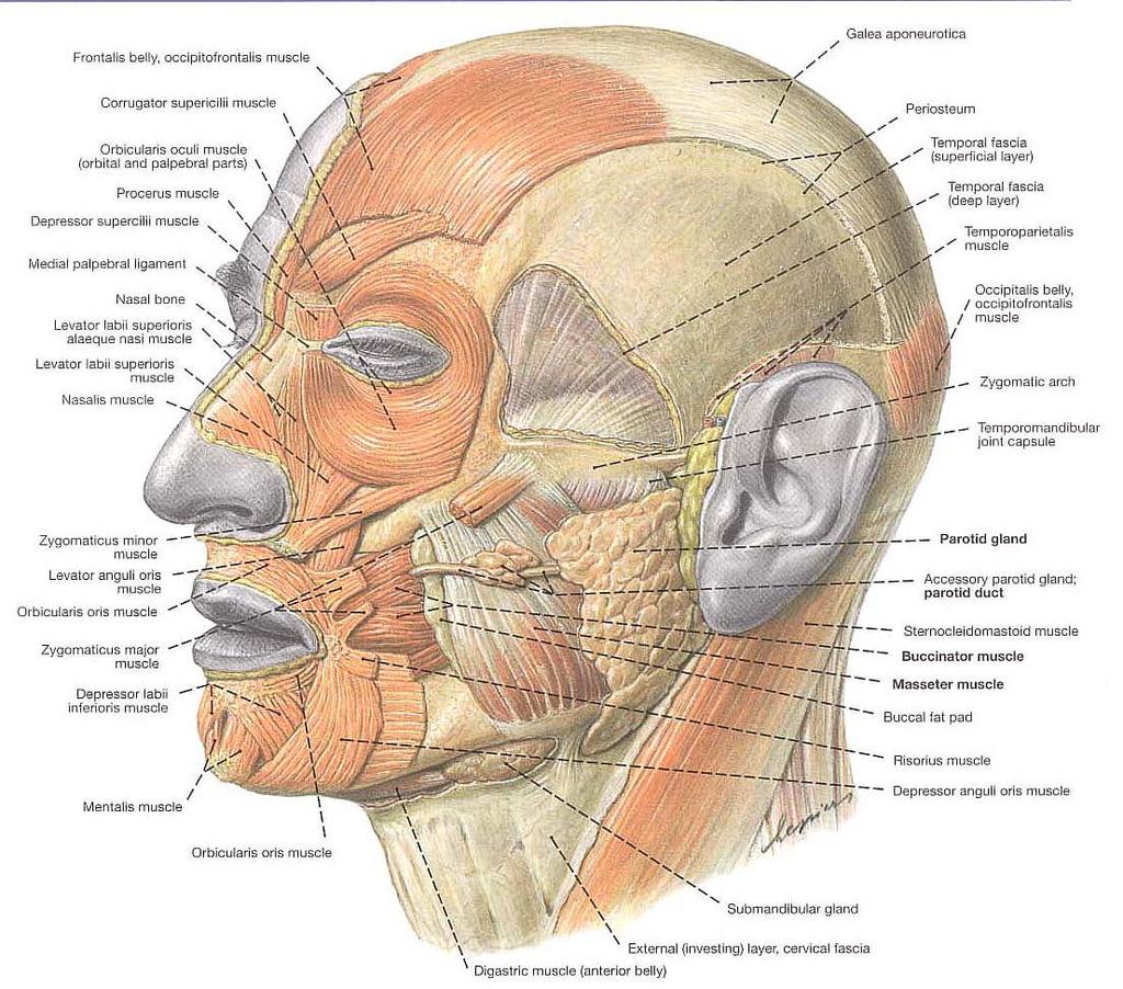
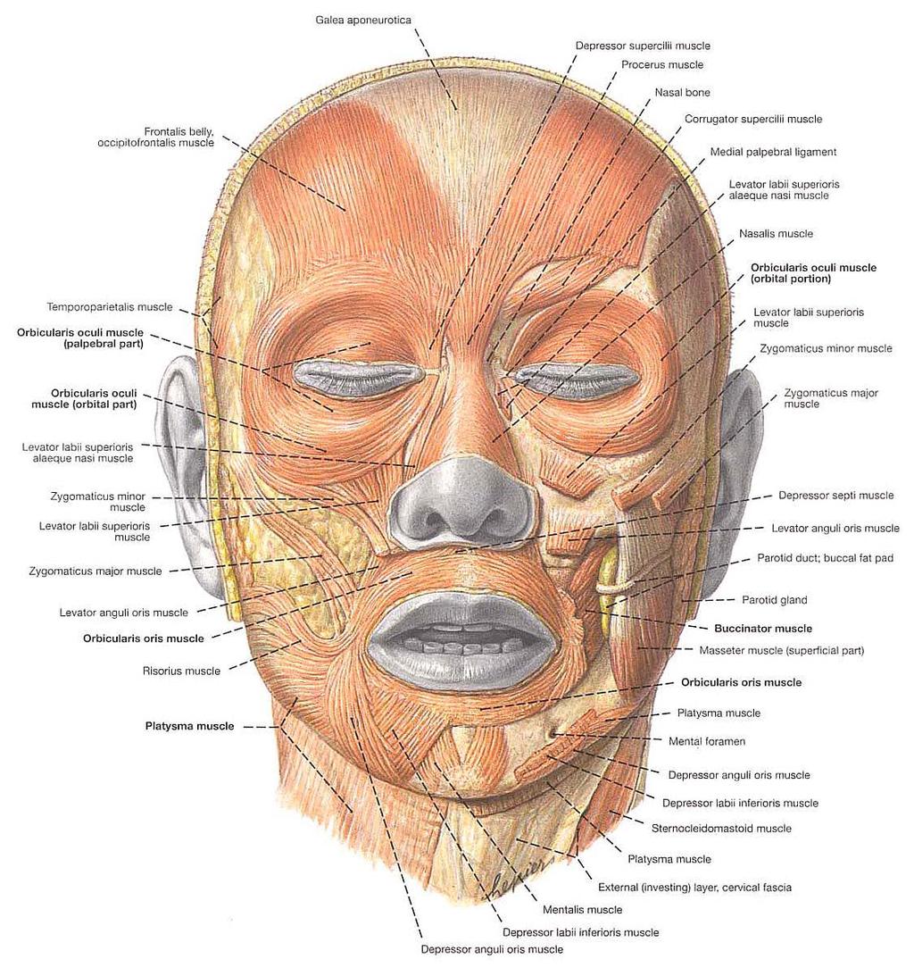
Face. Definition: The area between the two ears and from the chin to the eye brows. The muscles of the face
 Face Definition: The area between the two ears and from the chin to the eye brows. The muscles of the face The muscle of facial expression (include the muscle of the face and the scalp). All are derived
Introduction to Occlusion and Mechanics of Mandibular Movement
 Introduction to Occlusion and Mechanics of Mandibular Movement Dr. Pauline Hayes Garrett Department of Endodontics, Prosthodontics, and Operative Dentistry University of Maryland, Baltimore Assigned reading
*in general the blood supply of the nose comes from branches of the internal and external carotid arteries.
 In the previous lecture we talked about the anatomy of the nasal cavity, today we will talk about its blood supply, venous drainage, innervations, and finally about the paranasal sinuses. When we describe
Bones of the Skull Lateral View
 Bones of the Skull Lateral View Frontal Bone Parietal Bone Occipital Bone Temporal Bone Sphenoid Bone Pterion Sutures of the Skull Lateral View Coronal Suture Lambdoid Suture Squamous Suture Sutures of
Dr.Noor Hashem Mohammad Lecture (5)
") Dr.Noor Hashem Mohammad Lecture (5) 2016-2017 If the mandible is discarded, the anterior part of this aspect of the skull is seen to be formed by the hard palate. The palatal processes of the maxillae
Dr.Noor Hashem Mohammad Lecture (5) 2016-2017 If the mandible is discarded, the anterior part of this aspect of the skull is seen to be formed by the hard palate. The palatal processes of the maxillae
HEAD & NECK BY NUMBERS THIRD EDITION Copyright 2013, Anatomy Numbers (Phoenix, AZ) All rights reserved
 All rights reserved") HEAD & NECK BY NUMBERS THIRD EDITION Copyright 2013, Anatomy Numbers (Phoenix, AZ) All rights reserved No part of this publication may be reproduced or transmitted in any form or by any means, electronic
HEAD & NECK BY NUMBERS THIRD EDITION Copyright 2013, Anatomy Numbers (Phoenix, AZ) All rights reserved No part of this publication may be reproduced or transmitted in any form or by any means, electronic
Human Anatomy and Physiology - Problem Drill 07: The Skeletal System Axial Skeleton
 Human Anatomy and Physiology - Problem Drill 07: The Skeletal System Axial Skeleton Question No. 1 of 10 Which of the following statements about the axial skeleton is correct? Question #01 A. The axial
Anatomy Skull and Spinal Cord
 1 Anatomy Skull and Spinal Cord 1. Skull The skull is anterior to the spinal column and is the bony structure that encases the brain. Its purpose is to protect the brain and allow attachments for the facial
Functional components
 Facial Nerve VII cranial nerve Emerges from Pons Two roots Functional components: 1. GSA (general somatic afferent) 2. SA (Somatic afferent) 3. GVE (general visceral efferent) 4. BE (Special visceral/branchial
THE SKELETAL SYSTEM. Focus on the Skull
 THE SKELETAL SYSTEM Focus on the Skull Review Anatomical Terms Anterior/Posterior Dorsal/Ventral Medial/Lateral Superior/Inferior Bone Markings - Review Projections for attachment of muscles, ligaments
HEAD & NECK ANATOMY - MCQ HEAD & NECK ANATOMY
 . ' HEAD & NECK ANATOMY I. Deep investing layer of cervical fascia splits to enclose: A. Sternocleidomastoid B. Trapezius C. Parotid gland D. Omohyoid 2. Regarding the prevertebral fascia, the following
Biology 218 Human Anatomy. Adapted from Martini Human Anatomy 7th ed. Chapter 6 The Skeletal System: Axial Division
 Adapted from Martini Human Anatomy 7th ed. Chapter 6 The Skeletal System: Axial Division Introduction The axial skeleton: Composed of bones along the central axis of the body Divided into three regions:
Cranium Facial bones. Sternum Rib
 Figure 7.1 The human skeleton. Skull Thoracic cage (ribs and sternum) Cranium Facial bones Sternum Rib Bones of pectoral girdle Vertebral column Sacrum Vertebra Bones of pelvic girdle (a) Anterior view
3-Deep fascia: is absent (except over the parotid gland & buccopharngeal fascia covering the buccinator muscle)
") The Face 1-Skin of the Face The skin of the face is: Elastic Vascular (bleed profusely however heal rapidly) Rich in sweat and sebaceous glands (can cause acne in adults) It is connected to the underlying
The Face 1-Skin of the Face The skin of the face is: Elastic Vascular (bleed profusely however heal rapidly) Rich in sweat and sebaceous glands (can cause acne in adults) It is connected to the underlying
CN I Olfactory. CN II Optic. CN III Oculomotor. Special Sensory Efferent fibers to Olfactory Bulb. Cribiform Plate of Ethmoid
 CN I Olfactory Efferent fibers to Olfactory Bulb Olfactory Tract Olfactory Bulb Cribiform Plate of Ethmoid Anosmia Loss of sense of smell Uncinate Fits olfactory hallucinations To Olfactory Epithelium
The Ear The ear consists of : 1-THE EXTERNAL EAR 2-THE MIDDLE EAR, OR TYMPANIC CAVITY 3-THE INTERNAL EAR, OR LABYRINTH 1-THE EXTERNAL EAR.
 The Ear The ear consists of : 1-THE EXTERNAL EAR 2-THE MIDDLE EAR, OR TYMPANIC CAVITY 3-THE INTERNAL EAR, OR LABYRINTH 1-THE EXTERNAL EAR Made of A-AURICLE B-EXTERNAL AUDITORY MEATUS A-AURICLE It consists
Gross Anatomy of the. TEMPORAL BONE, EXTERNAL EAR, and MIDDLE EAR
 Gross Anatomy of the TEMPORAL BONE, EXTERNAL EAR, and MIDDLE EAR M1 Gross and Developmental Anatomy 9:00 AM, December 11, 2008 Dr. Milton M. Sholley Professor of Anatomy and Neurobiology Assignment: Head
Veins of the Face and the Neck
 Veins of the Face and the Neck Facial Vein The facial vein is formed at the medial angle of the eye by the union of the supraorbital and supratrochlear veins. connected through the ophthalmic veins with
The Neck the lower margin of the mandible above the suprasternal notch and the upper border of the clavicle
 The Neck is the region of the body that lies between the lower margin of the mandible above and the suprasternal notch and the upper border of the clavicle below Nerves of the neck Cervical Plexus Is formed
Bony orbit Roof The orbital plate of the frontal bone Lateral wall: the zygomatic bone and the greater wing of the sphenoid
 Bony orbit Roof: Formed by: The orbital plate of the frontal bone, which separates the orbital cavity from the anterior cranial fossa and the frontal lobe of the cerebral hemisphere Lateral wall: Formed
The Seventh Cranial Nerve The Facial By Prof. Dr. Muhammad Imran Qureshi
 The Seventh Cranial Nerve The Facial By Prof. Dr. Muhammad Imran Qureshi Functional Components: SVE: Fibers originate from nucleus of facial nerve, and supply facial muscles GVE: Fibers derived from superior
Subdivided into Vestibule & Oral cavity proper
 Extends from the lips to the oropharyngeal isthmus The oropharyngeal isthmus: Is the junction of mouth and pharynx. Is bounded: Above by the soft palate and the palatoglossal folds Below by the dorsum
Gross Anatomy of the. TEMPORAL BONE, EXTERNAL EAR, and MIDDLE EAR. Assignment: Head to Toe Temporomandibular Joint (TMJ)
") Gross Anatomy the TEMPORAL BONE, EXTERNAL EAR, and MIDDLE EAR M1 Gross and Developmental Anatomy 9:00 AM, December 11, 2008 Dr. Milton M. Sholley Pressor Anatomy and Neurobiology Assignment: Head to Toe
Gross Anatomy the TEMPORAL BONE, EXTERNAL EAR, and MIDDLE EAR M1 Gross and Developmental Anatomy 9:00 AM, December 11, 2008 Dr. Milton M. Sholley Pressor Anatomy and Neurobiology Assignment: Head to Toe
Skull-2. Norma Basalis Interna. Dr. Heba Kalbouneh Assistant Professor of Anatomy and Histology
 Skull-2 Norma Basalis Interna Dr. Heba Kalbouneh Assistant Professor of Anatomy and Histology Norma basalis interna Base of the skull- superior view The interior of the base of the skull is divided into
APRIL
 APRIL - 2003 OCTOBER - 2003 February 2009 [KU 652] Sub. Code : 4131 FIRST B.D.S DEGREE EXAMINATION (Modified Regulations III) Paper I HUMAN ANATOMY, HISTOLOGY AND EMBRYOLOGY Time : Three hours
The sebaceous glands (glands of Zeis) open directly into the eyelash follicles, ciliary glands (glands of Moll) are modified sweat glands that open
 open directly into the eyelash follicles, ciliary glands (glands of Moll) are modified sweat glands that open") The Orbital Region The orbits are a pair of bony cavities that contain the eyeballs; their associated muscles, nerves, vessels, and fat; and most of the lacrimal apparatus upper eyelid is larger and more
The Orbital Region The orbits are a pair of bony cavities that contain the eyeballs; their associated muscles, nerves, vessels, and fat; and most of the lacrimal apparatus upper eyelid is larger and more
-Ibrahim Al-Naser. -Dr Al- Muhtaseb. 1 P a g e
 -1 -Ibrahim Al-Naser - -Dr Al- Muhtaseb 1 P a g e The Digestive System The doctor started the lecture by talking about the class rules. The GI system is an organ system, it is divided into: The Alimentary
Salivary Glands and Teeth. Dr. Nabil Khouri MD, Ph.D
 Salivary Glands and Teeth Dr. Nabil Khouri MD, Ph.D Anatomy and Histology of salivary glands Functions of Saliva Keeps the mouth moist Aids in swallowing Aids in speech Keeps the mouth and teeth clean
EXAM NUMBER STRUCTURAL BASIS OF MEDICAL PRACTICE EXAMINATION 7 October 28, PART l. Answer in the space provided. (9 pts)
") STRUCTURAL BASIS OF MEDICAL PRACTICE EXAMINATION 7 October 28, 2005 PART l. Answer in the space provided. (9 pts) 1. Identify the structures. (3 pts) a. _Frontal Sinus b. _Lateral Posterior Inferior Nasal
STRUCTURAL BASIS OF MEDICAL PRACTICE EXAMINATION 7 October 28, 2005 PART l. Answer in the space provided. (9 pts) 1. Identify the structures. (3 pts) a. _Frontal Sinus b. _Lateral Posterior Inferior Nasal
ANTERIOR CERVICAL TRIANGLE (Fig. 2.1 )
") 2 Neck Anatomy ANTERIOR CERVICAL TRIANGLE (Fig. 2.1 ) The boundaries are: Lateral: sternocleidomastoid muscle Superior: inferior border of the mandible Medial: anterior midline of the neck This large triangle
2 Neck Anatomy ANTERIOR CERVICAL TRIANGLE (Fig. 2.1 ) The boundaries are: Lateral: sternocleidomastoid muscle Superior: inferior border of the mandible Medial: anterior midline of the neck This large triangle
Skeletal System -Axial System. Chapter 7 Part A
 Skeletal System -Axial System Chapter 7 Part A Skeleton Learn: Names of the s. Identify specific landmarks that allow: Bones to fit into each other, Organs to fit into the cavities, Muscles to attach,
A. The supraclavicular nerves supply sensory fibers to the skin of the clavicular area
 YR 1 GROSS ANATOMY WRITTEN EXAM 2 -- October 10, 1997. CHOOSE THE SINGLE BEST ANSWER FOR QUESTIONS 1-42. 1. Each of the following statements is CORRECT EXCEPT: A. The supraclavicular nerves supply sensory
Temporomandibular Joint
 6 Temporomandibular Joint Most human bones are joined with one another. These connections of bones to each other are termed articulations or joints. Some joints are immovable as in the articulations of
Basic Anatomy and Physiology of the Lips and Oral Cavity. Dr. Faghih
 Basic Anatomy and Physiology of the Lips and Oral Cavity Dr. Faghih It is divided into seven specific subsites : 1. Lips 2. dentoalveolar ridges 3. oral tongue 4. retromolar trigone 5. floor of mouth 6.
Oral cavity landmarks
 By: Dr. Ahmed Rabah Oral cavity landmarks The knowledge of oral anatomy and physiology will help the operator and provides enough landmarks to act as positive guide during denture construction. This subject
Head & Neck Radiology (I) Waseem Jerjes
 Waseem Jerjes") Head & Neck Radiology (I) Waseem Jerjes Lamboid Suture Frontal Sinus Left Orbit (roof) Left Supraorbital Margin Left Frontozygomatic Suture Left Superior Orbital Fissure Rt Petrous Ridge of Temporal Bone
Head & Neck Radiology (I) Waseem Jerjes Lamboid Suture Frontal Sinus Left Orbit (roof) Left Supraorbital Margin Left Frontozygomatic Suture Left Superior Orbital Fissure Rt Petrous Ridge of Temporal Bone
AXIAL SKELETON SKULL
 AXIAL SKELETON SKULL CRANIAL BONES (8 total flat bones w/ 2 paired) 1. Frontal forms forehead & upper portion of eyesocket (orbital) 2. Parietal paired bones; form superior & lateral walls of cranium 3.
Mohammad Mohtaseb. Nour Hussein. Faisal Nimri
 2 Mohammad Mohtaseb Nour Hussein Faisal Nimri Muscles of the tongue The tongue is a muscular organ and contains intrinsic and extrinsic muscles. The intrinsic muscle contains vertical, oblique, and transverse
Neck of Condylar. Process. Anterior Border of Ramus. Mandibular. Foramen. Posterior Border of Ramus Incisive Fossa.
 Learning Outcomes The Mandible Surface Anatomy Muscle Attachments The (FOM) Muscles of the FOM The Tongue Muscles of the Tongue The Submandibular Region Submandibular Gland Sublingual Gland Lingual The
Anatomy of Oral Cavity DR. MAAN AL-ABBASI
 Anatomy of Oral Cavity DR. MAAN AL-ABBASI By the end of this lecture you should be able to: 1. Differentiate different parts of the oral cavity 2. Describe the blood and nerve supply of mucosa and muscles