Paraspinal Venous Malformation Joseph Junewick, MD FACR
|
|
|
- Thomasina Hamilton
- 5 years ago
- Views:
Transcription
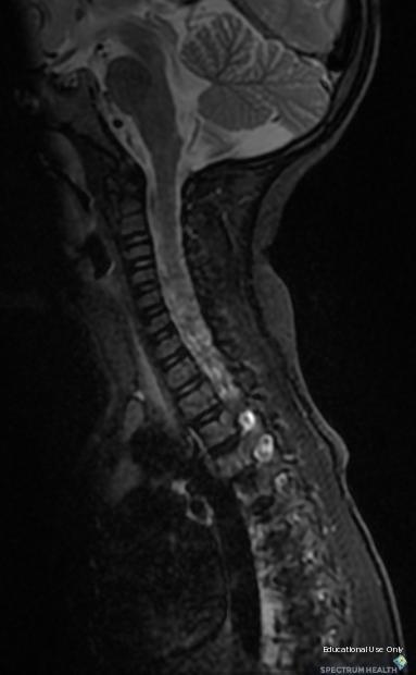
1 Paraspinal Venous Malformation Joseph Junewick, MD FACR 06/04/2010 History 2 year old with history of fall. Rule out spinal injury. Diagnosis Paraspinal Venous Malformation Additional Clinical CT of the brain revealed a non-displaced right parietal skull fracture and small subdural hematoma. Discussion A hemangioma is a abnormal congenital collection of blood vessels which may be found in one or several organs (skin, lungs, GI tract, eyes, and brain). However these benign, vascular neoplasms make up less than 0.5% of all mediastinal masses. Like other mediastinal masses of childhood, these might manifest by nonspecific symptoms (cough, dyspnea and chest pain) or symptoms related to their anatomic location and extent of invasion but most often, they are asymptomatic and are incidentally detected. The vast majority of mediastinal hemangiomas are located in the anterior mediastinum (68%). The posterior mediastinal location is very rare. Involvement of the middle mediastinum is seen as a continuation of disease from anterior or posterior locations. Most of the posterior mediastinal tumors in children are of neurogenic origin (neurofibroma, neurilemoma, neurenteric cyst, neuroblastoma, ganglioneuroblastoma, pheochromocytoma and neurofibrosarcoma). A posterior mediastinal mass in a young child is most often of neurogenic origin, with neuroblastoma being the diagnosis of import. The avid enhancement of a hemangioma mitigates against thoracic neuroblastoma, which typically enhances much less. The identification of metastatic disease suggests neuroblastoma; however, metastases are less common with thoracic neuroblastoma. Lack of calcification is not helpful in narrowing the diagnosis, since hemangiomas and thoracic neuroblastomas often lack calcification. Some neurogenic tumors, namely pheochromocytomas and paragangliomas, might demonstrate MR characteristics similar to those of a hemangioma hypointense on T1 weighted images and very hyperintense on T2-weighted images. Findings MR-Variably enhancing, minimally T1 hyperintense and markedly T2 hyperintense left paraspinal lesion insinuating into neural foramina and the intraspinal extradural space. Reference Sabharwal GK, Strouse PJ. Posterior mediastinal hemangiomas. Pediatr Radiol (2005); 35:

2

3

4
5
6

7

8

9

10
11
12 Sponsored By Disclaimer This teaching site is partially funded by an educational grant from GE Healthcare and Advanced Radiology Services, PC. The material on this site is independently controlled by Advanced Radiology Services, PC, and GE Healthcare and Spectrum Health have no influence over the content of this site Content Download Agreement The cases and images on this website are owned by Spectrum Health. Permission is granted (for nonprofit educational purposes) to download and print materials to distribute for the purpose of facilitating the education of health professionals. The authors retain all rights to the material and users are requested to acknowledge the source of the material. Site Disclaimer This site is developed to reach healthcare professionals and medical students. Nothing this site should be considered medical advice. Only your own doctor can help you make decisions about your medical care. If you have a specific medical question or are seeking medical care, please contact your physician. The information in this website is provided for general medical education purposes only and is not meant to substitute for the independent medical judgment of a physician relative to diagnostic and treatment options of a specific medical condition. The viewpoints expressed in these cases are those of the authors. They do not represent an endorsement. In no event will Advanced Radiology Associates, PC, Spectrum Health Hospitals (Helen Devos Children's Hospital) or GE Healthcare be liable for any decision made or action taken in reliance upon the information provided through this website.
Presacral Neuroblastoma Joseph Junewick, MD FACR
 Presacral Neuroblastoma Joseph Junewick, MD FACR 01/12/2010 History 16 month old male with irritability. Diagnosis Presacral Neuroblastoma Additional Clinical Initial US to evaluate for intussusception
Neuroblastoma Joseph Junewick, MD FACR
 Neuroblastoma Joseph Junewick, MD FACR 03/18/2011 History 15 month old with anemia. Diagnosis Neuroblastoma Discussion Neuroblastic tumors derive from primordial neural crest cells destined for sympathetic
Spinal LCH Joseph Junewick, MD FACR
 Spinal LCH Joseph Junewick, MD FACR 05/16/2009 History 16 year old female with multiply recurrent Langerhans Cell Histiocytosis now with severe left sided neck pain. Diagnosis Langerhans Cell Histiocytosis
Diskitis Joseph Junewick, MD FACR
 Diskitis Joseph Junewick, MD FACR 09/20/2010 History 2 year old with fever, back pain and elevated sedimentation rate. Diagnosis Diskitis Discussion Diskitis is an inflammatory process of the intervertebral
Transverse Dural Sinus Thrombosis Joseph Junewick, MD FACR
 Transverse Dural Sinus Thrombosis Joseph Junewick, MD FACR 03/19/2010 History Child with headache and otomastoiditis. Diagnosis Dural venous thrombosis secondary to mastoiditis Discussion The cerebral
Pleural Empyema Joseph Junewick, MD FACR
 Pleural Empyema Joseph Junewick, MD FACR 03/19/2010 History Teenager with persistent fever and cough. Pneumonia diagnosed 1 week ago. Diagnosis Pleural Empyema Additional Clinical Surgery-Clear fluid with
Vein of Galen Malformation Joseph Junewick, MD FACR
 Vein of Galen Malformation Joseph Junewick, MD FACR 04/14/2018 History Midline cystic intracranial mass on prenatal ultrasound. Diagnosis Vein of Galen Malformation Discussion In normal fetal development,
Bilateral Retinoblastoma Joseph Junewick, MD FACR
 Bilateral Retinoblastoma Joseph Junewick, MD FACR 06/11/2010 History 17 month old adopted female with proptosis. Diagnosis Bilateral Retinoblastoma Discussion Retinoblastoma is the most common pediatric
Retroperitoneal Teratoma Heather Borders, MD
 Retroperitoneal Teratoma Heather Borders, MD 03/04/2012 History Newborn with congenitally diagnosed mass. No other clinical symptoms. Diagnosis Retroperitoneal Teratoma; Immature teratoma, grade 1, with
Chiari III Joseph Junewick, MD FACR
 Chiari III Joseph Junewick, MD FACR 07/02/2010 History Newborn with suboccipital mass. Diagnosis Chiari III Additional Clinical Surgery-Skin covered suboccipital cystic mass confined by the dura. Pathology-Leptomeningeal
Radiation Pneumonitis Joseph Junewick, MD FACR
 Radiation Pneumonitis Joseph Junewick, MD FACR 03/19/2010 History 16 year old with history of relapsed stage IV-A Hodgkin disease. Prior pulmonary involvement was irradiated. Diagnosis Radiation Pneumonitis
Pituitary Macroadenoma Joseph Junewick, MD FACR
 Pituitary Macroadenoma Joseph Junewick, MD FACR 08/13/2010 History 12 year old female with headache and visual disturbance. Diagnosis Pituitary Macroadenoma Additional Clinical Markedly elevated growth
Scrofula Joseph Junewick, MD FACR
 Scrofula Joseph Junewick, MD FACR 06/20/2012 History 4 year old male with refractory cervical adenopathy Diagnosis Scrofula Additional Clinical Positive PPD skin test. Discussion Scrofula refers to tuberculous
Term Hypoxic Ischemic Injury Joseph Junewick, MD FACR
 Term Hypoxic Ischemic Injury Joseph Junewick, MD FACR 08/11/2010 History Term infant with perinatal distress and attempted forceps delivery. Diagnosis Term Hypoxic Ischemic Injury Discussion Encephalopathy
Chance Fracture Joseph Junewick, MD FACR
 Chance Fracture Joseph Junewick, MD FACR 08/02/2010 History Restrained teenager involved in motor vehicle accident. Diagnosis Chance Fracture (Hyperflexion-Distraction Injury) Discussion Chance-type spinal
Tuberculous Meningitis Joseph Junewick, MD FACR
 Tuberculous Meningitis Joseph Junewick, MD FACR 08/11/2010 History 14 month old with fever and increasing lethargy. Diagnosis Tuberculous Meningitis Additional Clinical Grandmother with active tuberculosis.
Ulcerative Colitis Joseph Junewick, MD FACR
 Ulcerative Colitis Joseph Junewick, MD FACR 06/04/2010 History 16 year old male with hematochezia and anemia. Diagnosis Ulcerative Colitis Additional Clinical History of ulcerative colitis. Discussion
Atlanto-occipital Dislocation Joseph Junewick, MD FACR
 Atlanto-occipital Dislocation Joseph Junewick, MD FACR 09/23/2009 History 12 year old male restrained back seat passenger in a car hit by a snowplow. Diagnosis Atlanto-occipital Dislocation Discussion
Thymic Involvement in Chronic Granulomatous Disease of Childhood
 Thymic Involvement in Chronic Granulomatous Disease of Childhood Joseph Junewick, MD FACR 07/16/2010 History 3 year old male with multifocal osteomyelitis. Diagnosis Thymic Involvement in Chronic Granulomatous
Gastrointestinal Hemangiomatosis Joseph Junewick, MD FACR
 Gastrointestinal Hemangiomatosis Joseph Junewick, MD FACR 03/06/2010 History 3 month old with protuberant abdomen and anemia. Diagnosis Gastrointestinal Hemangiomatosis Discussion Gastrointestinal hemangiomatosis
Testicular Microlithiasis related to McCune-Albright Syndrome Joseph Junewick, MD FACR
 Testicular Microlithiasis related to McCune-Albright Syndrome Joseph Junewick, MD FACR 04/25/2010 History 12 year old with McCune-Albright syndrome. Diagnosis Testicular Microlithiasis related to Mcune-Albright
Posterior Slipped Capital Femoral Epiphysis Joseph Junewick, MD FACR
 Posterior Slipped Capital Femoral Epiphysis Joseph Junewick, MD FACR 08/11/2010 History 6 year old male with intermittent hip pain for several months, acutely worsened after climbing the sand dunes. Diagnosis
Fallopian tube torsion and paratubal cyst Heather Borders, MD
 Fallopian tube torsion and paratubal cyst Heather Borders, MD 01/24/2012 History 13 year old female with one week of pelvic pain Diagnosis Fallopian tube torsion with paratubal cyst Additional Clinical
Imaging Of Cystic Paravertebral Masses:
 Imaging Of Cystic Paravertebral Masses: Differential Diagnosis and Key Discriminators John P. Lichtenberger III, MD, Maj, USAF, MC Brent McCarragher, MD, CPT, USA John R. Dryden, MD, LT, USN P. Gabriel
Mediastinal Tumors: Imaging
 Mediastinal Tumors: Imaging References Imaging in Oncology, Husband and Reznek Computed Tomography and Magnetic Resonance of the thorax, Naidich, Zerhouni, Siegelman, Mediastinal compartments Anterior:
Neckmasses in infancy and childhood: Clinical and radiological classification and imaging approaches M. Mearadji
 Neckmasses in infancy and childhood: Clinical and radiological classification and imaging approaches M. Mearadji International Foundation for Pediatric Imaging Aid Introduction Neck masses are a frequent
Pediatric Spine Tumors (and other masses)
") Pediatric Spine Tumors (and other masses) Francisco A Perez, MD, PhD Assistant Professor Neuroradiology and Pediatric Radiology Seattle Children s Hospital University of Washington, Seattle Commercial
Pediatric Spine Tumors (and other masses) Francisco A Perez, MD, PhD Assistant Professor Neuroradiology and Pediatric Radiology Seattle Children s Hospital University of Washington, Seattle Commercial
ORIGINAL ARTICLE. Abstract. Aim. Materials and methods. Introduction. Results
 Is anatomical distribution helpful for differentiating TB spondylitis from neoplastic causes of extradural spinal cord compression in children? A pilot study Reena George, MD, MMed Rad, FRCR (UK) Savvas
ADRENAL MEDULLARY DISORDERS: PHAEOCHROMOCYTOMAS AND MORE
 ADRENAL MEDULLARY DISORDERS: PHAEOCHROMOCYTOMAS AND MORE DR ANJU SAHDEV READER AND CONSULTANT RADIOLOGIST QUEEN MARY UNIVERSITY AND ST BARTHOLOMEW S HOSPITAL BARTS HEALTH, LONDON, UK DISCLOSURE OF CONFLICT
Pathology of Mediastinal Tumors
 SAMO Meeting Lucerne 2009 Pathology of Mediastinal Tumors Alex Soltermann Most common lesions (adults) Clinical presentation 50% of the patients are asymptomatic, lesion discovered incidentally Symptoms
A Nervous Breakdown: Multimodality Imaging of Thoracic Neurogenic Tumors
 A Nervous Breakdown: Multimodality Imaging of Thoracic Neurogenic Tumors John P. Lichtenberger III, MD, Maj, USAF, MC Assistant Professor, Dept. or Radiology Uniformed Services University of the Health
1/9/2013 EXTRAMEDULLARY TUMORS OF THE PEDIATRIC SPINE. Introduction. Classification for Extramedullary Tumors
 EXTRAMEDULLARY TUMORS OF THE PEDIATRIC SPINE Eugene Wang 1/20/12 Dent Neurologic Institute Introduction 2/3 of all intraspinal tumors of childhood are extramedullary 50% Extradural 10-15% Intradural Back
A Journey Down The Canal
 A Journey Down The Canal Radiological Assessment of Spinal Cord Masses John Berry-Candelario HMS III Gillian Lieberman, MD BIDMC Objectives Patient review Anatomy of the spine Imaging techniques Classification
Spinal Neoplasms. First Things First!! Localize the Lesion!! Ependymomas. Common Intramedullary Lesions
 Acta Radiológica Portuguesa, Vol.XXIII, nº 90, pág. 101-114, Abr.-Jun., 2011 Spinal Neoplasms Bruno A Policeni University of Iowa Hospitals and Clinics Assistant Professor of Radiology Disclosure of Commercial
CTA/MRA of Pediatric Hepatic Masses Radiology-Pathology Correlation
 Acta Radiológica Portuguesa, Vol.XVIII, nº70, pág. 41-50, Abr.-Jun., 2006 CTA/MRA of Pediatric Hepatic Masses Radiology-Pathology Correlation Marilyn J. Siegel Mallinckrodt Institute of Radiology, Washington
Intrathoracic neural tumours
 Intrathoracic neural tumours K. G. DAVIDSON, P. R. WALBAUM, AND R. J. M. McCORMACK From the Thoracic Surgery Department, City Hospital, Edinburgh, UK Thorax, 1978, 33, 359-367 Davidson, K. G., Walbaum,
Small lesions involving scalp and skull in pediatric age.
 Small lesions involving scalp and skull in pediatric age. Poster No.: C-1149 Congress: ECR 2013 Type: Educational Exhibit Authors: M. J. Yi, J. H. Yoo; Seoul/KR Keywords: Education and training, Education,
Small lesions involving scalp and skull in pediatric age.
 Small lesions involving scalp and skull in pediatric age. Poster No.: C-1149 Congress: ECR 2013 Type: Educational Exhibit Authors: M. J. Yi, J. H. Yoo; Seoul/ Keywords: Education and training, Education,
Open surgery for posterior mediastinal neurogenic tumors
 Review Article Page 1 of 5 Open surgery for posterior mediastinal neurogenic tumors Erkan Kaba 1, Mazen Rasmi Alomari 2, Alper Toker 2 1 Department of Thoracic Surgery, Istanbul Bilim University Medical
BONE METASTASIS OF MEDIASTINAL PARAGANGLIOMA: CASE REPORT
 BONE METASTASIS OF MEDIASTINAL PARAGANGLIOMA: CASE REPORT Dr. Abhinandan Gupta 1*, Kong Long 1, Prof. Huang Jing Bai 1, Dr. Deepikal Dhakal 1, Dr. Sunil Shrestha 1, Dr. Roshan Kumar Yadav 2 and Dr. Shashi
A Case of Pediatric Plasma Cell Granuloma
 August 2001 A Case of Pediatric Plasma Cell Granuloma Nii Tetteh, Harvard Medical School Year IV Our Patient 8 year old male with history of recurrent left lower lobe and lingular pneumonias since 1994.
Mediastinal Paraganglioma: a challenge to the echocardiographic
 Case - based learning from ESC Cardiologists of Tomorrow Look for the answer outside the heart Mediastinal Paraganglioma: a challenge to the echocardiographic 1 diagnosis and endovascular treatment 1 On
Thoracoscopic surgical resection of thoracic neurogenic tumors
 Neurosurg Focus 7 (5):Article 1, 1999 Thoracoscopic surgical resection of thoracic neurogenic tumors Patrick P. Han, M.D., and Curtis A. Dickman, M.D. Division of Neurological Surgery, Barrow Neurological
Imaging features of orbital neoplasm developed in pediatrics
 Imaging features of orbital neoplasm developed in pediatrics Poster No.: C-1119 Congress: ECR 2015 Type: Educational Exhibit Authors: J. H. Yoo; Seoul/KR Keywords: Eyes, Head and neck, Paediatric, CT,
Incidental Esophageal Findings on Chest CT. Amira Hussien, MD, Elliot Fishman, MD, Bouchra Younes, MD, Ahmed Hatw. Johns Hopkins Medical Institution
 Incidental Esophageal Findings on Chest CT Amira Hussien, MD, Elliot Fishman, MD, ouchra Younes, MD, Ahmed Hatw. Johns Hopkins Medical Institution I have nothing to disclose. DISCLOSURE INTRODUCTION Although
Characterization of adrenal lesions on CT and MRI: all that a radiologist must know
 Characterization of adrenal lesions on CT and MRI: all that a radiologist must know Poster No.: C-2476 Congress: ECR 2013 Type: Educational Exhibit Authors: N. Benzina, S. MAJDOUB, C. H. ZARRAD, H. Zaghouani,
Adrenal masses in infancy and childhood: A clinical and radiological overview M. Mearadji
 Adrenal masses in infancy and childhood: A clinical and radiological overview M. Mearadji International Foundation for Pediatric Imaging Aid Introduction Neoplastic adrenal masses usually originate from
Imaging Work-Up of a Neck Mass - Adults & Children
 Disclosures Imaging Work-Up of a Neck Mass - Adults & Children I have nothing to disclose Christine M Glastonbury MBBS Professor of Radiology & Biomedical Imaging Otolaryngology-Head & Neck Surgery and
Anterior Mediastinal Masses: The 4 T s
 May 2001 Anterior Mediastinal Masses: The 4 T s Rachel Van Sambeek, Harvard Medical School, Year III 1 Mediastinal Compartments 3 arbitrary divisions that do not correlate with anatomic planes: Anterior
Thymic Tumors. Feiran Lou MD. MS. Kings County Hospital Department of Surgery
 Thymic Tumors Feiran Lou MD. MS. Kings County Hospital Department of Surgery Case HPI 53 yo man referred from OSH for anterior mediastinal mass. Initially presented with leg weakness and back pain for
CT of Acute Thoracic Aortic Syndromes Stuart S. Sagel, M.D.
 CT of Acute Thoracic Aortic Syndromes Stuart S. Sagel, M.D. Thoracic Aortic Aneurysms Atherosclerotic Dissection Penetrating ulcer Mycotic Inflammatory (vasculitis) Traumatic Aortic Imaging Options Catheter
IMAGING OF A CASE OF SPINAL MENINGIOMA- A CASE REPORT
 IMAGING OF A CASE OF SPINAL MENINGIOMA- A CASE REPORT Ramneet Wadi 1, Anil Kumar Shukla 2, Seetha Pramila V. V 3, Sabyasachi Basu 4, Sonam Sanjay 5 1Postgraduate Student, Department of Radiodiagnosis,
Update on RECIST and Staging of Common Pediatric Tumors Ethan A. Smith, MD
 Update on RECIST and Staging of Common Pediatric Tumors Ethan A. Smith, MD Section of Pediatric Radiology C.S. Mott Children s Hospital University of Michigan ethans@med.umich.edu Disclosures No relevant
Primary mediastinal tumours
 Primary mediastinal tumours Thorax (1974), 29, 475. YOUSF D. AL-NAAMAN, MOHAMAD S. AL-AN, and MUAYYAD M. AL-OMER Department of Thoracic and Cardiovascular Surgery, College of Medicine, University of Baghdad,
Dr. Pratik Mukherjee, MMed, FRCR Dr. Ashish Chawla, MD, ABR (USA) Khoo Teck Puat Hospital, Singapore
 Khoo Teck Puat Hospital, Singapore") Dr. Pratik Mukherjee, MMed, FRCR Dr. Ashish Chawla, MD, ABR (USA) Khoo Teck Puat Hospital, Singapore The authors declare no financial disclosures. To revisit the basics of approach to mediastinal masses
Dr. Pratik Mukherjee, MMed, FRCR Dr. Ashish Chawla, MD, ABR (USA) Khoo Teck Puat Hospital, Singapore The authors declare no financial disclosures. To revisit the basics of approach to mediastinal masses
Imaging in neurofibromatosis type 1: An original research article with focus on spinal lesions
 Original Research Article Imaging in neurofibromatosis type 1: An original research article with focus on spinal lesions Kalpesh Patel 1*, Siddharth Zala 2, C. Raychaudhuri 3 1 Assistant Professor, 2 1
ADRENAL LESIONS 10/09/2012. Adrenal + lesion. Introduction. Common causes. Anatomy. Financial disclosure. Dr. Boraiah Sreeharsha. Nothing to declare
 ADRENAL LESIONS Financial disclosure Nothing to declare Dr. Boraiah Sreeharsha MBBS;FRCR;FRCPSC Introduction Adrenal + lesion Adrenal lesions are common 9% of the population Increase in the detection rate
Evaluation of 95 Cases with Mediastinal Tumors
 Evaluation of 95 Cases with Reza Bagheri 1, Reza Afghani 2 *, Seyed Ziaollah Haghi 1, Seyed Hossein Fattahi Masoum 3, Soroush Zarehparvar Moghaddam 4, Saeed Akhlaghi 5 1 Thoracic Surgeon, Cardio Thoracic
Primitive Neuroectodermal Tumor of Mediastinum in an Adult: A Case Report 1
 Primitive Neuroectodermal Tumor of Mediastinum in an Adult: A Case Report 1 Young Jae Sung, M.D., Jeung Sook Kim, M.D. A peripheral primitive neuroectodermal tumor (PNET) is a rare and aggressive malignant
SAMPLE. Radiology Essential links from CPT codes to ICD-10-CM and HCPCS ICD-10. Cross Coder
 Cross Coder www.optumcoding.com Radiology Essential links from CPT codes to ICD-10-CM and HCPCS 2017 a ICD-10 A full suite of resources including the latest code set, mapping products, and expert training
Essentials of Clinical MR, 2 nd edition. 51. Primary Neoplasms
 51. Primary Neoplasms As with spinal central canal neoplasms in other regions, those of the lumbar spine may be classified as extradural, intradural extramedullary, and medullary. If an extradural lesion
UCLA General Surgery Residency Program Rotation Educational Policy Goals and Objectives
 UPDATED: July 2009 ROTATION: THORACIC SURGERY UCLA General Surgery Residency Program ROTATION DIRECTOR: Mary Maish, M.D. CHIEF OF CARDIAC SURGERY: Robert Cameron, M.D. SITES: UCLA Medical Center - Westwood
Cardiac Tumors Sharon S. Brouha, MD
 Cardiac Tumors Sharon S. Brouha, MD CARDIAC TUMORS Imaging techniques Sharon Sudarshan Brouha, MD, MPH Assistant Clinical Professor Cardiothoracic Imaging Section University of California San Diego Cardiac
HOW TO IMAGE AND DESCRIBE CONGENITAL LUNG MALFORMATIONS
 HOW TO IMAGE AND DESCRIBE CONGENITAL LUNG MALFORMATIONS Paul Thacker, MD Assistant Professor Departments of Radiology and Pediatrics Medical University of South Carolina DISCLOSURES I have no relevant
Congenital Lung Malformations: Radiologic-Pathologic Correlation
 Acta Radiológica Portuguesa, Vol.XVIII, nº 70, pág. 51-60, Abr.-Jun., 2006 Congenital Lung Malformations: Radiologic-Pathologic Correlation Marilyn J. Siegel Mallinckrodt Institute of Radiology, Washington
Tumors. Chapter 3. Primary neurogenic tumors. Tumors 27
 Tumors 27 Chapter 3 Tumors MR imaging of the brachial plexus is frequently requested to rule out a tumor in or near the brachial plexus, or to evaluate the extension of a known tumor in the region of the
Spine. Neuroradiology. Spine. Spine Pathology. Distribution of fractures. Radiological algorithm. Role of radiology 18/11/2015
 Spine Neuroradiology Spine Prof.Dr.Nail Bulakbaşı X Ray: AP/L/Oblique Vertebra & disc spaces CT & CTA Vertebra, discs, vessels MRI & MRA Vertebra, disc, vessels, meninges Spinal cord & nerves Myelography
Vascular Imaging in the Pediatric Abdomen. Jonathan Swanson, MD
 Vascular Imaging in the Pediatric Abdomen Jonathan Swanson, MD Goals and Objectives To understand the imaging approach, appearance, and clinical manifestations of the common pediatric abdominal vascular
DIAGNOSTIC IMAGING OF THE MEDIASTINUM MASSES. ABSTRACT OF Ph.D Thesis
 UNIVERSITY OF MEDICINE AND PHARMACY CRAIOVA DIAGNOSTIC IMAGING OF THE MEDIASTINUM MASSES ABSTRACT OF Ph.D Thesis SCIENTIFIC COORDINATOR, PROF. UNIV. DR. ANDREI BONDARI PhD student Dr. DAN VASILE MOROŞANU
Vertebral and Paravertebral Diseases
 Department of Radiology University of California San Diego Vertebral and Paravertebral Diseases John R. Hesselink, M.D. Vertebral / Paravertebral Disease (Extradural) Metastatic disease Primary bone tumors
Imaging The Turkish Saddle. Russell Goodman, HMS III Dr. Gillian Lieberman
 Imaging The Turkish Saddle Russell Goodman, HMS III Dr. Gillian Lieberman Learning Objectives Review the anatomy of the sellar region Discuss the differential diagnosis of sellar masses Discuss typical
Imaging the Spinal Cord & Intradural Disease
 Department of Radiology University of California San Diego Imaging the Spinal Cord & Intradural Disease John R. Hesselink, M.D. Spinal Cord Diseases Tumors Syringohydromyelia Trauma Ischemia / Infarction
Daniela Faivovich K., MS VII Universidad de Chile Gillian Lieberman, MD Harvard Medical School
 Daniela Faivovich K., MS VII Universidad de Chile Gillian Lieberman, MD Harvard Medical School May 21st, 2010 56 year old male patient History of hypertension, hyperlipidemia and insulin-resistance 2009:
Radiology Pathology Conference
 Radiology Pathology Conference Nadia F. Yusaf, M.D. PGY-3 1/29/2010 Presentation material is for education purposes only. All rights reserved. 2010 URMC Radiology Page 1 of 90 Case 1 60 year- old man presents
Large mediastinal masses - etiology, imaging findings, differential diagnosis
 Large mediastinal masses - etiology, imaging findings, differential diagnosis Poster No.: C-2464 Congress: ECR 2012 Type: Educational Exhibit Authors: V. Urban, M. Djosev, T. Nastasic, B. Begenisic, N.
Learning Radiology: Recognizing the Basics. Text with Student Consult Online Access Code
 Learning Radiology: Recognizing the Basics. Text with Student Consult Online Access Code Herring, W ISBN-13: 9780323074445 Table of Contents 1. Recognizing Anything The "colorful" world of radiology A
PREAMBLE GENERAL DIAGNOSTIC RADIOLOGY
 PREAMBLE The General Diagnostic Radiology category is intended to cover the body of knowledge a practicing board certified Diagnostic Radiologist should know. Since the range of content relevant to the
Guideline for the Management of Fever and Neutropenia in Children with Cancer and/or Undergoing Hematopoietic Stem-Cell Transplantation
 Guideline for the Management of Fever Neutropenia in Children with Cancer /or Undergoing Hematopoietic Stem-Cell Transplantation COG Supportive Care Endorsed Guidelines Click here to see all the COG Supportive
Medical Review Guidelines Magnetic Resonance Angiography
 Medical Review Guidelines Magnetic Resonance Angiography Medical Guideline Number: MRG2001-05 Effective Date: 2/13/01 Revised Date: 2/14/2006 OHCA Reference OAC 317:30-5-24. Radiology. (f) Magnetic Resonance
Pediatric Abdominal Masses. Andrew Phelps MD Assistant Professor of Pediatric Radiology UCSF Benioff Children's Hospital
 Pediatric Abdominal Masses Andrew Phelps MD Assistant Professor of Pediatric Radiology UCSF Benioff Children's Hospital No Disclosures Take Home Message All you need to remember are the 5 common masses
CALGARY ZONE PULMONARY REFERRAL QUICK REFERENCE
 CALGARY ZONE PULMONARY REFERRAL QUICK REFERENCE EMERGENCY (Patient needs to be seen immediately) Hemoptysis (Active & 2 TBSP per day) Hypoxemia (if resting O2 SAT 85%) Pulmonary embolism (Acute - known
Vishnu Sharma M 1*, Janso Kollanur 1, Manjunath. M 1, Alka C Bhat 1, V.Viswambhar 2
 e - ISSN - 2349-8005 INTERNATIONAL JOURNAL OF ADVANCES IN CASE REPORTS Journal homepage: www.mcmed.us/journal/ijacr ELDERLY SMOKER WITH LEFT SIDED CHEST PAIN Vishnu Sharma M 1*, Janso Kollanur 1, Manjunath.
Oncologic Emergencies
 Oncologic Emergencies Peter Bjerkerot RN, OCN 1339 Normandy Drive Atlanta, GA 30306-2574 404.754.5952 WebPage http://boyrn.com peter.bjerkerot@mindspring.com Full Disclosure Statement Celgene Nurse Advisory
Pediatric TB Intensive Houston, Texas
 Pediatric TB Intensive Houston, Texas November 13, 2009 Radiographic Manifestations of Pediatric TB Susan D. John, MD, FACR November 13, 2009 Radiologic Presentation of Childhood TB Susan D. John, MD,
Pulmonary Sequestration
 July 26, 2004 Pulmonary Sequestration Jonathan Shaw, Harvard Medical School Year IV What do these two patients have in common? Patient 1: 50 y.o. non-smoking female with several months cough and hemoptysis;
PDF created with pdffactory Pro trial version
 Neuroblastoma Tumor derived from neural crest cell that form the sympathetic ganglia&adrenal medulla. Causes *unknown. *familial neuroblastoma has been reported but is rare. * The incidence is 1:100,000
Primary Retroperitoneal Myxofibrosarcoma: a case report and review of the literature
 J Radiol Sci 2014; 39: 57-62 Primary Retroperitoneal Myxofibrosarcoma: a case report and review of the literature Chih-Yu Chen 1 Yueh-Min Lin 2 Shang-Yun Ho 1 Kwo-Whei Lee 1 Ching Hsueh 1 Department of
Giant Cell Tumor of the Thoracic Spine Presenting as a Posterior Mediastinal Tumor with Benign Pulmonary Metastases: A Case Report 1
 Giant Cell Tumor of the Thoracic Spine Presenting as a Posterior Mediastinal Tumor with Benign Pulmonary Metastases: A Case Report 1 Tae Hun Kim, M.D., Byung Hak Rho, M.D. 2, Young Eun Bahn, M.D. 2, Won
Ultrasound of soft-tissue vascular anomalies
 Ultrasound of soft-tissue vascular anomalies Oscar M. Navarro Associate Professor, University of Toronto Dept. of Diagnostic Imaging, The Hospital for Sick Children Toronto, Canada Declaration of Disclosure
Concomitant Traumatic Spinal Subdural Hematoma and Hemorrhage from Intracranial Arachnoid Cyst Following Minor Injury
 Chin J Radiol 2005; 30: 173-177 173 Concomitant Traumatic Spinal Subdural Hematoma and Hemorrhage from Intracranial Arachnoid Cyst Following Minor Injury HUI-YI CHEN 1 YING-SHYUAN LI 1 CHUNG-HO CHEN 1
Pre-hospital Response to Trauma and Brain Injury. Hans Notenboom, M.D. Asst. Medical Director Sacred Heart Medical Center
 Pre-hospital Response to Trauma and Brain Injury Hans Notenboom, M.D. Asst. Medical Director Sacred Heart Medical Center Traumatic Brain Injury is Common 235,000 Americans hospitalized for non-fatal TBI
Retroperitoneal Ganglioneuroma Encasing the Celiac and Superior Mesenteric Arteries
 Case Study TheScientificWorldJOURNAL (2004) 4, 974 977 ISSN 1537-744X; DOI 10.1100/tsw.2004.198 Retroperitoneal Ganglioneuroma Encasing the Celiac and Superior Mesenteric Arteries Justin K. Nelms, Eric
Retroperitoneal Sarcomas - A pictorial review
 Retroperitoneal Sarcomas - A pictorial review Poster No.: C-1409 Congress: ECR 2013 Type: Educational Exhibit Authors: D. Douraghi-Zadeh, K. L. Shahabuddin, R. H. Thomas, E. Moskovic; London/UK Keywords:
Paravertebral calcification as a potential indicator for nonaccidental trauma
 Paravertebral calcification as a potential indicator for nonaccidental trauma Katsuaki Kojima 1*, Jennifer Nimtz 1, Steven W Martin 1, Stephen R Guertin 1, Ellen C Cavenagh 2 1. Department of Pediatrics
Traumatic and Non Traumatic Adrenal Emergencies
 Traumatic and Non Traumatic Adrenal Emergencies Michael N. Patlas, MD, FRCPC (1), Christine O. Menias, MD (2), Douglas S. Katz, MD, FACR (3), Ania Z. Kielar, MD, FRCPC (4), Alla M. Rozenblit, MD (5), Jorge
Role of imaging in RCC. Ultrasonography. Solid lesion. Cystic RCC. Solid RCC 31/08/60. From Diagnosis to Treatment: the Radiologist Perspective
 Role of imaging in RCC From Diagnosis to Treatment: the Radiologist Perspective Diagnosis Staging Follow up Imaging modalities Limitations and pitfalls Duangkamon Prapruttam, MD Department of Therapeutic
Case Based Fetal Lung Masses
 Case Based Fetal Lung Masses Advances in Fetal and Neonatal Imaging Course Orlando, Florida, January 28, 2017 Leann E. Linam, MD Associate Professor Radiology University of Arkansas for Medical Sciences/
Spinal tumour: primary cervical extradural meningioma at an unusual location
 Case Report Spinal tumour: primary cervical extradural meningioma at an unusual location Ishita Pant 1, Vinod Kumar Singh Gautam 2, Rima Kumari 3, Sujata Chaturvedi 1 1 Department of Pathology, 2 Department
Role of MRI Diffusion in Assessment of Mediastinal Lymphadenopathy
 Med. J. Cairo Univ., Vol. 85, No. 3, June: 925-931, 2017 www.medicaljournalofcairouniversity.net Role of MRI Diffusion in Assessment of Mediastinal Lymphadenopathy YOUSSRIAH Y. SABRI, M.D.*; MARIAN FAYEK,
Pitfalls of the Pediatric Chest and Abdomen SPR 2017
 Pitfalls of the Pediatric Chest and Abdomen SPR 2017 Richard I. Markowitz, MD, FACR Children s Hospital of Philadelphia Perelman School of Medicine University of Pennsylvania No Disclosures Cognitive Perceptual