Pathology of Mediastinal Tumors
|
|
|
- Marsha Robbins
- 5 years ago
- Views:
Transcription

1 SAMO Meeting Lucerne 2009 Pathology of Mediastinal Tumors Alex Soltermann

2 Most common lesions (adults)
3 Clinical presentation 50% of the patients are asymptomatic, lesion discovered incidentally Symptoms from compression or invasion of adjacent structures, including chest pain, cough, dyspnea Superior vena cava syndrome usually due to malignancy In adults metastatic lung carcinoma and malignant lymphoma In children malignant lymphoma and acute leukemia
4 Metastatic tumors May mimic primary mediastinal neoplasm, primarily in the middle mediastinum where most lymphnodes are situated Direct mediastinal extension or nodal metastases: Lung carcinoma, e.g. SCLC with huge mediastinal mass but small bronchial lesion Tumors of esophagus, pleura, chest wall, vertebra or trachea Metastases of breast, thyroid, nasopharynx, larynx, kidney, prostate, testicular germ cell tumors and malignant melanoma
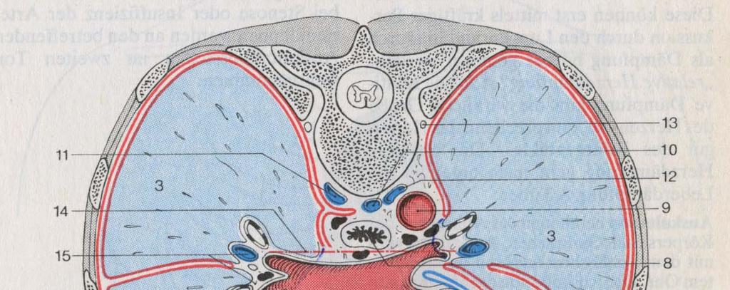

5 Anatomical borders


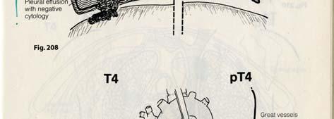
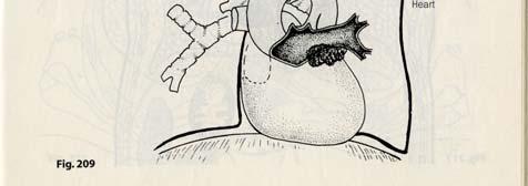
6 Staging problems of NSCLC Lymph node dissection: additional tissue shell
7 Acute mediastinitis Inflammatory diseases Predominantly posterior mediastinum Perforation of esophagus, descent of infection from within the neck, spread from chest wall infection, after heart surgery Chronic mediastinitis Anterior mediastinum Mycotic (histoplasmosis) or tuberculous Idiopathic fibrosing mediastinitis Associated with retroperitoneal fibrosis, inflammatory pseudotumor of the orbit, etc. Cave: DD Hodgkin s lymphoma
8 Cysts Pericardial (coelomic cysts) Foregut cysts: Bronchial, esophageal, gastric, enteric, pancreatic Thyroid and parathyroid lesions Struma with nodular hyperplasia, pulled down into the anterior prevascular or the retrotracheal compartment (posterior descending goiter) 7% of parathyroid adenomas found in superior mediastinum


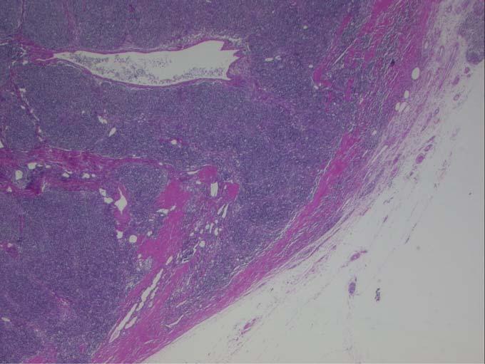
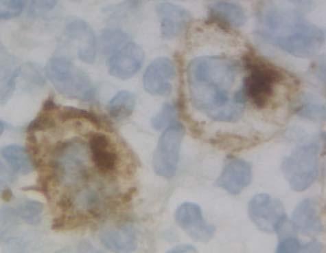
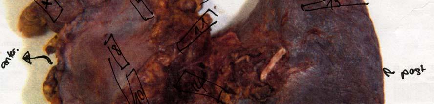
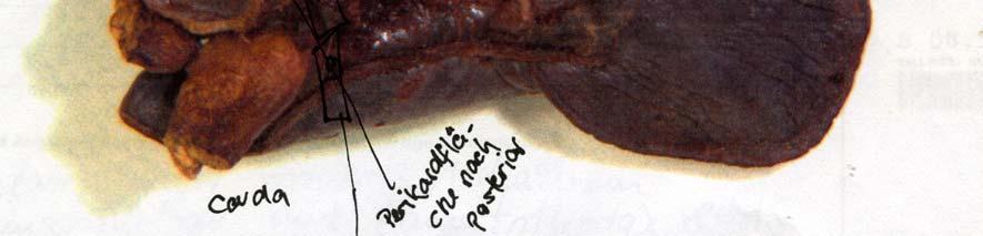
9 Pat. 41 yrs, tumor left pericardial apical Calretinin Benign coelomic cyst
")


10 Thymus Unilateral (developmental) or multilateral thymic cyst (reactive) Acute thymic involution (stress, HIV) Thymic follicular hyperplasia of B-lymphocytes (Myasthenia, HIV) Myasthenia gravis: Normal, follicular hyperplasia or thymoma
11 Thymoma Neoplasm of epithelial cells, admixed with immature thymocytes Type A: when epithelial cells have spindle/oval shape Type B: when epithelial cells have dendritic, plump, epithelioid shape Type C: overt atypia such as carcinoma ABC rule: A B C atrophic (spindle cell thymic cell of adult life) bioactive (biologically active organ of the fetus and infant) carcinoma A, AB, B1, B2, B3, C


12 Thymoma Type A Type B2 Capsular infiltration
13 Masaoka and TNM stage I II 1 II 2 III IVa IVb macroscopically completely encapsulated and microscopically no capsular invasion Macroscopic invasion into surrounding fatty tissue or mediastinal pleura or Microscopic invasion into capsule Macroscopic invasion into neighbouring organ, i. e. lung, pericardium or great vessels Pleural or pericardial dissemination Lymphogenous or hematogenous metastasis Masaoka stage T N M I II III IVa IVb Any T N1 M1 1. Encapsulated: T1 2. Minimally invasive: T2 3. Widely invasive or pleural or pericardial implants: T3/4 4. Metastatic: N1-2, M1
14 Thymoma prognosis Stage (single most important prognosticator) Histologic type: A<AB<B1<B2<B3<C Completeness of excision Myasthenia gravis
15 Malignant lymphoma Anterior, superior or middle mediastinum; thymus or nodes 1. Hodgkin s lymphoma: young females, nodular sclerosis, cysts Cave: multilocular thymic cyst, sclerosing mediastinitis 2. Lymphoblastic lymphoma: Usually immature T-cell type 3. Large cell lymphoma: Large vesicular irregular nuclei 4. Marginal zone B-cell lymphoma: Sjögren s disease, IgA type
 Send fresh tissue immediately on ice and gauze 1.")
 2.")
16 Lymphoma Service (M. Tinguely) Send fresh tissue immediately on ice and gauze 1. Fix Morphology Immunophenotyping by IHC ISH, FISH, PCR (DNA, RNA) 2. Freeze Molecular diagnostic PCR, also long distance Southern blot 3. FACS Immunophenotyping Co-expression


17 Frozen section (Schnellschnitt) CD15
18 Germ cell tumors 20% of mediastinal tumors and cysts Origin from extragonadal germ cells, related to the thymus Seminoma (always within thymus) Teratoma (mature, immature, with somatic type malignancy) Embryonal carcinoma Yolk sac tumor Choriocarcinoma Mixed germ cell tumors


19 Pat. 26 yrs, tumor anterior mediastinum Hematothorax right side: Lobectomy RUL and thymectomy

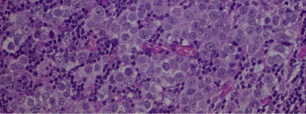


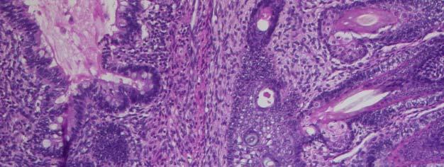
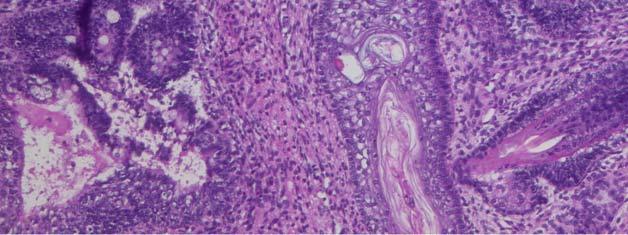
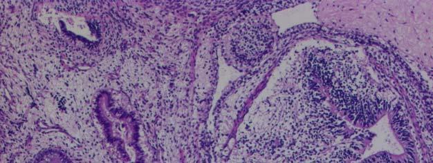
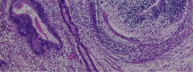
20 Mixed malignant seminomatous non seminomatous germ cell tumor with immature teratoma moiety
21 Mesenchymal tumors Benign Lipoma: above diaphragm, DD thymolipoma or lipomatosis (Cushing, steroid, obesity) Lymphangioma and Hemangioma Solitary fibrous tumor (SFT) Malignant Liposarcoma Synovial sarcoma Other sarcoma


22 Pat. 44 yrs, multilocular thymic cyst D2-40 Cystic lymphangioma with dysplastic vessels
23 Neurogenic tumors: Posterior Tumors of sympathetic nervous system (< 10 years) Neuroblastoma, ganglioneuroblastoma, ganglioneuroma Tumor of peripheral nerves (> 20 years) Schwannoma, neurofibroma, malignant peripheral nerve sheath tumor (MPNST), with rhabdomyoblastic features called Triton tumor Neuroendocrine tumors (thymus) Typical carcinoid (TC), atypical carcinoid (AC), small cell neuroendocrine carcinoma (SCLC), large cell neuroendocrine carcinoma (LCNEC) In lung mostly TC and SCLC, in thymus mostly AC
24 Summary Mediastinal lesions: cysts, benign or malignant tumors Frequency dependent on location: superior, anterior, middle and posterior mediastinum Metastases may mimic mediastinal primary Histology A<AB<B1<B2<B3<C, stage and margins major prognosticators of thymoma
25 SAMO Meeting Lucerne 2009 Thank you!
Mediastinal Tumors: Imaging
 Mediastinal Tumors: Imaging References Imaging in Oncology, Husband and Reznek Computed Tomography and Magnetic Resonance of the thorax, Naidich, Zerhouni, Siegelman, Mediastinal compartments Anterior:
Thymic Tumors. Feiran Lou MD. MS. Kings County Hospital Department of Surgery
 Thymic Tumors Feiran Lou MD. MS. Kings County Hospital Department of Surgery Case HPI 53 yo man referred from OSH for anterior mediastinal mass. Initially presented with leg weakness and back pain for
SCLEROSING LESIONS OF THE MEDIASTINUM
 SCLEROSING LESIONS OF THE MEDIASTINUM Mark R. Wick, MD Division of Surgical Pathology University of Virginia Health System Charlottesville, VA, USA SCLEROSING MEDIASTINITIS A slowly-evolving tumefactive
Karoline Nowillo, MD. February 1, 2008
 Case Presentation Karoline Nowillo, MD SUNY Downstate t February 1, 2008 Case Presentation Chief complaint enlarging goiter x 8 months History of present illness shortness of breath, heaviness in chest
Protocol for the Examination of Specimens From Patients With Thymic Tumors
 Protocol for the Examination of Specimens From Patients With Thymic Tumors Version: Protocol Posting Date: June 2017 Includes ptnm requirements from the 8 th Edition, AJCC Staging Manual For accreditation
Case Report Two Invasive Thymomas Incidentally Found during Coronary Artery Bypass Graft Surgery
 Case Reports in Pathology Volume 2016, Article ID 1516521, 4 pages http://dx.doi.org/10.1155/2016/1516521 Case Report Two Invasive Thymomas Incidentally Found during Coronary Artery Bypass Graft Surgery
ARRO Case Thymoma. Jordan Kharofa, MD Elizabeth Gore, MD Medical College of Wisconsin
 ARRO Case Thymoma Jordan Kharofa, MD Elizabeth Gore, MD Medical College of Wisconsin History HPI 54 yo male who presented to PCP with complaining subacute shortness of breath and chest pain. Pain increased
Tumors of the Thvmus and Thee
 Tumors of the Thvmus Thee Region: 111. Clinic&pathological Skdies on Teratornas Tumors of Germ Cell Type N. P. Bergh, M.D., P. Gatzinsky, M.D., S. Larsson, M.D., P. Lundin, M.D., B. Ridell, M.D. ABSTRACT
Mediastinal Germ Cell Tumors
 Mediastinal Germ Cell Tumors Anja C. Roden, M.D. Department of Laboratory Medicine and Pathology, Mayo Clinic, Rochester, MN, USA 2018 MFMER slide-1 Disclosure I have no relevant financial relationships
I. SCLEROSING LESIONS OF THE MEDIASTINUM
 I. SCLEROSING LESIONS OF THE MEDIASTINUM Mark R. Wick, MD Division of Surgical Pathology University of Virginia Health System Charlottesville, VA, USA SCLEROSING MEDIASTINITIS A slowly-evolving tumefactive
Dr. Pratik Mukherjee, MMed, FRCR Dr. Ashish Chawla, MD, ABR (USA) Khoo Teck Puat Hospital, Singapore
 Khoo Teck Puat Hospital, Singapore") Dr. Pratik Mukherjee, MMed, FRCR Dr. Ashish Chawla, MD, ABR (USA) Khoo Teck Puat Hospital, Singapore The authors declare no financial disclosures. To revisit the basics of approach to mediastinal masses
Dr. Pratik Mukherjee, MMed, FRCR Dr. Ashish Chawla, MD, ABR (USA) Khoo Teck Puat Hospital, Singapore The authors declare no financial disclosures. To revisit the basics of approach to mediastinal masses
Anterior Mediastinal Masses: The 4 T s
 May 2001 Anterior Mediastinal Masses: The 4 T s Rachel Van Sambeek, Harvard Medical School, Year III 1 Mediastinal Compartments 3 arbitrary divisions that do not correlate with anatomic planes: Anterior
Neoplasia part I. Dr. Mohsen Dashti. Clinical Medicine & Pathology nd Lecture
 Neoplasia part I By Dr. Mohsen Dashti Clinical Medicine & Pathology 316 2 nd Lecture Lecture outline Review of structure & function. Basic definitions. Classification of neoplasms. Morphologic features.
Sectional Anatomy Quiz II
 Sectional Anatomy II Rashid Hashmi Rural Clinical School, University of New South Wales, Wagga Wagga, New South Wales, Australia A R T I C L E I N F O Article type: Article history: Received: 3 Aug 2017
Presentation material is for education purposes only. All rights reserved URMC Radiology Page 1 of 98
 Presentation material is for education purposes only. All rights reserved. 2011 URMC Radiology Page 1 of 98 Radiology / Pathology Conference February 2011 Brooke Koltz, Cytopathology Resident Presentation
Clinical Usefulness of the WHO Histological Classification of Thymoma
 Original Article Clinical Usefulness of the WHO Histological Classification of Thymoma Satoshi Sonobe, MD, 1 Hideaki Miyamoto, MD, 1 Hiroshi Izumi, MD, 2 Bunsei Nobukawa, MD, 2 Toshiro Futagawa, MD, 1
Pulmonary Neoplasms & Chest Disorders Ahmed Mahmoud
 Pulmonary Neoplasms & Chest Disorders Ahmed Mahmoud Lung Ca ;Pathological types Adenocarcinoma More common in females Peripheral location Spread mainly by blood(brain..) Incidence is increasing (the most
Assessing the lung and mediastinum in cancer-is tissue the issue? George Santis
 1 Assessing the lung and mediastinum in cancer-is tissue the issue? George Santis Optimal management of Cancer Histological diagnosis & accurate staging at presentation Molecular analysis of primary tumour
Thyroid INTRODUCTION ANATOMY SUMMARY OF CHANGES
 AJC 7/14/06 1:19 PM Page 67 Thyroid C73.9 Thyroid gland SUMMARY OF CHANGES Tumor staging (T) has been revised and the categories redefined. T4 is now divided into T4a and T4b. Nodal staging (N) has been
Myasthenia: Is Medical Therapy in the Grave? Katy Marino, PGY-5
 Myasthenia: Is Medical Therapy in the Grave? Katy Marino, PGY-5 Disclosures Outline History of Thymus Anatomy of Thymus Pathophysiology of Myasthenia Gravis Medical Management of Myasthenia Gravis Surgical
Note: The cause of testicular neoplasms remains unknown
 - In the 15- to 34-year-old age group, they are the most common tumors of men. - Tumors of the testis are a heterogeneous group of neoplasms that include: I. Germ cell tumors : 95%; all are malignant.
THYMIC CARCINOMAS AN UPDATE
 THYMIC CARCINOMAS AN UPDATE Mark R. Wick, M.D. University of Virginia Medical Center Charlottesville, VA CARCINOMA OF THE THYMUS General Clinical Features No apparent gender predilection Age range of 35-75
Thymic Epithelial Tumors
 Thymic Epithelial Tumors What are the Challenges & What is New? Anja C. Roden, M.D. Department of Laboratory Medicine and Pathology, Mayo Clinic, Rochester, MN, USA No disclosures Disclosure Thymic Epithelial
TNM classifications have been established for various
 Lymphogenous and Hematogenous Metastasis of Thymic Epithelial Tumors Kazuya Kondo, MD, PhD, and Yasumasa Monden, MD, PhD Department of Oncological and Regenerative Surgery, School of Medicine, University
Basic Data. Sex:Male 31 years old Occupation: 搬家工人
 Basic Data Sex:Male 31 years old Occupation: 搬家工人 Chief Complaint Intermittent chest pain with shortness of breath for 2-3 months. Present Illness 4 months ago, he started having occasional chest pain
Pelvic tumor in childhood Classification, imaging approach and radiological findings
 Pelvic tumor in childhood Classification, imaging approach and radiological findings M. Mearadji International Foundation for Pediatric Imaging Aid Rotterdam, The Netherlands Solid pelvic masses in childhood
Mediastinoscopy, Mediastinotomy And Thoracoscopy For Mediastinal Lesions. Alper Toker, MD
 Mediastinoscopy, Mediastinotomy And Thoracoscopy For Mediastinal Lesions Alper Toker, MD Istanbul University, Istanbul Medical School Department of Thoracic Surgery The mediastinum is a complex anatomic
incidence rate x 100,000/year
 Tier R=rare C=common Cancer Entity European crude and age adjusted incidence by cancer, years of diagnosis 2000 and 2007 Analisys based on 83 population-based cancer registries * applying the European
Large mediastinal masses - etiology, imaging findings, differential diagnosis
 Large mediastinal masses - etiology, imaging findings, differential diagnosis Poster No.: C-2464 Congress: ECR 2012 Type: Educational Exhibit Authors: V. Urban, M. Djosev, T. Nastasic, B. Begenisic, N.
6. Cervical Lymph Nodes and Unknown Primary Tumors of the Head and Neck
 1 Terms of Use The cancer staging form is a specific document in the patient record; it is not a substitute for documentation of history, physical examination, and staging evaluation, or for documenting
Thymic epithelial tumors (TETs), including thymomas,
, including thymomas,") Evolution of Classification of Thymic Epithelial Tumors in the Era of Dr Thomas V. Colby Anja C. Roden, MD Context. Numerous histomorphologic and staging classifications of thymic epithelial tumors (TETs)
Evolution of Classification of Thymic Epithelial Tumors in the Era of Dr Thomas V. Colby Anja C. Roden, MD Context. Numerous histomorphologic and staging classifications of thymic epithelial tumors (TETs)
DETERMINATION OF A LYMPHOID PROCESS
 Chapter 2 Applications of Touch Preparation Cytology to Intraoperative Consultations: Lymph Nodes and Extranodal Tissues for Evaluation of Hematolymphoid Disorders INTRODUCTION As discussed in Chap. 1,
6 th Reprint Handbook Pages AJCC 7 th Edition
 6 th Reprint Handbook Pages AJCC 7 th Edition AJCC 7 th Edition Errata for 6 th Reprint Table 1 Handbook No Significant Staging Clarifications for 6 th Reprint AJCC 7 th Edition Errata for 6 th Reprint
Gross appearance of peritoneal cysts. They have a thin, translucent wall and contain a clear fluid.
 Gross appearance of peritoneal cysts. They have a thin, translucent wall and contain a clear fluid. So-called multicystic benign mesothelioma. A, Gross appearance. So-called multicystic benign mesothelioma.
Differential Diagnosis of Oral Masses. Palatal Lesions
 Differential Diagnosis of Oral Masses Palatal Lesions Palatal Masses Periapical Abscess Torus Palatinus Mucocele Lymphoid Hyperplasia Adenomatous Hyperplasia Benign Salivary Neoplasms Malignant Salivary
Diplomate of the American Board of Pathology in Anatomic and Clinical Pathology
 A 33-year-old male with a left lower leg mass. Contributed by Shaoxiong Chen, MD, PhD Assistant Professor Indiana University School of Medicine/ IU Health Partners Department of Pathology and Laboratory
Vishnu Sharma M 1*, Janso Kollanur 1, Manjunath. M 1, Alka C Bhat 1, V.Viswambhar 2
 e - ISSN - 2349-8005 INTERNATIONAL JOURNAL OF ADVANCES IN CASE REPORTS Journal homepage: www.mcmed.us/journal/ijacr ELDERLY SMOKER WITH LEFT SIDED CHEST PAIN Vishnu Sharma M 1*, Janso Kollanur 1, Manjunath.
Contents. Basic Ultrasound Principles and Terminology. Ultrasound Nodule Characteristics
 Contents Basic Ultrasound Principles and Terminology Basic Ultrasound Principles... 1 Ultrasound System... 2 Linear Transducer for Superficial Images and Ultrasound-Guided FNA... 3 Scanning Planes... 4
Non-neoplastic Conditions in the Mediastinum
 2017 WCTI Non-neoplastic Conditions in the Mediastinum Mi Young Kim, MD, PhD Hyun Jung Koo, MD Jae Woo Song, MD, PhD Department of Radiology and Research Institute of Radiology Asan Medical Center, University
FDG PET/CT in Lung Cancer Read with the experts. Homer A. Macapinlac, M.D.
 FDG PET/CT in Lung Cancer Read with the experts Homer A. Macapinlac, M.D. Patient with suspected lung cancer presents with left sided chest pain T3 What is the T stage of this patient? A) T2a B) T2b C)
DIAGNOSTIC IMAGING OF THE MEDIASTINUM MASSES. ABSTRACT OF Ph.D Thesis
 UNIVERSITY OF MEDICINE AND PHARMACY CRAIOVA DIAGNOSTIC IMAGING OF THE MEDIASTINUM MASSES ABSTRACT OF Ph.D Thesis SCIENTIFIC COORDINATOR, PROF. UNIV. DR. ANDREI BONDARI PhD student Dr. DAN VASILE MOROŞANU
Adrenal masses in infancy and childhood: A clinical and radiological overview M. Mearadji
 Adrenal masses in infancy and childhood: A clinical and radiological overview M. Mearadji International Foundation for Pediatric Imaging Aid Introduction Neoplastic adrenal masses usually originate from
ONCOLOGY. Csaba Bödör. Department of Pathology and Experimental Cancer Research november 19., ÁOK, III.
 ONCOLOGY Csaba Bödör Department of Pathology and Experimental Cancer Research 2018. november 19., ÁOK, III. bodor.csaba1@med.semmelweis-univ.hu ONCOLOGY Characteristics of Benign and Malignant Neoplasms
A Nervous Breakdown: Multimodality Imaging of Thoracic Neurogenic Tumors
 A Nervous Breakdown: Multimodality Imaging of Thoracic Neurogenic Tumors John P. Lichtenberger III, MD, Maj, USAF, MC Assistant Professor, Dept. or Radiology Uniformed Services University of the Health
Case Presentation HPI. PMHx: HTN, BPH, psychiatric disorder, negative cardiac stress test 4/2010. Allergies: NKDA
 Case Presentation HPI 62 year-old male presents with several episodes of anterior chest pain. Full cardiac evaluation was negative for ischemia. CT scan revealed a 4cm anterior mediastinal mass. Pt denies
WHO Histologic Classification is a Prognostic Indicator in Thymoma
 WHO Histologic Classification is a Prognostic Indicator in Thymoma Kazuya Kondo, MD, PhD, Kiyoshi Yoshizawa, MD, PhD, Masaru Tsuyuguchi, MD, PhD, Suguru Kimura, MD, PhD, Masayuki Sumitomo, MD, Junji Morita,
The thymus: A pictorial review of thymic pathology
 The thymus: A pictorial review of thymic pathology Poster No.: C-1692 Congress: ECR 2013 Type: Educational Exhibit Authors: D. J. Conces, S. Rissing, P. J. Loehrer; Indianapolis, IN/US Keywords: Cancer,
Contents Part I Introduction 1 General Description 2 Natural History: Importance of Size, Site, Histopathology
 Contents Part I Introduction 1 General Description... 3 1.1 Introduction... 3 1.2 Incidence and Prevalence... 5 1.3 Predisposing and Genetic Factors... 8 References... 16 2 Natural History: Importance
Neoplasia literally means "new growth.
 NEOPLASIA Neoplasia literally means "new growth. A neoplasm, defined as "an abnormal mass of tissue the growth of which exceeds and is uncoordinated with that of the normal tissues and persists in the
Principles and Practice of Radiation Oncology. 4 th edition. Chapter 45. Mediastinum and Trachea
 Principles and Practice of Radiation Oncology 4 th edition Chapter 45 Mediastinum and Trachea Tony Y. Eng 1, Todd J. Scarbrough 2, Charles R. Thomas, Jr. 3 1 Associate Professor 3 Associate Professor and
Anterior Mediastinal Masses
 Residents Section Pattern of the Month Shahrzad et al. nterior Mediastinal Masses Residents Section Pattern of the Month Maryam Shahrzad 1 Thi Som Mai Le Mario Silva lexander. ankier Ronald L. Eisenberg
SESSION 1: GENERAL (BASIC) PATHOLOGY CONCEPTS Thursday, October 16, :30am - 11:30am FACULTY COPY
 PATHOLOGY CONCEPTS Thursday, October 16, :30am - 11:30am FACULTY COPY") SESSION 1: GENERAL (BASIC) PATHOLOGY CONCEPTS Thursday, October 16, 2008 9:30am - 11:30am FACULTY COPY GOAL: Describe the basic morphologic (structural) changes which occur in various pathologic conditions.
SESSION 1: GENERAL (BASIC) PATHOLOGY CONCEPTS Thursday, October 16, 2008 9:30am - 11:30am FACULTY COPY GOAL: Describe the basic morphologic (structural) changes which occur in various pathologic conditions.
Primary mediastinal tumours
 Primary mediastinal tumours Thorax (1974), 29, 475. YOUSF D. AL-NAAMAN, MOHAMAD S. AL-AN, and MUAYYAD M. AL-OMER Department of Thoracic and Cardiovascular Surgery, College of Medicine, University of Baghdad,
Protocol for the Examination of Specimens From Patients With Thymoma and Thymic Carcinoma
 Protocol for the Examination of Specimens From Patients With Thymoma and Thymic Carcinoma Protocol applies to thymic epithelial tumors located in any area of the mediastinum. No AJCC/UICC TNM Staging System
-The cause of testicular neoplasms remains unknown
 - In the 15- to 34-year-old age group, they are the most common tumors of men. - include: I. Germ cell tumors : (95%); all are malignant. II. Sex cord-stromal tumors: from Sertoli or Leydig cells; usually
Mediastinum It is a thick movable partition between the two pleural sacs & lungs. It contains all the structures which lie
 Dr Jamila EL medany OBJECTIVES At the end of the lecture, students should be able to: Define the Mediastinum. Differentiate between the divisions of the mediastinum. List the boundaries and contents of
Mediastinal assessment: More than the 4 "T"
 Mediastinal assessment: More than the 4 "T" Poster No.: C-1434 Congress: ECR 2011 Type: Educational Exhibit Authors: S. Juanpere, N. Cañete, G. Sanchez, S. Martinez, M. Turell, P. Ortuño Muro, A. Villar
Take Home Quiz 1 Please complete the quiz below prior to the session. Use the Multiple Primary and Histology Rules
 Take Home Quiz 1 Please complete the quiz below prior to the session. Use the Multiple Primary and Histology Rules Case 1 72 year old white female presents with a nodular thyroid. This was biopsied in
Pier Luigi FILOSSO, MD FECTS Associate Professor University of Torino Dept Thoracic Surgery
 Thank you for viewing this presentation. We would like to remind you that this material is the property of the author. It is provided to you by the ERS for your personal use only, as submitted by the author.
CANCER Uncontrolled Cell Division
 CANCER Uncontrolled Cell Division What is cancer? Why does it occur? Where does it occur? Benign vs. Malignant? Types of Cancer (3 main groups) There are over 200 different types of cancer 1) Carcinomas
List of Available TMAs in the PRN
 TMA RPCI_BrainCa01 RPCI_BrCa03 RPCI_BrCa04 RPCI_BrCa05 RPCI_BrCa0 RPCI_BrCa07 RPCI_BrCa08 RPCI_BrCa15 RPCI_BrCa1 RPCI_BrCa17 RPCI_BrCa18 RPCI_BrCa19 RPCI_BrCa20 RPCI_BrCa21 RPCI_BrCa24 RPCI_BrCa25 RPCI_BrCa2
PATHOLOGICAL COMPARATIVE ASSESSMENT OF TWO CASES OF THYMIC CYST AND CYSTIC THYMOMA AND REVIEW OF THE LITERATURE
 Rev. Med. Chir. Soc. Med. Nat., Iaşi 2012 vol. 116, no. 3 INTERNAL MEDICINE - PEDIATRICS CASE REPORTS PATHOLOGICAL COMPARATIVE ASSESSMENT OF TWO CASES OF THYMIC CYST AND CYSTIC THYMOMA AND REVIEW OF THE
Spindle Cell Lesions Of The Breast. Emad Rakha Professor of Breast Pathology and Consultant Pathologist
 Spindle Cell Lesions Of The Breast Emad Rakha Professor of Breast Pathology and Consultant Pathologist * SCLs comprise a wide spectrum of diseases, ranging from reactive processes to aggressive malignant
Respiratory Interactive Session. Elaine Borg
 Respiratory Interactive Session Elaine Borg Case 1 Respiratory Cytology 55 year old gentleman Anterior mediastinal mass EBUS FNA Case 1 Respiratory Cytology 55 year old gentleman with anterior mediastinal
3 cell types in the normal ovary
 Ovarian tumors 3 cell types in the normal ovary Surface (coelomic epithelium) the origin of the great majority of ovarian tumors (neoplasms) 90% of malignant ovarian tumors Totipotent germ cells Sex cord-stromal
CODING TUMOUR MORPHOLOGY. Otto Visser
 CODING TUMOUR MORPHOLOGY Otto Visser INTRODUCTION The morphology describes the tissue of the tumour closest to normal tissue Well differentiated tumours are closest to normal Undifferentiated tumours show
Surveys and Anatomic Pathology Education Programs
 Surveys and Anatomic Pathology Education Programs Performance Improvement Program in Surgical Pathology PIP/PIPW-C 2018 Participant Summary 2018 College of American Pathologists. The College does not permit
ROLE OF COMPUTED TOMOGRAPHY IN EVALUATING MEDIASTINAL MASSES. Dr. M. VENU MADHAV
 ROLE OF COMPUTED TOMOGRAPHY IN EVALUATING MEDIASTINAL MASSES By Dr. M. VENU MADHAV DISSERTATION SUBMITTED TO THE RAJIV GANDHI UNIVERSITY OF HEALTH SCIENCES, KARNATAKA, BANGALORE In partial fulfillment
Pathology Mystery and Surprise
 Pathology Mystery and Surprise Tim Smith, MD Director Anatomic Pathology Medical University of South Carolina Disclosures No conflicts to declare Some problem cases Kidney tumor Scalp tumor Bladder tumor
Performance Improvement Program in Surgical Pathology
 PIP-A Performance Improvement Program in Surgical Pathology PARTICIPANT SUMMARY 2009 College of American Pathologists. The College does not permit reproduction of any substantial portion of the material
Methoden / Methods inc. ICCC-3 105
 Methoden / Methods inc. ICCC-3 105 Internationale Klassifikation der Krebserkrankungen bei Kindern (ICCC-3) Zuordnung von ICD-O-3-Codes für Morphologie und Topographie zu diagnostischen Kategorien International
Case Report A case report of sclerosing thymoma of the anterior mediastinum: an exceedingly rare morphology
 Int J Clin Exp Pathol 2015;8(4):4233-4237 www.ijcep.com /ISSN:1936-2625/IJCEP0006183 Case Report A case report of sclerosing thymoma of the anterior mediastinum: an exceedingly rare morphology Shogo Tajima
Region: 11. Clinic6patholo cal Studies
 Tumors of the Thvmus and ThVIIliC Region: 11. Clinic6patholo cal Studies on Hodgkin s Disease of Re Thymus N. P. Bergh, M.D., P. Gatzinsky, M.D., S. Larsson, M.D., P. Lundin, M.D., and B. Ridell, M.D.
Renal Parenchymal Neoplasms
 Renal Parenchymal Neoplasms د. BENIGN TUMORS : Benign renal tumors include adenoma, oncocytoma, angiomyolipoma, leiomyoma, lipoma, hemangioma, and juxtaglomerular tumors. Renal Adenomas : The adenoma is
Thyroid Gland. Protocol applies to all malignant tumors of the thyroid gland, except lymphomas.
 Thyroid Gland Protocol applies to all malignant tumors of the thyroid gland, except lymphomas. Procedures Cytology (No Accompanying Checklist) Partial Thyroidectomy Total Thyroidectomy With/Without Lymph
Standardized definitions and policies of minimally invasive thymoma resection
 Perspective Standardized definitions and policies of minimally invasive thymoma resection Alper Toker 1,2 1 Department of Thoracic Surgery, Istanbul Medical School, Istanbul University, Istanbul, Turkey;
Atypical Palisaded Myofibroblastoma of Lymph Node: Report of a rare case.
 ISPUB.COM The Internet Journal of Pathology Volume 10 Number 1 Atypical Palisaded Myofibroblastoma of Lymph Node: Report of a rare case. V Kinnera, R Nandyala, M Yootla, K Mandyam Citation V Kinnera, R
10/14/2018 Dr. Shatarat
 2018 Objectives To discuss mediastina and its boundaries To discuss and explain the contents of the superior mediastinum To describe the great veins of the superior mediastinum To describe the Arch of
Update in Lymphoma Imaging
 Update in Lymphoma Imaging Victorine V. Muse, MD Lymphoma Update in Lymphoma Imaging Victorine V Muse, MD Heterogeneous group of lymphoid neoplasms divided into two broad histological categories Hodgkin
CAP Cancer Protocol and ecc Summary of Changes for August 2014 Thyroid Agile Release
 CAP Cancer Protocol and ecc Summary of Changes for August 2014 Thyroid Agile Release 2 REVISION HISTORY Date Author / Editor Comments 5/19/2014 Jaleh Mirza Created the document 8/12/2014 Samantha Spencer/Jaleh
Impact of immunostaining of pulmonary and mediastinal cytology
 Impact of immunostaining of pulmonary and mediastinal cytology Harman Sekhon MD, PhD Director of Cytopathology Head of Ottawa-site Ontario Tumour Bank June 20, 2014 Disclaimer Pfizer: Honorarium-Advisory
number Done by Corrected by Doctor Maha shomaf
 number 17 Done by Ahmad rawajbeh Corrected by أسامة الخضر Doctor Maha shomaf 0 P a g e In this lecture, we are going to: complete the differentiation between benign and malignant tumors. - -start to study
NEOPLASIA-I CANCER. Nam Deuk Kim, Ph.D.
 NEOPLASIA-I CANCER Nam Deuk Kim, Ph.D. 1 2 Tumor in the hieroglyphics of the Edwin Smith papyrus (1,600 B.C., Breasted s translation 1930) 3 War on Cancer (National Cancer Act, 1971) 4 Cancer Acts in Korea
Malignant Cardiac Tumors Rad-Path Correlation
 Malignant Cardiac Tumors Rad-Path Correlation Vincent B. Ho, M.D., M.B.A. 1 Jean Jeudy, M.D. 2 Aletta Ann Frazier, M.D. 2 1 Uniformed Services University of the Health Sciences 2 University of Maryland
Thymoma and Thymic Carcinoma
 Thymoma and Thymic Carcinoma Protocol applies to thymic epithelial tumors located in any area of the mediastinum. Procedures Biopsy Resection Protocol revision date: January 2004 No AJCC/UICC staging system
TUMOR,NEOPLASM. Pathology Department, Zhejiang University School of Medicine,
 TUMOR,NEOPLASM Pathology Department, Zhejiang University School of Medicine, 马丽琴,maliqin198@zju.edu.cn The points in this chapter What is a neoplasm (conception) Morphology of neoplasm Macroscopy of Neoplasm
Male Genital Cancers in the US in Frequency of Types
 Germ Cell Tumors of the Testis Pathology, Immunohistochemistry, and the Often Confusing Appearance of Their Metastases Charles Zaloudek, MD Department of Pathology UCSF Male Genital Cancers in the US in
Neckmasses in infancy and childhood: Clinical and radiological classification and imaging approaches M. Mearadji
 Neckmasses in infancy and childhood: Clinical and radiological classification and imaging approaches M. Mearadji International Foundation for Pediatric Imaging Aid Introduction Neck masses are a frequent
In the 1960s, thymomas were classified into two categories: Staging System of Thymoma MALIGNANCIES OF THE THYMUS. Akira Masaoka, MD, PhD S304
 MALIGNANCIES OF THE THYMUS Akira Masaoka, MD, PhD Introduction: Thirty years have gone by since the Masaoka staging system of thymoma was proposed in 1981. Although the Masaoka staging system has been
LYMPHATIC DRAINAGE AXILLARY (MOSTLY) INTERNAL MAMMARY SUPRACLAVICULAR
 INTERNAL MAMMARY SUPRACLAVICULAR") BREAST LYMPHATIC DRAINAGE AXILLARY (MOSTLY) INTERNAL MAMMARY SUPRACLAVICULAR HISTOLOGY LOBE: (10 in whole breast) LOBULE: (many per lobe) ACINUS/I, aka ALVEOLUS/I: (many per lobule) DUCT(S): INTRA- or
BREAST LYMPHATIC DRAINAGE AXILLARY (MOSTLY) INTERNAL MAMMARY SUPRACLAVICULAR HISTOLOGY LOBE: (10 in whole breast) LOBULE: (many per lobe) ACINUS/I, aka ALVEOLUS/I: (many per lobule) DUCT(S): INTRA- or
Prof. Dr. med. Beata BODE-LESNIEWSKA Institute of Pathology and Molecular Pathology University Hospital; Zurich
 Prof. Dr. med. Beata BODE-LESNIEWSKA Institute of Pathology and Molecular Pathology University Hospital; Zurich 32 year old man 2 months history of growing left supraclavicular lymph nodes Antibiotic treatment
Pitfalls in thyroid tumor pathology. Prof.Valdi Pešutić-Pisac MD, PhD
 Pitfalls in thyroid tumor pathology Prof.Valdi Pešutić-Pisac MD, PhD Too many or... Tumour herniation through a torn capsule simulating capsular invasion fibrous capsule with a sharp discontinuity, suggestive
CURRENT REVIEW. Tumors
 CURRENT REVIEW Primary Malignant of the Mediastinum Tumors David M. Conkle, M.D., and R. Benton Adkins, Jr., M.D. ABSTRACT Forty-three patients with primary malignant tumors of the rnediastinum are reviewed.
Appendix E SEER Program Coding and Staging Manual DRAFT Reportable Examples
 Appendix E1 2018 SEER Program Coding and Staging Manual DRAFT Reportable Examples Malignant # Diagnosis/Condition Notes 1 Atypical fibroxanthoma (superficial malignant fibrous histiocytoma) 2 Positive
FOLLICULARITY in LYMPHOMA
 FOLLICULARITY in LYMPHOMA Reactive Follicular Hyperplasia Follicular Hyperplasia irregular follicles Follicular Hyperplasia dark and light zones Light Zone Dark Zone Follicular hyperplasia MIB1 Follicular
MEDICAL POLICY Gene Expression Profiling for Cancers of Unknown Primary Site
 POLICY: PG0364 ORIGINAL EFFECTIVE: 04/22/16 LAST REVIEW: 07/26/18 MEDICAL POLICY Gene Expression Profiling for Cancers of Unknown Primary Site GUIDELINES This policy does not certify benefits or authorization
From Morphology to Molecular Pathology: A Practical Approach for Cytopathologists Part 1-Cytomorphology. Songlin Zhang, MD, PhD LSUHSC-Shreveport
 From Morphology to Molecular Pathology: A Practical Approach for Cytopathologists Part 1-Cytomorphology Songlin Zhang, MD, PhD LSUHSC-Shreveport I have no Conflict of Interest. FNA on Lymphoproliferative
4/22/2010. Hakan Korkmaz, MD Assoc. Prof. of Otolaryngology Ankara Dıșkapı Training Hospital-Turkey.
 Management of Differentiated Thyroid Cancer: Head Neck Surgeon Perspective Hakan Korkmaz, MD Assoc. Prof. of Otolaryngology Ankara Dıșkapı Training Hospital-Turkey Thyroid gland Small endocrine gland:
the urinary system pathology Dr. Fairoz A Eltorgman
 the urinary system pathology Dr. Fairoz A Eltorgman Tumors of the renal pelvis & kidney Benign tumors of the renal pelvis: Hemangioma Leiomyoma Malignant tumors: Transitional cell carcinoma Squamous cell
A neoplasm is defined as "an abnormal tissue proliferation, which exceeds that of adjacent normal tissue. This proliferation continues even after
 NEOPLASIA Neoplasia is a very important topic in pathology because neoplasms are both common and serious diseases. A neoplasm literally means a new growth, and this term is used interchangeably with a