SKIN SONOGRAPHY IN CHILDREN. CRISTIAN J. GARCIA MD Santiago, Chile
|
|
|
- Darlene Francis
- 5 years ago
- Views:
Transcription


1 SKIN SONOGRAPHY IN CHILDREN CRISTIAN J. GARCIA MD Santiago, Chile
2 I HAVE NO DISCLOSURES
3 OBJECTIVES RELEVANCE OF SKIN LESIONS IN CHILDREN ROLEN OF THE RADIOLOGIST CLINICAL CORRELATION US TECHNIQUE NORMAL ANATOMY EXAMPLES OF MOST COMMON LESIONS VASCULAR ANOMALIES WILL NOT BE DISCUSSED
4 INTRODUCTION Superficial soft tissue lesions are not uncommon in children Often a matter of concern Malignant lesions are very rare in this age group Clinical findings may be nonspecific
5 INTRODUCTION High resolution ultrasonography (US) with color-doppler study could be of help in the characterization of skin lesions and can suggest the specific diagnosis, when clinically the diagnosis is unclear Significant and progressive increase in the number of studies However, in many cases the final diagnosis is made histologically
6 INTRODUCTION US can give information about the size, extension, shape, depth, involves layers and vascularity of the lesion Solid, cystic, complex Calcifications
7 INTRODUCTION There is an incredible number of different entities, based on pathological findings US : Different lesions may look similar Clinical correlation is extremely important US - histologic correlation Close relation with pathologists and dermatologists
8 DERMATOPATHOLOGIST DERMATOLOGIST DIAGNOSIS RADIOLOGIST
9 DERMATOPATHOLOGIST DERMATOLOGIST DIAGNOSIS RADIOLOGIST
10 DERMATOPATHOLOGIST DERMATOLOGIST DIAGNOSIS RADIOLOGIST
11 DERMATOPATHOLOGIST DERMATOLOGIST DIAGNOSIS RADIOLOGIST depth involved layers vascular flow cystic-solidcomplex inflammation rupture
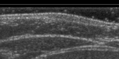
12 DERMATOPATHOLOGIST DERMATOLOGIST DIAGNOSIS RADIOLOGIST
13 DERMATOPATHOLOGIST DERMATOLOGIST DIAGNOSIS RADIOLOGIST
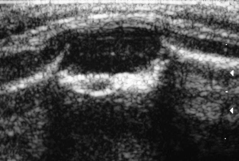
14 LEARNING CURVE ACCURACY NUMBER OF CASES
15
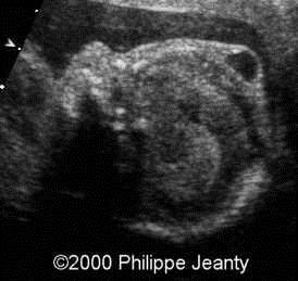
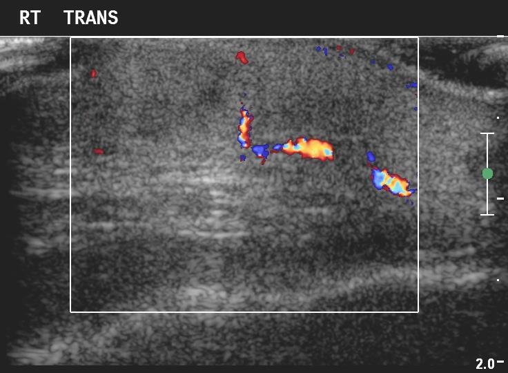
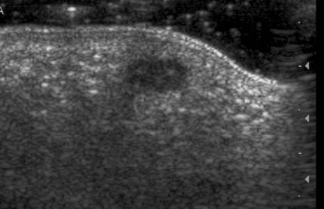
16

17 NORMAL ANATOMY Wortsman, X. - EPIDERMIS - DERMIS -HYPODERMIS OF SUBCUTANEOUS TISSUE


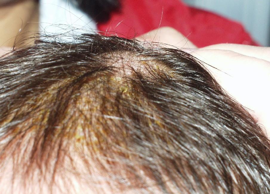
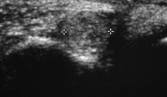
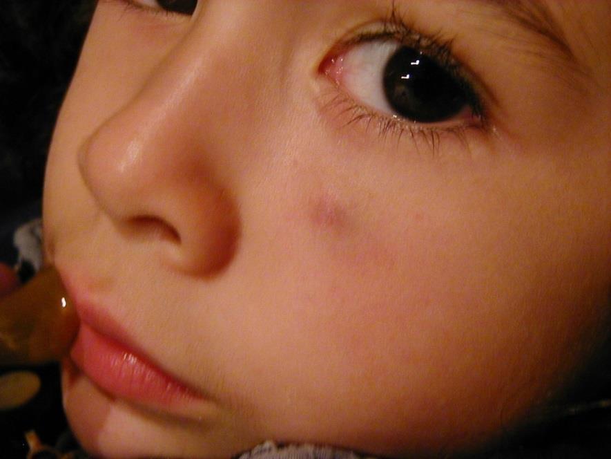
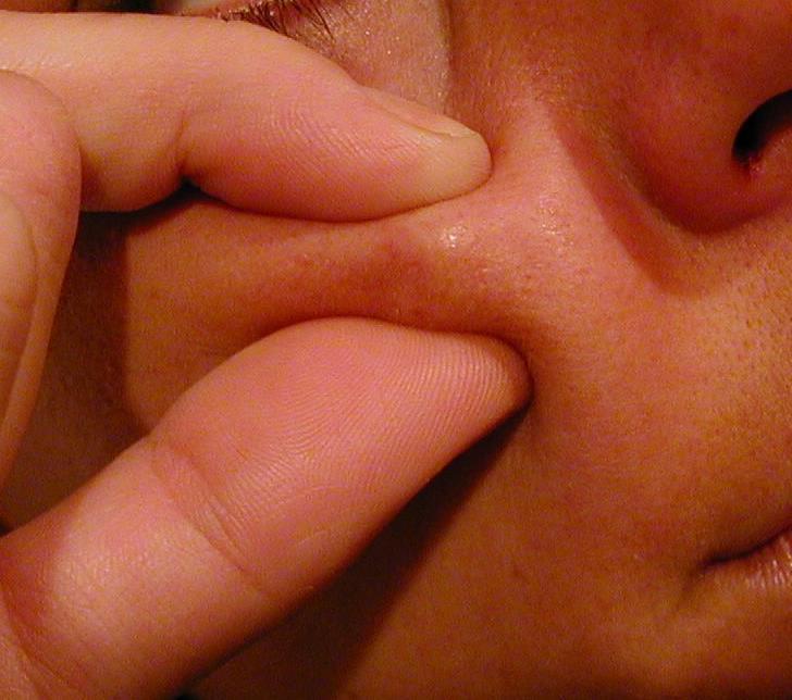
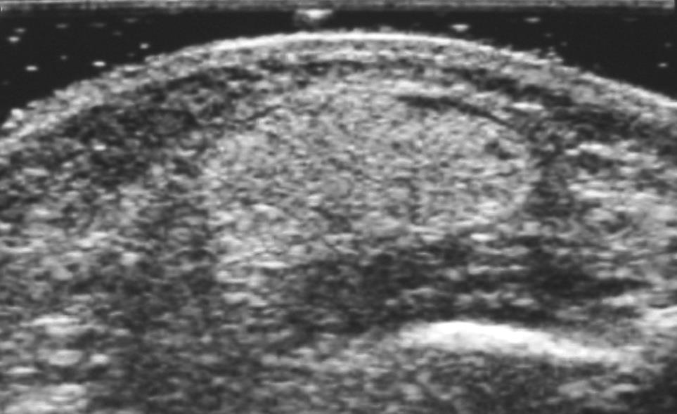
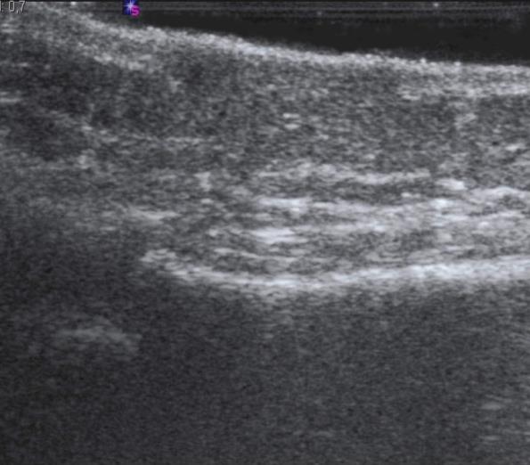
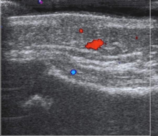
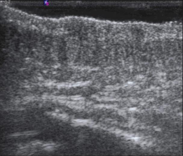
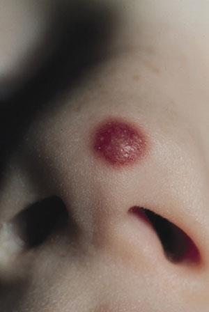
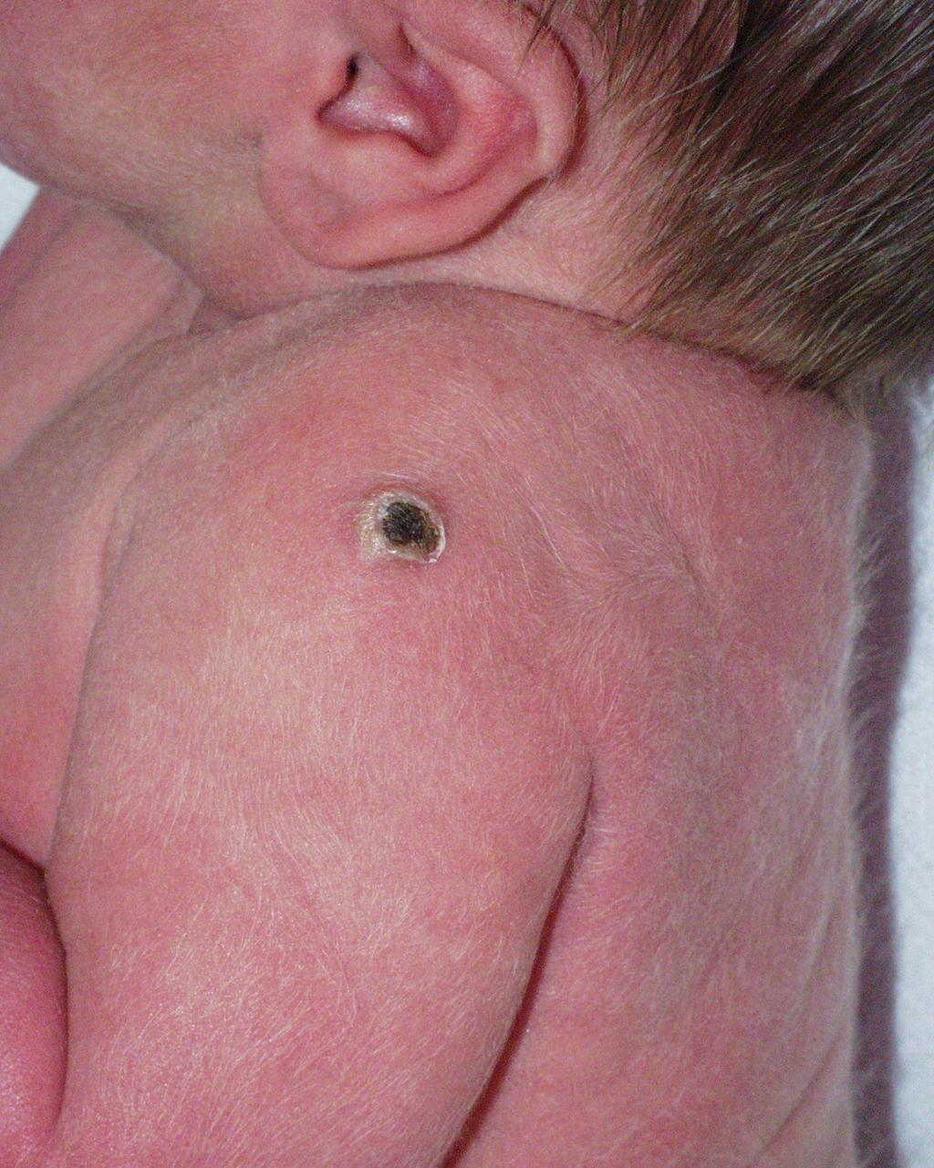
18 NORMAL ANATOMY E D H D H Wortsman, X. E M D E H

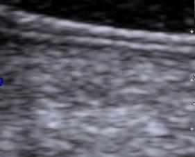
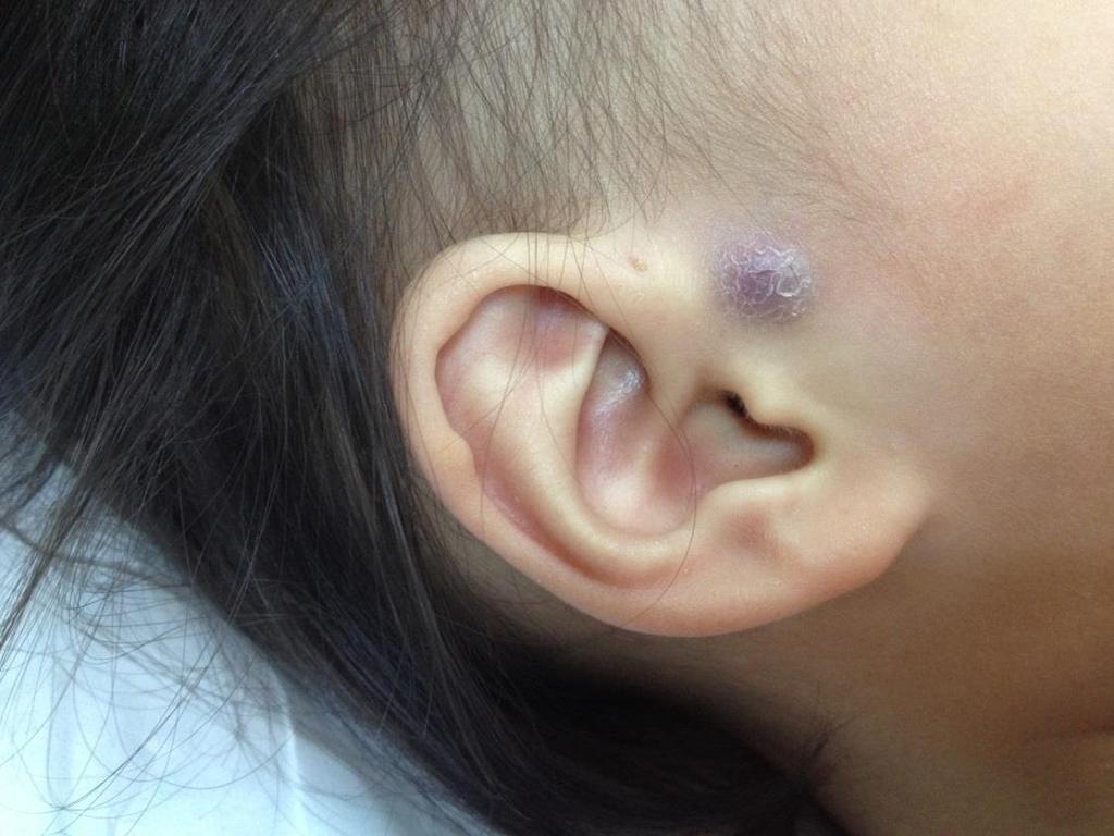
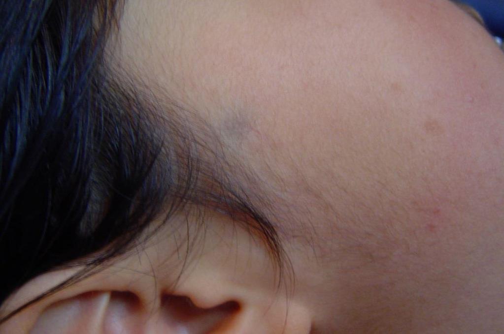
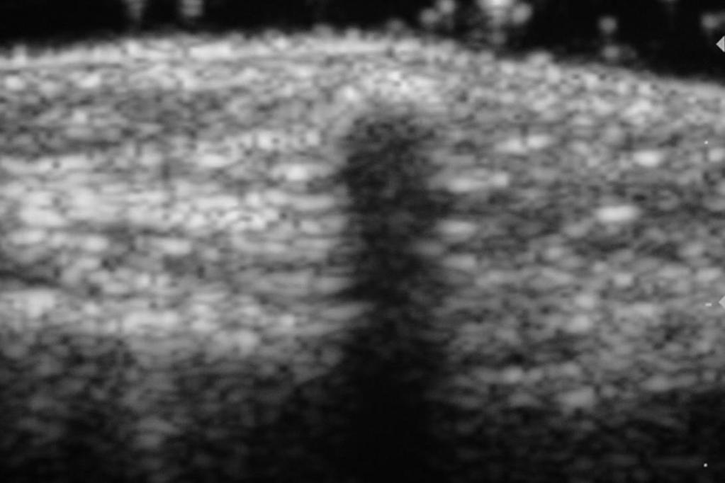
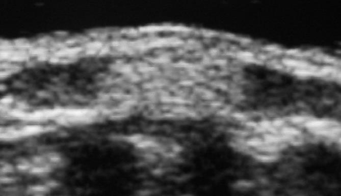
19 TECHNIQUE High-resolution linear transducers mhz Color Doppler US Jelly pad No sedation Spontaneous sleep if necessary


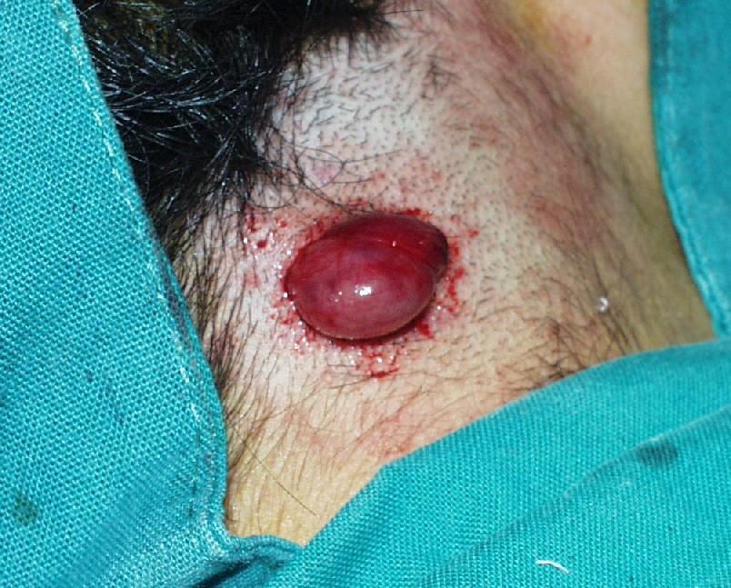
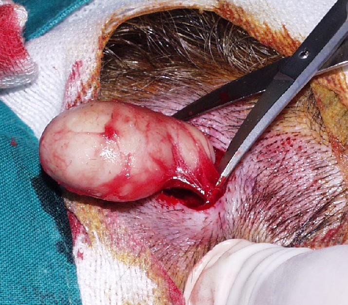
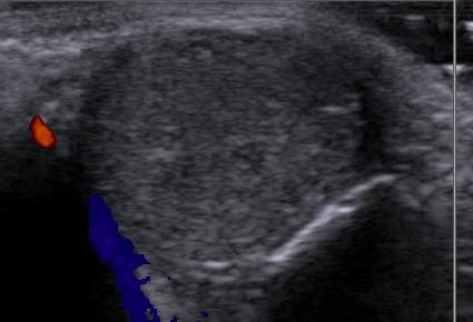
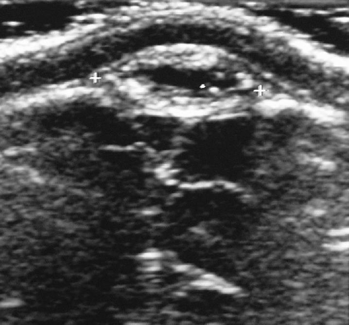
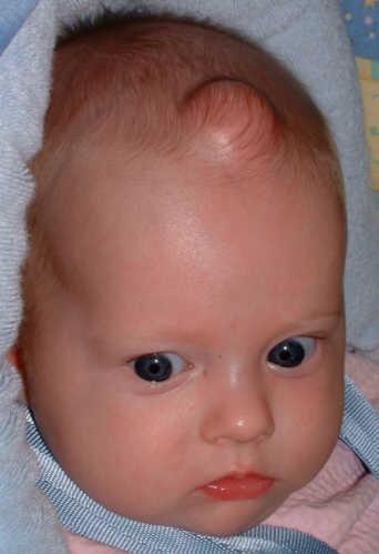
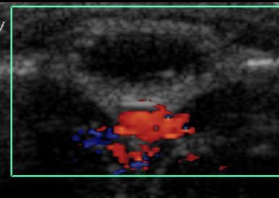
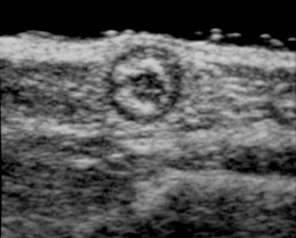
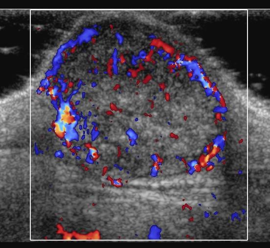
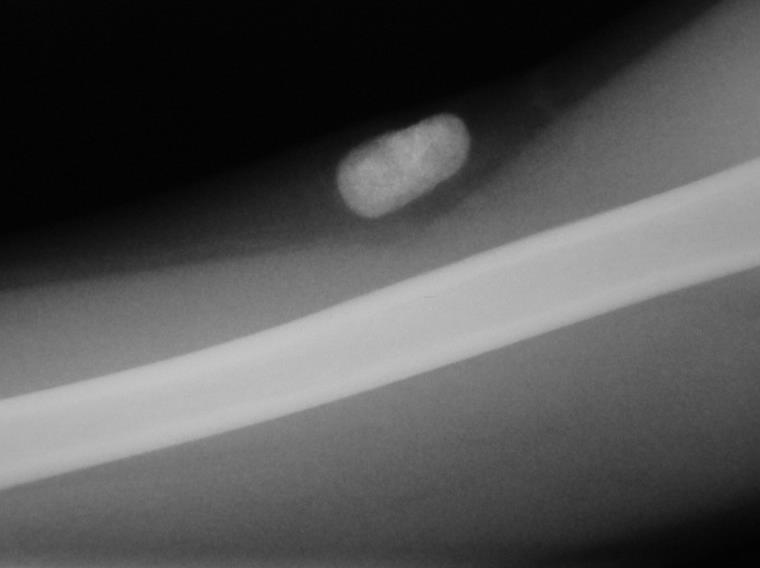
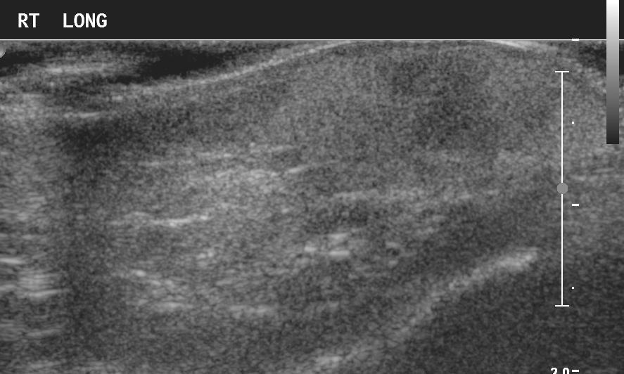
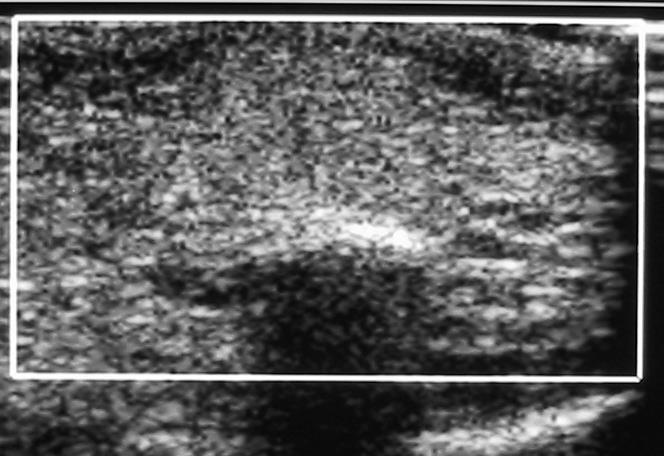
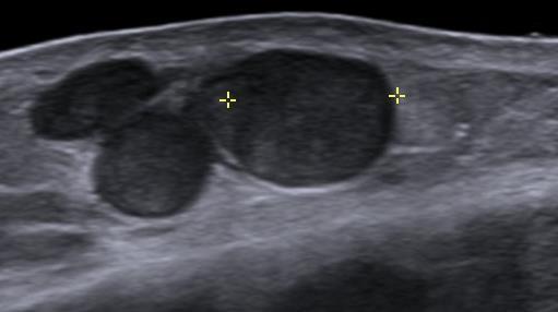
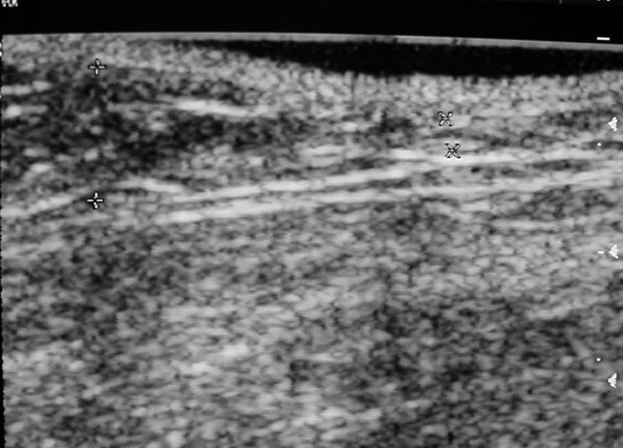
20 14 y/o. Posterior cervical mass

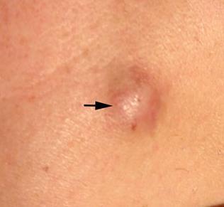
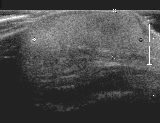
21 EPIDERMAL CYST
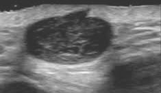
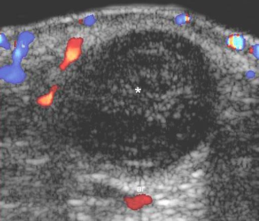
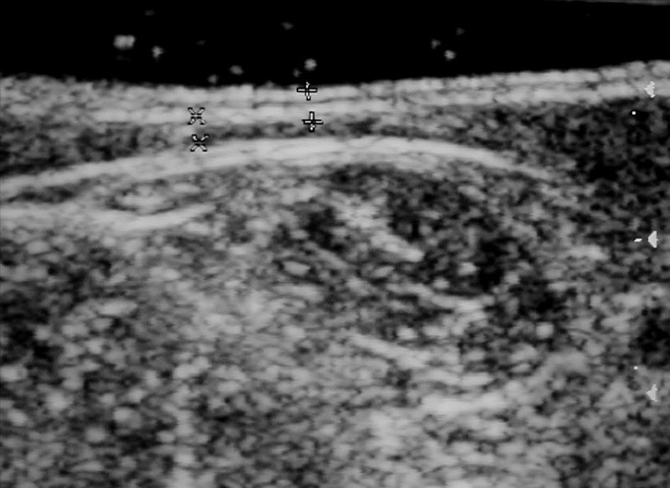
22 EPIDERMAL CYST Epidermal - epidermal inclusion cyst is the most common type of follicular cyst (80 %) Implantation of epidermal components in the dermis and subcutaneous tissue Congenital, traumatic, related to previous surgery or unknown cause Histologically composed of stratified squamous epithelium without dermal components Therefore, the term sebaceous cyst is not correct and confusing
23 EPIDERMAL CYST More common in head, face, neck, back Any age, more common around puberty Infection is common 1-6 cm Most cases (96%) with acoustic enhancement Avascular Ruptured or inflammed cyst could show peripheral flow Most are hypoechoic and may contain scattered internal echoes or keratinous debris

24 Wortsman X. Springer, 2013


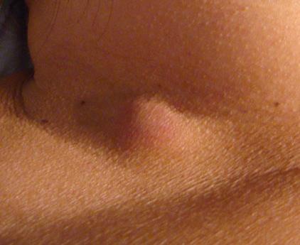
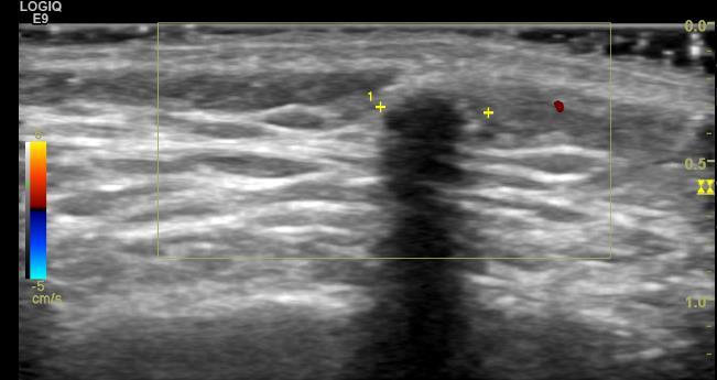
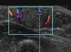
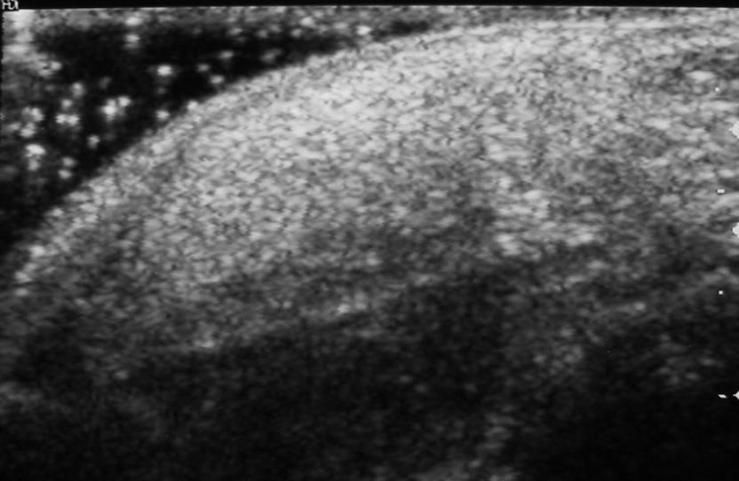
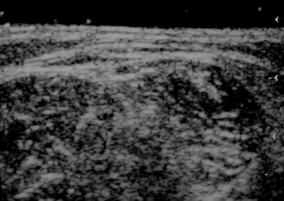
25 2 y/o. History of facial trauma 6 months ago Epidermal inclusion cyst

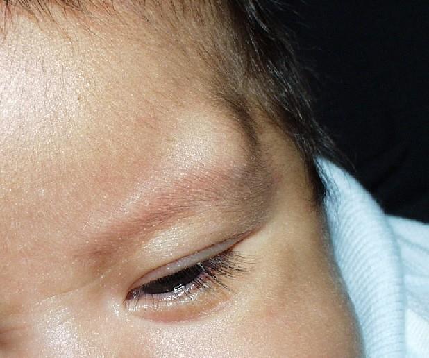
26 EYEBROW TAIL NODULE
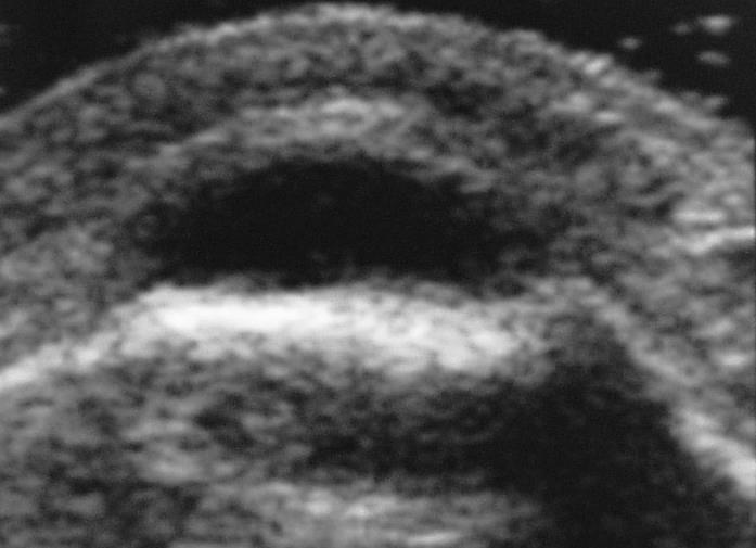
27 ANTERIOR FONTANELLE

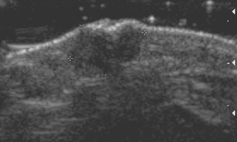
28 SKULL DERMOID CYST
29 DERMOID CYST Includes epidermal and dermal elements Most cases congenital 50% at birth Deep, below the hypodermis May have bone involvement Skull, orbit, midline, anterior fontanelle, nose, anterior neck Can grow up to a size of 1-4 cm. May persist unchanged in size or grow later in life. US : Hypo hyperechoic
30
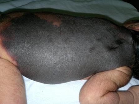
31
32

33
34 PILOMATRICOMA Also called calcifying epithelioma of Malherbe or trichomatricoma Benign dermal and subcutaneous tumor One of the most commonly excised superficial masses in children. More common in < 15 y. Most are solitary nodule Head and neck (50-70%), upper limbs (25 30 %), and less commonly on the trunk and legs (15 20 %)
35 1-mont-old male infant. Posterior neck mass

36 SUBCUTANEOUS FAT NECROSIS
37 SUBCUTANEOUS FAT NECROSIS OF THE First month of life NEWBORN Tender or asymptomatic indurated nodules or plaques with or without erythema Thought to be secondary to ischemic injury from trauma of the delivery US : echogenic subcutaneous lesion Epidermis and dermis may be thickened


38 14 y/o female. 3- month history of soft tissue swelling of the right knee. Treated with anti-inflammatory with no changes Courtesy Dr. Marcos Silva. Chile.


39 Courtesy Dr. Marcos Silva. Chile.
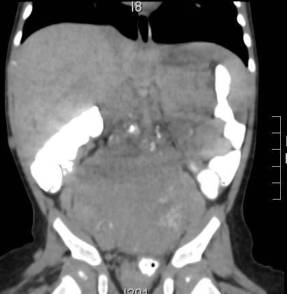
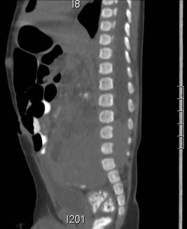
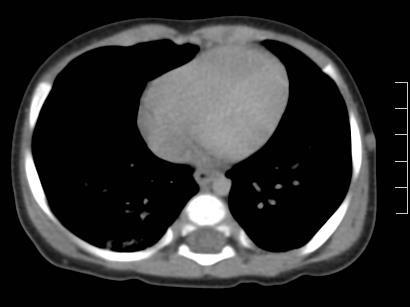
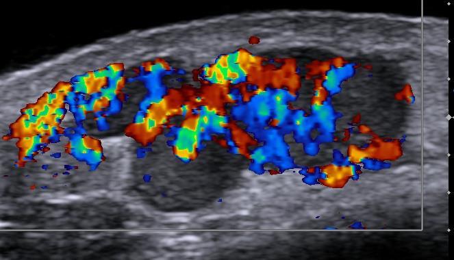
40 DIFFERENTIAL DIAGNOSIS Paniculitis Edema Fat necrosis Insect bite Lupus Lipoma Lipoblastoma Lymphoma
41 DIFFERENTIAL DIAGNOSIS Paniculitis Edema Fat necrosis Insect bite Lupus Lipoma Lymphoma
42 Extranodal NK/T-cell lymphoma Rare type of non-hodgkin lymphoma Extranodal NK/T-cell lymphoma, nasal type, can develop in either T cells or natural killer (NK) cells Most commonly involves upper airway nasal May involve primarily the skin Occurs in all age groups Strongly linked to Epstein-Barr virus Eritematous plaques, sometimes ulcerated Bad prognosis Kimura, Blood 2012; 119:


43 6 m/o. Multiple soft tissue nodules occipital anterior abd wall axillary mamary
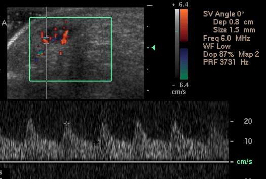
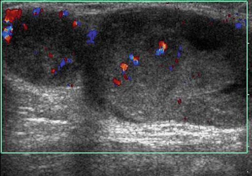
44 MULTIPLE SOFT TISSUE NODULES Infantile myofibromatosis Metastatic sarcoma Epithelioid sarcoma Metatastatic melanoma Granuloma annulare Septic emboli Metastatic neuroblastoma


45 6 m/o. Multiple soft tissue nodules

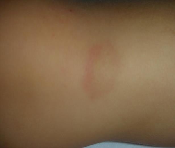
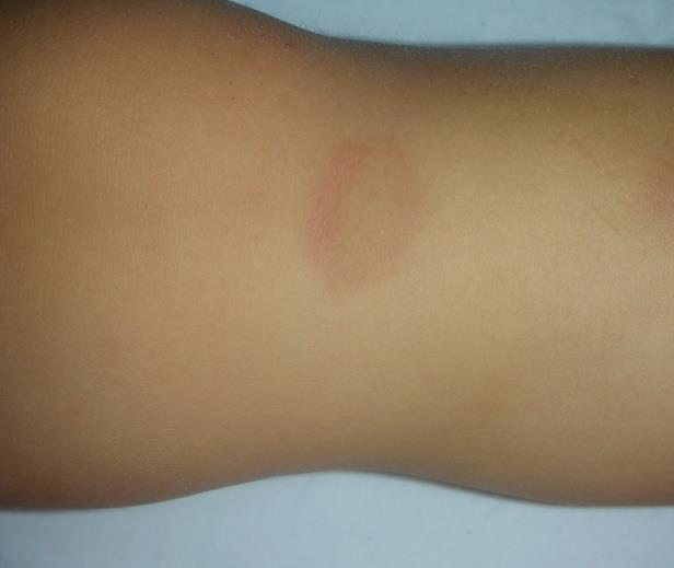
46
47 METASTATIC NEUROBLASTOMA


48 18 y/o female. Painful, non inflammatory nodules in the right thigh
49 CLINICAL DIFFERENTIAL DIAGNOSIS ENGLAND Espiradenoma (E) Neurofibroma (N) Glomangioma (G) Leiomioma (L) Angioleomioma (A) Neurilemoma (N) Dermatofibroma (D)
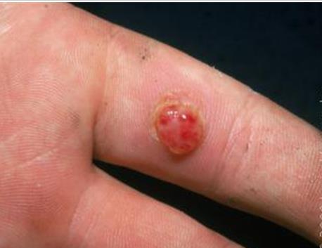
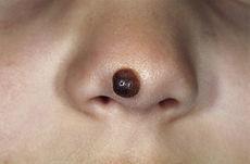
50 CLINICAL DIFFERENTIAL DIAGNOSIS ENGLAND Espiradenoma (E) Neurofibroma (N) Glomangioma (G) Leiomioma (L) Angioleomioma (A) Neurilemoma (N) Dermatofibroma (D)
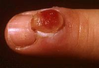
51 ESPIRADENOMA Benign neoplasm of sweat glands Usually a solitary multinodular lesion Deep dermal or subcutaneous location Trunk and proximal extremities No predilection for sex nor age Usually painful Hypervascular The presence of pain supports the diagnosis
52 3 y/o girl. Asymptomatic lesion at the posterior aspect of the right knee (Courtesy Dr. JD Arce, Chile)


53 5 y/o girl, palpable lesion left knee GRANULOMA ANNULARE

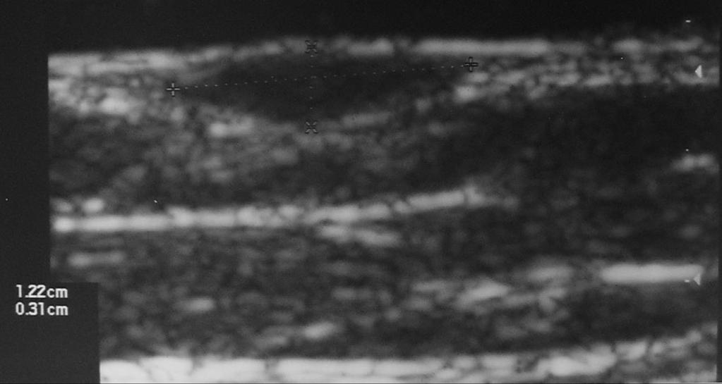
54 GRANULOMA ANNULARE
55 GRANULOMA ANNULARE Benign inflammatory papular dermal and annular plaques. Relatively common All age groups, rare in infancy Cause unknown Usually asymptomatic, may improve in winter and worsen in summer
56 GRANULOMA ANNULARE LOCALIZED GRANULOMA ANNULARE Most common form Children and adults younger than 30 years. Groups of 1-2 mm annular papules over distal extremities. Most common locations: feet, hands, fingers, arms, legs. GENERALIZED GRANULOMA ANNULARE (10%) Bimodal age distribution, occurring in patients younger than 10 years and in patients aged years.


57 9 y/o. Red nodule left sole


58 PYOGENIC GRANULOMA
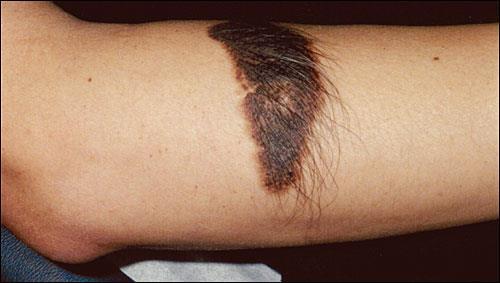
59 PYOGENIC GRANULOMA Benign vascular lesion Most comm in children: fingers and hands Originally, thought to be caused by bacterial infection pyogenic granuloma Etiology unknown Histology: capillary hemangioma History of previous minor trauma is frequent. Usually solitary lesions. Might bleed with little or no trauma


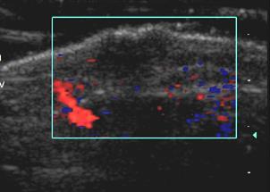
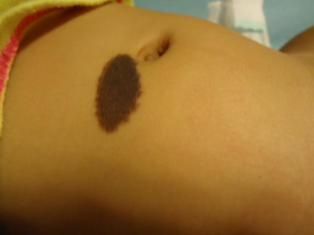
60 9 y/o girl. Indurated depressed lesion of the right thigh
61 MORPHEA
62 MORPHEA Localized scleroderma Self limited disorder, lasting 3-5 years Often history of trauma Superficial erythema, hardening of the skin, loss of hair, hypo or hyperpigmentation, sclerosis,atrophy US mostly used for f/u after treatment
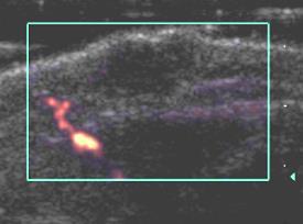
63
64
65 CONGEN ITAL HISTIOCYTOSIS (Hashimoto-Pritzker) Unusual form of histiocytosis Self-healing reticulohistiocytosis Isolated involvemet of the skin Presents with papules and nodules that frequently ulcerate Affected infants are otherwise well and the skin lesions tend to involute spontaneously within weeks to months.
66 CONGENITAL MELANOCYTIC NEVUS


67 8 m/o. nevus anterior abd wall CONGENITAL MELANOCYTIC NEVUS
68
69 CONGENITAL MELANOCYTIC NEVUS Found at birth 1% of infants Located in the area of the head and neck 15% May be small (less than 2 cm), medium-sized (more than 2 cm) or giant (more than 40 cm) May have hairy patches Can have histological characteristics resembling melanomasl Risk of malignant transformation specially the giant form Because of the premalignant potential, early surgery should ne cosndered the nevocytic overload.


70 MELANOMA Wortsman X, Springer, 2013
71 PRIMARY CUTANEOUS MELANOMA Very rare in children Incidence may be increasing PREDISPOSING CONDITIONS Transplacental melanoma, transmitted from the mother with melanoma to the fetus in utero. Giant congenital melanocytic nevus Congenital conditions: xeroderma pigmentosum, dysplastic nevus syndrome, albinism Development from a preexisting nevus
72 PRIMARY CUTANEOUS MELANOMA ULTRASONOGRAPHY Usually well defined Oval- or fusiform-shaped, homogeneous, hypoechoic Smooth borders, increased acoustic transmission Variable degrees of vascularity, most commonly hypervascular on color Doppler Assessment of the vascularity, including the peak systolic velocity of the arterial vessels, may provide an idea of the angiogenic power of the tumor that can correlate with the metastatic potential
73 Wortsman, X. Dermatologic US with Clinical and Histologic Correlation. Springer 2013
74 CONCLUSIONS US can be useful in the characterization and diagnosis of skin lesions in children when clinical findings are nonspecific US can be also helpful as a complement of clinical findings when the diagnosis is already known Most skin lesions are benign in children Radiologists should be familiar with the clinical and sonographic appearance of the most common lesions
75 CONCLUSIONS Interaction with Dermatologists and Pathologists is important Other studies such as MRI might be needed in very specific cases, mostly in larger and deeper lesions In many cases the final diagnosis is made histologically


76 THANKS FOR YOUR ATTENTION
Objectives. 1. Recognizing benign skin lesions. 2.Know which patients will likely need surgical intervention.
 The Joy of Pediatric Skin Dr. Claire Sanger University of Kentucky Plastic & Reconstructive Surgery Objectives 1. Recognizing benign skin lesions 2.Know which patients will likely need surgical intervention.
Contents. Basic Ultrasound Principles and Terminology. Ultrasound Nodule Characteristics
 Contents Basic Ultrasound Principles and Terminology Basic Ultrasound Principles... 1 Ultrasound System... 2 Linear Transducer for Superficial Images and Ultrasound-Guided FNA... 3 Scanning Planes... 4
Sonography of soft-tissue vascular lesions
 Sonography of soft-tissue vascular lesions Oscar M. Navarro Associate Professor, University of Toronto Dept. of Diagnostic Imaging, The Hospital for Sick Children Toronto, Canada Declaration of Disclosure
Ultrasound imaging of vascular anomalies: pearls and pitfalls
 Ultrasound imaging of vascular anomalies: pearls and pitfalls Oscar Navarro, MD Dept. of Medical Imaging, University of Toronto Dept. of Diagnostic Imaging, The Hospital for Sick Children Declaration of
Ultrasound of soft-tissue vascular anomalies
 Ultrasound of soft-tissue vascular anomalies Oscar M. Navarro Associate Professor, University of Toronto Dept. of Diagnostic Imaging, The Hospital for Sick Children Toronto, Canada Declaration of Disclosure
المركب النموذج--- سبيتز وحمة = Type Spitz's Nevus, Compound SPITZ NEVUS 1 / 7
 SPITZ NEVUS 1 / 7 Epidemiology An annual incidence rate of 1.4 cases of Spitz nevus per 100,000 individuals has been estimated in Australia, compared with 25.4 per 100,000 individuals for cutaneous melanoma
Neckmasses in infancy and childhood: Clinical and radiological classification and imaging approaches M. Mearadji
 Neckmasses in infancy and childhood: Clinical and radiological classification and imaging approaches M. Mearadji International Foundation for Pediatric Imaging Aid Introduction Neck masses are a frequent
أملس عضلي غرن = Leiomyosarcoma. Leiomyosarcoma 1 / 5
 Leiomyosarcoma 1 / 5 EPIDEMIOLOGY Exact incidence is unknown, but older studies suggest that leiomyosarcomas comprise approximately 3 percent of soft-tissue sarcomas. Superficial leiomyosarcoma occurs
Ultrasound Evaluation of Masses
 Ultrasound Evaluation of Masses Jon A. Jacobson, M.D. Professor of Radiology Director, Division of Musculoskeletal Radiology University of Michigan Disclosures: Consultant: Bioclinica Advisory Panel: GE,
Hemangiomas and Other Vascular Tumors
 facebook.com/cincykidsrad Hemangiomas and Other Vascular Tumors Disclosures No relevant financial disclosures Bernadette L. Koch, M.D. Departments of Radiology and Pediatrics Cincinnati Children s Hospital
Pediatric Ocular Sonography
 Pediatric Ocular Sonography Cicero J Torres A Silva, MD Associate Professor of Radiology 2016 SPR Pediatric Ultrasound Course Yale University School of Medicine None Disclosures Objectives of Presentation
11/8/2012. Chapter 6 Part 1 Objectives: Skin = Integument = Cutaneous Membrane. The Structure of Skin. Epidermis
 Chapter 6 Part 1 Objectives: Define organ, and associate the skin as an organ of the integumentary system. List the general functions of the skin. Describe the structure of the layers of the skin. Summarize
Principles of Anatomy and Physiology
 Principles of Anatomy and Physiology 14 th Edition CHAPTER 5 The Integumentary System Introduction The organs of the integumentary system include the skin and its accessory structures including hair, nails,
Dermatopathology: The tumor is composed of keratinocytes which show atypia, increase mitoses and abnormal mitoses.
 Squamous cell carcinoma (SCC): A common malignant tumor of keratinocytes arising in the epidermis, usually from a precancerous condition: 1- UV induced actinic keratosis, usually of low grade malignancy.
Sonographic imaging of pediatric thyroid disorders in childhood. Experiences and report in 150 cases
 Sonographic imaging of pediatric thyroid disorders in childhood. Experiences and report in 150 cases M. Mearadji International Foundation for Pediatric Imaging Aid Sonographic technique. Use of high frequency
Lid Lesions: Relax or Refer
 Lid Lesions: Relax or Refer Blair Lonsberry, MS, OD, MEd., FAAO Professor of Optometry Pacific University College of Optometry blonsberry@pacificu.edu Agenda Benign vs. Malignant lesions Benign Eyelid
Dermoscopy: Recognizing Top Five Common In- Office Diagnoses
 Dermoscopy: Recognizing Top Five Common In- Office Diagnoses Vu A. Ngo, DO Department of Family Medicine and Dermatology Choctaw Nation Health Services Authority Learning Objectives Introduction to dermoscopy
Birthmarks: When to worry, when to reassure
 Birthmarks: When to worry, when to reassure Aimee Smidt, MD, FAAD, FAAP Associate Professor, Depts of Dermatology and Pediatrics University of New Mexico School of Medicine November 2016 Goals and Objectives
THE INTEGUMENTARY SYSTEM. Body Membranes & Skin
 THE INTEGUMENTARY SYSTEM Body Membranes & Skin TYPES OF MEMBRANES Epithelial Membranes includes layer of epithelial cells and connective tissue Serous Cutaneous Mucous Connective Tissue Membranes solely
Human Anatomy & Physiology
 PowerPoint Lecture Slides prepared by Barbara Heard, Atlantic Cape Community College Ninth Edition Human Anatomy & Physiology C H A P T E R 5 Annie Leibovitz/Contact Press Images 2013 Pearson Education,
Skin and Body Membranes
 Essentials of Human Anatomy & Physiology Elaine N. Marieb Seventh Edition Chapter 4 Skin and Body Membranes Slides 4.1 4.32 Lecture Slides in PowerPoint by Jerry L. Cook Skin and Body Membranes Function
Atlas of Eyelid and Conjunctival Tumors
 Atlas of Eyelid and Conjunctival Tumors Jerry A. Shields, M.D. Director, Ocular Oncology Service Wills Eye Hospital Professor of Ophthalmology Thomas Jefferson University Philadelphia, Pennsylvania Carol
Skin and Body Membranes Body Membranes Function of body membranes Cover body surfaces Line body cavities Form protective sheets around organs
 Skin and Body Membranes Body Membranes Function of body membranes Cover body surfaces Line body cavities Form protective sheets around organs Classification of Body Membranes Epithelial membranes Cutaneous
Skin and Body Membranes
 4 Skin and Body Membranes PowerPoint Lecture Slide Presentation by Jerry L. Cook, Sam Houston University ESSENTIALS OF HUMAN ANATOMY & PHYSIOLOGY EIGHTH EDITION ELAINE N. MARIEB Skin and Body Membranes
Benign and malignant epithelial lesions: Seborrheic keratosis: A common benign pigmented epidermal tumor occur in middle-aged or older persons more
 Benign and malignant epithelial lesions: Seborrheic keratosis: A common benign pigmented epidermal tumor occur in middle-aged or older persons more common on the trunk; but extremities, head and neck are
Swelling. Size: measure exact size in cm using a tape measure (measure longitudinal and transverse axis and if possible the depth)
") Swelling Inspection Site: exact anatomic position Number: single or multiple Shape: spherical, oval, kidney-shaped or irregular Size: measure exact size in cm using a tape measure (measure longitudinal
Swelling Inspection Site: exact anatomic position Number: single or multiple Shape: spherical, oval, kidney-shaped or irregular Size: measure exact size in cm using a tape measure (measure longitudinal
DERMATOLOGY ROTATION: COMPETENCY-BASED GOALS AND OBJECTIVES
 UNC DIVISION OF PLASTIC AND RECONSTRUCTIVE SURGERY DERMATOLOGY ROTATION: COMPETENCY-BASED GOALS AND OBJECTIVES MEDICAL KNOWLEDGE A. Anatomy/Physiology/Embryology Goal: The resident will have knowledge
SESSION 1: GENERAL (BASIC) PATHOLOGY CONCEPTS Thursday, October 16, :30am - 11:30am FACULTY COPY
 PATHOLOGY CONCEPTS Thursday, October 16, :30am - 11:30am FACULTY COPY") SESSION 1: GENERAL (BASIC) PATHOLOGY CONCEPTS Thursday, October 16, 2008 9:30am - 11:30am FACULTY COPY GOAL: Describe the basic morphologic (structural) changes which occur in various pathologic conditions.
SESSION 1: GENERAL (BASIC) PATHOLOGY CONCEPTS Thursday, October 16, 2008 9:30am - 11:30am FACULTY COPY GOAL: Describe the basic morphologic (structural) changes which occur in various pathologic conditions.
Clinical characteristics
 Skin Cancer Fernando Vega, MD Seattle Healing Arts Clinical characteristics Precancerous lesions Common skin cancers ACTINIC KERATOSIS Precancerous skin lesions Actinic keratoses Dysplastic melanocytic
Imaging Of Cutaneous and Subcutaneous Nodules in Oncology Patient
 Imaging Of Cutaneous and Subcutaneous Nodules in Oncology Patient Poster No.: C-2263 Congress: ECR 2013 Type: Educational Exhibit Authors: A. Youssef, U. I. S. E. N. Salem, M. E. M. abdelsalam, M. 1 1
AACE/ACE Principles of Endocrine Neck Sonography Course
 AACE/ACE Principles of Endocrine Neck Sonography Course Primary objective of thyroid ultrasound: assess for malignant disease Nodular Disease Benign Malignant Goiter Iodine deficient Thyroiditis Organification
Skin lesions The Good and the Bad. Dr Virginia Hubbard Ipswich Hospital NHS Trust Barts and the London School of Medicine and Dentistry
 Skin lesions The Good and the Bad Dr Virginia Hubbard Ipswich Hospital NHS Trust Barts and the London School of Medicine and Dentistry Case 1 32 year old woman Australian Lesion on back New hair growing
Doctors of Optometry Course Notes
 Doctors of Optometry Course Notes OD19 1CE COPE: 43871-AS Eyelid Lumps and Bumps Sunday, February 26, 2017 2:40 pm 3:30 pm Regency C 3 rd Floor Presenter: Blair Lonsberry, OD, FAAO Dr. Lonsberry is a Full
Introduction. Skin and Body Membranes. Cutaneous Membranes Skin 9/14/2017. Classification of Body Membranes. Classification of Body Membranes
 Introduction Skin and Body Membranes Body membranes Cover surfaces Line body cavities Form protective and lubricating sheets around organs Classified in 5 categories Epithelial membranes 3 types- cutaneous,
What are the functions of the integumentary system? What are some disorders of the integumentary system?
 Essential Questions: What are the functions of the integumentary system? What are some disorders of the integumentary system? How are integumentary system disorders treated? How do you relate the integumentary
Due next week in lab - Scientific America Article Select one article to read and complete article summary
 Due in Lab 1. Skeletal System 33-34 2. Skeletal System 26 3. PreLab 6 Due next week in lab - Scientific America Article Select one article to read and complete article summary Cell Defenses and the Sunshine
Musculoskeletal Sarcomas
 Musculoskeletal Sarcomas Robert C. Orth, M.D., Ph.D. Edward B. Singleton Department of Pediatric Radiology Texas Children s Hospital Page 0 xxx00.#####.ppt 9/23/2012 9:01:18 AM No disclosures Page 1 xxx00.#####.ppt
PowerPoint Lecture Slide Presentation by Patty Bostwick-Taylor, Florence-Darlington Technical College Skin and Body Membranes
 PowerPoint Lecture Slide Presentation by Patty Bostwick-Taylor, Florence-Darlington Technical College Skin and Body Membranes 4 Body Membranes Function of body membranes Cover body surfaces Line body cavities
Ultrasound Physics & Doppler
 Ultrasound Physics & Doppler Endocrine University 2018 Mark Lupo, MD, FACE, ECNU Objectives Review the essential components of ultrasound physics in neck sonography Demonstrate the importance of ultrasound
Role of imaging in RCC. Ultrasonography. Solid lesion. Cystic RCC. Solid RCC 31/08/60. From Diagnosis to Treatment: the Radiologist Perspective
 Role of imaging in RCC From Diagnosis to Treatment: the Radiologist Perspective Diagnosis Staging Follow up Imaging modalities Limitations and pitfalls Duangkamon Prapruttam, MD Department of Therapeutic
ABCD rule. apocrine glands. arrector pili. ceruminous glands. contact dermatitis
 ABCD rule assessing moles: asymmetric, broder irregularity, color, diameter (larger than 6mm) apocrine glands arrector pili sweat glands in the pubic and underarm areas that secrete thicker sweat, that
Sonography of Facial Cutaneous Basal Cell Carcinoma
 SOUND JUDGMENT SERIES Sonography of Facial Cutaneous Basal Cell Carcinoma A First-line Imaging Technique Ximena Wortsman, MD Invited paper The Sound Judgment Series consists of invited articles highlighting
Integumentary System
 Integumentary System Physiology of Touch Skin: our most sensitive organ Touch: first sense to develop in embryos Most important but most neglected sense How many sensory receptors do we have? (We have
Anatomy Ch 6: Integumentary System
 Anatomy Ch 6: Integumentary System Introduction: A. Organs are body structures composed of two or more different tissues. B. The skin and its accessory organs make up the integumentary system. Types of
Describe the functions of the vertebrate integumentary system. Discuss the structure of the skin and how it relates to function.
 Chapter 5 Describe the functions of the vertebrate integumentary system. Discuss the structure of the skin and how it relates to function. Explain the basis for different skin colors. Describe the structure
NON-ATHEROSCLEROTIC PATHOLOGY OF THE CAROTID ARTERIES
 NON-ATHEROSCLEROTIC PATHOLOGY OF THE CAROTID ARTERIES Leslie M. Scoutt, MD, FACR Professor of Diagnostic Radiology & Surgery Vice Chair, Dept of Radiology & Biomedical Imaging Chief, Ultrasound Section
Selected Pseudomalignant Soft Tissue Tumors of the Skin and Subcutis
 Selected Pseudomalignant Soft Tissue Tumors of the Skin and Subcutis Andrew L. Folpe, M.D. Professor of Laboratory Medicine and Pathology Mayo Clinic, Rochester, MN folpe.andrew@mayo.edu 2016 MFMER slide-1
US in non-traumatic acute abdomen. Lalita, M.D. Radiologist Department of radiology Faculty of Medicine ChiangMai university
 US in non-traumatic acute abdomen Lalita, M.D. Radiologist Department of radiology Faculty of Medicine ChiangMai university Sagittal Orientation Transverse (Axial) Orientation Coronal Orientation Intercostal
Overview of Cutaneous Lymphomas: Diagnosis and Staging. Lauren C. Pinter-Brown MD, FACP Health Sciences Professor of Medicine and Dermatology
 Overview of Cutaneous Lymphomas: Diagnosis and Staging Lauren C. Pinter-Brown MD, FACP Health Sciences Professor of Medicine and Dermatology Definition of Lymphoma A cancer or malignancy that comes from
Integumentary System
 Integumentary System The integumentary system is commonly known as the Skin Largest organ of human body 10% total body weight and would cover over 20 square feet Functions of Skin 1. Protection Barrier
Periocular Malignancies
 Periocular Malignancies Andrew Gurwood, O.D., F.A.A.O., Dipl. Marc Myers, O.D., F.A.A.O. Drs. Myers and Gurwood have no financial interests to disclose. Course Description Discussion of the most common
Lumps and Bumps: The Dermatology of Lid Lesions
 Lumps and Bumps: The Dermatology of Lid Lesions Thomas J. Joly, MD, PhD Assistant Professor of Ophthalmology Eastern Virginia Medical School Ophthalmic Plastic Surgery Service Virginia Eye Consultants
Sonography of Pediatric Superficial Lumps and Bumps: Illustrative Examples from Head to Toe
 Sonography of Pediatric Superficial Lumps and umps: Illustrative Examples from Head to Toe nmol Gupta ansal, MD Henrietta Kotlus Rosenberg, MD, FCR, FP Mount Sinai Hospital Icahn School of Medicine at
Pimples and Boils!! Dr Nathan Harvey Anatomical Pathology, PathWest
 Pimples and Boils!! Dr Nathan Harvey Anatomical Pathology, PathWest Overview & Learning Objectives Review the cardinal signs/symptoms of acute inflammation Review the histological features of acute inflammation
Chapter 5 The Integumentary System. Copyright 2009, John Wiley & Sons, Inc. 1
 Chapter 5 The Integumentary System Copyright 2009, John Wiley & Sons, Inc. 1 Introduction The organs of the integumentary system include the skin and its accessory structures including hair, nails, and
The Oral Cavity. Image source:
 The Oral Cavity Anatomy Image source: http://anatomyforlayla.blogspot.co.za/2007/04/blog-post.html The major structures of the oral cavity are the lips, the teeth, the alveolar ridges (bony areas that
LUMPS AND BUMPS: AN ORGANIZED APPROACH TO DIAGNOSIS AND MANAGEMENT
 LUMPS AND BUMPS: AN ORGANIZED APPROACH TO DIAGNOSIS AND MANAGEMENT Tammy P. Than, M.S., O.D., F.A.A.O. The University of Alabama at Birmingham / School of Optometry 1716 University Blvd. Birmingham, AL
****************************************************************************************************** INTEGUMENTARY SYSTEM
 BIOLOGY 211: HUMAN ANATOMY & PHYSIOLOGY ****************************************************************************************************** INTEGUMENTARY SYSTEM ******************************************************************************************************
B. Incorrect! The ectoderm does not produce the dermis. C. Incorrect! The dermis is derived from the mesoderm.
 Human Anatomy - Problem Drill 04: The Integumentary System Question No. 1 of 10 Instructions: (1) Read the problem and answer choices carefully, (2) Work the problems on paper as 1. From the inner cell
Case 18. M75. Excision of mass on scalp. Clinically SCC. The best diagnosis is:
 Case 18 M75. Excision of mass on scalp. Clinically SCC. The best diagnosis is: A. Pilomatrical carcinoma B. Adnexal carcinoma NOS C. Metastatic squamous cell carcinoma D.Primary squamous cell carcinoma
The premammary layer at breast sonography imaging: normal appearance and disease with mammography correlation
 The premammary layer at breast sonography imaging: normal appearance and disease with mammography correlation Poster No.: C-0250 Congress: ECR 2012 Type: Educational Exhibit Authors: A. Fariña; Bilbao/ES
A 40-year old male with follicular papule and pustule at central face area for 3 months
 A 40-year old male with follicular papule and pustule at central face area for 3 months GMS- Neg AFB-Neg Fite stain - neg HISTOPATHOLOGICAL DIFFERENTIAL DIAGNOSIS CASEOUS GRANULOMA INFECTION -MYCOBACTERIUM
CHAPTER 5 INTEGUMENTARY
 CHAPTER 5 INTEGUMENTARY skin under the skin other stuff cutaneous layer hypodermis (subcutaneous) accessory structures Cutaneous layer = skin epithelial layers = connective tissue layer = dermis Subcutaneous
A Rare Case Report on Pilomatrixoma of the Arm Diagnosed Cytologically
 www.jmscr.igmpublication.org Impact Factor 3.79 ISSN (e)-2347-176x A Rare Case Report on Pilomatrixoma of the Arm Diagnosed Cytologically Authors Dr. Vinny Gupta 1, Dr. Mukesh Kumar Gupta 2, Dr. Laxmi
Patient Information. Age: 8 y/o Sex: Female. Date of Admission: Date of Discharge:
 Patient Information Age: 8 y/o Sex: Female Date of Admission: 92-10-08 Date of Discharge: 92-10-18 Chief Complaint Severe admominal pain and vomiting with dysuria since last afternoon Present Illness Lower
Ultrasonography of the Neck as an Adjunct to FNA. Nicole Massoll M.D.
 Ultrasonography of the Neck as an Adjunct to FNA Nicole Massoll M.D. Basic Features of Head and Neck Ultrasound and Anatomy Nicole Massoll M.D. University of Arkansas for Medical Sciences, Little Rock
High resolution ultrasound scanner for skin imaging
 High resolution ultrasound scanner for skin imaging Christine Turlat Sales Director Atys medical 17 Parc d Arbora 69510 SOUCIEU EN JARREST Atys company Principle of ultrasound imaging DERMCUP Normal image
CH 05 THE INTEGUMENTARY SYSTEM
 CH 05 THE INTEGUMENTARY SYSTEM This system consists of skin and its derivatives. The skin is one of the largest organs of the body in terms of surface area. The functions of the integumentary system include:
Unit 4 - The Skin and Body Membranes 1
 Unit 4 - The Skin and Body Membranes 1 I. Unit 4: Skin and Body Membranes A. Body Membranes 1. Function of body membranes a) Cover body surfaces b) Line body cavities c) Form protective sheets around organs
Introduction. Results. Discussion. Histopathologic and immunohistochemical findings. Results. conclusions,
 1/5 2/5 Carcinoma distinctive carcinoma. form erysipeloides (CE), metastasis. which clinically Itfrom has resembles been termed erysipelas, is an uncommon, but may extend It164 toclassically back, presents
Integumentary System. Study of the Skin
 Integumentary System Study of the Skin Skin is used to: Maintain homeostasis Provide a protective covering Slow down water loss from deeper tissues House sensory receptors Synthesize various biochemicals
Small lesions involving scalp and skull in pediatric age.
 Small lesions involving scalp and skull in pediatric age. Poster No.: C-1149 Congress: ECR 2013 Type: Educational Exhibit Authors: M. J. Yi, J. H. Yoo; Seoul/KR Keywords: Education and training, Education,
LUMPS AND BUMPS: EVALUATION AND MANAGEMENT OF SOFT TISSUE MASSES IN PEDIATRICS. By Elizabeth A. Paton, MSN, RN-BC, PPCNP-BC, FAEN
 LUMPS AND BUMPS: EVALUATION AND MANAGEMENT OF SOFT TISSUE MASSES IN PEDIATRICS By Elizabeth A. Paton, MSN, RN-BC, PPCNP-BC, FAEN I. Objectives II. By the end of this presentation, the learner will be able
INTEGUMENTARY SYSTEM CHAPTER 4
 INTEGUMENTARY SYSTEM CHAPTER 4 FUNCTIONS Waterproofs Protein called keratin Protection 1 st line of defense against pathogens, chemicals & abrasions Insulation Regulates heat loss by controlling blood
Small lesions involving scalp and skull in pediatric age.
 Small lesions involving scalp and skull in pediatric age. Poster No.: C-1149 Congress: ECR 2013 Type: Educational Exhibit Authors: M. J. Yi, J. H. Yoo; Seoul/ Keywords: Education and training, Education,
Conflicts. Objectives. University of Texas Health Science Center at San Antonio. Pediatrics Grand Rounds 24 August Pediatric Dermatology 101
 Pediatric Dermatology 101 John C. Browning, MD, FAAD, FAAP Conflicts Investigator: ViroXis Advisor: ViroXis Advisory Board: TopMD Speaker: Galderma Objectives Understand the meaning and importance of cutaneous
Ch. 4: Skin and Body Membranes
 Ch. 4: Skin and Body Membranes I. Body Membranes A. Function of body membranes 1. Cover body surfaces 2. Line body cavities 3. Form protective sheets around organs II. Classification of Body Membranes
A Practical Approach to Adnexal Masses
 A Practical Approach to Adnexal Masses Darcy J. Wolfman, MD Section Chief of Genitourinary Imaging American Institute for Radiologic Pathology Clinical Associate Johns Hopkins Community Radiology Division
Vascular Tumors in Children and Adults. Thuy Phung, MD, PhD Houston Methodist Hospital Texas Children s Hospital Baylor College of Medicine
 Vascular Tumors in Children and Adults Thuy Phung, MD, PhD Houston Methodist Hospital Texas Children s Hospital Baylor College of Medicine What are these lesions? (Marcelo Hochman, MD) What are these lesions?
Ch 4. Skin and Body Membranes
 Ch 4 Skin and Body Membranes TITLE HISTOLOGY SLIDES & NOTES ESSENTIAL QUESTION What tissues compose the integumentary system? Stratified Squamous Epithelium Stratified = several layers; Squamous = shape
Basal cell carcinoma 5/28/2011
 Goal of this Presentation A practical approach to the diagnosis of cutaneous carcinomas and their mimics Thaddeus Mully, MD University of California San Francisco To review common non-melanoma skin cancers
Integumentary System
 Chapter 5 Integumentary System 5-1 Skin: composed of dermis and epidermis Dermis. Gives structural strength. C.T. with many fibers, fibroblasts, macrophages. Some adipocytes and blood vessels. Contains
Differences in Sonographic Features of Ruptured and Unruptured
 ORIGINAL RESEARCH Differences in Sonographic Features of Ruptured and Unruptured Epidermal Cysts Wei-Hsin Yuan, MD, Hui-Chen Hsu, MD, Yi-Chen Lai, MD, Yi-Hong Chou, MD, Anna Fen-Yau Li, MD, PhD Received
Eccrine Spiradenoma Arising in the Breast Misdiagnosed as an Epidermal Inclusion Cyst
 Caee Report DOI: 10.3348/kjr.2011.12.2.256 pissn 1229-6929 eissn 2005-8330 Korean J Radiol 2011;12(2):256-260 Eccrine Spiradenoma Arising in the Breast Misdiagnosed as an Epidermal Inclusion Cyst Hyun
Chapter 4 Opener Pearson Education, Inc.
 Chapter 4 Opener Introduction The integumentary system is composed of: Skin Hair Nails Sweat glands Oil glands Mammary glands The skin is the most visible organ of the body Clinicians can tell a lot about
Integumentary System. Packet #12
 Integumentary System Packet #12 Introduction Skin/Integument Skin, considered an organ, is the major component of the integumentary system. The integumentary system is also composed of other accessory
Study of validity of ultrasonographic diagnosis in relation to Fine Needle Aspiration Cytology (FNAC) diagnosis
 diagnosis") Original article: Study of validity of ultrasonographic diagnosis in relation to Fine Needle Aspiration Cytology (FNAC) diagnosis *Dr Rajvi Matalia, ** Dr Y.P.Sachdev, ***Dr D.S.Kulkarni *Junior Resident,
Original article: Study of validity of ultrasonographic diagnosis in relation to Fine Needle Aspiration Cytology (FNAC) diagnosis *Dr Rajvi Matalia, ** Dr Y.P.Sachdev, ***Dr D.S.Kulkarni *Junior Resident,
PAPILLARY THYROID CARCINOMA PRESENTING AS A LATERAL NECK MASS MASS. Dr. Pamela Hanson DO PGY3
 PAPILLARY THYROID CARCINOMA PRESENTING AS A LATERAL NECK MASS MASS Dr. Pamela Hanson DO PGY3 MK CASE PRESENTATION 28 yo Female presented to the ENT Clinic in October 2016, with the complaint of chronic
Common Benign Lesions and Skin Cancers. 22nd May 2015 Dr Mark Foley
 Common Benign Lesions and Skin Cancers 22nd May 2015 Dr Mark Foley Thank you for downloading this file. This intended to supplement the presentation given at the NZ Wound Care Conference, it is not intended
PowerPoint Lecture Slide Presentation by Patty Bostwick-Taylor, Florence-Darlington Technical College Skin and Body Membranes
 PowerPoint Lecture Slide Presentation by Patty Bostwick-Taylor, Florence-Darlington Technical College Skin and Body Membranes 4 Body Membranes Function of body membranes Cover body surfaces Line body cavities
Vascular Imaging in the Pediatric Abdomen. Jonathan Swanson, MD
 Vascular Imaging in the Pediatric Abdomen Jonathan Swanson, MD Goals and Objectives To understand the imaging approach, appearance, and clinical manifestations of the common pediatric abdominal vascular
Malignant Peripheral Nerve Sheath Tumor
 C H A P T E R 120 Malignant Peripheral Nerve Sheath Tumor Currently, malignant peripheral nerve sheath tumor (MPNST) is the most commonly used generic name for the neoplasms known in the past as neurosarcoma,
Salivary ultrasound. Dr T J Beale Royal National Throat Nose & Ear and UCLH Hospitals London UK
 Salivary ultrasound Dr T J Beale Royal National Throat Nose & Ear and UCLH Hospitals London UK Two main groups of patients with presenting symptoms of: Obstructive or chronic inflammatory symptoms (salivary
Chief complaint. A mass at right chest
 Chief complaint A mass at right chest Present illness This 1-year-5-month-old girl had a mass at right side chest since one month ago. flat and not tender at first In the recent 2 days, the mass enlarged
Pelvic tumor in childhood Classification, imaging approach and radiological findings
 Pelvic tumor in childhood Classification, imaging approach and radiological findings M. Mearadji International Foundation for Pediatric Imaging Aid Rotterdam, The Netherlands Solid pelvic masses in childhood
PEDIATRICS WK 3 HEAD AND NECK ALISON WALLACE MD, PHD
 PEDIATRICS WK 3 HEAD AND NECK ALISON WALLACE MD, PHD Topics 1. Cervical lymphadenopathy 2. Lymphatic malformation 3. Thyroglossal duct cysts 4. Branchial cleft cysts 5. Thyroid masses CASE 1 Case 1 A 2
DERMCASE. A Shiny, Pink, Nose Lesion. Case 1
 Test your knowledge with multiple-choice cases This month 5 cases: 1. A Shiny, Pink, Nose Lesion p.43 2. A Red Patch on the Forehead p.44 3. An Ulcerated Nodule on the Thigh p.45 4. A Large Lump on the
Lagophthalmos. Lagophthalmos: signs. Lagophthalmos: clinical tips. Lagophthalmos: treatment plan. Madarosis
 Lagophthalmos Def: incomplete closure of the eyelid SX: FBS, irritation, red, burn, dry, chronic morning corneal irritation Lagophthalmos: signs 2-5 mm lid separation with slit lamp during blink can force
AACE 2018 Advanced Endocrine Neck Ultrasound and UGFNA Course
 AACE 2018 Advanced Endocrine Neck Ultrasound and UGFNA Course Describe the sonographic appearance of diffuse thyroid diseases: autoimmune thyroid disease Review non thyroidal findings that can be encountered