Chapter 9. Nervous System
|
|
|
- Alfred Williamson
- 5 years ago
- Views:
Transcription
1 Chapter 9 Nervous System 1
. 4.")
2 9.1 Introduction A. The nervous system is composed of neurons and neuroglia. 1. Neurons transmit nerve impulses along nerve fibers to other neurons. 2. Neurons typically have a cell body, an axon, and dendrites. 3. Nerves are made up of bundles of nerve fibers (axons). 4. Neuroglia carry out a variety of functions to aid and protect components of the nervous system. 2
3 B. The synapse 1. A small space lies between a neuron and the cell it is communicating with. 2. The nerve impulse can not jump over this space. 3. Neurotransmitters are released and convey the information across the synapse. 3
, made up of nerves that connect the CNS to the rest of the body. D.")
4 C. Organs of the nervous system can be divided into the central nervous system (CNS), made up of the brain and spinal cord, and the peripheral nervous system (PNS), made up of nerves that connect the CNS to the rest of the body. D. General functions of the nervous system sensory, integrative, and motor 4
5 9.2 General functions of the nervous system A. Sensory receptors at the ends of peripheral nerves gather information and convert it into nerve impulses. B. When sensory impulses are brought together, create sensation, add to memory, or produce thought in the brain as perceptions, this is the integrative function of the nervous system. C. Conscious or subconscious decisions follow, leading to motor functions via effectors (muscles or glands). D. Motor functions can be divided into the consciously controlled somatic nervous system and the unconscious autonomic system. 5
6 9.3 Neuroglia A. Neuroglial cells fill spaces, support neurons, provide structural frameworks, produce myelin, and carry on phagocytosis; they have the ability to divide. B. Central nervous system neuroglia 1. Microglial cells are small cells that phagocytize bacterial cells and cellular debris. 2. Oligodendrocytes form the myelin sheath in the brain and spinal cord. 6
7 Neuroglia, cont 3. Astrocytes are between blood vessels and neurons a. Provide support b. Regulate nutrients and ions c. Respond to brain injury by filling in spaces with scar tissue. d. Form the blood-brain barrier that protects brain tissue from chemical fluctuation and prevents entry of many substances 4. Ependymal cells line the inside of ventricles and covers choroid plexuses within the ventricles. C. Schwann cells are the myelin-producing neuroglia of the peripheral nervous system. 7
 Capillary")
8 Fluid-filled cavity of the brain or spinal cord Neuron Ependymal cell Oligodendrocyte Astrocyte Microglial cell Axon Myelin sheath (cut) Capillary Node 8
9 9.4 Neurons A. Neuron Structure 1. A neuron contains a cell body, tubular cytoplasm filled dendrites, and an axon a. The granular cell body contains mitochondria, lysosomes, a Golgi apparatus, chromatophilic substance (Nissl bodies similar to rough ER), neurofibrils, and a large nucleus with a nucleolus. b. Short, branching dendrites carry impulses from other neurons (or from receptors) toward the cell body. 9
10 Neuron structure, cont. c. The axon transmits the impulse away from the axon hillock of the cell body and may give off side branches. 2. Myelination of axons a. Larger axons are enclosed by myelin sheaths produced by Schwann cells and are called myelinated fibers. b. The outer layer of myelin is surrounded by a neurilemma (neurilemmal sheath) made up of the cytoplasm and nuclei of the Schwann cell. 10
11 Myelinated axons, cont. c. Narrow gaps in the myelin sheath between Schwann cells are called nodes of Ranvier. d. The smallest axons lack a myelin sheath and are unmyelinated fibers. e. White matter in the CNS is due to myelin sheaths in this area. f. Unmyelinated nerve tissue in the CNS comprise the gray matter. 11

12 12
13 Myelinated axons, cont. g. Peripheral neurons are able to regenerate because of the neurilemma but the CNS axons are myelinated by oligodendrocytes thus lacking neurilemma and usually do not regenerate. h. Neural stem cells in the hippocampus and near the ventricles can divide to produce new neurons or neuroglia. 13

14 B. Classification of Neurons 1. Structural classification a. Multipolar neurons have many dendrites and one axon arising from their cell bodies and are commonly found in the CNS. b. Bipolar neurons are found in the eyes, nose, and ears, and have a single axon and a single dendrite extending from opposite sides of the cell body. c. Unipolar neurons are found in ganglia outside the CNS and have one axon that divides; the peripheral process has dendrites near a peripheral body part and a central process that runs into the CNS. 14
15 2. Classification by function a. Sensory neurons (afferent neurons) conduct impulses from peripheral receptors to the CNS and are usually unipolar, although some are bipolar neurons. b. Interneurons (association or internuncial neurons) are multipolar neurons lying within the CNS that form links between other neurons. The cell bodies of some interneurons aggregate in specialized masses called nuclei. c. Motor neurons (efferent neurons) are multipolar neurons that conduct impulses from the CNS to effectors. 15
16 Peripheral Nervous System Sensory receptor
17 Peripheral Nervous System Sensory neuron Sensory (afferent) neurons transfer sensory information to neurons in the CNS Sensory receptor
18 Central Nervous System Interneuron Interneuron Interneurons transfer information from one part of the CNS to another Peripheral Nervous System Sensory neuron Sensory (afferent) neurons transfer sensory information to neurons in the CNS Sensory receptor
19 Central Nervous System Interneuron Interneuron Motor neuron Interneurons transfer information from one part of the CNS to another Peripheral Nervous System Sensory neuron Sensory (afferent) neurons transfer sensory information to neurons in the CNS Motor (efferent) neurons transfer instructions from the CNS to effectors Sensory receptor
20 Central Nervous System Interneuron Interneuron Motor neuron Interneurons transfer information from one part of the CNS to another Peripheral Nervous System Sensory neuron Sensory (afferent) neurons transfer sensory information to neurons in the CNS Motor (efferent) neurons transfer instructions from the CNS to effectors Sensory receptor Effector (muscle or gland)

21 9.5 The synapse A. The synapse is the junction between two communicating neurons; there exists a synaptic cleft between them across which the impulse must be conveyed. 1. The neuron sending the impulse is the presynaptic neuron. 2. The neuron receiving the impulse is the postsynaptic neuron. 21

22 B. Synaptic transmission is the process whereby the message crosses the synaptic cleft. 1. Neurotransmitters are biochemicals that carry out the synaptic transmission process in the synaptic cleft. 2. Distal end of axons have extensions called synaptic knobs which contain synaptic vesicles filled with neurotransmitters. 3. The action of the neurotransmitters are either excitatory or inhibitory. 22
23 9.6 Cell membrane potential A. A cell membrane is usually polarized, with an excess of negative charges on the inside of the membrane; polarization is important to the conduction of nerve impulses (action potentials). 23

24 B. Distribution of Ions 1. The distribution of ions is determined by the membrane channel proteins that are selective for certain ions. 2. Potassium ions pass through the membrane more readily than do sodium ions, making potassium ions a major contributor to membrane polarization. 3. There is a greater concentration of sodium ions on the outside and a greater concentration of potassium ions, along with large negatively charged 24 ions on the inside.
25 C. Resting Potential 1. Due to active transport, the cell maintains a greater concentration of sodium ions outside and a greater concentration of potassium ions inside the membrane. 2. The inside of the membrane has excess negative charges, while the outside has more positive charges. 3. This separation of charge, or potential difference, is called the resting potential and has a value of -70mV. 4. The Na + /K + pump actively transports Na + out and K + in to maintain the resting potential. 25


26 26
 5.")
27 D. Potential Changes 1. Stimulation of a membrane can locally affect its resting potential. 2. When the membrane potential becomes less negative, the membrane is depolarized. 3. If sufficiently strong depolarization occurs, a threshold potential is achieved as ion channels open. 4. At threshold, an action potential is reached. (-55mV) 5. Local potentials are graded the magnitude of change to the resting potential is directly proportional to the intensity of the stimulation. 27

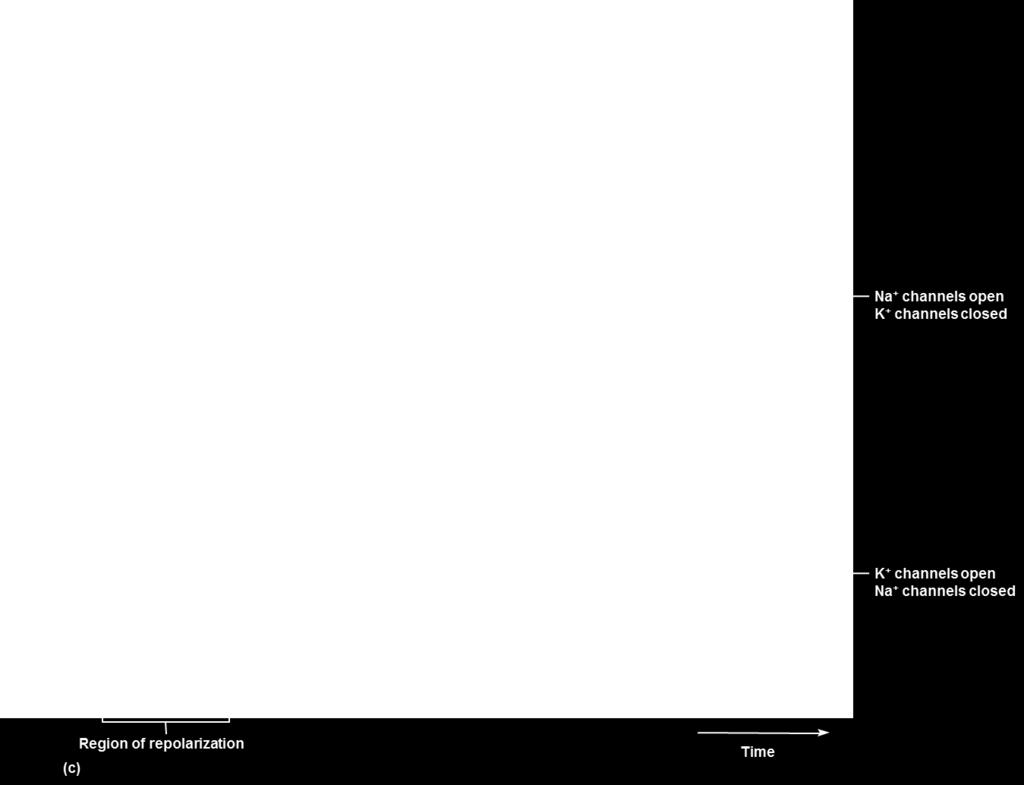
28 E. Action Potential 1. At threshold potential, membrane permeability to sodium suddenly changes at the axon hillock, called the trigger zone. 2. As sodium channels open, sodium ions rush in, and the membrane potential changes and becomes depolarized. 3. At the same time, potassium channels open to allow potassium ions to leave the cell, the membrane becomes repolarized, and resting potential is reestablished. 4. The membrane may become overly negative for a short time which is hyperpolarization. 28


29 Action potential, cont. 5. This rapid sequence of events is the action potential. 6. The active transport mechanism then works to maintain the original concentrations of sodium and potassium ions. 29

30 30

31 9.7 Impulse conduction A. Only axons are capable of action potentials 1. A nerve impulse is conducted as an action potential is reached at the trigger zone. 2. An action potential at the trigger zone causes a bioelectrical current to flow to adjacent regions of the axon membrane. 3. This spreads by a local current flowing down the fiber that stimulates the next region continuing down the axon to its end. 31

32 32
33 B. Impulse Conduction 1. Unmyelinated fibers conduct impulses over their entire membrane surface. 2. Myelinated fibers conduct impulses from one Node of Ranvier to the next, a phenomenon called saltatory conduction. 3. Saltatory conduction is many times faster than conduction on unmyelinated neurons. 4. Speed of impulse conduction is proportional to the diameter of the axon a. Thick, myelinated motor axons conduct at 120 m/s b. Thin, unmyelinated sensory axons conduct at 0.5 m/s 33
34 C. All-or-None Response 1. If a nerve fiber responds at all to a stimulus of threshold or above, it responds completely by conducting an impulse (all-or-none response). 2. All action potentials are of the same strength. 3. Greater intensity of stimulation triggers more impulses per second, not stronger impulses. 4. Refractory period short period of time after an action potential when a threshold stimulus will NOT trigger another action potential. a. Limits frequency b. Ensures the impulse is only transmitted in one direction down the axon. 34
35 9.8 Synaptic transmission A. Excitatory and Inhibitory Actions 1. Neurotransmitters that increase postsynaptic membrane permeability to sodium ions may trigger impulses and are thus excitatory. 2. Other neurotransmitters may decrease membrane permeability to sodium ions, reducing the chance that it will reach threshold, and are thus inhibitory. 3. When an impulse reaches the synaptic knobs of an axon, synaptic vesicles release a neurotransmitter into the synaptic cleft. 4. The neurotransmitter reacts with specific receptors on the postsynaptic membrane. 35
36 B. Neurotransmitters 1. More than 100 neurotransmitters are produced by the nervous system, most of which are synthesized in the cytoplasm of the synaptic knobs and stored in synaptic vesicles. 2. Types: acetylcholine, biogenic amines, amino acids, and more than 50 neuropeptides. 36

37 37


38 C. Synaptic transmission 1. When an action potential reaches the synaptic knob, calcium ions rush inward and, in response, some synaptic vesicles fuse with the membrane and release their contents to the synaptic cleft. 2. Enzymes in synaptic clefts and on postsynaptic membranes rapidly decompose the neurotransmitters after their release. (Acetylcholinesterase breaks down acetylcholine) 3. Destruction or removal of the neurotransmitter prevents 38 continuous stimulation of the postsynaptic neuron.

39 39
40 9.9 Impulse processing A. How impulses are processed is dependent upon how neurons are organized in the brain and spinal cord. B. Neuronal Pools 1. Neurons within the CNS are organized into neuronal pools with varying numbers of cells that work together. 2. Each pool receives input from afferent nerves and processes the information according to the special characteristics of the pool. 40
41 C. Facilitation 1. A particular neuron of a pool may receive excitatory or inhibitory stimulation; if the net effect is excitatory but subthreshold, the neuron becomes more excitable to incoming stimulation (a condition called facilitation). 2. This second stimulus may bring the postsynaptic cell to threshold and an action potential is created. 41
42 D. Convergence 1. A single neuron within a pool may receive impulses from two or more fibers (convergence), which makes it possible for the neuron to summate impulses from different sources. E. Divergence 1. Impulses leaving a neuron in a pool may be passed into several output fibers (divergence), a pattern that serves to amplify an impulse. 42
 held together by layers of")

43 9.10 Types of nerves A. A nerve is a bundle of nerve fibers (axons) held together by layers of connective tissue. B. Connective tissue wraps 1. Epineurium outer covering of a nerve 2. Perineurium covering of fascicles of nerve fibers (bundles) 3. Endoneurium covering of individual nerve fibers (axons) 43
44 C. Types of nerves 1. Sensory or afferent nerves bring information to the CNS. 2. Motor or efferent nerves carry impulses from the CNS to effectors. 3. Mixed nerves carry sensory and motor fibers; most nerves are of this type. 44
 4. Motor neuron that carries a response to effectors 5.")
45 9.11 Nerve pathways A. The routes nerve impulses travel are called pathways, the simplest of which is a reflex arc. B. Reflex arc components 1. A sensory receptor that detects changes. 2. Sensory neuron that carries the information toward the CNS 3. An interneuron in the CNS (reflex center) 4. Motor neuron that carries a response to effectors 5. An effector that responds to the initial change. 45
46 C. Reflex Behavior 1. Reflexes are automatic responses to changes (stimuli) in or out of the body, that help maintain homeostasis (heart rate, blood pressure, etc.) and carry out automatic responses (vomiting, sneezing, swallowing, etc.). 2. The patellar reflex (knee-jerk reflex) tendon is an example of a simple reflex (no interneuron). a. Helps maintain upright posture 46
47 Spinal cord Receptor associated with dendrites of sensory neuron Patella Patellar ligament
48 Axon of sensory neuron Spinal cord Cell body of sensory neuron Receptor associated with dendrites of sensory neuron Patella Direction of impulse conduction Patellar ligament

49 Axon of sensory neuron Cell body of sensory neuron Spinal cord Axon of motor neuron Cell body of motor neuron Receptor associated with dendrites of sensory neuron Patella Direction of impulse conduction Patellar ligament
 Receptor associated with dendrites of sensory neuron Patella Direction of impulse conduction Patellar")
50 Axon of sensory neuron Cell body of sensory neuron Spinal cord Axon of motor neuron Cell body of motor neuron Effector (extensor muscles contract and cause a kicking motion) Receptor associated with dendrites of sensory neuron Patella Direction of impulse conduction Patellar ligament
51 Reflex behavior, cont. 3. The withdrawal reflex involves sensory neurons, interneurons, and motor neurons. a. Motor signals are sent to flexor muscles to contract. b. At the same time, the antagonistic extensor muscles are inhibited and a message is sent to the brain. c. Serves a protective function 51
52 Cell body of sensory neuron Direction of impulse conduction Axon of sensory neuron Spinal cord Dendrite of sensory neuron Pain receptor in skin Tack
53 Cell body of sensory neuron Direction of impulse conduction Axon of sensory neuron Interneuron Spinal cord Dendrite of sensory neuron Pain receptor in skin Tack


54 Cell body of sensory neuron Direction of impulse conduction Axon of sensory neuron Interneuron Dendrite of sensory neuron Effector (flexor muscles contract and withdraw part being stimulated) Axon of motor neuron Spinal cord Cell body of motor neuron Pain receptor in skin Tack

55
56 9.12 Meninges A. The brain and spinal cord are surrounded by membranes called meninges that lie between the bone and the soft tissues. B. The outermost meninx is made up of tough, white dense connective tissue, contains many blood vessels, and is called the dura mater. 1. It forms the inner periosteum of the skull bones. 2. In some areas, the dura mater forms partitions between lobes of the brain, and in others, it forms dural sinuses. 3. The sheath around the spinal cord is separated from the vertebrae by an epidural space. 56
. D. The innermost pia mater is thin and contains many blood vessels and nerves. 1.")
57 C. The middle meninx, the arachnoid mater, is thin and lacks blood vessels. 1. It does not follow the convolutions of the brain. 2. Between the arachnoid and pia mater is the subarachnoid space containing cerebrospinal fluid (CSF). D. The innermost pia mater is thin and contains many blood vessels and nerves. 1. It is attached to the surface of the brain and spinal cord and follows their contours.


58 9.13 Spinal cord A. The spinal cord begins at the base of the brain at the foramen magnum and extends as a slender cord to the level of the intervertebral disc between the first and second lumbar vertebrae.
59 B. Structure of the Spinal Cord 1. The spinal cord consists of 31 segments, each of which gives rise to a pair of spinal nerves. 2. A cervical enlargement gives rise to nerves leading to the upper limbs, and a lumbar enlargement gives rise to those innervating the lower limbs. 3. Two deep longitudinal grooves (anterior median fissure and posterior median sulcus) divide the cord into right and left halves. 4. White matter, made up of bundles of myelinated nerve fibers (nerve tracts), surrounds a butterfly-shaped core of gray matter housing interneurons and cell bodies. 59


60 Spinal cord structure, cont. 5. The upper and lower wings of gray matter are the posterior and anterior horns and between them is the lateral horn. 6. The gray matter divides the white matter into three regions - anterior, lateral and posterior funiculi (columns), each consisting of longitudinal bundles called tracts. 7. A central canal contains cerebrospinal fluid. 60
61 C. Functions of the Spinal Cord 1. The spinal cord has two major functions: to transmit impulses to and from the brain, and to house spinal reflexes. 2. Tracts carrying sensory information to the brain are called ascending tracts; descending tracts carry motor information from the brain. 3. The names that identify nerve tracts identify the origin and termination of the fibers in the tract. a. Spinothalamic tracts carry sensory information to the thalamus b. Corticospinal tracts (pyramidal tracts) carry motor impulses from the cerebral cortex, down the spinal cord 61


62 Tracts of the Spinal Cord c. Corticospinal tracts are also called pyramidal tracts because they pass through and cross in the pyramids of the medulla oblongata. d. Other descending tracts dealing with posture and balance are extra-pyramidal tracts and do not pass through the medullary pyramids. 62

, the cerebellum (coordinates")
63 9.14 The brain A. Introduction 1. The brain is the largest, most complex portion of the nervous system, containing 100 billion multipolar neurons. 2. The brain can be divided into the cerebrum (largest portion and associated with higher mental functions), the diencephalon (processes sensory input and controls many homeostatic processes), the cerebellum (coordinates muscular activity), and the brain stem (coordinates and regulates visceral activities). 63
64 B. Structure of the Cerebrum 1. The cerebrum is the largest portion of the mature brain, consisting of two cerebral hemispheres. 2. A flat bundle of nerve fibers called the corpus callosum connects the hemispheres. 3. The surface of the brain is marked by gyri, sulci, and fissures (longitudinal, transverse). 64
65 Structure of the cerebrum, cont. 4. Four lobes of the brain are named according to the bones they underlie and include the frontal lobe, parietal lobe, temporal lobe, occipital lobe, and the fifth lobe is the insula. 5. A thin layer of gray matter, the cerebral cortex, lies on the outside of the cerebrum and contains 75% of the cell bodies in the nervous system. 6. Beneath the cortex lies a mass of white matter made up of myelinated nerve fibers connecting the cell bodies of the cerebral cortex with the rest of the nervous system. 65
66 C. Functions of the Cerebrum 1. The cerebrum provides higher brain functions, such as interpretation of sensory input, initiating voluntary muscular movements, memory, and integrating information for reasoning. 66
67 2. Functional Regions of the Cerebral Cortex a. The functional areas of the brain overlap, but the cortex can generally be divided into motor, sensory, and association areas. b. The sensory areas are located in several areas of the cerebrum and interpret sensory input, producing feelings or sensations. 1) Cutaneous senses anterior parietal lobe 2) Visual area posterior occipital lobe 3) Auditory area temporal lobe 4) Taste area base of central sulcus 5) Smell area - insula 67
68 Functional regions, cont. c. The various association areas of the brain analyze and interpret sensory impulses and function in reasoning, judgment, emotions, verbalizing ideas, and storing memory. 1) Association areas of the frontal lobe control a number of higher intellectual processes. 2) Association areas next to sensory areas are important for analyzing that sensory input 3) A general interpretive area is found at the junction of the parietal, temporal, and occipital lobes, and plays a primary role in complex thought processing. 68
69 Functional areas, cont. d. The primary motor areas lie in the posterior frontal lobes, anterior to the central sulcus. 1) This region includes the pyramidal cells that are also called upper motor neurons. They synapse with lower motor neurons that exit the spinal cord and reach the skeletal muscles. 2) Broca s motor speech area is in the frontal lobe. 3) Frontal eye field controls voluntary eye movements 69

70 Functional areas, cont. e. Sensory and motor fibers alike cross over in the spinal cord or brain stem so centers in the right hemisphere are interpreting or controlling the left side of the body, and vice versa. 70
71 3. Hemisphere Dominance a. Both cerebral hemispheres function in receiving and analyzing sensory input and sending motor impulses to the opposite side of the body, but one side is dominant. b. For most people, the left hemisphere is dominant for the language-related activities of speech, writing, and reading, as well as complex intellectual functions. c. Some individuals have the right hemisphere as dominant and others show equal dominance in both hemispheres. 71
72 Hemisphere dominance, cont. d. The non-dominant hemisphere specializes in nonverbal functions and controls emotions and intuitive thinking. e. The nerve fibers of the corpus callosum connect the two hemispheres. 72

73 4. The basal nuclei are masses of gray matter located deep within the cerebral hemispheres that relay motor impulses from the cerebrum and help to control motor activities by producing the inhibitory neurotransmitter, dopamine. a. Basal nuclei include the caudate nucleus, the putamen, and the globus pallidus. b. The neurons of the basal nuclei interact with the motor cortex, thalamus, and cerebellum c. Altered activity of these nuclei neurons produces the signs of Parkinson disease and Huntington disease. 73
74 D. Ventricles and Cerebrospinal Fluid 1. The ventricles are a series of connected cavities within the cerebral hemispheres and brain stem. 2. Names: 2 Lateral ventricles, third ventricle, and fourth ventricle. The interventricular foramen connects the lateral ventricles to the third ventricle and the cerebral aqueduct connects the third and fourth ventricles. 3. The ventricles are continuous with the central canal of the spinal cord and the subarachnoid space, and are filled with cerebrospinal fluid. 74


75 75


76 4. Choroid plexuses, specialized capillaries from the pia mater, secrete cerebrospinal fluid into the ventricles; most cerebrospinal fluid arises in the lateral ventricles. 5. Cerebrospinal fluid has nutritive as well as protective (cushioning) functions. 6. CSF circulates through the ventricles and connecting passageways into the subarachnoid space where it is reabsorbed back into the blood. 76
77 E. Diencephalon 1. The diencephalon lies between the cerebral hemispheres and above the midbrain. 2. The main parts are the thalamus and hypothalamus. 3. Other portions of the diencephalon are the optic tracts and optic chiasma, the infundibulum (attachment for the pituitary), the posterior pituitary, mammillary bodies, and the pineal gland. 77
78 4. The thalamus functions in sorting and directing sensory information arriving from other parts of the nervous system, performing the services of both messenger and editor. a. Pinpoints the origin of the sensory input. b. Produces general awareness of the sensation. c. Descending fibers from the cerebral cortex also communicate with the thalamus. 78
79 5. The hypothalamus maintains homeostasis by regulating a wide variety of visceral activities and by linking the endocrine system with the nervous system. a. Regulates heart rate and arterial blood pressure b. Regulates body temperature, water and electrolyte balance, hunger and body weight c. Controls movements and secretions of the digestive tract. d. Helps to regulate sleep and wakefulness. e. Stimulates the posterior pituitary gland to secrete stored hormones. 79
80 6. The limbic system, in the area of the diencephalon, controls emotional experience and expression. By generating pleasant or unpleasant feelings about experiences, the limbic system guides behavior that may enhance the chance of survival. 80
81 F. Brain Stem 1. The brain stem, consisting of the midbrain, pons, and medulla oblongata, lies at the base of the cerebrum, and connects the brain to the spinal cord. 81
82 2. Midbrain a. The midbrain, located between the diencephalon and pons, contains bundles of myelinated nerve fibers that convey impulses to and from higher parts of the brain. b. It also contains masses of gray matter that serve as centers for auditory and visual reflexes. 82
83 3. Pons a. The pons, lying between the midbrain and medulla oblongata, transmits impulses between the brain and spinal cord, and contains centers that help regulate the rate and depth of breathing. b. The ventral surface has transverse fibers that communicate with the cerebellum. 83
84 4. Medulla Oblongata a. The medulla oblongata transmits all ascending and descending impulses between the brain and spinal cord. b. Most of these tracts will cross in the pyramids of the medulla oblongata. c. The medulla oblongata also houses nuclei that control visceral functions, including the cardiac center that controls heart rate, the vasomotor center for blood pressure control, and the respiratory center that works, along with the pons, to control the rate and depth of breathing. 84

85 Medulla oblongata, cont. d. Other nuclei in the medulla oblongata are associated with coughing, sneezing, swallowing, and vomiting. 85
86 5. Reticular Formation a. Throughout the brain stem, hypothalamus, cerebrum, cerebellum, and basal ganglia, is a complex network of nerve fibers connecting tiny islands of gray matter; this network is the reticular formation. b. Decreased activity in the reticular formation results in sleep; increased activity results in wakefulness. c. The reticular formation filters incoming sensory impulses. 86

87 G. Cerebellum 1. The cerebellum is located beneath the posterior part of the cerebrum and on top of the brain stem. 2. The cerebellum is made up of two hemispheres connected by the vermis. 3. A thin layer of gray matter called the cerebellar cortex lies outside a core of white matter called the arbor vitae. 87
88 Cerebellum, cont. 4. The cerebellum communicates with other parts of the central nervous system through cerebellar peduncles. 5. The cerebellum functions to integrate sensory information about the position of body parts and coordinates skeletal muscle activity and maintains posture. 88
89 9.15 Peripheral nervous system A. The peripheral nervous system (PNS) consists of the cranial and spinal nerves that arise from the central nervous system and travel to the remainder of the body. B. The PNS is made up of the somatic nervous system that oversees voluntary activities, and the autonomic nervous system that controls involuntary activities. 89

90 90
, optic (II), oculomotor (III), trochlear (IV), trigeminal (V), abducens (VI), facial (VII), vestibulocochlear (VIII),")
91 C. Cranial Nerves 1. Twelve pairs of cranial nerves arise from the underside of the brain, most of which are mixed nerves. 2. The 12 pairs are designated by number and name and include the olfactory (I), optic (II), oculomotor (III), trochlear (IV), trigeminal (V), abducens (VI), facial (VII), vestibulocochlear (VIII), glossopharyngeal (IX), vagus (X), accessory (XI), and hypoglossal nerves (XII). 91

92 92

93 D. Spinal Nerves 1. Thirty-one pairs of mixed nerves make up the spinal nerves. 2. Spinal nerves are grouped according to the level from which they arise and are numbered in sequence, beginning with 8 pairs of cervical nerves, then 12 pairs of thoracic nerves, 5 pairs of lumbar nerves, 5 pairs of sacral nerves and 1 pair of coccygeal nerves. 3. Each spinal nerve arises from two roots: a sensory dorsal root, and a motor ventral root. a. The dorsal root has the dorsal root ganglion 4. Roots join and exit through intervertebral foramen. 93
94 5. The main branches of most spinal nerves form networks called plexuses. a. Cervical Plexuses (C1-C5) lie on either side of the neck and supply muscles and skin of the neck, and also includes the phrenic nerves which serve the diaphragm. b. Brachial Plexuses arise from lower cervical and upper thoracic nerves (C6-T1) and lead to the upper limbs including the musculocutaneous, ulnar, median, radial, and axillary nerves. 94
95 Plexuses, cont. c. Lumbosacral Plexuses arise from the lower spinal cord (T12-S5) and lead to the lower abdomen, external genitalia, buttocks, and legs, including the obturator, femoral, and sciatic nerves. 6. Anterior branches of the thoracic spinal nerves do not form plexuses but become the intercostal nerves. 95
96 9.16 Autonomic nervous system A. The autonomic nervous system has the task of maintaining homeostasis of visceral activities without conscious effort. 1. Supplies smooth muscle, cardiac muscle, and glands. 2. Helps maintain homeostasis, responds to emotional stress, and prepares the body for strenuous activity. 96
97 B. General Characteristics 1. Autonomic activities are regulated by visceral reflexes. 2. The autonomic nervous system includes two divisions: the sympathetic and parasympathetic divisions, which exert opposing effects on target organs. a. The parasympathetic division operates under normal, restful conditions. b. The sympathetic division operates under conditions of stress or emergency (fight or flight). 97

98 C. Autonomic Neurons 1. In the autonomic motor system, motor pathways include two fibers: a preganglionic fiber that leaves the CNS, and a postganglionic fiber that innervates the effector. The two fibers communicate through a ganglion. 98

99 2. Sympathetic Division a. Short preganglionic fibers in the sympathetic division arise from the thoracic and lumbar regions of the spinal cord, and synapse in paravertebral ganglia close to the vertebral column. b. Long postganglionic axons lead to an effector organ (most will travel with a spinal nerve). 99

100 3. Parasympathetic Division a. Long, preganglionic fibers in the parasympathetic division arise from the brainstem and sacral region of the spinal cord, and synapse in ganglia close to the effector organ. b. Short, postganglionic fibers continue into the effector organs.
101 D. Autonomic Neurotransmitters 1. Preganglionic fibers of both sympathetic and parasympathetic divisions release acetylcholine and are called cholinergic fibers. 2. Parasympathetic postganglionic fibers are cholinergic fibers and release acetylcholine. 3. Sympathetic postganglionic fibers are adrenergic and release norepinephrine. 4. The effects of these two divisions, based on the effects of releasing different neurotransmitters to the effector, are generally antagonistic. 101

102 Autonomic Neurotransmitters, cont. 5. Most organs receive innervation from both divisions. 6. Some effectors are innervated by only one division; example blood vessels only have sympathetic nerves. 102
103 E. Control of Autonomic Activity 1. The autonomic nervous system is largely controlled by reflex centers in the brain and spinal cord. 2. The limbic system and cerebral cortex alter the reactions of the autonomic nervous system through emotional influence. 103

104 104
Chapter 9. Nervous System
 Chapter 9 Nervous System Central Nervous System (CNS) vs. Peripheral Nervous System(PNS) CNS Brain Spinal cord PNS Peripheral nerves connecting CNS to the body Cranial nerves Spinal nerves Neurons transmit
Chapter 9 Nervous System
 Chapter 9 Nervous System Introduction: A. The nervous system is composed of neurons and neuroglia. 1. Neurons transmit nerve impulses to other neurons. 2. Nerves are made up of bundles of nerve fibers
Unit Three. The brain includes: cerebrum, diencephalon, brain stem, & cerebellum. The brain lies within the cranial cavity of the skull.
 Human Anatomy & Physiology 11 Divisions of the Nervous System Karen W. Smith, Instructor Unit Three BRAIN & SPINAL CORD Refer to the following URLs. Be sure to study these along with your book. http://www.sirinet.net/~jgjohnso/nervous.html
Hole s Essentials of Human Anatomy & Physiology
 Hole s Essentials of Human Anatomy & Physiology David Shier Jackie Butler Ricki Lewis Created by Dr. Melissa Eisenhauer Trevecca Nazarene University Amended by John Crocker Chapter 9 1 Chapter 9 Nervous
Laboratory Manual for Comparative Anatomy and Physiology Figure 15.1 Transparency Master 114
 Neuron Capillary Astrocyte Microglial cell Neuron Fluid-filled cavity Process of oligodendrocyte Ependymal cells Brain or spinal cord tissue Myelin sheath Nerve fibers Figure 15.1 Transparency Master 114
Nervous and Endocrine System Exam Review
 Directions: Read each question and complete the statement using the multiple choice responses I. Nervous System 1. The interpretation of olfactory receptor information would fall under which general function
Nervous System. Human Anatomy & Physiology P. Wilson
 Nervous System Human Anatomy & Physiology P. Wilson 1 2 Types of cells in the nervous system: Neurons & Neuroglial cells Neuroglial (aka glial) cells perform functions that are vital to neurons by filling
Organization of The Nervous System PROF. SAEED ABUEL MAKAREM
 Organization of The Nervous System PROF. SAEED ABUEL MAKAREM Objectives By the end of the lecture, you should be able to: List the parts of the nervous system. List the function of the nervous system.
The Nervous System PART A
 7 The Nervous System PART A PowerPoint Lecture Slide Presentation by Jerry L. Cook, Sam Houston University ESSENTIALS OF HUMAN ANATOMY & PHYSIOLOGY EIGHTH EDITION ELAINE N. MARIEB Structural Classification
Chapter 8 Nervous System
 Chapter 8 Nervous System Two message centers: Functions of these systems: 1. * 2. * Overview of the Nervous System Parts: General Functions: Functions Sensory input: Sensation via nerves Integration: interpretation
Unit Three. I. General Functions of the Nervous System. I. General Functions of the Nervous System
 10 Refer to the following URLs. It is a good idea to print them and bring them to class. Be sure to study these along with your book. http://www.sirinet.net/~jgjohnso/nervous.html http://faculty.washington.edu/chudler/ap.html
Chapter 17 Nervous System
 Chapter 17 Nervous System 1 The Nervous System Two Anatomical Divisions Central Nervous System (CNS) Brain and Spinal Cord Peripheral Nervous System (PNS) Two Types of Cells Neurons Transmit nerve impulses
action potential afferent neuron Weblike; specifically, the weblike middle layer of the three meninges. arachnoid astrocytes autonomic nervous system
 action potential A large transient depolarization event, including polarity reversal, that is conducted along the membrane of a muscle cell or a nerve fiber. afferent neuron Nerve cell that carries impulses
10.1: Introduction. Cell types in neural tissue: Neurons Neuroglial cells (also known as neuroglia, glia, and glial cells) Dendrites.
 Dendrites.") 10.1: Introduction Copyright The McGraw-Hill Companies, Inc. Permission required for reproduction or display. Cell types in neural tissue: Neurons Neuroglial cells (also known as neuroglia, glia, and glial
10.1: Introduction Copyright The McGraw-Hill Companies, Inc. Permission required for reproduction or display. Cell types in neural tissue: Neurons Neuroglial cells (also known as neuroglia, glia, and glial
Chapter 7 Nervous System
 Chapter 7 Nervous System Two message centers: Functions of these systems: 1. * 2. * Overview of the Nervous System Parts: General Functions: Functions Sensory input: Sensation via nerves Integration: interpretation
Central N.S. Peripheral N.S. 2) List the functional subdivisions. 1) List the anatomical subdivisions.
 List the functional subdivisions. 1) List the anatomical subdivisions.") S T U D Y G U I D E 8 1. Divisions of the Nervous System 1) List the anatomical subdivisions. Central N.S. Peripheral N.S. 2) List the functional subdivisions. Somatic N.S. Autonomic N.S. 2. Nerve Tissue
S T U D Y G U I D E 8 1. Divisions of the Nervous System 1) List the anatomical subdivisions. Central N.S. Peripheral N.S. 2) List the functional subdivisions. Somatic N.S. Autonomic N.S. 2. Nerve Tissue
Good Morning! Take out your notes and vocab 1-10! Copyright 2003 Pearson Education, Inc. publishing as Benjamin Cummings
 Good Morning! Take out your notes and vocab 1-10! Functions of the Nervous System 1. Sensory input gathering information To monitor changes occurring inside and outside the body (changes = stimuli) 2.
The neurvous system senses, interprets, and responds to changes in the environment. Two types of cells makes this possible:
 NERVOUS SYSTEM The neurvous system senses, interprets, and responds to changes in the environment. Two types of cells makes this possible: the neuron and the supporting cells ("glial cells"). Neuron Neurons
CHAPTER 11: NERVOUS SYSTEM II: DIVISIONS OF THE NERVOUS SYSTEM. 1. Outline the major divisions of the nervous system.
 CHAPTER 11: NERVOUS II: DIVISIONS OF THE NERVOUS OBJECTIVES: 1. Outline the major divisions of the nervous system. NERVOUS CENTRAL NERVOUS (BRAIN & SPINAL CORD) (INTERNEURONS) PERIPHERAL NERVOUS (CRANIAL
NOTES CHAPTER 9 (Brief) The Nervous System LECTURE NOTES
 The Nervous System LECTURE NOTES") NOTES CHAPTER 9 (Brief) The Nervous System LECTURE NOTES I. Divisions of the Nervous System two major divisions A. Central Nervous System (CNS) 1. brain 2. spinal cord B. Peripheral Nervous System (PNS)
NOTES CHAPTER 9 (Brief) The Nervous System LECTURE NOTES I. Divisions of the Nervous System two major divisions A. Central Nervous System (CNS) 1. brain 2. spinal cord B. Peripheral Nervous System (PNS)
Neurology study of the nervous system. nervous & endocrine systems work together to maintain homeostasis
 Nervous System Neurology study of the nervous system nervous & endocrine systems work together to maintain homeostasis Nervous System works very fast Uses electrical signals called nerve impulses Short-lived
1. 01/20/15 Ch 8: Muscular System /09/15 Ch 9: Nervous System 16
 Table of Contents # Date Title Page # 1. 01/20/15 Ch 8: Muscular System 1 2. 02/09/15 Ch 9: Nervous System 16 i 1 Anatomy and Physiology Sem 2 Ch 9 Nervous System.notebook 02/09/15 Ch. 9 Nervous System
Major Structures of the Nervous System. Brain, cranial nerves, spinal cord, spinal nerves, ganglia, enteric plexuses and sensory receptors
 Major Structures of the Nervous System Brain, cranial nerves, spinal cord, spinal nerves, ganglia, enteric plexuses and sensory receptors Nervous System Divisions Central Nervous System (CNS) consists
Organization of The Nervous System PROF. MOUSAED ALFAYEZ & DR. SANAA ALSHAARAWY
 Organization of The Nervous System PROF. MOUSAED ALFAYEZ & DR. SANAA ALSHAARAWY Objectives At the end of the lecture, the students should be able to: List the parts of the nervous system. List the function
SOME BASIC TERMINOLOGY CNS: Central Nervous System: Brain + Spinal Cord
 SOME BASIC TERMINOLOGY CNS: Central Nervous System: Brain + Spinal Cord CEREBROSPINAL FLUID (CSF): The fluid filling the ventricles, cerebral aqueduct, central canal, and subarachnoid space. It is a filtrate
Lesson 14. The Nervous System. Introduction to Life Processes - SCI 102 1
 Lesson 14 The Nervous System Introduction to Life Processes - SCI 102 1 Structures and Functions of Nerve Cells The nervous system has two principal cell types: Neurons (nerve cells) Glia The functions
Nervous System The Brain and Spinal Cord Unit 7b
 Nervous System The Brain and Spinal Cord Unit 7b Chetek High School Mrs. Michaelsen 9.12 Meninges A. Meninges 1. The organs of the CNS are covered by membranes a. The meninges are divided into 3 layers:
Week 7 and 8 Master Worksheet
 The Nervous System Week 7 and 8 Master Worksheet 1. Complete the chart regarding the 3 functions of the nervous system: Sensory input What does it do? Integration Motor output 2. Complete the chart: Component
Dispose of debris Nervous Tissue: Support Cells Ependymal cells Line cavities of the brain and spinal cord Circulate cerebrospinal fluid Nervous
 The Nervous System Functions of the Nervous System Sensory input gathering information To monitor changes occurring inside and outside the body Changes = stimuli Integration To process and interpret sensory
Primary Functions. Monitor changes. Integrate input. Initiate a response. External / internal. Process, interpret, make decisions, store information
 NERVOUS SYSTEM Monitor changes External / internal Integrate input Primary Functions Process, interpret, make decisions, store information Initiate a response E.g., movement, hormone release, stimulate/inhibit
The Nervous System. Functions of the Nervous System input gathering To monitor occurring inside and outside the body Changes =
 The Nervous System Functions of the Nervous System input gathering To monitor occurring inside and outside the body Changes = To process and sensory input and decide if is needed output A response to integrated
Dendrites Receive impulse from the axon of other neurons through synaptic connection. Conduct impulse towards the cell body Axon
 Dendrites Receive impulse from the axon of other neurons through synaptic connection. Conduct impulse towards the cell body Axon Page 22 of 237 Conduct impulses away from cell body Impulses arise from
Chapter 17. Nervous System Nervous systems receive sensory input, interpret it, and send out appropriate commands. !
 Chapter 17 Sensory receptor Sensory input Integration Nervous System Motor output Brain and spinal cord Effector cells Peripheral nervous system (PNS) Central nervous system (CNS) 28.1 Nervous systems
Nervous System. Student Learning Objectives:
 Nervous System Student Learning Objectives: Identify the primary parts of the neuron Identify the major structures of the central nervous system Identify the major structures of the peripheral nervous
The Nervous System. PowerPoint Lecture Slides C H A P T E R 7. Prepared by Patty Bostwick-Taylor, Florence-Darlington Technical College
 PowerPoint Lecture Slides Prepared by Patty Bostwick-Taylor, Florence-Darlington Technical College C H A P T E R 7 The Nervous System NERVOUS SYSTEM OVERVIEW Essential Question: What are the primary functions
Chapter 7 The Nervous System
 Chapter 7 The Nervous System Fxns of the Nervous System 1. Sensory input gathering information To monitor Δs occurring inside and outside the body (Δs = stimuli) 2. Integration to process and interpret
Nervous System: An Introduction. HAP Susan Chabot Lemon Bay High School
 Nervous System: An Introduction HAP Susan Chabot Lemon Bay High School Function of the Nervous System 3 overlapping functions SENSORY INPUT - Monitor changes inside and outside of the body; these changes
Biology 218 Human Anatomy
 Chapter 17 Adapted form Tortora 10 th ed. LECTURE OUTLINE A. Overview of the Nervous System (p. 537) 1. The nervous system and the endocrine system are the body s major control and integrating centers.
Functional Organization of the Central Nervous System
 Functional Organization of the Central Nervous System Hierarchical orgnization CNS consists of the brain and the spinal cord The brain analyzes and interprets the information Response messages are
Chapter 11 Introduction to the Nervous System and Nervous Tissue Chapter Outline
 Chapter 11 Introduction to the Nervous System and Nervous Tissue Chapter Outline Module 11.1 Overview of the Nervous System (Figures 11.1-11.3) A. The nervous system controls our perception and experience
Unit 7 - The Nervous System 1
 Unit 7 - The Nervous System 1 I. Unit 7: The Nervous System A. Functions of the Nervous System 1. Sensory input - gathering information a) To monitor changes occurring inside and outside the body b) Changes
ACTIVITY2.15 Text:Campbell,v.8,chapter48 DATE HOUR NERVOUS SYSTEMS NEURON
 AP BIOLOGY ACTIVITY2.15 Text:Campbell,v.8,chapter48 NAME DATE HOUR NERVOUS SYSTEMS NEURON SIMPLE REFLEX RESTING POTENTIAL ACTION POTENTIAL ACTION POTENTIAL GRAPH TRANSMISSION ACROSS A SYNAPSE QUESTIONS:
Warm-Up. Label the parts of the neuron below.
 Warm-Up Label the parts of the neuron below. A B C D E F G Warm-Up 1. One neuron transmits a nerve impulse at 40 m/s. Another conducts at the rate of 1 m/s. Which neuron has a myelinated axon? 2. List
b. The groove between the two crests is called 2. The neural folds move toward each other & the fuse to create a
 Chapter 13: Brain and Cranial Nerves I. Development of the CNS A. The CNS begins as a flat plate called the B. The process proceeds as: 1. The lateral sides of the become elevated as waves called a. The
CHAPTER 13&14: The Central Nervous System. Anatomy of the CNS
 CHAPTER 13&14: The Central Nervous System Anatomy of the CNS in human consists of brain and spinal cord as stated earlier neurons have little support from their extracellular matrix and depend on glial
Chapter 11: Nervous System and Nervous Tissue
 Chapter 11: Nervous System and Nervous Tissue I. Functions and divisions of the nervous system A. Sensory input: monitor changes in internal and external environment B. Integrations: make decisions about
Nervous Systems: Diversity & Functional Organization
 Nervous Systems: Diversity & Functional Organization Diversity of Neural Signaling The diversity of neuron structure and function allows neurons to play many roles. 3 basic function of all neurons: Receive
Unit 3 : Nervous System
 Unit 3 : Nervous System Mind Map Structural Classification The nervous Tissue Disorders of The nervous system Nervous System Central Nervous System Peripheral Nervous System The brain Spinal Cord Sensory
Human Anatomy - Problem Drill 11: The Spinal Cord and Spinal Nerves
 Human Anatomy - Problem Drill 11: The Spinal Cord and Spinal Nerves Question No. 1 of 10 Instructions: (1) Read the problem statement and answer choices carefully, (2) Work the problems on paper as needed,
Nervous System Dr. Naim Kittana Department of Biomedical Sciences Faculty of Medicine & Health Sciences An-Najah National University
 Nervous System Department of Biomedical Sciences Faculty of Medicine & Health Sciences An-Najah National University Declaration The content and the figures of this seminar were directly adopted from the
ANATOMY & PHYSIOLOGY ONLINE COURSE - SESSION 7 THE NERVOUS SYSTEM
 ANATOMY & PHYSIOLOGY ONLINE COURSE - SESSION 7 THE NERVOUS SYSTEM Introduction The nervous system is the major controlling, regulatory, and communicating system in the body. It is the center of all mental
Human Anatomy. Spinal Cord and Spinal Nerves
 Human Anatomy Spinal Cord and Spinal Nerves 1 The Spinal Cord Link between the brain and the body. Exhibits some functional independence from the brain. The spinal cord and spinal nerves serve two functions:
Chapter 11: Functional Organization of Nervous Tissue
 Chapter 11: Functional Organization of Nervous Tissue I. Functions of the Nervous System A. List and describe the five major nervous system functions: 1. 2. 3. 4. 5. II. Divisions of the Nervous System
Nervous System - PNS and CNS. Bio 105
 Nervous System - PNS and CNS Bio 105 Outline I. Central Nervous System vs Peripheral Nervous System II. Peripheral Nervous System A. Autonomic Nervous Systems B. Somatic Nervous Systems III. Autonomic
Note: Please refer to handout Spinal Plexuses and Representative Spinal Nerves for
 Chapter 13 Outline Note: Please refer to handout Spinal Plexuses and Representative Spinal Nerves for what you need to know from Exhibits 13.1 13.4 I. INTRODUCTION A. The spinal cord and spinal nerves
3/15/17. Outline. Nervous System - PNS and CNS. Two Parts of the Nervous System
 Nervous System - PNS and CNS Bio 105 Outline I. Central Nervous System vs Peripheral Nervous System II. Peripheral Nervous System A. Autonomic Nervous Systems B. Somatic Nervous Systems III. Autonomic
The Nervous System: Neural Tissue Pearson Education, Inc.
 13 The Nervous System: Neural Tissue Introduction Nervous System Characteristics Controls and adjust the activity of the body Provides swift but brief responses The nervous system includes: Central Nervous
Fundamentals of the Nervous System and Nervous Tissue. Nervous System. Basic Divisions of the Nervous System C H A P T E R 12.
 C H A P T E R 12 Fundamentals of the Nervous System and Nervous Tissue Nervous System Sensory input Integration Motor output Figure 12.1 Basic Divisions of the Nervous System Brain CNS Spinal cord Nerves
SHORT ANSWER. Write the word or phrase that best completes each statement or answers the question.
 Exam Name 1) A change in the conditions in the synaptic terminal can influence the soma as a result of axoplasmic transport. 2) The nervous system is composed of the brain and spinal cord. A) efferent
The Nervous System. Chapter 7. Essentials of Human Anatomy & Physiology. Elaine N. Marieb. Seventh Edition
 Essentials of Human Anatomy & Physiology Elaine N. Marieb Seventh Edition Chapter 7 The Nervous System Functions of the Nervous System 1. Sensory input gathering information To monitor changes occurring
Principles of Anatomy and Physiology
 Principles of Anatomy and Physiology 14 th Edition CHAPTER 14 The Brain and Cranial Nerves Introduction The purpose of the chapter is to: 1. Understand how the brain is organized, protected, and supplied
BIOH111. o Cell Module o Tissue Module o Integumentary system o Skeletal system o Muscle system o Nervous system o Endocrine system
 BIOH111 o Cell Module o Tissue Module o Integumentary system o Skeletal system o Muscle system o Nervous system o Endocrine system Endeavour College of Natural Health endeavour.edu.au 1 TEXTBOOK AND REQUIRED/RECOMMENDED
Peripheral Nervous system messages via spinal and cranial nerves
 Biol 067: Section 13 - Nervous System A. Overview of the nervous system: 1. 2 parts of the Nervous System: Nervous system Central Nervous System (CNS) Peripheral Nervous System (PNS) 2. How CNS and PNS
The Nervous System. Chapter 9
 The Nervous System Chapter 9 Objectives To identify the basic structure of a neuron. To explain the main components of the nervous system. To compare and contrast the central nervous system and the peripheral
Unit 7: The Nervous System
 Unit 7: The Nervous System I. Functions of the Nervous System A. Sensory input gathering information 1. To monitor changes occurring inside and outside the body 2. Changes = stimuli B. Integration 1. To
II. Nervous System (NS) Organization: can be organized by location/ structure or by function A. Structural Organization 1. Central N.S.
 Organization: can be organized by location/ structure or by function A. Structural Organization 1. Central N.S.") Nervous System I. Nervous system Functions A. Detect Changes in the environment (stimuli) B. Interpret/evaluate those stimuli C. Initiate responses (trigger muscle contractions or glandular response) II.
Nervous System I. Nervous system Functions A. Detect Changes in the environment (stimuli) B. Interpret/evaluate those stimuli C. Initiate responses (trigger muscle contractions or glandular response) II.
Nervous System C H A P T E R 2
 Nervous System C H A P T E R 2 Input Output Neuron 3 Nerve cell Allows information to travel throughout the body to various destinations Receptive Segment Cell Body Dendrites: receive message Myelin sheath
The Nervous System. Nervous System Functions 1. gather sensory input 2. integration- process and interpret sensory input 3. cause motor output
 The Nervous System Nervous System Functions 1. gather sensory input 2. integration- process and interpret sensory input 3. cause motor output The Nervous System 2 Parts of the Nervous System 1. central
BIOL2010 Huaman A&P I -- Exam XX -- Form A
 BIOL2010 Huaman A&P I -- Exam 5 -- 20XX -- Form A Name: 1. Axons A. have a distal portion that branches to form the presynaptic terminals. B. are numerous extensions from each neuron. C. do not have a
Nervous System CHAPTER 9. Copyright 2016 by Elsevier, Inc.
 Nervous System CHAPTER 9 Copyright 2016 by Elsevier, Inc. Neurons and Supporting Cells Copyright 2016 by Elsevier, Inc. 2 Communication and Control Systems nervous system endocrine system uses chemicals
Bellringer: The central nervous system is comprised of: What is the name of the outermost layer of the brain? a. Brain. b.
 Bellringer: The central is comprised of: a. Brain b. Spinal cord c. Sensory receptors d. Both a and b What is the name of the outermost layer of the brain? a. Pia mater b. Dura mater c. Arachnoid d. Pons
The functional Anatomy of the Nervous System. DR. OKSANA PETRICHKO Department of Human Anatomy
 The functional Anatomy of the Nervous System DR. OKSANA PETRICHKO Department of Human Anatomy Coordination and Regulation of Body Systems Nervous system. Conducts nerve impulses maintaining homeostasis
2. When a neuron receives signals, an abrupt, temporary the inside becomes more positive in the polarity is generated (an action potential).
.") Chapter 34 Integration and Control: Nervous Systems I. Neurons The Communication Specialists A. Functional Zones of a Neuron 1. The contains the nucleus and metabolic machinery for protein synthesis. 2.
Chapter 34 Integration and Control: Nervous Systems I. Neurons The Communication Specialists A. Functional Zones of a Neuron 1. The contains the nucleus and metabolic machinery for protein synthesis. 2.
Chapter 9 The Nervous System: The Spinal Cord and Spinal Nerves
 Chapter 9 The Nervous System: The Spinal Cord and Spinal Nerves Copyright 2015 Wolters Kluwer Health Lippincott Williams & Wilkins Overview Key Terms acetylcholine motor presynaptic action potential nerve
The Nervous System & Nervous tissue. Dr. Ali Ebneshahidi
 The Nervous System & Nervous tissue Dr. Ali Ebneshahidi Functions of the Nervous System 1. Nervous system and endocrine system are the chief control centers in maintaining body homeostasis. 2. Nervous
Axon Nerve impulse. Axoplasm Receptor. Axomembrane Stimuli. Schwann cell Effector. Myelin Cell body
 Nervous System Review 1. Explain a reflex arc. 2. Know the structure, function and location of a sensory neuron, interneuron, and motor neuron 3. What is (a) Neuron Axon Nerve impulse Axoplasm Receptor
NERVOUS SYSTEM. Chapter 48-49
 NERVOUS SYSTEM Chapter 48-49 Nervous System Function: coordinates and controls bodily functions with nerves and electrical impulses The system is composed of different types of nerve cells called neurons
meninges Outermost layer of the meninge dura mater arachnoid mater pia mater membranes located between bone and soft tissue of the nervous system
 membranes located between bone and soft tissue of the nervous system meninges Outermost layer of the meninge dura mater middle layer of the meninges, contains no blood vessels arachnoid mater Innermost
Nervous system. Made up of. Peripheral nervous system. Central nervous system. The central nervous system The peripheral nervous system.
 Made up of The central nervous system The peripheral nervous system Nervous system Central nervous system Peripheral nervous system Brain Spinal Cord Cranial nerve Spinal nerve branch from the brain connect
Human Anatomy. Autonomic Nervous System
 Human Anatomy Autonomic Nervous System 1 Autonomic Nervous System ANS complex system of nerves controls involuntary actions. Works with the somatic nervous system (SNS) regulates body organs maintains
CHAPTER 48: NERVOUS SYSTEMS
 CHAPTER 48: NERVOUS SYSTEMS Name I. AN OVERVIEW OF NERVOUS SYSTEMS A. Nervous systems perform the three overlapping functions of sensory input, integration, and motor output B. Networks of neurons with
Synapse Homework. Back page last question not counted. 4 pts total, each question worth 0.18pts. 26/34 students answered correctly!
 Synapse Homework Back page last question not counted 26/34 students answered correctly! 4 pts total, each question worth 0.18pts Business TASS hours extended! MWF 1-2pm, Willamette 204 T and Th 9:30-10:30am,
The Nervous System 7PART B. PowerPoint Lecture Slide Presentation by Patty Bostwick-Taylor, Florence-Darlington Technical College
 PowerPoint Lecture Slide Presentation by Patty Bostwick-Taylor, Florence-Darlington Technical College The Nervous System 7PART B What is a reflex? What is a reflex? What is meant by the statement that
1. NERVOUS SYSTEM FUNCTIONS OF THE NERVOUS SYSTEM. FUNCTION The major function of the nervous system can be summarized as follows (Figure 1-1).
.") 1. NERVOUS SYSTEM FUNCTION The major function of the nervous system can be summarized as follows (Figure 1-1). Sensory input. Multiple signals from both, internal and external environment are detected
1. NERVOUS SYSTEM FUNCTION The major function of the nervous system can be summarized as follows (Figure 1-1). Sensory input. Multiple signals from both, internal and external environment are detected
Nervous tissue, charachteristics, neurons, glial cells
 Nervous tissue, charachteristics, neurons, glial cells Functional Organization of Nervous Tissue The Nervous System Components Brain, spinal cord, nerves, sensory receptors Responsible for Sensory perceptions,
Functional Organization of Nervous Tissue. Nervous tissue, charachteristics, neurons, glial cells. The Nervous System. The Nervous System 21/12/2010
 Nervous tissue, charachteristics, neurons, glial cells Functional Organization of Nervous Tissue The Nervous System Components Brain, spinal cord, nerves, sensory receptors Responsible for Sensory perceptions,
Functions of Nervous System Neuron Structure
 Chapter 10 Nervous System I Divisions of the Nervous System Cell Types of Neural Tissue neurons neuroglial cells Central Nervous System brain spinal cord Peripheral Nervous System nerves cranial nerves
Nervous System and Brain Review. Bio 3201
 Nervous System and Brain Review Bio 3201 Dont worry about: glial cells Oligodendrocytes Satelite cells etc Nervous System - Vital to maintaining homeostasis in organisms - Comprised of : brain, spinal
The Nervous System: Central Nervous System
 The Nervous System: Central Nervous System I. Anatomy of the nervous system A. The CNS & the body by: 1. monitoring of the body 2. & information between parts of the body 3. acting as a to gather, store,
sensory input receptors integration Human Anatomy motor output Ch. 7 effectors Structural classification
 Human Anatomy Ch. 7 I. The Nervous System A. General characteristics 1. body s control & communication center a. 3 overlapping functions 1) sensory input: receptors monitor stimuli 2) integration: processes,
Model 3-50B or 3-88 III VIII. Olfactory Nerve. Optic Nerve. Oculomotor Nerve. Trochlear Nerve. Trigeminal Nerve. Abducens Nerve.
 Model 3-50B or 3-88 I Olfactory Nerve II Optic Nerve Oculomotor Nerve III IV Trochlear Nerve Trigeminal Nerve V VI Abducens Nerve Glossopharyngeal Nerve IX VII Facial Nerve VIII Vestibocochlear Nerve or
The Nervous System. Lab Exercise 29. Objectives. Introduction
 Lab Exercise The Nervous System Objectives -You should be able to recognize a neuron and identify its components. - Be able to identify the principal components of the brain and be able to name at least
Biological Bases of Behavior. 3: Structure of the Nervous System
 Biological Bases of Behavior 3: Structure of the Nervous System Neuroanatomy Terms The neuraxis is an imaginary line drawn through the spinal cord up to the front of the brain Anatomical directions are
Biology 3201 Unit 1: Maintaining Dynamic Equilibrium II
 Biology 3201 Unit 1: Maintaining Dynamic Equilibrium II Ch. 12 The Nervous System (Introduction and Anatomy) The Nervous System - Introduction Cells, tissues, organs and organ systems must maintain a biological
DO NOW: ANSWER ON PG 73
 DO NOW: ANSWER ON PG 73 1. Name 1 neurotransmitter that we have learned about. 2. Draw a basic graph of a neuron action potential. Label resting potential, threshold, depolarization, and repolarization
The Nervous System PART B
 7 The Nervous System PART B PowerPoint Lecture Slide Presentation by Jerry L. Cook, Sam Houston University ESSENTIALS OF HUMAN ANATOMY & PHYSIOLOGY EIGHTH EDITION ELAINE N. MARIEB Central Nervous System
Human Nervous System. The nervous system has three functions
 Chapter 37 Human Nervous System The nervous system has three functions 1) Receive sensory input from both external and internal stimuli 2) Perform integration. (Coordinating all of the various inputs of
Chapter 7. The Nervous System: Structure and Control of Movement
 Chapter 7 The Nervous System: Structure and Control of Movement Objectives Discuss the general organization of the nervous system Describe the structure & function of a nerve Draw and label the pathways
Chapter 7. Objectives
 Chapter 7 The Nervous System: Structure and Control of Movement Objectives Discuss the general organization of the nervous system Describe the structure & function of a nerve Draw and label the pathways