Lecture 10. JOINTS of the FOOT. Dr Farooq Khan Aurakzai. Dated:
|
|
|
- Louisa Lester
- 5 years ago
- Views:
Transcription


1 Lecture 10 JOINTS of the FOOT. BY Dr Farooq Khan Aurakzai Dated:
2
3

4

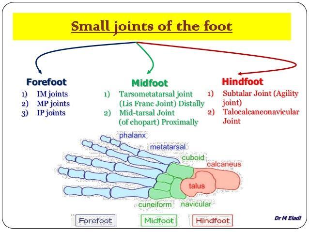
5 The joints of the foot are numerous. They are classified: A. Intertarsals B. Tarso metatarsals C. Intermetatarsals D. Metatarsophalangeal E. Interphalangeal.
6 Articulations 1. Intertarsal joints; (subtalar joint) Talocalcaneal joint Talocalcaneonavicular joint Calcaneocuboid joint SMALLAR Joints are Cunonavicular Cubiodonavicular Intercuniform Cuneocuboid joint. 2. Tarso metatarsal joints 3. Meta tarsophalangeal joints 4. Inter phalangeal joints
7 Articulations
8
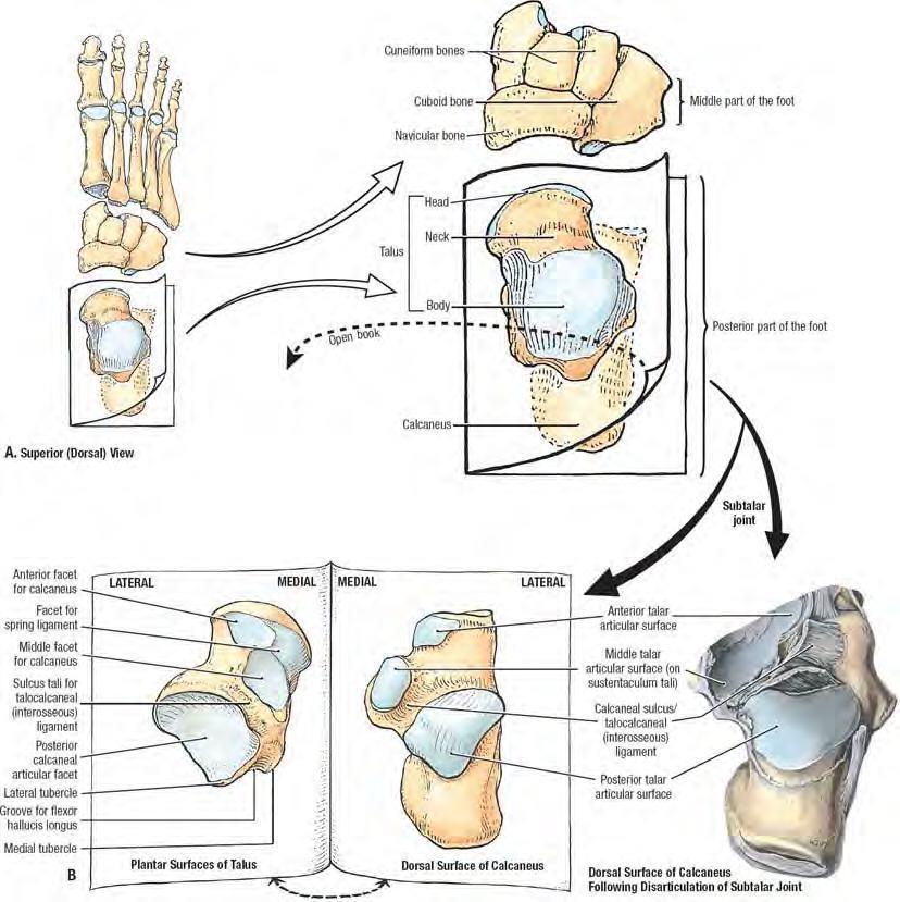
 A.TalocalcaneanL joint Articulation: convex posterior articular facet of calcaneum with concave articular facet on the under surface of talus.")
9 Intertarsal joints On the under surface of talus there are 2 separate joints; The talocalcanean joint at the back The talocalcaneonavicular joint at the front which is more complicated 1.(subtalar joint) A.TalocalcaneanL joint Articulation: convex posterior articular facet of calcaneum with concave articular facet on the under surface of talus. The articular surfaces are covered with hyaline cartilage.
10

11 Type : plane synovial joint. SUBTALAR JOINT Capsule : encloses the joint and attached to the margins of articular areas of the two bones. Synovial membrane : lines the capsule Ligaments: Medial talocalcaneal ligament Lateral talocalcaneal ligament Interosseous talocalcaneal ligament Movements : inversion and eversion of foot.
12 Medial ligament Lateral ligament and interosseous ligament


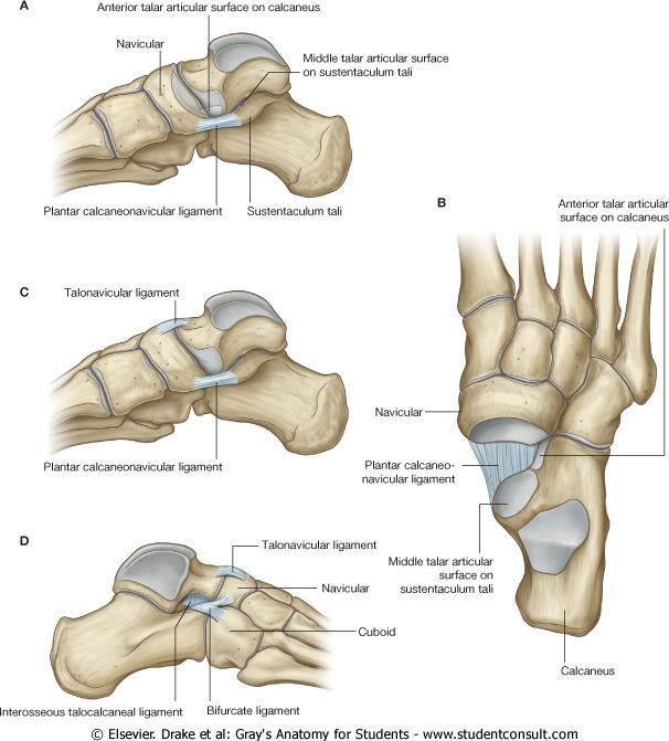
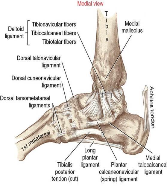
13 B.Talocalcaneonavicular joint Articulation : Head of talus with navicular Under surface of head of talus with upper surface of sustantaculam tali and the spring ligament Articular surface is covered with hyaline cartilage.

14 Talocalcaneonavicular joint Type : synovial joint Capsule : incompletely encloses the joint. Ligaments : Synovial membrane : have a single synovial cavity with the subtalar joint. Movements : Gliding and rotatory movements are possible.

15 Subtalar joint Clinical significance Mobility Shock absorption Stability Function.. Inversion and eversion of foot
 Collective")

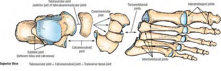
16 3.Transverse tarsal joints (midtarsal joints) Collective term for : a. Talonavicular joint and b. calcaniocuboidal joint

17 a. Talonavicular joint Articulation: Between the head of Talus and the posterior surface of the navicular bone. Type : Fibrous joint Ligaments : supported by dorsal, planter and interosseous ligaments.

18 b. Calcaneocuboid joint Articulation : anterior end of calcanium and posterior surface of cuboid. Articular surfaces covered with hyaline cartilage. Type : plane synovial joint Capsule : encloses the joint. Synovial membrane : It lines the capsule.

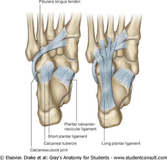
19 Bifurcate ligament
20
21
22 4.Cuneonavicular joint Articulation: between three cuniform and navicular Type : synovial gliding joint Capsule: strenghtened by planter and dorsal ligaments

23 5.Cuboidonavicular joint Articulation : between cuboid and navicular Type : fibrous joint Ligaments : supported by dorsal, planter and interosseous ligaments.

24 JOINT Cavities of FOOT. There are only six joint cavities in the intertarsal, Tarsometatarsal and intermetatarsal joints. These are: 1. Talocalcaneal joint. 2. Talocalcaneonavicular joint. 3. Calcaneocuboid 4. First Cuneometatarsal. 5. Cubometatarsal 6. Cuneonavicular with extension i.e: Navicular with three cuneiforms and second and third cuneometatarsals. Subtalar Joints

25 Tarsometatarsal joints and intermetatarsal joints Type : synovial plane joints Capsule : The first tarsometatarsal joint has it`s own capsule and synovial membrane. Rest of the tarsometatarsal joints are united by articular capsule. Ligaments : They are reinforced by planter,dorsal and interosseous ligaments.
26 Metatarsophalangeal joints Articulation : between head of metatarsal and the base of proximal phalynx. Capsule : they have articular capsule. Type : they are condyloid Ligaments : synovial joints. It is reinforced by planter and collateral ligament.
27
 and two collateral ligaments.")
28 Inter Phalangeal Joint Articulation The interphalangeal articulations of the foot are the joints between the phalanges (bones) of the toes. Type.. They are hinge joints. Ligaments Each has a plantar (underside) and two collateral ligaments. (medial and lateral)
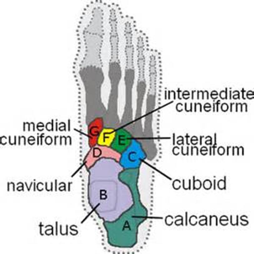
 Intermediate cuneiform (2) Lateral cuneiform (3)")
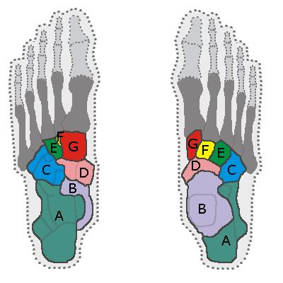
29 Mnemonic for Learning Tarsal Bones: Tiger Cubs Need M I L C Talus Calcaneus Navicular A boat It sails on the Cs Medial cuneiform (1) Intermediate cuneiform (2) Lateral cuneiform (3) Cuboid
30
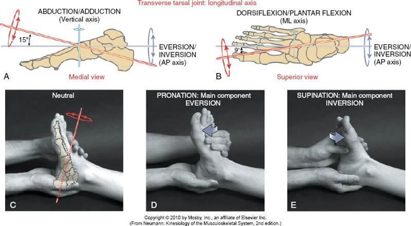
31 FUNCTIONS OF FOOT 1. Accept vertical forces during heel strike. 2. Absorb and dissipate these forces across a flexible mid- and forefoot during inversion. 3. Provide propulsion as the foot becomes a rigid lever with eversion and toe-off


32 Buddy wrapping Buddy wrapping or buddy taping is the act of bandaging a damaged or particularly a fractured finger or toe together with a healthy one. The bandage or medical tape is usually stiff, not allowing the digits to move; the healthy digit acts as a splint, keeping the damaged one in a natural position for healing. Rest plays a major role in the healing process. Buddy wrapping may also be used for sprains, dislocations, and other injuries.

33 Thank You
Introduction. The primary function of the ankle and foot is to absorb shock and impart thrust to the body during walking.
 The ankle 1 Introduction The primary function of the ankle and foot is to absorb shock and impart thrust to the body during walking. OSTEOLOGRY The term ankle refers primarily to the talocrural joint,
The Lower Limb VII: The Ankle & Foot. Anatomy RHS 241 Lecture 7 Dr. Einas Al-Eisa
 The Lower Limb VII: The Ankle & Foot Anatomy RHS 241 Lecture 7 Dr. Einas Al-Eisa Ankle joint Synovial, hinge joint Allow movement of the foot in the sagittal plane only (1 degree of freedom): dorsiflexion:
Joints and muscles of the foot. Architecture of the foot. Sándor Katz M.D.,Ph.D.
 Joints and muscles of the foot. Architecture of the foot. Sándor Katz M.D.,Ph.D. Ankle (talocrural) joint type: hinge Talocrural joint - medial collateral ligament Medial collateral = deltoid ligament
The Dance Hall by Vincent van Gogh,1888
 The Dance Hall by Vincent van Gogh,1888 Articulations of the pelvic girdle Lumbosacral joints, sacroiliac joints & pubic symphysis The remaining joints of the lower limb Hip joint Knee joint Tibiofibular
Copyright 2012 by The McGraw-Hill Companies, Inc. All rights reserved. McGraw-Hill/Irwin
 CHAPTER 8: THE LOWER EXTREMITY: KNEE, ANKLE, AND FOOT KINESIOLOGY Scientific Basis of Human Motion, 12 th edition Hamilton, Weimar & Luttgens Presentation Created by TK Koesterer, Ph.D., ATC Humboldt State
Clarification of Terms
 Clarification of Terms The plantar aspect of the foot refers to the role or its bottom The dorsal aspect refers to the top or its superior portion The ankle and foot perform three main functions: 1. shock
Dr Nabil khouri MD. MSc. Ph.D
 Dr Nabil khouri MD. MSc. Ph.D Foot Anatomy The foot consists of 26 bones: 14 phalangeal, 5 metatarsal, and 7 tarsal. Toes are used to balance the body. Metatarsal Bones gives elasticity to the foot in
The University Of Jordan Faculty Of Medicine FOOT. Dr.Ahmed Salman Assistant Prof. of Anatomy. The University Of Jordan
 The University Of Jordan Faculty Of Medicine FOOT Dr.Ahmed Salman Assistant Prof. of Anatomy. The University Of Jordan Tarsal Tunnel Syndrome Due to compression of Tibial nerve as it travels through the
Pelvic cavity. Gross anatomy of the lower limb. Walking. Sándor Katz M.D.,Ph.D.
 Pelvic cavity. Gross anatomy of the lower limb. Walking. Sándor Katz M.D.,Ph.D. Lower limb Pelvic girdle Free lower extremity Hip bone Definitive fusion of the Y- shaped growth plate occurs 16th -18th
Pelvic Girdle
 ARTICULATIONS OF LOWER EXTREMITY Pages 429-437 Pelvic Girdle formed by connection of the hip bones and the sacrum Sacroiliac Joints compound joints synovial joint - anterior, between the auricular surfaces
بسم هللا الرحمن الرحيم
 بسم هللا الرحمن الرحيم Laboratory RHS 221 Manual Muscle Testing Theory 1 hour practical 2 hours Dr. Ali Aldali, MS, PT Department of Physical Therapy King Saud University Talocrural and Subtalar Joint
radiologymasterclass.co.uk
 http://radiologymasterclass.co.uk Hip X-ray anatomy - Normal AP (anterior-posterior) Shenton's line is formed by the medial edge of the femoral neck and the inferior edge of the superior pubic ramus Loss
MIDFOOT INJURIES-ARE WE UNDERTREATING IT? Mr Rajiv Limaye Mr Prasad Karpe University Hospital of North Tees 3 rd Foot and Ankle Symposium
 MIDFOOT INJURIES-ARE WE UNDERTREATING IT? Mr Rajiv Limaye Mr Prasad Karpe University Hospital of North Tees 3 rd Foot and Ankle Symposium Introduction Increasing sports injuries RTA and traumatic injuries
Anatomy of Foot and Ankle
 Anatomy of Foot and Ankle Surface anatomy of the ankle & foot Surface anatomy of the ankle & foot Medial orientation point medial malleous sustentaculum tali tuberosity of navicular TA muscle TP muscle
Foot Injuries. Dr R B Kalia
 Foot Injuries Dr R B Kalia Overview Dramatic impact on the overall health, activity, and emotional status More attention and aggressive management Difficult appendage to study and diagnose. Aim- a stable
Hip joint Type: Articulating bones:
 Ana (242 ) Hip joint Type: Synovial, ball & socket Articulating bones: Formed between head of femur and lunate surface of acetabulum of hip bone. Capsule: it is a strong fibrous sleeve connecting the articulating
Main Menu. Ankle and Foot Joints click here. The Power is in Your Hands
 1 The Ankle and Foot Joints click here Main Menu Copyright HandsOn Therapy Schools 2009 K.8 http://www.handsonlineeducation.com/classes/k8/k8entry.htm[3/27/18, 1:40:03 PM] Ankle and Foot Joint 26 bones
First & second layers of muscles of the sole
 The FOOT First & second layers of muscles of the sole introduction The muscles acting on the foot can be divided into two distinct groups; extrinsic and intrinsic muscles. The extrinsic muscles arise from
ANKLE JOINT ANATOMY 3. TALRSALS = (FOOT BONES) Fibula. Frances Daly MSc 1 CALCANEUS 2. TALUS 3. NAVICULAR 4. CUBOID 5.
 Fibula. Frances Daly MSc 1 CALCANEUS 2. TALUS 3. NAVICULAR 4. CUBOID 5.") ANKLE JOINT ANATOMY The ankle joint is a synovial joint of the hinge type. The joint is formed by the distal end of the tibia and medial malleolus, the fibula and lateral malleolus and talus bone. It is
ANKLE JOINT ANATOMY The ankle joint is a synovial joint of the hinge type. The joint is formed by the distal end of the tibia and medial malleolus, the fibula and lateral malleolus and talus bone. It is
 P R E S E N T S Dr. Mufa T. Ghadiali is skilled in all aspects of General Surgery. His General Surgery Services include: General Surgery Advanced Laparoscopic Surgery Surgical Oncology Gastrointestinal
Foot. Dr. Heba Kalbouneh Associate Professor of Anatomy and Histology
 Foot Dr. Heba Kalbouneh Associate Professor of Anatomy and Histology Dorsum of the Foot Sole of the Foot Plantar aponeurosis It is a triangular thickening of deep fascia in the sole of the foot Attachments:
Introduction to Human Osteology Chapter 3: Hands and Feet
 Introduction to Human Osteology Chapter 3: Hands and Feet Roberta Hall Kenneth Beals Holm Neumann Georg Neumann Gwyn Madden Revised in 1978, 1984, and 2008 Bones of the Hand Eight carpal bones, in two
CHAPTER 80 BASIC CONSIDERATIONS
 Página 1 de 32 Copyright 2001 Lippincott Williams & Wilkins Loeser, John D. Bonica's Management of Pain, 3rd Edition CHAPTER 80 BASIC CONSIDERATIONS Part of "CHAPTER 80 - Pain in the Leg, Ankle, and Foot"
BONES JOINTS MUSCLES OF THE LOWER LIMB
 BONES JOINTS MUSCLES OF THE LOWER LIMB LOWER LIMB: BONES LOWER LIMB GLUTEAL REGION consists of 6 major segments: FEMORAL REGION (THIGH) KNEE REGION LEG REGION TALOCRURAL REGION (ANKLE) FOOT REGION LOWER
What Happens to the Paediatric Flat Foot? Peter J Briggs Freeman Hospital Newcastle upon Tyne
 What Happens to the Paediatric Flat Foot? Peter J Briggs Freeman Hospital Newcastle upon Tyne We don t know!! Population Studies 2300 children aged 4-13 years Shoe wearers Flat foot 8.6% Non-shoe wearers
LEVEL 3 DIPLOMA IN AROMATHERAPY MODULE 10 KNOWLEDGE OF ANATOMY, PHYSIOLOGY & PATHOLOGY FOR COMPLEMENTARY THERAPIES THE ARTICULAR SYSTEM COURSE MANUAL
 LEVEL 3 DIPLOMA IN AROMATHERAPY MODULE 10 KNOWLEDGE OF ANATOMY, PHYSIOLOGY & PATHOLOGY FOR COMPLEMENTARY THERAPIES THE ARTICULAR SYSTEM COURSE MANUAL CHRISTINA LYNE christina@aromalyne.com 1 THE ARTICULAR
Biology 325 Fall 2003
 Name: pre-lab exercise due at beginning of your lab session Matching a. fibrous joints b. cartilaginous joints c. synovial joints 1. exhibit a joint cavity 2. types are sutures and syndesmoses 3. bones
ABC of Emergency Radiology
 l ja ) $% _2) < j> ~~~~~~~~~~~~~~~~~foot ABC of Emergency Radiology THE FOOT D A Nicholson, D O'Keeffe, P A Driscoll Accurate clinical assessment of injuries to the foot will avoid unnecessary exposure
BRAD G. SAMOJLA LIMB DEVELOPMENT
 BRAD G. SAMOJLA LIMB DEVELOPMENT There are many different types of deformities that may present in the foot. Many congenital deformities can be explained by problems occurring during development of the
Anatomy of Ankle & Foot. Chang-Hyung Lee, M.D., Ph.D. Physical Medicine & Rehabilitation Samsung Medical Center
 Anatomy of Ankle & Foot Chang-Hyung Lee, M.D., Ph.D. Physical Medicine & Rehabilitation Samsung Medical Center Ankle Introduction Most frequently injured major joint 3 main articulation: distal tibiofibular
Anatomy of the lower limb
 Anatomy of the lower limb Arches & sole of the foot Dr. Hayder ARCHES OF THE FOOT The foot as a mechanical unit performs two major functions: - It acts as a pliable platform to support the body weigh during
Exercise 13. Articulations and Body Movements
 Exercise 13 Articulations and Body Movements Articulations Articulations, or joints, are points where a bone is connected to one or more other bones. Articulations hold the skeleton together. Articulations
The plantar aponeurosis
 Anatomy of the foot The plantar aponeurosis Is a triangular thickening of the deep fascia Its apex is attached to the medial and lateral tubercles of the calcaneum. The base of the aponeurosis divides
musculoskeletal system anatomy muscles of foot sheet done by: dina sawadha & mohammad abukabeer
 musculoskeletal system anatomy muscles of foot sheet done by: dina sawadha & mohammad abukabeer Extensor retinaculum : A- superior extensor retinaculum (SER) : originates from the distal ends of the tibia
Feet First. Michael K. Cooper, DO FACOFP Family Practice/OMM St John Clinic - Claremore OOA 2018 Annual Convention
 Feet First Michael K. Cooper, DO FACOFP Family Practice/OMM St John Clinic - Claremore OOA 2018 Annual Convention Disclaimer I have no conflict of interest. I am not on any pharmaceutical company payroll
To describe he knee joint, ligaments, structure & To list the main features of other lower limb joints
 To describe he knee joint, ligaments, structure & neurovascular supply To demonstrate the ankle joint anatomy To list the main features of other lower limb joints To list main groups of lymph nodes in
Section Three: The Leg, Ankle, and Foot Lecture: Review of Clinical Anatomy, Patterns of Dysfunction and Injury, and
 Section Three: The Leg, Ankle, and Foot Lecture: Review of Clinical Anatomy, Patterns of Dysfunction and Injury, and Treatment Implications for the Leg, Ankle, and Foot Levels I and II Demonstration and
The study of the internal workings of the human body and how it moves. A user s guide
 DEFINITION The study of the internal workings of the human body and how it moves. A user s guide OUR FOCUS Bones: structure, protection, levers Joints: allow for movement Muscles: cause movement Anatomical
Biokinesiology of the Ankle Complex
 Rehabilitation Considerations Following Ankle Fracture: Impact on Gait & Closed Kinetic Chain Function Disclosures David Nolan, PT, DPT, MS, OCS, SCS, CSCS I have no actual or potential conflict of interest
حسام أبو عوض. - Ahmad. 1 P a g e
 - 9 حسام أبو عوض - - Ahmad 1 P a g e In the last lecture, we finished discussing the superficial part of the posterior compartment and the popliteus muscle of the deep layer[reminder: The entire posterior
CHAPTER 3 What Is Anatomy?
 CHAPTER 3 What Is Anatomy? Kinesiology Books Publisher 1 TABLE OF CONTENTS The Language of Anatomy Anatomical Position Directional Terms Body Planes Movements Musculoskeletal System Human Skeleton Types
Answers to Pre-Lab Quiz (p. 171) Answers to Activity Questions
 Answers to Activity Questions") Answers to Pre-Lab Quiz (p. 171) 1. Holds bones together; allows the rigid skeleton some flexibility so that gross body movements can occur 2. c, amount of movement allowed by the joint 3. synovial 4.
Answers to Pre-Lab Quiz (p. 171) 1. Holds bones together; allows the rigid skeleton some flexibility so that gross body movements can occur 2. c, amount of movement allowed by the joint 3. synovial 4.
Understanding Leg Anatomy and Function THE UPPER LEG
 Understanding Leg Anatomy and Function THE UPPER LEG The long thigh bone is the femur. It connects to the pelvis to form the hip joint and then extends down to meet the tibia (shin bone) at the knee joint.
Section 4: Tarsal Coalitions
 Case H (Figure 2): PedCat CBCT transverse plane reconstruction of right Lisfranc midfoot dislocation compared to normal left foot. Clinical Relevance of the PedCat Study: The weight bearing CBCT study
Anatomy. Anatomy deals with the structure of the human body, and includes a precise language on body positions and relationships between body parts.
 Anatomy deals with the structure of the human body, and includes a precise language on body positions and relationships between body parts. Proper instruction on safe and efficient exercise technique requires
OTM Lecture Gait and Somatic Dysfunction of the Lower Extremity
 OTM Lecture Gait and Somatic Dysfunction of the Lower Extremity Somatic Dysfunction Tenderness Asymmetry Range of Motion Tissue Texture Changes Any one of which must be present to diagnosis somatic dysfunction.
Skeletal System. Supplementary Information
 Skeletal System Supplementary Information COMMON ANATOMICAL TERMS Planes run through the body side to side and front to back eg. median plane Surfaces of the body are also named eg. anterior surface This
10/12/2010. Upper Extremity. Pectoral (Shoulder) Girdle. Clavicle (collarbone) Skeletal System: Appendicular Skeleton
 Girdle. Clavicle (collarbone) Skeletal System: Appendicular Skeleton") Skeletal System: Appendicular Skeleton Pectoral girdle Pelvic girdle Upper limbs Lower limbs 8-1 Pectoral (Shoulder) Girdle Consists of scapula and clavicle Clavicle articulates with sternum (Sternoclavicular
Skeletal System: Appendicular Skeleton Pectoral girdle Pelvic girdle Upper limbs Lower limbs 8-1 Pectoral (Shoulder) Girdle Consists of scapula and clavicle Clavicle articulates with sternum (Sternoclavicular
Dr.Israa H. Mohsen. Lecture 5. The vertebral column
 Anatomy Lecture 5 Dr.Israa H. Mohsen The vertebral column The vertebral column a flexible structure consisting of 33 vertebrae holds the head and torso upright, serves as an attachment point for the legs,
LISFRANC FRACTURE-DISLOCATION
 LISFRANC FRACTURE-DISLOCATION Napoleon at Mont St. Bernard, Jacques-Louis David, 1800, Oil on Canvas, Musee du Louvre, Paris. This is Jacques-Louis David s immortal depiction of a young Napoleon Bonaparte,
Arthrology the study of joint structure, function and dysfunction. Sentenced to Life in the Joint
 Arthrology Arthrology the study of joint structure, function and dysfunction Sentenced to Life in the Joint Kinesiology study of musculo-skeletal movement Articulations any point where two bones meet (joint)
Joints. Articulations Arthroses
 Joints Articulations Arthroses 1 Joints, defined Points of contact between Two bones Bone and teeth Joint classification: 2 schemes Functional classification degree of movement permitted Structural classification
Therapeutic Foot Care Certificate Program Part I: Online Home Study Program
 Therapeutic Foot Care Certificate Program Part I: Online Home Study Program 1 Anatomy And Terminology Of The Lower Extremity Joan E. Edelstein, MA, PT, FISPO Associate Professor of Clinical Physical Therapy
Traumatic Injuries to the Foot and Ankle
 Traumatic Injuries to the Foot and Ankle Dr. Joseph N. Daniel Clinical Associate Professor of Orthopaedic Surgery Foot and Ankle Service, The Rothman Institute Thomas Jefferson University Hospital Philadelphia,
Pectoral (Shoulder) Girdle
 Girdle") Chapter 8 Skeletal System: Appendicular Skeleton Pectoral girdle Pelvic girdle Upper limbs Lower limbs 8-1 Pectoral (Shoulder) Girdle Consists of scapula and clavicle Clavicle articulates with sternum
Chapter 8 Skeletal System: Appendicular Skeleton Pectoral girdle Pelvic girdle Upper limbs Lower limbs 8-1 Pectoral (Shoulder) Girdle Consists of scapula and clavicle Clavicle articulates with sternum
Assignment 2: Human Anatomy
 Assignment 2: Human Anatomy Chapter 2 Quiz: How Much Do You Know About Anatomy? 1. Which of the following is not a feature of the anatomical position: A) The body stands erect. B) The body is facing forward.
Joints. Agenda. Joints. Structural and Functional Classification of Articulations
 Joints Structural and Functional Classification of Articulations Agenda Joint Basics Classification Structural Joint Details Joint Stability Movements of Synovial Joints Shape Classification of Synovial
pedcat Clinical Case Studies
 pedcat Clinical Case Studies C u r v e B e a m 1 7 5 T i t u s A v e, S u i t e 3 0 0 W a r r i n g t o n, P A 1 8 9 7 6 267-4 8 3-8081 w w w. c u r v e b e a m. c o m PedCAT: Clinical Evidence of diagnostic
Bones of Lower Limb. Dr. Heba Kalbouneh Associate Professor of Anatomy and Histology
 Bones of Lower Limb Dr. Heba Kalbouneh Associate Professor of Anatomy and Histology Bones of the lower limb Hip Bone Made up of 3 bones: 1) Ilium (flat), superior in position 2) Ischium (L), postero-inferior
 17.2 A-P Lower Leg Measure: A-P at mid-lower leg Protection: Apron draped over pelvis SID: 40 Table top No Tube Angle Film: 7 x17 I.D. down or diagonal 14 x 17 www.fisiokinesiterapia.biz A-P Lower Leg
Chapter 8 The Skeletal System: The Appendicular Skeleton. Copyright 2009 John Wiley & Sons, Inc.
 Chapter 8 The Skeletal System: The Appendicular Skeleton Appendicular Skeleton It includes bones of the upper and lower limbs Girdles attach the limbs to the axial skeleton The pectoral girdle consists
Muscle Tissue. Isometric Contraction. Isotonic Contractions 11/22/2016. Muscles. Anatomy Two Joints And Movements
 Muscles Anatomy Two Joints And Movements Structure of a Muscle Organ Copyright 2008 by Saunders Muscle Tissue Highly elastic and vascularized, produces movement through elongation and contraction Types
Evidence-Based Examination of the Foot Presented by Alexis Wright, PT, PhD, DPT, FAAOMPT Practice Sessions/Skill Check-offs
 Evidence-Based Examination of the Foot Presented by Alexis Wright, PT, PhD, DPT, FAAOMPT Practice Sessions/Skill Check-offs Module Five: Movement Assessment of the Foot/Ankle (1 hour CEU Time) Skilled
Chapter 09 Articulations Pearson Education, Inc.
 Chapter 09 Articulations An Introduction to Articulations Articulations Body movement occurs at joints (articulations) where two bones connect Joint Structure Determines direction and distance of movement
BLUE SKY SCHOOL OF PROFESSIONAL MASSAGE AND THERAPEUTIC BODYWORK Musculoskeletal Anatomy & Kinesiology KNEE & ANKLE MUSCLES
 BLUE SKY SCHOOL OF PROFESSIONAL MASSAGE AND THERAPEUTIC BODYWORK Musculoskeletal Anatomy & Kinesiology KNEE & ANKLE MUSCLES MSAK201-I Session 3 1) REVIEW a) THIGH, LEG, ANKLE & FOOT i) Tibia Medial Malleolus
THE JOURNAL OF NUCLEAR MEDICINE Vol. 56 No. 3 March 2015 Rauscher et al.
 Supplemental Figure 1 Correlation analysis of tracer between and subsequent as assessed by SUV max in focal lesions (A). x-axis displays quantitative values as obtained by, and y-axis displays corresponding
Section 3: Foot Subluxations and Dislocations
 Section 3: Foot Subluxations and Dislocations Case Study F: Lisfranc s Midfoot Dislocation Clinical History: J.K. a 28 year old female presents complaining of a painful right foot. She sustained an acute
Amy Warenda Czura, Ph.D. 1 SCCC BIO130 Lab 7 Appendicular Skeleton & Articulations
 The Skeletal System II: Appendicular Skeleton and Articulations Exercises 11, 13 (begins: page 145 in 9 th and 10 th editions) Exercises 10, 11 (begins: page 147 in 11 th edition, page 149 in 12 th edition)
PowerPoint Lecture Slides prepared by Janice Meeking, Mount Royal College C H A P T E R. Joints: Part A. Copyright 2010 Pearson Education, Inc.
 PowerPoint Lecture Slides prepared by Janice Meeking, Mount Royal College C H A P T E R 8 Joints: Part A Warm Up 11/28/16 Happy Thanksgiving welcome back! J (be ready to share something fun you did over
Skeletal System: Articulations (Chapter 9) Lecture Materials for Amy Warenda Czura, Ph.D. Suffolk County Community College Eastern Campus
 Lecture Materials for Amy Warenda Czura, Ph.D. Suffolk County Community College Eastern Campus") Skeletal System: Articulations (Chapter 9) Lecture Materials for Amy Warenda Czura, Ph.D. Suffolk County Community College Eastern Campus Primary Sources for figures and content: Marieb, E. N. Human Anatomy
Skeletal System: Articulations (Chapter 9) Lecture Materials for Amy Warenda Czura, Ph.D. Suffolk County Community College Eastern Campus Primary Sources for figures and content: Marieb, E. N. Human Anatomy
Practical Applications of Manual Therapy for the Ankle and Foot
 Practical Applications of Manual Therapy for the Ankle and Foot PHATS Annual Meeting 2014 Orlando, Florida Outline! Objectives! Case Study! What is Manual Therapy?! Joint Mobilization! Joint Mobilization
Tarsal Coalition On MR
 Tarsal Coalition On MR By William Renner, M.D. This and other topics will be discussed in Tarsal coalition is a congenital anomaly with fusion of the tarsal bones. The fusion may be bony, fibrous or cartilaginous.
Dynamics of the Foot in Locomotion
 Continuing Dynamics of the Foot in Locomotion A new way of looking at functional dynamics. ORTHOTICS & BIOMECHANICS Objectives After reading this article, the podiatric physician should be able to: 1)
Joints Dr. Ali Ebneshahidi
 Joints Dr. Ali Ebneshahidi Function of Joints 1. Serve as functional junctions between bones. 2. Bind bones, strokes, and other related tissues together. 3. Allow bone growth to occur. 4. Permit certain
Human Anatomy & Physiology I Dr. Sullivan Unit IX Arthrology (joints) - Chapter 9
 - Chapter 9") Human Anatomy & Physiology I Dr. Sullivan Unit IX Arthrology (joints) - Chapter 9 I. Joints: aka Articulations a) Joints are points of contact between two or more bones. Joints may be moveable or may not
Human Anatomy & Physiology I Dr. Sullivan Unit IX Arthrology (joints) - Chapter 9 I. Joints: aka Articulations a) Joints are points of contact between two or more bones. Joints may be moveable or may not
Introduction. Physiology. Classification of Bones. Anatomy of a Long Bone. Anatomy of a Long Bone. Skeletal System and Joint Movements.
 Chapter 13 Skeletal System and Joint Movements Susan G. Salvo Introduction Skeletal system is composed of bones, cartilage, ligaments, and joints 206 bones in the body Bone is living tissue Skeletal system
Arthrokinematics and Selected Joint Techniques
 The Foot and Ankle: An Overview of Arthrokinematics and Selected Joint Techniques Janice K. Loudon, PhD, PT, ATC, SCS; Stephania L. Bell, MS, PT ABSTRACT: Limited range of motion of the ankle is common
CHAPTER 8: THE BIOMECHANICS OF THE HUMAN LOWER EXTREMITY
 CHAPTER 8: THE BIOMECHANICS OF THE HUMAN LOWER EXTREMITY _ 1. The hip joint is the articulation between the and the. A. femur, acetabulum B. femur, spine C. femur, tibia _ 2. Which of the following is
The Lower Limb VI: The Leg. Anatomy RHS 241 Lecture 6 Dr. Einas Al-Eisa
 The Lower Limb VI: The Leg Anatomy RHS 241 Lecture 6 Dr. Einas Al-Eisa Muscles of the leg Posterior compartment (superficial & deep): primary plantar flexors of the foot flexors of the toes Anterior compartment:
Joints Outline 8.1 Joints are classified into three structural and three functional categories (p. 251; Table 8.1) A. Joints are classified by
 A. Joints are classified by") Joints Outline 8.1 Joints are classified into three structural and three functional categories (p. 251; Table 8.1) A. Joints are classified by structure and by function: Structural classification focuses
Joints Outline 8.1 Joints are classified into three structural and three functional categories (p. 251; Table 8.1) A. Joints are classified by structure and by function: Structural classification focuses
EDL EHL. Extensor Hallucis Longus L5 Extensor Digitorum longus L5,1 Peroneus Tertius L5 1 Extensor Digitorum Brevis S1,2 [like intrinsic muscle]
![EDL EHL. Extensor Hallucis Longus L5 Extensor Digitorum longus L5,1 Peroneus Tertius L5 1 Extensor Digitorum Brevis S1,2 [like intrinsic muscle]](/thumbs/78/77875930.jpg "EDL EHL. Extensor Hallucis Longus L5 Extensor Digitorum longus L5,1 Peroneus Tertius L5 1 Extensor Digitorum Brevis S1,2 [like intrinsic muscle]") ANATOMY OF ANKLE AND FOOT Lateral aspect: [Dorsal medial to lateral= dorsal under extensor retinaculum] Tibialis Anterior EHL Artery [Dorsal pedal A] and Anterior tibial N EDL Peroneus Tertius Behind the
ANATOMY OF ANKLE AND FOOT Lateral aspect: [Dorsal medial to lateral= dorsal under extensor retinaculum] Tibialis Anterior EHL Artery [Dorsal pedal A] and Anterior tibial N EDL Peroneus Tertius Behind the
Radiographic Assessment of Pediatric Foot Alignment: Self-Assessment Module
 1.5 CME AJR Integrative Imaging LIFELONG LEARNING FOR RADIOLOGY Radiographic Assessment of Pediatric Foot Alignment: Self-Assessment Module Mahesh M. Thapa 1,2, Sumit Pruthi 1,2, Felix S. Chew 2 ABSTRACT
Pathology & Primary Treatment of Clubfoot
 Pathology & Primary Treatment of Clubfoot Hyun-Dae Shin, MD, PhD. Department of Orthopedic Surgery, School of Medicine, Chungnam National University, Daejeon, Korea Introduction The affected foot Restricted
Exercise Science Section 4: Joint Mechanics and Joint Injuries
 Exercise Science Section 4: Joint Mechanics and Joint Injuries An Introduction to Health and Physical Education Ted Temertzoglou Paul Challen ISBN 1-55077-132-9 Types of Joints Fibrous joint Cartilaginous
~, /' ~::'~ EXTENSOR HALLUCIS LONGUS. Leg-anterolateral :.:~ / ~\,
 TIBIALIS ANTERIOR Lateral condyle of tibia, upper half of lateral surface of tibia, interosseous membrane Medial side and plantar surface of medial cuneiform bone, and base of first metatarsal bone Dorsiflexes
It is formed by fusion of 3 bones: I. Ilium (superior bone). II. Pubis (antero-inferior bone). III. Ischium (postero-inferior bone).
. II. Pubis (antero-inferior bone). III. Ischium (postero-inferior bone).") It is formed by fusion of 3 bones: I. Ilium (superior bone). II. Pubis (antero-inferior bone). III. Ischium (postero-inferior bone). Pubis Acetabulum Ana (242 ) The three constituent of bones of the hip
It is formed by fusion of 3 bones: I. Ilium (superior bone). II. Pubis (antero-inferior bone). III. Ischium (postero-inferior bone). Pubis Acetabulum Ana (242 ) The three constituent of bones of the hip
Effect of Fixation Using Locked Compression Plate versus Lag Screws on Biomechanics of Talonavicular Joint: A Human Cadaveric Foot Model
 University of Tennessee Health Science Center UTHSC Digital Commons Theses and Dissertations (ETD) College of Graduate Health Sciences 12-2010 Effect of Fixation Using Locked Compression Plate versus Lag
Managing Tibialis Posterior Tendon Injuries
 Managing Tibialis Posterior Tendon Injuries by Thomas C. Michaud, DC Published April 1, 2015 by Dynamic Chiropractic Magazine Tibialis posterior is the deepest, strongest, and most central muscle of the
MECHANICS OF MOVEMENT
 MECHANICS OF MOVEMENT Tissues and Structures Involved Muscle Nerve Bone Cartilage What are Tendons? Role of Joints Mechanics of Joints Making it all work Nerve and Muscle--the Motor Unit Skeletal muscles
the interosseous tibio-fibular ligaments. The capsule is innervated by twigs from
 THE SPRAINED ANKLE by H. Davis, Ch. M., M. Sch., Orth., F. R. C. S., England Consultant Orthopaedic Surgeon The ankle joint is a synovial joint of typical hinge pattern. The lower ends of the tibia and
A. Incorrect! The appendicular skeleton includes bones of the shoulder, arm, hand, pelvis, leg and foot.
 Anatomy and Physiology - Problem Drill 08: The Skeletal System III No. 1 of 10 1. Which of the following statements about the appendicular skeleton is correct? A. The appendicular skeleton includes bones
Ligaments of Elbow hinge: sagittal plane so need lateral and medial ligaments
 Ligaments of Elbow hinge: sagittal plane so need lateral and medial ligaments Ulnar Collateral ligament on medial side; arising from medial epicondyle and stops excess valgus movement (lateral movement)
The evaluation and management of acute musculoskeletal
 ONLINE EXCLUSIVE George E. Eldayrie, MD; Kristy Smith, MD; Michael Seth Smith, MD, CAQSM, PharmD Department of Community Health and Family Medicine (Drs. Eldayrie and K. Smith) and Department of Orthopedics
Peggers Super Summaries: Foot Injuries
 Lisfranc Injury ANATOMY Roman arch with recessed 2 nd MT base AP medial side of intermediate cuneiform to 2 nd MT base Oblique medial side of lateral cuneiform with 3 rd MT base and 4 th with medial boarder
Skeletal Considerations for Movement. Kinesiology RHS 341 Lecture 2 Dr. Einas Al-Eisa
 Skeletal Considerations for Movement Kinesiology RHS 341 Lecture 2 Dr. Einas Al-Eisa The Skeletal System Bones, cartilage, ligaments, & joints Consists of approximately 20% of total body weight Bone constitutes
Case 1 7 yo male Right elbow injury 3 months ago Medial elbow pain and tenderness over medial epicondyle Long arm cast given but off himself 1 month a
 Case presentations Case 1 7 yo male Right elbow injury 3 months ago Medial elbow pain and tenderness over medial epicondyle Long arm cast given but off himself 1 month after Progressive limited elbow flexion
Understand the skeletal system:
 Understand the skeletal system: Including axial and appendicular skeleton All joints in the body All major bones Development of bones & bone growth Training effects on the skeletal system All movements
Anatomy of the lower limb
 Anatomy of the lower limb 1. Bones of the lower limb Pelvis Hip bone/coxal bone Acetabulum o Acetabular margin o Acetabular fossa o Acetabular notch o Lunate surface Ischiopubic ramus Obturator foramen
Principles of Anatomy and Physiology
 Principles of Anatomy and Physiology 14 th Edition CHAPTER 8 The Skeletal System: The Appendicular Skeleton The Appendicular Skeleton The 126 bones of the appendicular skeleton are primarily concerned
ANKLE PLANTAR FLEXION
 ANKLE PLANTAR FLEXION Evaluation and Measurements By Isabelle Devreux 1 Ankle Plantar Flexion: Gastrocnemius and Soleus ROM: 0 to 40-45 A. Soleus: Origin: Posterior of head of fibula and proximal1/3 of