MR imaging spectrum in intracranial meningiomas-a retrospective study
|
|
|
- Claud McBride
- 5 years ago
- Views:
Transcription
1 IOSR Journal of Dental and Medical Sciences (IOSR-JDMS) e-issn: , p-issn: Volume 17, Issue 3 Ver.7 March. (2018), PP MR imaging spectrum in intracranial meningiomas-a retrospective study Dr Madhusudhan C 1., Dr Moorthy N.L.N 2., Dr Balaji Gopal 3, Dr Ravikumar 4 Dr Dileep 5 Dr Chandramma 6. DrMadhusudhan C associate prof radiodiagnosis DrMoorthy N.L.N. Prof of radio diagnosis DrBalaji Gopal associate prof radio diagnosis DrRavikumar M asst prof radio diagnosis DrDileepKothareddy senior resident DrChandramma senior radiologist Dept of radio diagnosis, Apollo medical college CHITTOOR A.P Corresponding author: Dr NLN Moorthy Abstract: Meningiomas are one of the most common benign intracranial tumors seen in females after 3 rd decade. They are well defined extra axial masses with characteristic CT and MR imaging features. In the present study we retrospectively evaluated the MR imaging in 67 operated cases of intracranial meningiomas in our institute. Key Words : Meningioma-MRI-dural masses Date of Submission: Date of acceptance: I. Introduction Meningiomas are slow growing benign tumors that arise from the meningothelial cells of the arachnoid membrane. Majority of the tumors present in middle age or above. The clinical signs are varied and are due to compression of brain structures or obstruction of ventricular system. About 90 % of meningiomas belong to benign WHO Grade1 lesions. However a small percentage represent WHO Grade 2 ( atypical 5-7 % ) and WHO Grade 3 ( anaplastic 1-3 % ) types 1.These extra axial tumors are located mostly along the parasagittal planes and along the lateral cerebral convexity, sphenoid wing, posterior fossa. They also seen in the middle cranial fossa, along the olfactory groove, sella turcica, tentorium, CP angle, lateral ventricle and optic nerve sheath. Meningiomas are mostly solitary and multiple meningiomas are seen in neurofibromatosis type 2 2. On MRI, meningioma typically show the following features. Iso to hypointensity on T1 weighted and iso to slightly hyperintensity on T2 weighted, FLAIR sequences, varying degrees of restriction on diffusion weighted imaging with homogenous enhancement on contrast administration. Rarely they show calcification which is better appreciated on CT scan and appear as low intensity signal on T2 weighted sequences. Although most of the meningiomas show homogenous enhancement with contrast,heterogenous and peripheral enhancement is seen in rare microcystic type of meningioma. One of the characteristic feature of meningioma is the presence of dural tail ( due to enhancement of dura infiltrating away from the lesion) which is seen in 72 % of cases 3. The presence of CSF cleft between the tumor and the underlying brain cortex is also one of the characteristic features of meningioma due to its extra axial location 4. Encasement of arteries and veins are usually seen in meningiomas that are located along the skull base or parasagittal planes. Meningiomas, especially enplaque type invades the underlying bone causing hyperostosis or lytic destruction. This finding is seen in 20% of cases of meningiomas and better evaluated on CT scan than MRI. MR Spectroscopy findings are mostly non specific. Meningioma may show increased choline ( 3.2ppm), decreased creatine ( 3.0ppm), presence of lactate. Presence of alanine which was thought as specific is not detectable always 2. II. Case Results : Magnetic resonance imaging findings of 67 operated cases of intracranial meningiomas were analysed. The study was done mainly to look into the following points. Age incidence, sex of the patients, location of the tumor, T1 weighted sequence, T2 weighted sequence, contrast enhancement pattern, for any evidence of calcification, cysts, peritumoral edema, hemorrhage, MR spectroscopy findings. Findings : The age varied between 17 years and 82 years with peak incidence between years for females and above 60 years for males. The tumors are more common in female patients 49 out of 67 and the rest are males. Almost all locations described in the literature are affected with parasagittal tumors are the most common DOI: / Page
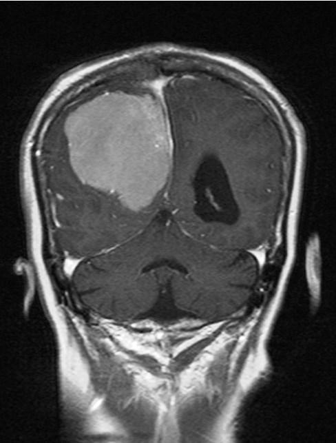
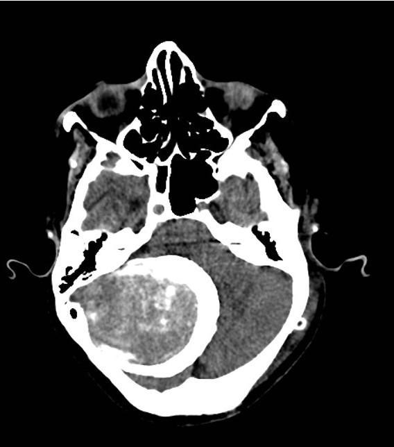
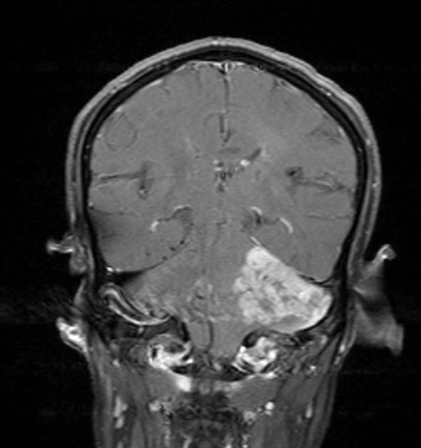
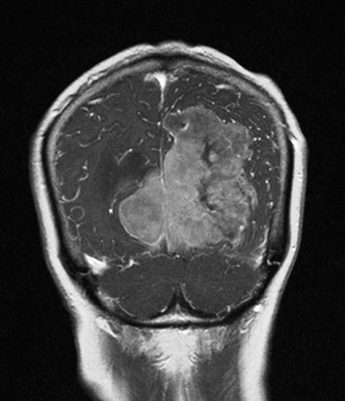
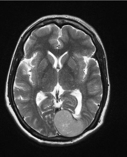
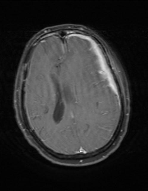
2 site in our study 23 out of 67 (22.9%) followed by lateral hemisphere convexity closely.( Fig 1-9 ). Meningiomas more common on left side 39, and remaining 28 were right side( table 1) Table 1.) Location of meningiomas in the present study Parasagittal 23 Lateral hemisphere convexity 20 Sphenoid wing 5 Middle cranial fossa 4 Posterior fossa 4 Sellaturcica 3 CP angle 2 Olfactory groove 2 Others ( I case of enplaque meningioma, tentoroium, two cases of recurrent meningiomas ) Total cases = 67 Majority of the tumors followed the classical pattern of signal intensities on MR imaging. They are either homogenously hypo or isointense on T1 weighted sequences and slightly hyper intense on T2 weighted sequences, Fluid attenuated sequences and restricted on DW imaging. The contrast enhancement pattern was homogenous in many cases. 11 cases showed heterogenous signal intensities, focal or diffuse calcification and heterogenous enhancement after contrast administration, cysts, necrotic areas suggesting atypical type of meningiomas. Dural tail sign and peritumoral edema were noted in more than one third cases.hyperostosis of underlying was more common than osteolysis in our study seen in 19 cases. Increased peaks of choline and creatine, presence of alanine on MR spectroscopy was observed in 10 cases only and the rest are nonspecific. Four cases showed intratumoral and peritumoral hemorrhage and in one case there was associated spontaneous intracerebral hemorrhage indicating anaplastic subtype of meningioma which was confirmed on histopathology (Fig 10 ). Encasement of adjacent carotid arteries and dural venous sinuses was seen in four cases. Two patients presented with recurrent meningiomas after surgery with typical findings (Fig 11). One case of enplaque meningioma and another case of meningioma arising from falx and tentorium also seen in our study ( Fig 12 and fig 13 ). ( Table 2 ) Table 2.) list of salient features of meningioma on MRI Findings number of cases Peritumoral edema 32 Dural tail sign 24 Bone hyperostosis /erosion 19 Cystic changes 8 Abnormal MR spectroscopy 10 Intra/peritumoral haemorrhage 4 Vascular encasement 4 Calcification 4 III. Discussion: Meningothelial, fibrous, transitional subtypes constitute more than 80 % of meningiomas, considered as WHO Grade 1, which have typical imaging features of meningiomas described above. There are other small group of histological subtypes of meningiomas labeled as WHO grade 2 and WHO grade 3 which have different and atypical imaging findings extensively described 5. Meningothelial meningioma, the commonest subtype of meningioma shows typical MR imaging findings- hyperintensity on T2 weighted sequence, homogenous contrast enhancement with dural tail sign and bone hyperostosis. Fibrous meningioma shows relative T2 hypointensity due to the presence of collagen. Transitional meningioma shows mixed iso- and hyperintensity on T2 weighted sequences. Psammomatous meningioma is characterized by the presence of calcifications besides the typical imaging features of WHO grade1 meningioma. Angiomatous meningioma shows the presence of multiple flow voids due to hypervascularity along with prominent peritumoral edema. Reticular type contrast enhancement with peritumoral edema characterize microcystic meningioma. Secretory meningioma commonly arise at skull base, shows high signal intensity on T2weighted sequence with high ADC values unlike typical meningioma along with significant peritumoral edema. Lymphoplasmacyte-rich meningioma seen in young patients shows typical irregular tumor margin with invasion to adjacent brain. The mass shows heterogenous contrast enhancement with significant peritumoral edema. Clear cell meningioma typically seen in young patients and located at cerebello pontine angles. Recurrence are common with this type. Heterogenous contrast enhancement, presence of cysts, peritumoral edema and lytic bone destruction are also common. MR imaging DOI: / Page
3 of atypical and anaplastic meningioma : common in males. These tumors show irregular tumor margin, indistinct tumor brain interface, heterogenous signal intensities 6, restriction on diffusion weighted sequence. Underlying bone destruction, peritumoral edema, necrosis and calcification also seen. Metaplastic meningioma contain cartilaginous, osseous or lipomatous tissue and the imaging findings depend on the composition of the tumor. Though extra axial about 60 % of meningiomas are associated with vasogenic cerebral edema. In our study 32 cases showed brain edema. Meningiomas are rarely associated with cystic changes which can be extratumoral or intratumoral7. Spontaneous or intratumoral hemorrhage are occasionally seen in meningiomas. We had 4 cases of intratumoral hemorrhage of which one was associated with spontaneous intracerebral hemorrhage. Though avid and homogenous contrast enhancement is typical of meningioma, but sometimes it can be heterogenous or ring enhancement also. Many studies were reported in the literature correlating the MR imaging pattern with histopathological subtypes of meningioma 5,8. NORIAKI TOMURA et al 9 evaluated the neuroradiological findings in 8 cases of atypical meningiomas and found certain signs like absent dural tail sign, large perilesional edema,heterogenous contrast enhancement with heterogenous signal intensities of the tumor, early venous filling on angiography (not seen in typical benign meningiomas). EMERSON L GASPARETTO et al 10 retrospectively analysed the MR imaging in 78 operated cases of meningiomas and found that most of the tumors showed heterogenous low signals on T1 weighted sequences with vasogenic edema in 90 % of cases. Based on MR imaging characteristics JASON M. HOVER 11 was able to differentiate soft and suckable or firm and fibrous meningiomas which guides the surgeon during the operation. He concluded that soft meningiomas are hypointense on T1W and hyperintense on T2weighted sequences where as firm or fibrous tumors are isointense on T1W and hypointense on T2Weighted imaging. Based on clinical and MR imaging parameters JOHN VARLOTTO 12 found that it was possible to distinguish grade 1 meningioma from high grade meningioma without biopsy in 85 patients. ANDREW T.HALE and others 13 retrospectively evaluated MR imaging findings in 128 patients to differentiate meningioma grading. Presence of tumor necrosis, increased peritumoral edema, location along the falx, convexity, presence of draining vein, tumor volume are predictors of high grade meningioma. Meningiomas have to be differentiated from similar extra axial dural based conditions like dural metastases( hyperintensity on T2Weighted images), hemangiopericytoma ( no bone hyperostosis, presence of prominent internal flow voids), neurosarcoid ( besides dura, also involve leptomeninges, perivascular subarachnoid spaces ), CNS lymphoma ( hypointense on T2weighted sequence, multiple lesions, involve leptomeninges). From the present study we were able to correlate the MR imaging findings with the diagnosis of meningiomas in all cases. IV. Conclusions : MR imaging is the most important preoperative investigation in the diagnosis and grading of meningioma and it also helps to differentiate it from other conditions that mimic meningioma. References : [1]. Whittle IR,et al. meningiomas. Lancet.2004 ; 363 (9420) : ( PubMed) [2]. J.Watts,G.Box,A.Galvin,P.Brotchie,N.Trost and T.Sutherland Magnetic resonance imaging of meningiomas: a pictorial review Insights Imaging 2014 feb; 5(1): [3]. O Leary S, et al. atypical imaging appearences of intracranial meningiomas. ClinRadiol 2007; 62(1): ( PubMed) [4]. CananErzen, Alp Dincer Chapter 14: MRI evaluation of meningiomas Clinical gate.commar [5]. Akira Kunimatsu, NatsukoKunimatsu,KouheiKamiya,MasakiKatsura, HarushiMori,KuniOhtomo. Variants of meningiomas: a review of imaging findings and clinical features Japanese journal of Radiology july 2016 volume 34 issue 7, pp [6]. Kawahara Y, Nakada M, Hayashi Y, Kai Y, Hayashi Y, Uchiyama N, et al. Prediction of high-grade meningioma by preoperative MRI assessment. J Neurooncol. 2012;108(1): [7]. Chen TY, et al. magnetic resonance imaging and iffusion weighted images of cystic meningioma:correlating with histopathology Clin Imaging 2004;28(1) : ( PubMed ) [8]. Francesco Maiuri,Giorgio Laconetta,Oreste de Divitis, SossioCirillo, Francesco Di Salle,Maria Laura De Caro Intracranial meningiomas: correlations between MRR imaging and history European journal of radiology july 1999 volume 31 issue 1 p [9]. NoriakiTomura, Satoshi Takahashi, IkuoSakuma,KoichiOmachi, JiroWatarai, Toshio Sasajima,Hiroyuki Kinouchi,Kazuo Mizoi. Neuroradiological findings of atypical menigiomas Computerized medical imaging and graphics cases 28(2004) [10]. EmersonL.Gasparetto; Claudia da Costa Leite;LeandroT.Lucato; Cristiano Ventorin de Barros; SueliK.N.Marie; PedroSantana;Paulo Henrique Pires de Aguiar; Sergio Rosemberg. Intracranial meningiomas: magnetic resonance imaging findings in 78 cases Arq.Neuro-Psiquiatrvol 65 no 3a sao Paulo sept 2007 [11]. Jason M.Hoover,JonathanM.Morris, Fredric B.Meyer Use of preoperative magnetic resonance imaging T1 and T2 sequences to determine intraoperative meningioma consistency Surg Neurol Int 12 oct 2011; 2: 142 [12]. John Varlotto,John Flickinger, Martin T.Pavelic Distinguishing grade 1 meningioma from higher grade meningiomas without biopsy Impact journals.com oncotarget oct 2015 [13]. Andrew T.Hale,Li Wang, Megan K.Strother,LolaB.Chambless Differentiating meningioma grade by imaging features on MRI J Neurol Surg B (S 01) : S1-S156 DOI: / Page
 DOI: 10.")
4 Fig 1.A) Fig 1.B) DOI: / Page

5 Fig 2.,A) Fig2.B) DOI: / Page

6 Fig 3,A) Fig 3.B) DOI: / Page
7 Fig 4.A) Fig 4.B) DOI: / Page
8 Fig 4.C) Fig 5.A) DOI: / Page
 DOI: 10.")
9 Fig 5.B) Fig 6.A) DOI: / Page

10 Fig 6.B) Fig 6.C) DOI: / Page
 DOI: 10.")
11 Fig 7.A) Fig 7.B) DOI: / Page
 DOI: 10.")
12 Fig8.A) Fig 8.B) DOI: / Page

13 Fig 9.A) Fig 9.B) DOI: / Page

14 Fig 10. A) Fig 10.B) DOI: / Page
 DOI: 10.")
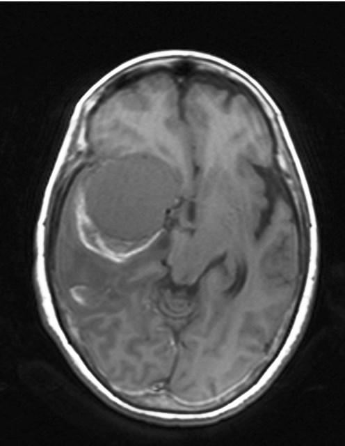
15 Fig10.C) Fig 10.D) DOI: / Page
 DOI: 10.")
16 Fig11.A) Fig 11.B) DOI: / Page
 DOI: 10.")
17 Fig12.A) Fig12..B) DOI: / Page
18 Fig 13.) LEGENDS: Fig 1 A & B: Contrast MR in axial & coronal planes show parasagittal meningiomas in two different cases. Fig 2 A & B: Contrast MR show lateral convexity meningiomas in two different cases Fig 3 A & B: contrast MR shows sphenoid wing meningioma Fig 4 A,B.C: Axial MR show meningioma in middle cranial fossa in different cases Fig 5 A & B: Meningioma at Sella turcica Fig 6 A & B: Peripherally calcified posterior fossa meningioma Fig 6 C: Left posterior fossa meningioma Fig 7 A & B: Left CP angle meningioma Fig 8 A & B: Meningioma from falx& tentorium Fig 9 A & B: Bifrontal Meningioma Fig 10. A& B: Parasagittal meningioma with peripheral hemorrhage Fig 10).C & D: Right middle cranial fossa Meningioma with intra tumoral hemorrhage & intracerebral hemorrhage Fig 11.) A &B : left parasagittal Recurrent Meningioma Fig 12 ) A & B: Left frontoparietal enplaque meningioma Fig 13) :Meningioma with cystic changes & hyperostosis Dr NLN Moorthy "MR imaging spectrum in intracranial meningiomas-a retrospective study." IOSR Journal of Dental and Medical Sciences (IOSR-JDMS), vol. 17, no. 3, 2018, pp DOI: / Page
Role of Diffusion weighted Imaging in the Evaluation of Intracranial Tumors
 IOSR Journal of Dental and Medical Sciences (IOSR-JDMS) e-issn: 2279-0853, p-issn: 2279-0861.Volume 15, Issue 12 Ver. IX (December. 2016), PP 99-104 www.iosrjournals.org Role of Diffusion weighted Imaging
Structural and functional imaging for the characterization of CNS lymphomas
 Structural and functional imaging for the characterization of CNS lymphomas Cristina Besada Introduction A few decades ago, Primary Central Nervous System Lymphoma (PCNSL) was considered as an extremely
Intracranial Lesions: MRI Signs for Localization
 Intracranial Lesions: MRI Signs for Localization Poster No.: C-1574 Congress: ECR 2017 Type: Educational Exhibit Authors: M. Cucos, A. Puiu, S. Manole ; Cluj-Napoca/RO, Cluj napoca/ RO Keywords: Cerebrospinal
MRI Findings Of An Atypical Cystic Meningioma A Rare Case
 ISPUB.COM The Internet Journal of Radiology Volume 14 Number 1 MRI Findings Of An Atypical Cystic Meningioma A Rare Case D Saxena, P Rout, K Pavan, B Philip Citation D Saxena, P Rout, K Pavan, B Philip.
CT & MRI Evaluation of Brain Tumour & Tumour like Conditions
 CT & MRI Evaluation of Brain Tumour & Tumour like Conditions Dr. Anjana Trivedi 1, Dr. Jay Thakkar 2, Dr. Maulik Jethva 3, Dr. Ishita Virda 4 1 M.D. Radiology, Professor and Head, P.D.U. Medical College
Dr. T. Venkat Kishan Asst. Prof Department of Radiodiagnosis
 Dr. T. Venkat Kishan Asst. Prof Department of Radiodiagnosis Schwannomas (also called neurinomas or neurilemmomas) constitute the most common primary cranial nerve tumors. They are benign slow-growing
IMAGING OF A CASE OF SPINAL MENINGIOMA- A CASE REPORT
 IMAGING OF A CASE OF SPINAL MENINGIOMA- A CASE REPORT Ramneet Wadi 1, Anil Kumar Shukla 2, Seetha Pramila V. V 3, Sabyasachi Basu 4, Sonam Sanjay 5 1Postgraduate Student, Department of Radiodiagnosis,
CNS TUMORS. D r. Ali Eltayb ( U. of Omdurman. I ). M. Path (U. of Alexandria)
. M. Path (U. of Alexandria)") CNS TUMORS D r. Ali Eltayb ( U. of Omdurman. I ). M. Path (U. of Alexandria) CNS TUMORS The annual incidence of intracranial tumors of the CNS ISmore than intraspinal tumors May be Primary or Secondary
CNS TUMORS D r. Ali Eltayb ( U. of Omdurman. I ). M. Path (U. of Alexandria) CNS TUMORS The annual incidence of intracranial tumors of the CNS ISmore than intraspinal tumors May be Primary or Secondary
Posterior fossa tumors: clues to differential diagnosis with case-based review
 Posterior fossa tumors: clues to differential diagnosis with case-based review Poster No.: C-0323 Congress: ECR 2017 Type: Educational Exhibit Authors: H. A. Aboughalia, M. Abdelhady; Doha/QA Keywords:
Meningioma tumor. Meningiomas are named according to their location (Fig. 1) and cause various symptoms: > 1
 and cause various symptoms: > 1") Meningioma tumor Overview A meningioma is a type of tumor that grows from the protective membranes, called meninges, which surround the brain and spinal cord. Most meningiomas are benign (not cancer) and
Meningioma tumor Overview A meningioma is a type of tumor that grows from the protective membranes, called meninges, which surround the brain and spinal cord. Most meningiomas are benign (not cancer) and
MRI imaging in meningeal diseases
 Original article MRI imaging in meningeal diseases 1Dr. Narendrakumar M Shah, 2 Dr Vaishali D M 1Associate professor, Department of Radiodiagnosis, SDM Medical college, Dharwad 2Consultant radiologist,
TABLES. Imaging Modalities Evidence Tables Table 1 Computed Tomography (CT) Imaging. Conclusions. Author (Year) Classification Process/Evid ence Class
 Imaging. Conclusions. Author (Year) Classification Process/Evid ence Class") TABLES Imaging Modalities Evidence Tables Table 1 Computed Tomography (CT) Imaging Author Clark (1986) 9 Reformatted sagittal images in the differential diagnosis meningiomas and adenomas with suprasellar
TABLES Imaging Modalities Evidence Tables Table 1 Computed Tomography (CT) Imaging Author Clark (1986) 9 Reformatted sagittal images in the differential diagnosis meningiomas and adenomas with suprasellar
RING ENCHANCING LESION BY M.S. HEMHNATH
 RING ENCHANCING LESION BY M.S. HEMHNATH A 21 YRS FEMALE CAME WITH H/O HEADACHE AND SEIZURE FOR THE PAST ONE MONTH. NO OTHER FOCAL NEUROLOGICAL DEFICIT. DIFFERENTIAL DIAGNOSIS For this case are Neurocysticerosis
General: Brain tumors are lesions that have mass effect distorting the normal tissue and often result in increased intracranial pressure.
 1 Lecture Objectives Know the histologic features of the most common tumors of the CNS. Know the differences in behavior of the different tumor types. Be aware of the treatment modalities in the various
Corresponding author - Dr.Krishnakumar M
 Chettinad Health City Medical Journal Original Article Karthikeyan KV*, Krishnakumar M**, Ramesh VG***, Siddarth G****, Jayendrapalan**** * Senior Consultant, Department of Neurosurgery, Chettinad Superspeciality
The Common Radiological Features of Meningiomas on CT scan and MRI among Patients at Major Hospitals in Eldoret, Kenya
 VOLUME 2 ISSUE 2 ISSN: 2320-4818 JOURNAL OF SCIENTIFIC & INNOVATIVE RESEARCH ORIGINAL RESEARCH ARTICLE The Common Radiological Features of Meningiomas on CT scan and MRI among Patients at Major Hospitals
The central nervous system
 Sectc.qxd 29/06/99 09:42 Page 81 Section C The central nervous system CNS haemorrhage Subarachnoid haemorrhage Cerebral infarction Brain atrophy Ring enhancing lesions MRI of the pituitary Multiple sclerosis
Pediatric Spine Tumors (and other masses)
") Pediatric Spine Tumors (and other masses) Francisco A Perez, MD, PhD Assistant Professor Neuroradiology and Pediatric Radiology Seattle Children s Hospital University of Washington, Seattle Commercial
Pediatric Spine Tumors (and other masses) Francisco A Perez, MD, PhD Assistant Professor Neuroradiology and Pediatric Radiology Seattle Children s Hospital University of Washington, Seattle Commercial
Year 2003 Paper two: Questions supplied by Tricia
 question 43 A 42-year-old man presents with a two-year history of increasing right facial numbness. He has a history of intermittent unsteadiness, mild hearing loss and vertigo but has otherwise been well.
Primary Meningeal Angiosarcoma in a Child: Imaging Findings
 Primary Meningeal Angiosarcoma in a Child: Imaging Findings Qamar malabeh MD*, Alaa abed MD*, Tamara obeidat MD*, Razan Abudalu MD*, Ola Alwaqfi MD** ABSTRACT Primary CNS angiosarcoma is an extremely rare
Tumors of the Central Nervous System
 Tumors of the Central Nervous System 1 Financial Disclosures I have NO SIGNIFICANT FINANCIAL, GENERAL, OR OBLIGATION INTERESTS TO REPORT Introduction General: Brain tumors are lesions that have mass effect
Variants of meningiomas: a review of imaging findings and clinical features
 DOI 10.1007/s11604-016-0550-6 REVIEW Variants of meningiomas: a review of imaging findings and clinical features Akira Kunimatsu 1 Natsuko Kunimatsu 2 Kouhei Kamiya 3 Masaki Katsura 3 Harushi Mori 1 Kuni
Pediatric CNS Tumors. Disclosures. Acknowledgements. Introduction. Introduction. Posterior Fossa Tumors. Whitney Finke, MD
 Pediatric CNS Tumors Disclosures Whitney Finke, MD Neuroradiology Fellow PGY-6 University of Utah Health Sciences Center Salt Lake City, Utah None Acknowledgements Introduction Nicholas A. Koontz, MD Luke
Dynamic MRI of meningiomas and schwannomas: is differential diagnosis possible?
 Neuroradiology (1997) 39: 633 638 Springer-Verlag 1997 DIAGNOSTIC NEURORADIOLOGY I. Ikushima Y. Korogi J. Kuratsu T. Hirai S. Hamatake M. Takahashi Y. Ushio Dynamic MRI of meningiomas and schwannomas:
Case Report Multiple Intracranial Meningiomas: A Review of the Literature and a Case Report
 Case Reports in Surgery Volume 2013, Article ID 131962, 4 pages http://dx.doi.org/10.1155/2013/131962 Case Report Multiple Intracranial Meningiomas: A Review of the Literature and a Case Report F. Koech,
Case Report Cystic Meningioma Simulating Arachnoid Cyst: Report of an Unusual Case
 Case Reports in Radiology, Article ID 371969, 4 pages http://dx.doi.org/10.1155/2014/371969 Case Report Cystic Meningioma Simulating Arachnoid Cyst: Report of an Unusual Case Docampo Jorge, 1 Gonzalez
PRINCESS MARGARET CANCER CENTRE CLINICAL PRACTICE GUIDELINES
 PRINCESS MARGARET CANCER CENTRE CLINICAL PRACTICE GUIDELINES CENTRAL NERVOUS SYSTEM MENINGIOMA CNS Site Group Meningioma Author: Dr. Norm Laperriere Date: February 20, 2018 1. INTRODUCTION 3 2. PREVENTION
JMSCR Vol 05 Issue 08 Page August 2017
 www.jmscr.igmpublication.org Impact Factor 5.84 Index Copernicus Value: 83.27 ISSN (e)-2347-176x ISSN (p) 2455-0450 DOI: https://dx.doi.org/10.18535/jmscr/v5i8.19 Magnetic Resonance Imaging in Evaluation
Role of imaging in RCC. Ultrasonography. Solid lesion. Cystic RCC. Solid RCC 31/08/60. From Diagnosis to Treatment: the Radiologist Perspective
 Role of imaging in RCC From Diagnosis to Treatment: the Radiologist Perspective Diagnosis Staging Follow up Imaging modalities Limitations and pitfalls Duangkamon Prapruttam, MD Department of Therapeutic
RADIOLOGY TEACHING CONFERENCE
 RADIOLOGY TEACHING CONFERENCE John Athas, MD Monica Tadros, MD Columbia University, College of Physicians & Surgeons Department of Otolaryngology- Head & Neck Surgery September 27, 2007 CT SCAN IMAGING
EXTRACRANIAL MENINGIOMA PRESENTING AS INFRATEMPORAL FOSSA MASS: A CASE SERIES
 Case Series EXTRACRANIAL MENINGIOMA PRESENTING AS INFRATEMPORAL FOSSA MASS: A CASE SERIES Sunil Mathew * 1, Reddy Ravikanth 2, Vijaykishan B 3. ABSTRACT Extradural meningioma occurs as extracranial extension
Meningeal thickening in MRI: from signs to etiologies
 Meningeal thickening in MRI: from signs to etiologies Poster No.: C-1979 Congress: ECR 2016 Type: Educational Exhibit Authors: A. Hssine, N. Mallat, M. Limeme, H. Zaghouani, S. Majdoub, H. Amara, D. Bakir,
Case 7391 Intraventricular Lesion
 Case 7391 Intraventricular Lesion Bastos Lima P1, Marques C1, Cabrita F2, Barbosa M2, Rebelo O3, Rio F1. 1Neuroradiology, 2Neurosurgery, 3Neuropathology, Coimbra University Hospitals, Portugal. University
An Introduction to Imaging the Brain. Dr Amy Davis
 An Introduction to Imaging the Brain Dr Amy Davis Common reasons for imaging: Clinical scenarios: - Trauma (NICE guidelines) - Stroke - Tumours - Seizure - Neurological degeneration memory, motor dysfunction,
Supratentorial multiple little meningiomas with transitory stroke symptoms like. MRI case presentation
 114 Romanian Neurosurgery (2010) XVII 1: 114-121 Supratentorial multiple little meningiomas with transitory stroke symptoms like. MRI case presentation E. Moldovanu 1,2, Adriana Moldovanu 1,2, Carmen Gherman
A Study of Meningiomas in Tertiary Care Center in South India.
 IOSR Journal of Dental and Medical Sciences (IOSR-JDMS) e-issn: 2279-0853, p-issn: 2279-0861.Volume 15, Issue 10 Ver. XII (October. 2016), PP 07-12 www.iosrjournals.org A Study of Meningiomas in Tertiary
Laura Tormoehlen, M.D. Neurology and EM-Toxicology Indiana University
 Laura Tormoehlen, M.D. Neurology and EM-Toxicology Indiana University Disclosures! No conflicts of interest to disclose Neuroimaging 101! Plain films! Computed tomography " Angiography " Perfusion! Magnetic
Cerebro-vascular stroke
 Cerebro-vascular stroke CT Terminology Hypodense lesion = lesion of lower density than the normal brain tissue Hyperdense lesion = lesion of higher density than normal brain tissue Isodense lesion = lesion
Masses of the Corpus Callosum
 Masses of the Corpus Callosum Kesav Raghavan, HMS Year III Dr. Agenda Corpus Callosum Development and Anatomy Our Patient: Clinical Presentation Differential Diagnosis of Masses in the Corpus Callosum
Metastasis. 57 year old with progressive Headache and Right Sided Visual Loss
 Metastasis 1% of sellar/parasellar masses Usually occurs with known primary Can involve third ventricle, hypothalamus, infundibular stalk May be both supra-, intrasellar 57 year old with progressive Headache
Essentials of Clinical MR, 2 nd edition. 73. Urinary Bladder and Male Pelvis
 73. Urinary Bladder and Male Pelvis Urinary bladder carcinoma is best locally staged with MRI. It is important however to note that a thickened wall (> 5 mm) is a non-specific finding seen in an underfilled
Radiological Appearance of Extra-axial CNS Hemangioma
 Chin J Radiol 2002; 27: 183-190 183 Radiological Appearance of Extra-axial CNS Hemangioma MING-SHIANG YANG CLAYTON CHI-CHANG CHEN WEN-HSIEN CHEN HAO-CHUN HUNG SAN-KAN LEE Department of Radiology, Taichung
Intracranial Hemangiopericytomas: MR and CT Features
 Intracranial Hemangiopericytomas: MR and CT Features Maria V. Chiechi, James G. Smirniotopoulos, and Hernando Mena PURPOSE: To describe the MR and CT imaging features of hemangiopericytoma and to identify
Benign brain lesions
 Benign brain lesions Diagnostic and Interventional Radiology Hung-Wen Kao Department of Radiology, Tri-Service General Hospital, National Defense Medical Center Computed tomography Hounsfield unit (HU)
Difficulties in diagnosis of intracranial meningiomas by computed tomography
 Journal of Neurology, Neurosurgery, and Psychiatry, 1980, 43, 1022-1029 Difficulties in diagnosis of intracranial meningiomas by computed tomography P PULLICINO, B E KENDALL, AND J JAKUBOWSKI From the
Diffusion-weighted imaging and ADC mapping in the differentiation of intraventricular brain tumors
 Diffusion-weighted imaging and ADC mapping in the differentiation of intraventricular brain tumors Poster No.: C-2652 Congress: ECR 2010 Type: Educational Exhibit Topic: Neuro Authors: M. Gavrilov, T.
Case Studies in the Skull Base
 Case Studies in the Skull Base Amy C Tsai, MD Neuroradiology Fellow Department of Radiology and Imaging Sciences University of Utah Health Sciences Center Salt Lake City, Utah, USA No disclosures related
Part II - Revising the sellar and parasellar region: differential diagnosis of a sellar region mass
 Part II - Revising the sellar and parasellar region: differential diagnosis of a sellar region mass Poster No.: C-1390 Congress: ECR 2015 Type: Educational Exhibit Authors: I. Candelaria, C. Figueira,
1/5 (ID:15UK )
") 1/5 (ID:15UK02000531) Introduction Osteochondromas are benign bone tumors of mesenchymal and non mesenchymal types (1). Their presentation as intracranial mass is a very rare phenomenon (2). The incidence
1/5 (ID:15UK02000531) Introduction Osteochondromas are benign bone tumors of mesenchymal and non mesenchymal types (1). Their presentation as intracranial mass is a very rare phenomenon (2). The incidence
Magnetic resonance imaging findings in 78 cases
 Arq Neuropsiquiatr 2007;65(3-A):610-614 Intracranial meningiomas Magnetic resonance imaging findings in 78 cases Emerson L. Gasparetto 1, Claudia da Costa Leite 1, Leandro T. Lucato 1, Cristiano Ventorin
Mesenchymal Chondrosarcoma at the Falx Cerebri
 Chin J Radiol 2004; 29: 337-341 337 Mesenchymal Chondrosarcoma at the Falx Cerebri YI-LUAN HUANG 1,3 PING-HONG LAI 1,3 SHONG-LING LIN 2,3 MEI-KANG YUAN 1,3 HUAY-BEN PAN 1,3 Department of Radiology 1, Department
Supratentorial Gangliocytoma Mimicking Extra-axial Tumor: A Report of Two Cases
 Supratentorial Gangliocytoma Mimicking Extra-axial Tumor: A Report of Two Cases Ho Sung Kim, MD 1 Ho Kyu Lee, MD 1 Ae Kyung Jeong, MD 1 Ji Hoon Shin, MD 1 Choong Gon Choi, MD 1 Shin Kwang Khang, MD 2 We
DISCLOSURES LEARNING OBJECTIVES WE WILL NOT DISCUSS. CSB: Birdseye View MESSAGE NAVIGATING THE SELLA AND CENTRAL SKULL BASE
 NAVIGATING THE SELLA AND CENTRAL SKULL BASE Christopher P. Hess, M.D., Ph.D. DISCLOSURES Research Support, General Electric SLIDES: http://www.radiology.ucsf.edu/research/meetings/rsna LEARNING OBJECTIVES
Meninges and Ventricles
 Meninges and Ventricles Irene Yu, class of 2019 LEARNING OBJECTIVES Describe the meningeal layers, the dural infolds, and the spaces they create. Name the contents of the subarachnoid space. Describe the
Original Research Article
 Original Research Article Study Determining Correlation between Histopathological Diagnosis and MRI Findings of Posterior Fossa Tumors Srinivasarao S. Gummadidala 1, B. Jyothi 2 1 Assistant Professor,
Index. aneurysm, 92 carotid occlusion, 94 ICA stenosis, 95 intracranial, 92 MCA, 94
 A ADC. See Apparent diffusion coefficient (ADC) Aneurysm cerebral artery aneurysm, 93 CT scan, 93 gadolinium, 93 Angiography, 13 Anoxic brain injury, 25 Apparent diffusion coefficient (ADC), 7 Arachnoid
MRI XR, CT, NM. Principal Modality (2): Case Report # 2. Date accepted: 15 March 2013
: Case Report # 2. Date accepted: 15 March 2013") Radiological Category: Musculoskeletal Principal Modality (1): Principal Modality (2): MRI XR, CT, NM Case Report # 2 Submitted by: Hannah Safia Elamir, D.O. Faculty reviewer: Naga R. Chinapuvvula, M.D.
Radiological Category: Musculoskeletal Principal Modality (1): Principal Modality (2): MRI XR, CT, NM Case Report # 2 Submitted by: Hannah Safia Elamir, D.O. Faculty reviewer: Naga R. Chinapuvvula, M.D.
Five Most Common Problems in Surgical Neuropathology
 Five Most Common Problems in Surgical Neuropathology If the brain were so simple that we could understand it, we would be so simple that we couldn t Emerson Pugh What is your greatest difficulty in neuropathology?
Imaging The Turkish Saddle. Russell Goodman, HMS III Dr. Gillian Lieberman
 Imaging The Turkish Saddle Russell Goodman, HMS III Dr. Gillian Lieberman Learning Objectives Review the anatomy of the sellar region Discuss the differential diagnosis of sellar masses Discuss typical
SWI including phase and magnitude images
 On-line Table: MRI imaging recommendation and summary of key features Sequence Pathologies Visible Key Features T1 volumetric high-resolution whole-brain reformatted in axial, coronal, and sagittal planes
NEURORADIOLOGY Part I
 NEURORADIOLOGY Part I Vörös Erika University of Szeged Department of Radiology SZEGED DISEASES OF CNS BRAIN Developmental anomalies Cerebrovascular disorders Tumours Inflammatory diseases Trauma DISEASES
Essentials of Clinical MR, 2 nd edition. 51. Primary Neoplasms
 51. Primary Neoplasms As with spinal central canal neoplasms in other regions, those of the lumbar spine may be classified as extradural, intradural extramedullary, and medullary. If an extradural lesion
Rare case of multiple meningiomas in nonneurofibromatosis
 Romanian Neurosurgery Volume XXXI Number 2 2017 April-June Article Rare case of multiple meningiomas in nonneurofibromatosis patient at unusual locations Vikrant Setia, Deepashu Sachdeva, Shrinivas Odugoudar,
Meningiomas are the second most common primary intracranial
 Published April 16, 2015 as 10.3174/ajnr.A4309 ORIGINAL RESEARCH ADULT BRAIN Chordoid Meningioma: Differentiating a Rare World Health Organization Grade II Tumor from Other Meningioma Histologic Subtypes
Tumors of the Nervous System
 Tumors of the Nervous System Peter Canoll MD. PhD. What I want to cover What are the most common types of brain tumors? Who gets them? How do they present? What do they look like? How do they behave? 1
PITUITARY PARASELLAR LESIONS. Kim Learned, MD
 PITUITARY PARASELLAR LESIONS Kim Learned, MD DIFFERENTIALS Pituitary Sella Clivus, Sphenoid Sinus Suprasellar Optic chiasm, Hypothalamus, Circle of Willis Parasellar Cavernous Sinus Case 1 17 YEAR-OLD
HEAD AND NECK IMAGING. James Chen (MS IV)
") HEAD AND NECK IMAGING James Chen (MS IV) Anatomy Course Johns Hopkins School of Medicine Sept. 27, 2011 OBJECTIVES Introduce cross sectional imaging of head and neck Computed tomography (CT) Review head
HEAD AND NECK IMAGING James Chen (MS IV) Anatomy Course Johns Hopkins School of Medicine Sept. 27, 2011 OBJECTIVES Introduce cross sectional imaging of head and neck Computed tomography (CT) Review head
Spectrum of Radiological Findings in Bronchogenic Carcinoma A Retrospective Study
 IOSR Journal of Dental and Medical Sciences (IOSR-JDMS) e-issn: 2279-0853, p-issn: 2279-0861.Volume 17, Issue 01 Ver. VIII January. (2018), PP 43-59 www.iosrjournals.org Spectrum of Radiological Findings
NEURORADIOLOGY-NEUROPATHOLOGY CONFERENCE
 THE UNIVERSITY OF NORTH CAROLINA at CHAPEL HILL SEPTEMBER 2013 NEURORADIOLOGY-NEUROPATHOLOGY CONFERENCE Claudia da Costa Leite, MD, PhD Thomas Bouldin, MD CASE 1 6 y-o female with headaches and vomiting
NEURORADIOLOGY Part I
 NEURORADIOLOGY Part I Vörös Erika University of Szeged Department of Radiology SZEGED BRAIN IMAGING METHODS Plain film radiography Ultrasonography (US) Computer tomography (CT) Magnetic resonance imaging
STROKE - IMAGING. Dr RAJASEKHAR REDDY 2nd Yr P.G. RADIODIAGNOSIS KIMS,Narkatpalli.
 STROKE - IMAGING Dr RAJASEKHAR REDDY 2nd Yr P.G. RADIODIAGNOSIS KIMS,Narkatpalli. STROKE Describes a clinical event that consists of sudden onset of neurological symptoms Types Infarction - occlusion of
Joana Ramalho, MD C. Ryan Miller, MD, PhD
 Joana Ramalho, MD C. Ryan Miller, MD, PhD Case 1 56 year old female Presented with: 3-4 weeks of visual symptoms (asymmetric vision loss, blurry & dark vision, photosensitivity & decreased peripheral vision)
1/9/2013 EXTRAMEDULLARY TUMORS OF THE PEDIATRIC SPINE. Introduction. Classification for Extramedullary Tumors
 EXTRAMEDULLARY TUMORS OF THE PEDIATRIC SPINE Eugene Wang 1/20/12 Dent Neurologic Institute Introduction 2/3 of all intraspinal tumors of childhood are extramedullary 50% Extradural 10-15% Intradural Back
Brain Space Occupying Lesions by Magnetic Resonance Imaging: A Prospective Study
 Original Article DOI: 10.17354/ijss/2015/523 Brain Space Occupying Lesions by Magnetic Resonance Imaging: A Prospective Study Bulabai Karpagam 1, V Vadanika 2 1 Associate Professor, Department of Radiology,
Clinics in diagnostic imaging (175)
") Singapore Med J 2017; 58(3): 121-125 doi: 10.11622/smedj.2017017 CMEArticle Clinics in diagnostic imaging (175) Vijay Krishnan 1, MD, FRCR, Tze Chwan Lim 1, MBBS, FRCR, Francis Cho Hao Ho 2, MBBS, FRANZCR,
Singapore Med J 2017; 58(3): 121-125 doi: 10.11622/smedj.2017017 CMEArticle Clinics in diagnostic imaging (175) Vijay Krishnan 1, MD, FRCR, Tze Chwan Lim 1, MBBS, FRCR, Francis Cho Hao Ho 2, MBBS, FRANZCR,
Cystic Meningioma in the Inter-Hemisferic Space Location
 Case Cystic Meningioma in the Inter-Hemisferic Space Location Muhammad Zafrullah Arifin, Firman Priguna Tjahjono, Agung Budi Sutiono, Ahmad Faried Department of Neurosurgery, Faculty of Medicine, Universitas
Pediatric Retroperitoneal Masses Radiologic-Pathologic Correlation
 Acta Radiológica Portuguesa, Vol.XVIII, nº 70, pág. 61-70, Abr.-Jun., 2006 Pediatric Retroperitoneal Masses Radiologic-Pathologic Correlation Marilyn J. Siegel Mallinckrodt Institute of Radiology, Washington
Oligodendroglioma: imaging findings, radio-pathological correlation and evolution
 Oligodendroglioma: imaging findings, radio-pathological correlation and evolution Poster No.: C-2104 Congress: ECR 2013 Type: Authors: Keywords: DOI: Scientific Exhibit A. Hernandez Castro, M. D. Monedero
Laurie A. Loevner, MD
 Laurie A. Loevner, MD Chief, Division of Neuroradiology UPHS Professor of Radiology, Otorhinolaryngology: Head & Neck Surgery, Neurosurgery, and Ophthalmology University of Pennsylvania Health System Disclosures
Cross sectional imaging of Intracranial cystic lesions Abdel Razek A
 Cross sectional imaging of Intracranial cystic lesions Abdel Razek A Department of Radiology. Mansoura Faculty of Medicine, Mansoura. Egypt. arazek@mans.edu.eg Introduction Intracranial cystic lesions
CTA/MRA of Pediatric Hepatic Masses Radiology-Pathology Correlation
 Acta Radiológica Portuguesa, Vol.XVIII, nº70, pág. 41-50, Abr.-Jun., 2006 CTA/MRA of Pediatric Hepatic Masses Radiology-Pathology Correlation Marilyn J. Siegel Mallinckrodt Institute of Radiology, Washington
Retroperitoneal Sarcomas - A pictorial review
 Retroperitoneal Sarcomas - A pictorial review Poster No.: C-1409 Congress: ECR 2013 Type: Educational Exhibit Authors: D. Douraghi-Zadeh, K. L. Shahabuddin, R. H. Thomas, E. Moskovic; London/UK Keywords:
Pure Intracavernous Sinus Epidermoid Cyst: Diffusion-Weighted (DW) and Constructive Interference in Steady State (CISS) Images 1
 and Constructive Interference in Steady State (CISS) Images 1") Pure Intracavernous Sinus Epidermoid Cyst: Diffusion-Weighted (DW) and Constructive Interference in Steady State (CISS) Images 1 Suk Jin Park, M.D., In Kyu Yu, M.D., Min Sun Kim, M.D., Hyeon Mi Yoo, M.D.,
Pure Intracavernous Sinus Epidermoid Cyst: Diffusion-Weighted (DW) and Constructive Interference in Steady State (CISS) Images 1 Suk Jin Park, M.D., In Kyu Yu, M.D., Min Sun Kim, M.D., Hyeon Mi Yoo, M.D.,
Case 8 Soft tissue swelling
 Case 8 Soft tissue swelling 26-year-old female presented with a swelling on the back of the left knee joint since the last 6 months and chronic pain in the calf and foot since the last 2 months. Pain in
Spinal meningioma imaging
 Spinal meningioma imaging Poster No.: C-0448 Congress: ECR 2018 Type: Educational Exhibit Authors: M. Smoljan, D. Zadravec ; Zagreb/HR, Zageb/HR Keywords: Neoplasia, Imaging sequences, Education, MR, CT,
Peter Canoll MD. PhD.
 Tumors of the Nervous System Peter Canoll MD. PhD. What I want to cover What are the most common types of brain tumors? Who gets them? How do they ypresent? What do they look like? How do they behave?
Diverse Imaging Appearances and Locations of Meningioma.
 Diverse Imaging Appearances and Locations of Meningioma. Poster No.: C-1616 Congress: ECR 2013 Type: Educational Exhibit Authors: L. Fatahi Bandpey, P. J. Sanchez Santos, D. yago, F. O. 1 1 1 1 1 2 Lenghel,
RADIOANATOMY OF SELLA TURCICA
 RADIOANATOMY OF SELLA TURCICA O.BAKKACHA, H.MALAJATI, M.RHISSASSI, H. BENCHAABOUNE, N.CHAKIR, My R. EL HASSANI,M.JIDDANE Department of Neuroradiology specialties Hospital. Rabat Objective: New imaging
Brain Meninges, Ventricles and CSF
 Brain Meninges, Ventricles and CSF Lecture Objectives Describe the arrangement of the meninges and their relationship to brain and spinal cord. Explain the occurrence of epidural, subdural and subarachnoid
Astroblastoma: Radiologic-Pathologic Correlation and Distinction from Ependymoma
 AJNR Am J Neuroradiol 23:243 247, February 2002 Case Report Astroblastoma: Radiologic-Pathologic Correlation and Distinction from Ependymoma John D. Port, Daniel J. Brat, Peter C. Burger, and Martin G.
A CASE OF A Huge Submandibular Pleomorphic Adenoma
 ISPUB.COM The Internet Journal of Head and Neck Surgery Volume 4 Number 2 S VERMA Citation S VERMA.. The Internet Journal of Head and Neck Surgery. 2009 Volume 4 Number 2. Abstract Pleomorphic adenoma
Case Report Intracranial Capillary Hemangioma in the Posterior Fossa of an Adult Male
 Case Reports in Radiology Volume 2016, Article ID 6434623, 4 pages http://dx.doi.org/10.1155/2016/6434623 Case Report Intracranial Capillary Hemangioma in the Posterior Fossa of an Adult Male Jordan Nepute,
Rapid recurrence of a malignant meningioma: case report
 Romanian Neurosurgery Volume XXXI Number 2 2017 April-June Article Rapid recurrence of a malignant meningioma: case report Oguz Baran, Sima Sayyahmeli, Taner Tanriverdi, Pamir Erdincler TURKEY DOI: 10.1515/romneu-2017-0027
International Journal of Case Reports and Images (IJCRI)
") www.edoriumjournals.com CLINICAL IMAGE PEER REVIEWED OPEN ACCESS Porencephalic cyst Mugtaba Alghazali, Ikhlas Abdelaziz, Hatim Zain Alabdeen ABSTRACT Abstract is not required for Clinical Images International
www.edoriumjournals.com CLINICAL IMAGE PEER REVIEWED OPEN ACCESS Porencephalic cyst Mugtaba Alghazali, Ikhlas Abdelaziz, Hatim Zain Alabdeen ABSTRACT Abstract is not required for Clinical Images International
SKULL BASE LESIONS THAT MAY MIMICK DISEASE
 SKULL BASE LESIONS THAT MAY MIMICK DISEASE AUTHORS: MYERS, TANDBERG, LORENZO UNIVERSITY OF NEW MEXICO DIAGNOSTIC RADIOLOGY Learning Objectives The participant will identify normal anatomic variants that
Meningioma or meningioma-like mass - from the imaging signs to the diagnosis
 Meningioma or meningioma-like mass - from the imaging signs to the diagnosis Poster No.: C-2387 Congress: ECR 2017 Type: Educational Exhibit Authors: E. M. Preda, I. G. Lupescu; Bucharest/RO Keywords:
J of Evolution of Med and Dent Sci/ eissn , pissn / Vol. 3/ Issue 46/Sep 22, 2014 Page 11296
 CT SPECTRUM OF GIANT RETROPERITONEAL LIPOSARCOMAS WITH HISTOPATHOLOGICAL CORRELATION Shashikumar M. R 1, Rajendra Kumar N. L 2, C. P. Nanjaraj 3, Nishanth R. K 4, Vishwanath Joshi 5 HOW TO CITE THIS ARTICLE:
CNS Imaging. Dr Amir Monir, MD. Lecturer of radiodiagnosis.
 CNS Imaging Dr Amir Monir, MD Lecturer of radiodiagnosis www.dramir.net Types of radiological examinations you know Plain X ray X ray with contrast GIT : barium (swallow, meal, follow through, enema) ERCP
THE ROLE OF IMAGING IN DIAGNOSIS OF SUBDURAL HEMATOMA: REVIEW ARTICLE
 THE ROLE OF IMAGING IN DIAGNOSIS OF SUBDURAL HEMATOMA: REVIEW ARTICLE * Dr. Sumendra Raj Pandey, Prof. Dr. Liu Pei WU, Dr. Sohan Kumar Sah, Dr. Lalu Yadav, Md. Sadam Husen Haque and Rajan KR. Chaurasiya
RINGS N THINGS: Imaging Patterns in Differential Diagnosis. Anne G. Osborn, M.D.
 RINGS N THINGS: Imaging Patterns in Differential Diagnosis Anne G. Osborn, M.D. ExpDDxs: Intra-axial (Parenchymal) Lesions Ring-enhancing lesions, solitary 1 Ring-enhancing lesion crossing corpus callosum