RING ENCHANCING LESION BY M.S. HEMHNATH
|
|
|
- Carmella Little
- 6 years ago
- Views:
Transcription

1 RING ENCHANCING LESION BY M.S. HEMHNATH
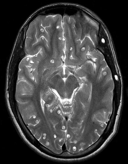
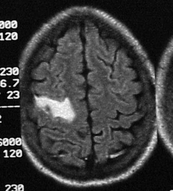
2 A 21 YRS FEMALE CAME WITH H/O HEADACHE AND SEIZURE FOR THE PAST ONE MONTH. NO OTHER FOCAL NEUROLOGICAL DEFICIT.
3 DIFFERENTIAL DIAGNOSIS For this case are Neurocysticerosis tuberculoma The dd For other ring enchancing lesion are toxoplasmosis Primary cns lymphoma Abcess Primary tumour Metastases Resolving infarct and haemotoma
4

5
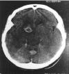
6 CECT OF TUBERCULOMA
7 CECT OF TUBERCULOMA

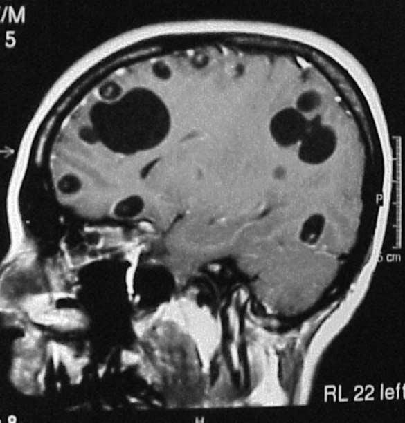
8 MRI T1 OF TUBERCULOMA Shows thick rim of slightly hyperintense with central isointense partially surrounded by aslightly hypointense rim.

9 MRI T2 OF TUBERCULOMA Shows a round slightly hetergenous isointense and hypointense mass adjcent multiple small conglomerate hypointense nodules. Diffuse surrounding edema seen.

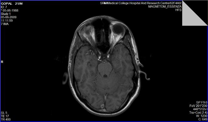
10 MRI T1(CONSTRAT) OF TUBERCULOMA shows a ring shaped homogenous enchacement and adjcent conglomerate nodules.
11 TUBERCULOMA IMMATURE STAGE NECT iso or hyperdense CECT shows ring or nodular irregular enchancement. MATURE STAGE NECT well delineated round or oval lesion Mass effect Moderate surrounding edema Central calcification.
12 TUBERCULOMA CECT homogenous enhacement ring blush with smooth or slightly shaggy margin and thick irregular wall around an isodense centre. Target sign central calcification in isodense lesion and ring blush. Homogenous blush enplaque along the dural plane.
13 TUBERCULOMA MRI T1 isointense to brain Marked enchancement on t1 T2 central hyperintense with hypointense rim on t2 NON CAESATING GRANULOMA hypointense to brain on t1 Hyperintense on t2 Homogenous nodular enhacement. CAESATING GRANULOMA ISOINTENSE TO MARKEDELY HYPERINTENSE ON T2

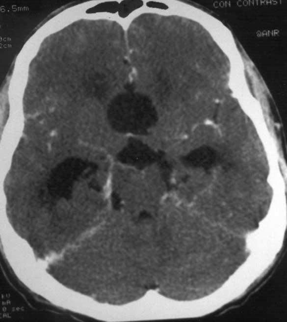
14 NECT OF BRAIN ABCESS (EARLY CEREBITIS ) There is a poorly ill defined area in left parietal lobe surrounding edema.


15 MRI T2 BRAIN ABCESS There is a well defined thin walled mass is noted in right frontal region. Surrounding edema and mass effect is noted.


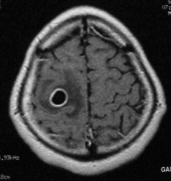
16 MRI T1 CONSTRAT ABCESS There is awell defined ring enchaning lesion is noted on right parietal region. Surrounding edema is present.


17 MRI OF T1 CONSTRAT OF ABCESS there is well defined ring enchasing mass is seen in the cp angle.


18 DIFFUSION WEIGHED IMAGE OF ABCESS There is markedly increased in intensity from the mass
19 BRAIN ABCESS (EARLY CEREBRITIS) NECT normal or show only a poorly marginated hypodense area. Gas within the lesion is a diagnostic of gas forming organism. CECT shows an illdefined constrat enchancing area with edematous region. MRI a ill defined hyperintense zone to be noted on T2. Constrat t1 poorly delineated enchancing area with iso to hypodense edematous region.
20 ABCESS ( LATE CEREBRITIS ) SHOWS IRREGULAR ENCHANCING RIM THAT SURROUNDS CENTRAL LOW DENSITY AREA IS TYPICAL. DELAYED SHOWS CONSTRAT FILLS THE CENTRAL AREA. PERIPERAL EDEMA. MASS EFFECT SUCLI OBLIERTATION APPERANT CENTRAL NECROTIC AREA IS TYPICAL HYPERINTENSE TO BRAIN ON T2 AND PROTON. THICK RIM APPEARS ISO TO HYPOINTENSE. THICK RIM APPEARS ISO TO HYPERINTENSE ON T1.
21 ABCESS ( EARLY & LATE CAPSULE 0N CT) EARLY CAPSULE A WELL DEFINED CAPSULE THAT ENCHANCE STRONGLY AND UNIFORMLY. WALL THICKEST NEAR THE CORTEX AND THINNER NEAR EPENDYMAL. MODERATE VASOGENIC EDEMA. LATE CAPSULE RIM ENCHACEMENT MAY PERSIST EDEMA SHRINKS MULTILOCULATION AND SUBADJCENT DAUGHTER ABCESS IN WHITE MATTER.
22 ABCESS ( EARLY AND LATE ON MRI) CAPSULE ENCHANCE ON T1. THIN WALLED HYPOINTENSE RIM ON T2.

23 MRI OF TOXOPLASMOSIS Constrat enchaced T1 weighed images showing well defined ring enchaning lesion is seen


24 PLAIN MRI T1 OF TOXOPLASMOSIS It shows a well defined hypointense mass in the right parieatal region. The mass compressing the right lateral ventricle.


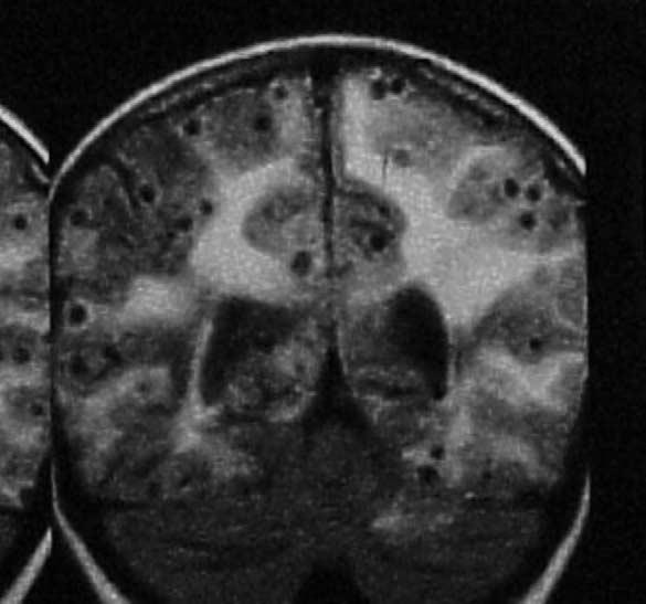
25 MRI T2 OF TOXOPLASMOSIS Shows large bilateral hyperintense lesion placed on either side of third ventricle and lateral ventricle. There is slight mass effect over the right ventricle.
26 toxoplasmosis CECT shows muliple or solitary ring enchancing masses wiith periperhal edema (target sign ). MRI t1 iso to hypointense t2 iso to hyperintense Restricted diffusion signal is high. Calcification and haemorrhage can be demonstrated.


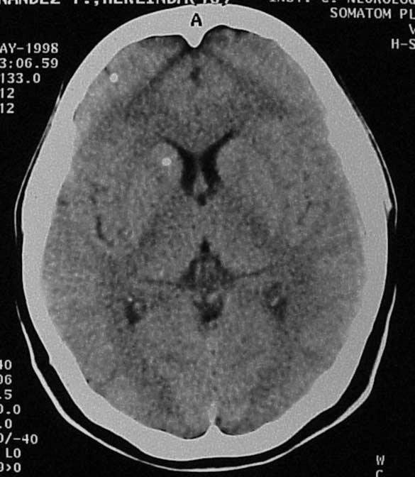
27 NECT OF LYMPHOMA There is a hyper dense mass in right parietal lobe surrounded by low density zone with vasogenic edema.

28 NECT OF LYMPHOMA There is a lobulated mass which is hyper attenuating the mass is extending across the splenium of corpus callosum.

29 CECT OF LYMPHOMA There is a homogenous enchacement of mass with surrounding edema.


30 MRI T2 OF LYMPHOMA There is a heterogenous mass with low signal intensity. Central linear hyper intensity suggesting necrosis. There is also vasogenic edema.

31 MRI (CONSRAT T1 ) 0F LYMPHOMA There is homogenous enchacement of the mass. There is also mass effect over the ventricle.

32 MRI T2 LYMPHOMA There is nodular well defined right frontal sub cortical lesion with central hypointense core.

33 MRI (CONSTRAT T1 ) OF LYMPHOMA There is enchanced soliatary ring like enchacement of mass and peripheral low signal intensity is seen.
34 LYMPHOMA CT NECT iso to hyperdense Little mass effect with paucity of peritumoural edema. CECT homogenous dense and well defined to irregular constrast enchancement. Thick walled ring enchancement
35 lymphoma MRI Well demarcated round/ oval/ gyral shaped mass. Relatively little mass effect. T1 iso/hypointense ring shaped enchancement. T2 iso/ hyperintense Ring pattern central necrosis with densely cellular rim seen hyperintense and edema.
36 lymphoma angio Avascular mass with neovascularity. Focal blusih in late arterial to capillary phase peristing well into the venous phase. Arterial encasement. Dilated deep medullary vien.


37 BRAIN METASTASES A WELL DEFINED RIM ENCHACEMENT IS SEEN SURROUNDING EDEMA.
38 CT METASTASES NECT mostly isodense to brain some will be hyperdense seen eg small round cell tumour and neoplasm with high nuclear and cystoplasmic ratio. Edema will be present. Calcification and cystic changes are rare. Haemorrhage also will be seen. CECT solid and ring enchance pattern are seen but not all metastases enchance strongly
39 METASTASES MRI SIGNAL INTENSITY VARIES T1 some non haemorrahgic metastases are hypointense to brain. some non haemorrahgic metastases are hyper intense to brain (melanoma ). T2 hyperintense (punctate high signal foci ) hypointense ( gastrointenstial adeno ca ). Constrat all metastases do not enchance. Solid rim and mixed enchancement pattern. High dose mostly useful.
40 CT Primary tumour astrocytoma NECT inhomogenous mixed density little discernible mass effect. Intra tumoural heamorrhage. Calcification is uncommon until malignat degeneration. CECT enchance strongly but non uniformly. Irregular rim enchancement.
41 MRI ASTROCYTOMA Poorly delineated lesion that have heterogenous signal intensties on t1&t2. Mixed iso to hypodense area are seen on t1. Central core of hyperintensity surrounded by an isointense rim with peripheral finger like high intensity projection secondary to vasogenic edema. CONSTRAT irregular peripheral rim like enchancement and edema surrounds mass.

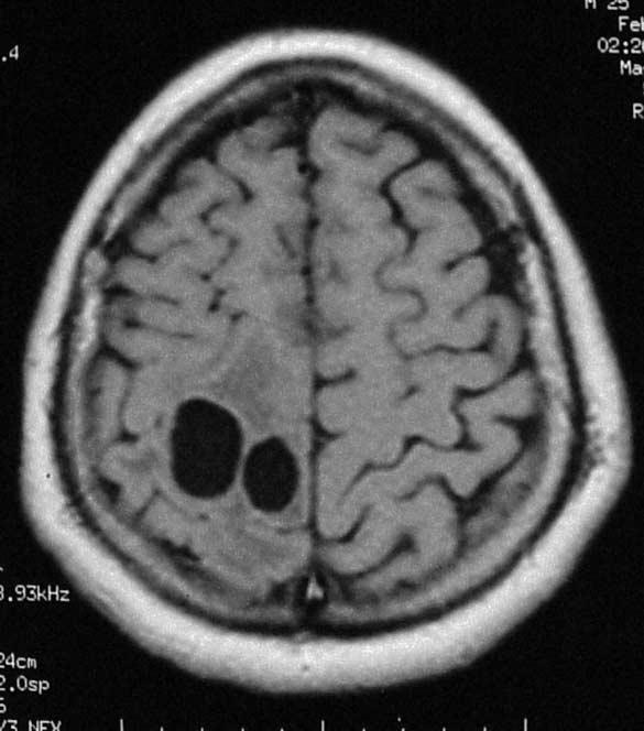
42 Vesicular stage of neurocysticerosis cect and plain mri

43 MASSIVE NEUROCYSTICEROSIS

44 COLLIDAL VESICULAR STAGE

45 CYSTICEROSIS WITH ENCEPHALITIS

46 CALCIFIED NEUROCYSTICERSOSIS ON CT WITHOUT EDEMA AND MRI WITH EDEMA

47 SUB ARCHNOID CYSTICEROSIS


48 VENTRICULAR NEUROCYSTICEROSIS It may produce obstructive hydrocephalus caused by blockage with various portion of ventricular systeam.
49 Vesicular stage Larvum appears as round csf like cyst with a mural nodule ( scolex ). Edema and enchancement are rare during this stage.
50 Collidal vesicular stage Ct During this stage ring enchancement is seen. Edema will be present. mri Cyst fluid will be hyperintense to csf. Ring capsule enchancing on t1. Hypointense mural nodule with strong homogenous enchancement on t2.
51 Granular nodular stage CT Nect isodense cyst and with a hyper dense calcified scolex. Edema will be present. Cect ring enchancement will persist. MRI T1 isointense to brain T2 hypointense Constrat nodular or ring enchancement.
52 Nodular calicifed stage CT NECT small calcified nodule without mass effect. CECT typical enchancement Bull eye appearance Rice like muscle calcification.
RINGS N THINGS: Imaging Patterns in Differential Diagnosis. Anne G. Osborn, M.D.
 RINGS N THINGS: Imaging Patterns in Differential Diagnosis Anne G. Osborn, M.D. ExpDDxs: Intra-axial (Parenchymal) Lesions Ring-enhancing lesions, solitary 1 Ring-enhancing lesion crossing corpus callosum
Cerebro-vascular stroke
 Cerebro-vascular stroke CT Terminology Hypodense lesion = lesion of lower density than the normal brain tissue Hyperdense lesion = lesion of higher density than normal brain tissue Isodense lesion = lesion
Masses of the Corpus Callosum
 Masses of the Corpus Callosum Kesav Raghavan, HMS Year III Dr. Agenda Corpus Callosum Development and Anatomy Our Patient: Clinical Presentation Differential Diagnosis of Masses in the Corpus Callosum
Disclosure. + Outline. Case-based approach to neurological emergencies that might present to the ED
 Kathleen R. Fink, MD University of Washington 5 th Nordic Emergency Radiology Course May 21, 2015 Disclosure My spouse receives research salary support from: Bracco BayerHealthcare Guerbet Outline Case-based
Role of Diffusion weighted Imaging in the Evaluation of Intracranial Tumors
 IOSR Journal of Dental and Medical Sciences (IOSR-JDMS) e-issn: 2279-0853, p-issn: 2279-0861.Volume 15, Issue 12 Ver. IX (December. 2016), PP 99-104 www.iosrjournals.org Role of Diffusion weighted Imaging
Interesting Cases from Liver Tumor Board. Jeffrey C. Weinreb, M.D.,FACR Yale University School of Medicine
 Interesting Cases from Liver Tumor Board Jeffrey C. Weinreb, M.D.,FACR Yale University School of Medicine jeffrey.weinreb@yale.edu Common Liver Diseases Hemangioma Cyst FNH Focal Fat/Sparing THID Non-Cirrhotic
CNS TUMORS. D r. Ali Eltayb ( U. of Omdurman. I ). M. Path (U. of Alexandria)
. M. Path (U. of Alexandria)") CNS TUMORS D r. Ali Eltayb ( U. of Omdurman. I ). M. Path (U. of Alexandria) CNS TUMORS The annual incidence of intracranial tumors of the CNS ISmore than intraspinal tumors May be Primary or Secondary
CNS TUMORS D r. Ali Eltayb ( U. of Omdurman. I ). M. Path (U. of Alexandria) CNS TUMORS The annual incidence of intracranial tumors of the CNS ISmore than intraspinal tumors May be Primary or Secondary
HEPATO-BILIARY IMAGING
 HEPATO-BILIARY IMAGING BY MAMDOUH MAHFOUZ MD PROF.OF RADIOLOGY CAIRO UNIVERSITY mamdouh.m5@gmail.com www.ssregypt.com CT ABDOMEN Indications Patient preparation Patient position Scanogram Fasting 4-6 hours
Renal masses - the role of diagnostic imaging
 Renal masses - the role of diagnostic imaging Poster No.: C-2471 Congress: ECR 2015 Type: Educational Exhibit Authors: V. Rai#; Bjelovar/HR Keywords: Cysts, Cancer, Structured reporting, Ultrasound, MR,
Pathologic Analysis of CNS Surgical Specimens
 2015 Kenneth M. Earle Memorial Neuropathology Review Pathologic Analysis of CNS Surgical Specimens Peter C. Burger, MD Interdisciplinary Quality Control Familiarity with entities Use of diagnostic algorithm
ISCHEMIC STROKE IMAGING
 ISCHEMIC STROKE IMAGING ผศ.พญ พญ.จ ร ร ตน ธรรมโรจน ภาคว ชาร งส ว ทยา คณะแพทยศาสตร มหาว ทยาล ยขอนแก น A case of acute hemiplegia Which side is the abnormality, right or left? Early Right MCA infarction
SWI including phase and magnitude images
 On-line Table: MRI imaging recommendation and summary of key features Sequence Pathologies Visible Key Features T1 volumetric high-resolution whole-brain reformatted in axial, coronal, and sagittal planes
NEURORADIOLOGY Part I
 NEURORADIOLOGY Part I Vörös Erika University of Szeged Department of Radiology SZEGED DISEASES OF CNS BRAIN Developmental anomalies Cerebrovascular disorders Tumours Inflammatory diseases Trauma DISEASES
Essentials of Clinical MR, 2 nd edition. 51. Primary Neoplasms
 51. Primary Neoplasms As with spinal central canal neoplasms in other regions, those of the lumbar spine may be classified as extradural, intradural extramedullary, and medullary. If an extradural lesion
Astroblastoma: Radiologic-Pathologic Correlation and Distinction from Ependymoma
 AJNR Am J Neuroradiol 23:243 247, February 2002 Case Report Astroblastoma: Radiologic-Pathologic Correlation and Distinction from Ependymoma John D. Port, Daniel J. Brat, Peter C. Burger, and Martin G.
Imaging in breast cancer. Mammography and Ultrasound Donya Farrokh.MD Radiologist Mashhad University of Medical Since
 Imaging in breast cancer Mammography and Ultrasound Donya Farrokh.MD Radiologist Mashhad University of Medical Since A mammogram report is a key component of the breast cancer diagnostic process. A mammogram
Metastasis. 57 year old with progressive Headache and Right Sided Visual Loss
 Metastasis 1% of sellar/parasellar masses Usually occurs with known primary Can involve third ventricle, hypothalamus, infundibular stalk May be both supra-, intrasellar 57 year old with progressive Headache
Cross sectional imaging of Intracranial cystic lesions Abdel Razek A
 Cross sectional imaging of Intracranial cystic lesions Abdel Razek A Department of Radiology. Mansoura Faculty of Medicine, Mansoura. Egypt. arazek@mans.edu.eg Introduction Intracranial cystic lesions
General Identification. Name: 江 X X Age: 29 y/o Gender: Male Height:172cm, Weight: 65kg Date of admission:95/09/27
 General Identification Name: 江 X X Age: 29 y/o Gender: Male Height:172cm, Weight: 65kg Date of admission:95/09/27 Chief Complaint Sudden onset of seizure for several minutes Present illness This 29-year
of Thyroid Lesions Comet Tail Crystals
 2 Ultrasound Features of Thyroid Lesions There are many different features indicating a certain benign or malignant tumor type, but many of these are overlapping signs. Combining several features is considered
CT 101 :Pancreas and Spleen
 CT 101 :Pancreas and Spleen Shikha Khullar,, MD, MPH Division of Radiology University of South Alabama The Pancreas Normal Pancreas 3 Phase Pancreatic CT Non contrast Arterial phase : 30-35 35 second
How to Analyse Difficult Chest CT
 How to Analyse Difficult Chest CT Complex diseases are:- - Large lesion - Unusual or atypical pattern - Multiple discordant findings Diffuse diseases are:- - Numerous findings in both sides 3 basic steps
Pediatric Retroperitoneal Masses Radiologic-Pathologic Correlation
 Acta Radiológica Portuguesa, Vol.XVIII, nº 70, pág. 61-70, Abr.-Jun., 2006 Pediatric Retroperitoneal Masses Radiologic-Pathologic Correlation Marilyn J. Siegel Mallinckrodt Institute of Radiology, Washington
Kathleen R. Fink, MD Virginia Mason Medical Center. 6 th Nordic Emergency Radiology Course 2017
 Kathleen R. Fink, MD Virginia Mason Medical Center 6 th Nordic Emergency Radiology Course 2017 Disclosure My spouse receives research salary support from: Guerbet Outline Indications for imaging CNS infections
For Emergency Doctors. Dr Suzanne Smallbane November 2011
 For Emergency Doctors Dr Suzanne Smallbane November 2011 A: Orbit B: Sphenoid Sinus C: Temporal Lobe D: EAC E: Mastoid air cells F: Cerebellar hemisphere A: Frontal lobe B: Frontal bone C: Dorsum sellae
An Introduction to Imaging the Brain. Dr Amy Davis
 An Introduction to Imaging the Brain Dr Amy Davis Common reasons for imaging: Clinical scenarios: - Trauma (NICE guidelines) - Stroke - Tumours - Seizure - Neurological degeneration memory, motor dysfunction,
X-Ray Corner. Imaging Approach to Cystic Liver Lesions. Pantongrag-Brown L. Solitary cystic liver lesions. Hepatic simple cyst (Figure 1)
") THAI J 136 Imaging Approach to Cystic Liver Lesions GASTROENTEROL 2013 X-Ray Corner Imaging Approach to Cystic Liver Lesions Pantongrag-Brown L Cystic liver lesions are common findings in daily practice
THAI J 136 Imaging Approach to Cystic Liver Lesions GASTROENTEROL 2013 X-Ray Corner Imaging Approach to Cystic Liver Lesions Pantongrag-Brown L Cystic liver lesions are common findings in daily practice
Key words: Brain, Cysticercosis, Neurocysticercosis, Parasites, Spinal Cord.
 JOURNAL OF CASE REPORTS 2015;5(2):438-442 Disseminated Spinal Cysticercosis: A Rare Intramedullary Ring Enhancing Lesion Avanti Gulhane, Sushil Kumar Kale Department of Radio-Diagnosis, Mahatma Gandhi
Brain Pain Infections of the CNS
 FRIDAY, OCTOBER 28, 2016 Brain Pain Infections of the CNS Suyash Mohan MD, PDCC Assistant Professor of Radiology & Neurosurgery Division of Neuroradiology, Department of Radiology Perelman School of Medicine
An Approach. to Brain. Infection. 37F found down. Disclosures. Approach to CNS Infection. Objectives. Parenchymal. None.
 An Approach Disclosures to Brain None. Infection Jason Shewchuk, MD Clinical Associate Professor Head of Neuroradiology UBC European Course in Neuroradiology 2018 Objectives Following this session the
Oligodendroglioma: imaging findings, radio-pathological correlation and evolution
 Oligodendroglioma: imaging findings, radio-pathological correlation and evolution Poster No.: C-2104 Congress: ECR 2013 Type: Authors: Keywords: DOI: Scientific Exhibit A. Hernandez Castro, M. D. Monedero
JMSCR Vol 04 Issue 12 Page December 2016
 www.jmscr.igmpublication.org Impact Factor 5.244 Index Copernicus Value: 83.27 ISSN (e)-2347-176x ISSN (p) 2455-0450 DOI: https://dx.doi.org/10.18535/jmscr/v4i12.113 Role of Computed Tomography in Evaluation
Vascular Malformations of the Brain: A Review of Imaging Features and Risks
 Vascular Malformations of the Brain: A Review of Imaging Features and Risks Comprehensive Neuroradiology: Best Practices October 27-30, 2016 Sudhakar R. Satti, MD Associate Director Neurointerventional
CT & MRI Evaluation of Brain Tumour & Tumour like Conditions
 CT & MRI Evaluation of Brain Tumour & Tumour like Conditions Dr. Anjana Trivedi 1, Dr. Jay Thakkar 2, Dr. Maulik Jethva 3, Dr. Ishita Virda 4 1 M.D. Radiology, Professor and Head, P.D.U. Medical College
The central nervous system
 Sectc.qxd 29/06/99 09:42 Page 81 Section C The central nervous system CNS haemorrhage Subarachnoid haemorrhage Cerebral infarction Brain atrophy Ring enhancing lesions MRI of the pituitary Multiple sclerosis
Case 7391 Intraventricular Lesion
 Case 7391 Intraventricular Lesion Bastos Lima P1, Marques C1, Cabrita F2, Barbosa M2, Rebelo O3, Rio F1. 1Neuroradiology, 2Neurosurgery, 3Neuropathology, Coimbra University Hospitals, Portugal. University
NEURO IMAGING 2. Dr. Said Huwaijah Chairman of radiology Dep, Damascus Univercity
 NEURO IMAGING 2 Dr. Said Huwaijah Chairman of radiology Dep, Damascus Univercity I. EPIDURAL HEMATOMA (EDH) LOCATION Seventy to seventy-five percent occur in temporoparietal region. CAUSE Most likely caused
The Many Faces of Central Nervous System Tuberculosis
 The Many Faces of Central Nervous System Tuberculosis Poster No.: C-2347 Congress: ECR 2013 Type: Educational Exhibit Authors: B. Alami, F. Belhoussine, O. Addou, M. Y. Alaoui Lamrani, M. 1 1 1 1 1 1 2
Clinics in diagnostic imaging (175)
") Singapore Med J 2017; 58(3): 121-125 doi: 10.11622/smedj.2017017 CMEArticle Clinics in diagnostic imaging (175) Vijay Krishnan 1, MD, FRCR, Tze Chwan Lim 1, MBBS, FRCR, Francis Cho Hao Ho 2, MBBS, FRANZCR,
Singapore Med J 2017; 58(3): 121-125 doi: 10.11622/smedj.2017017 CMEArticle Clinics in diagnostic imaging (175) Vijay Krishnan 1, MD, FRCR, Tze Chwan Lim 1, MBBS, FRCR, Francis Cho Hao Ho 2, MBBS, FRANZCR,
Essentials of Clinical MR, 2 nd edition. 65. Benign Hepatic Masses
 65. Benign Hepatic Masses Pulse sequences acquired for abdominal MRI typically consist of fast acquisition schemes such as single-shot turbo spin echo (i.e. HASTE) and gradient echo schemes such as FLASH
Attenuation value in HU From -500 To HU From -10 To HU From 60 To 90 HU. From 200 HU and above
 Brain Imaging Common CT attenuation values Structure Air Fat Water Brain tissue Recent hematoma Calcifications Bone Brain edema and infarction Normal liver parenchyma Attenuation value in HU From -500
41 year old female with headache. Elena G. Violari MD and Leo Wolansky MD
 41 year old female with headache Elena G. Violari MD and Leo Wolansky MD ? Dural Venous Sinus Thrombosis with Hemorrhagic Venous Infarct Acute intraparenchymal hematoma measuring ~3 cm in diameter centered
Brain Tumors. Medulloblastoma. Pilocytic astrocytoma: Ahmed Koriesh, MD. Pathological finding
 NeuroPathology Page 8 Brain Tumors Pathological finding Pseudorosette Rosenthal fibers Rosettes Wet Keratin Psammoma bodies Fried egg Tumor Ependymoma, SEGA Pilocytic astrocytoma Medulloblastoma Craniopharyngioma
Discovering the hippocampus with cranial-ct.
 Discovering the hippocampus with cranial-ct. Poster No.: C-0378 Congress: ECR 2018 Type: Educational Exhibit Authors: F. Pozo Piñon, A. B. Barba Arce, E. herrera romero, V. 1 2 3 1 3 3 Fernández Lobo,
Tumors of the Nervous System
 Tumors of the Nervous System Peter Canoll MD. PhD. What I want to cover What are the most common types of brain tumors? Who gets them? How do they present? What do they look like? How do they behave? 1
Neuroradiology of AIDS
 Neuroradiology of AIDS Frank Minja,, HMS IV Gillian Lieberman MD September 2002 AIDS 90% of HIV patients have CNS involvement 1 10% of AIDS patients present first with neurological symptoms 2 73-80% of
Acute stroke. Ischaemic stroke. Characteristics. Temporal classification. Clinical features. Interpretation of Emergency Head CT
 Ischaemic stroke Characteristics Stroke is the third most common cause of death in the UK, and the leading cause of disability. 80% of strokes are ischaemic Large vessel occlusive atheromatous disease
MR neuroimaging of HIV infected patients : A pictorial review
 MR neuroimaging of HIV infected patients : A pictorial review Poster No.: R-0198 Congress: 2014 CSM Type: Scientific Exhibit Authors: P. F. Kwan, R. Thomas, A. Dixon; SOUTH YARRA/AU Keywords: Neuroradiology
brain MRI for neuropsychiatrists: what do you need to know
 brain MRI for neuropsychiatrists: what do you need to know Christoforos Stoupis, MD, PhD Department of Radiology, Spital Maennedorf, Zurich & Inselspital, University of Bern, Switzerland c.stoupis@spitalmaennedorf.ch
Role of imaging in RCC. Ultrasonography. Solid lesion. Cystic RCC. Solid RCC 31/08/60. From Diagnosis to Treatment: the Radiologist Perspective
 Role of imaging in RCC From Diagnosis to Treatment: the Radiologist Perspective Diagnosis Staging Follow up Imaging modalities Limitations and pitfalls Duangkamon Prapruttam, MD Department of Therapeutic
Structural and functional imaging for the characterization of CNS lymphomas
 Structural and functional imaging for the characterization of CNS lymphomas Cristina Besada Introduction A few decades ago, Primary Central Nervous System Lymphoma (PCNSL) was considered as an extremely
Posterior fossa tumors: clues to differential diagnosis with case-based review
 Posterior fossa tumors: clues to differential diagnosis with case-based review Poster No.: C-0323 Congress: ECR 2017 Type: Educational Exhibit Authors: H. A. Aboughalia, M. Abdelhady; Doha/QA Keywords:
Table of Contents: SKULL AND BRAIN. Scalp, Skull. Anatomically Based Differentials. Skull Normal Variants. Scalp Mass, Child.
 Table of Contents: SKULL AND BRAIN Scalp, Skull Skull Normal Variants Scalp Mass, Child Scalp Mass, Adult Congenital Anomalies of Skull Base Sellar/Parasellar Mass With Skull Base Invasion "Hair on End"
Leonard M. Glassman MD
 BI-RADS The New BI-RADS Leonard M. Glassman MD FACR Former Chief of Breast Imaging American Institute for Radiologic Pathology Washington Radiology Associates, PC Breast Imaging Reporting and Data System
General: Brain tumors are lesions that have mass effect distorting the normal tissue and often result in increased intracranial pressure.
 1 Lecture Objectives Know the histologic features of the most common tumors of the CNS. Know the differences in behavior of the different tumor types. Be aware of the treatment modalities in the various
Characteristic features of CNS pathology. By: Shifaa AlQa qa
 Characteristic features of CNS pathology By: Shifaa AlQa qa Normal brain: - The neocortex (gray matter): six layers: outer plexiform, outer granular, outer pyramidal, inner granular, inner pyramidal, polymorphous
Criteria of Malignancy. Evaluation Score
 30 5 Diagnostic Criteria Criteria of Malignancy Table 5.2 lists criteria in contrast-enhancing MR mammography that strongly indicate the presence of malignancy or are unspecific. Unifactorial evaluation
Pediatric CNS Tumors. Disclosures. Acknowledgements. Introduction. Introduction. Posterior Fossa Tumors. Whitney Finke, MD
 Pediatric CNS Tumors Disclosures Whitney Finke, MD Neuroradiology Fellow PGY-6 University of Utah Health Sciences Center Salt Lake City, Utah None Acknowledgements Introduction Nicholas A. Koontz, MD Luke
Infratentorial and Intraparenchymal Subependymoma in the Cerebellum: Case Report
 Case Report Neuroimaging and Head and Neck http://dx.doi.org/10.3348/kjr.2014.15.1.151 pissn 1229-6929 eissn 2005-8330 Korean J Radiol 2014;15(1):151-155 Infratentorial and Intraparenchymal Subependymoma
Brain Imaging. IC calcifications. Mamdouh mahfouz MD
 Brain Imaging IC calcifications www.ssregypt.com Mamdouh mahfouz MD mamdouh.m5@gmail.com CT Hyper dense [ more than100 HU ] MRI Low signal in T1 and T2 WIs [non mobile protons] Exceptions Minute calcifications
Year 2003 Paper two: Questions supplied by Tricia
 question 43 A 42-year-old man presents with a two-year history of increasing right facial numbness. He has a history of intermittent unsteadiness, mild hearing loss and vertigo but has otherwise been well.
Pictorial Essay. Primary Lymphoma of the Central Nervous System: Typical and Atypical CT and MR Imaging Appearances
 Downloaded from www.ajronline.org by 37.44.197.38 on 02/11/18 from IP address 37.44.197.38. opyright RRS. For personal use only; all rights reserved Primary Lymphoma of the entral Nervous System: Typical
Nonfunctioning Islet Cell Tumors of the Pancreas: Computed Tomography Findings
 Chin J Radiol 2002; 27: 239-243 239 Nonfunctioning Islet Cell Tumors of the Pancreas: Computed Tomography Findings CHAO-HSUAN YEN 1 JEN-HWEY CHIANG 1 JEN-I HUANG 3 CHENG-SHI SU 2 YI-YOU CHIOU 1 CHENG-YEN
PULMONARY TUBERCULOSIS RADIOLOGY
 PULMONARY TUBERCULOSIS RADIOLOGY RADIOLOGICAL MODALITIES Medical radiophotography Radiography Fluoroscopy Linear (conventional) tomography Computed tomography Pulmonary angiography, bronchography Ultrasonography,
Question 1 History. Likely Diagnosis Differential. Further Investigation or Management. Requires Paediatric Surgical referral for laparotomy
 Question 1 Male newborn spilling green tinged vomit day 1 of life Imaging Abdominal X-Rays performed on 03/05/2012 Upper and lower gastrointestinal contrast studies performed on 03/05/2012 Abdominal X-Rays
BI-RADS Update. Martha B. Mainiero, MD, FACR, FSBI Brown University Rhode Island Hospital
 BI-RADS Update Martha B. Mainiero, MD, FACR, FSBI Brown University Rhode Island Hospital No Disclosures BI-RADS History 1980s Quality Issues ACR Accreditation BI-RADS 1994 2003 4 th Edition MRI, US January
Personal data. Age : 63 Gender : male
 Personal data Age : 63 Gender : male Chief complain No specific symptom or discomfort A hepatic mass, found by abdominal sonography of routine health exam on 88-12-08 Past history 1984-3-3 Old CVA with
Bacterial, viral, protoozal and fungal infections of the CNS
 Bacterial, viral, protoozal and fungal infections of the CNS Prof. Isidro Ferrer, Institut Neuropatologia, Servei Anatomia Patològica, IDIBELL-Hospital Universitari de Bellvitge, Universitat de Barcelona,
CT and MRI Findings of Intracranial Lymphoma
 Neuroradiology- Slone et al. CT and MRI of Intracranial Lymphoma Downloaded from www.ajronline.org by 37.44.204.233 on 02/10/18 from IP address 37.44.204.233. Copyright RRS. For personal use only; all
STROKE - IMAGING. Dr RAJASEKHAR REDDY 2nd Yr P.G. RADIODIAGNOSIS KIMS,Narkatpalli.
 STROKE - IMAGING Dr RAJASEKHAR REDDY 2nd Yr P.G. RADIODIAGNOSIS KIMS,Narkatpalli. STROKE Describes a clinical event that consists of sudden onset of neurological symptoms Types Infarction - occlusion of
The Incidental Renal lesion
 The Incidental Renal lesion BACKGROUND Increase in abdominal CT/US in last 15 years Resulted in detection of many (small) renal lesions 50% > 50yrs has at least 1 lesion majority simple cysts Renal lesions
IMAGING OF INTRACRANIAL INFECTIONS
 IMAGING OF INTRACRANIAL INFECTIONS Dr Carolina Kachramanoglou LYSHOLM DEPARTMENT OF NEURORADIOLOGY NATIONAL HOSPITAL FOR NEUROLOGY AND NEUROSURGERY Plan Introduce MR sequences that are useful in the diagnosis
Original Research Article
 Original Research Article Role of in Correlation with Mr Spectroscopy R. Archana 1, P. Sunil Kumar 2, Anurudh Kishore 3 1 Associate Professor, 2 Assistant Professor, 3 Post Graduate, Department of Radiodiagnosis:
SPECIAL SLIDE SEMINAR CASE 3
 SPECIAL SLIDE SEMINAR CASE 3 Tihana Džombeta, MD Leo Pažanin, MD, PhD Department of Pathology, School of Medicine, University of Zagreb Department of Pathology, Clinical Hospital Centre Sestre milosrdnice
Armed Forces Institute of Pathology.
 Armed Forces Institute of Pathology www.radpath.com Armed Forces Institute of Pathology Breast Disease www.radpath.org Armed Forces Institute of Pathology Interpretation of Breast MRI Leonard M. Glassman
MRI Findings Of An Atypical Cystic Meningioma A Rare Case
 ISPUB.COM The Internet Journal of Radiology Volume 14 Number 1 MRI Findings Of An Atypical Cystic Meningioma A Rare Case D Saxena, P Rout, K Pavan, B Philip Citation D Saxena, P Rout, K Pavan, B Philip.
Category Term Definition Comments 1 Major Categories 1a
 Working Lexicon Categories, Terms & Definitions Category Term Definition Comments 1 Major Categories 1a Physiologic Category (consistent with normal ovarian physiology) Follicle Simple 3 cm in premenopausal
Recently role of non-invasive diagnostics methods
 CERTAIN ASPECTS OF NLS-DIAGNOSTICS OF LIVER FOCAL PATHOLOGY A.Y. Shvack, V.I. Nesterov, N.L. Ogluzdina This article contains information about NLS-graphy application in diagnostics of liver focal affections:
Pearls and Pitfalls in Neuroradiology of Cerebrovascular Disease The Essentials with MR and CT
 Pearls and Pitfalls in Neuroradiology of Cerebrovascular Disease The Essentials with MR and CT Val M. Runge, MD Wendy R. K. Smoker, MD Anton Valavanis, MD Control # 823 Purpose The focus of this educational
NEURORADIOLOGY DIL part 5
 NEURORADIOLOGY DIL part 5 Masses and tumors K. Agyem MD, G. Hall MD, D. Palathinkal MD, Alexandre Menard March/April 2015 OVERVIEW Introduction to Neuroimaging - DIL part 1 Basic Brain Anatomy - DIL part
Original Research Article
 Original Research Article Study Determining Correlation between Histopathological Diagnosis and MRI Findings of Posterior Fossa Tumors Srinivasarao S. Gummadidala 1, B. Jyothi 2 1 Assistant Professor,
DES 9 janvier P. David. Clinic of Neuroradiology Erasme Hospital Université Libre de Bruxelles Belgium
 DES 9 janvier 2015 P. David Clinic of Neuroradiology Erasme Hospital Université Libre de Bruxelles Belgium CNS Infections Early recognition in children, infants Longterm effects on the brain :devastating
CTA/MRA of Pediatric Hepatic Masses Radiology-Pathology Correlation
 Acta Radiológica Portuguesa, Vol.XVIII, nº70, pág. 41-50, Abr.-Jun., 2006 CTA/MRA of Pediatric Hepatic Masses Radiology-Pathology Correlation Marilyn J. Siegel Mallinckrodt Institute of Radiology, Washington
ADRENAL MEDULLARY DISORDERS: PHAEOCHROMOCYTOMAS AND MORE
 ADRENAL MEDULLARY DISORDERS: PHAEOCHROMOCYTOMAS AND MORE DR ANJU SAHDEV READER AND CONSULTANT RADIOLOGIST QUEEN MARY UNIVERSITY AND ST BARTHOLOMEW S HOSPITAL BARTS HEALTH, LONDON, UK DISCLOSURE OF CONFLICT
Vascular Pattern in Tumours
 Acta Radiologica ISSN: 0001-6926 (Print) (Online) Journal homepage: https://www.tandfonline.com/loi/iaro20 Vascular Pattern in Tumours To cite this article: (1957) Vascular Pattern in Tumours, Acta Radiologica,
PITUITARY PARASELLAR LESIONS. Kim Learned, MD
 PITUITARY PARASELLAR LESIONS Kim Learned, MD DIFFERENTIALS Pituitary Sella Clivus, Sphenoid Sinus Suprasellar Optic chiasm, Hypothalamus, Circle of Willis Parasellar Cavernous Sinus Case 1 17 YEAR-OLD
Optic Pathway Gliomas, Germinomas, Spinal Cord Tumours. Colin Kennedy March 2015
 Optic Pathway Gliomas, Germinomas, Spinal Cord Tumours Colin Kennedy March 2015 Glioma of the optic chiasm. T1-weighted MRI with gadolinium enhancement, showing intense irregular uptake of contrast. The
Cystic Meningioma in the Inter-Hemisferic Space Location
 Case Cystic Meningioma in the Inter-Hemisferic Space Location Muhammad Zafrullah Arifin, Firman Priguna Tjahjono, Agung Budi Sutiono, Ahmad Faried Department of Neurosurgery, Faculty of Medicine, Universitas
Part II - Revising the sellar and parasellar region: differential diagnosis of a sellar region mass
 Part II - Revising the sellar and parasellar region: differential diagnosis of a sellar region mass Poster No.: C-1390 Congress: ECR 2015 Type: Educational Exhibit Authors: I. Candelaria, C. Figueira,
CNS pathology Third year medical students. Dr Heyam Awad 2018 Lecture 5: disturbed fluid balance and increased intracranial pressure
 CNS pathology Third year medical students Dr Heyam Awad 2018 Lecture 5: disturbed fluid balance and increased intracranial pressure ILOs Understand causes and symptoms of increased intracranial pressure.
MRI imaging in meningeal diseases
 Original article MRI imaging in meningeal diseases 1Dr. Narendrakumar M Shah, 2 Dr Vaishali D M 1Associate professor, Department of Radiodiagnosis, SDM Medical college, Dharwad 2Consultant radiologist,
CT - Brain Examination
 CT - Brain Examination Submitted by: Felemban 1 CT - Brain Examination The clinical indication of CT brain are: a) Chronic cases (e.g. headache - tumor - abscess) b) ER cases (e.g. trauma - RTA - child
CT & MRI of Benign Liver Neoplasms Srinivasa R Prasad
 CT & MRI of Benign Liver Neoplasms Srinivasa R Prasad No financial disclosures Acknowledgements Many thanks to Drs. Heiken, Narra & Menias (MIR) Dr. Sahani (MGH) for sharing images Benign Liver Tumors:
ACG Clinical Guideline: Diagnosis and Management of Focal Liver Lesions
 ACG Clinical Guideline: Diagnosis and Management of Focal Liver Lesions Jorge A. Marrero, MD, 1 Joseph Ahn, MD, FACG, 2 K. Rajender Reddy, MD, FACG 3 1 University of Texas at Southwestern, Dallas, Texas,
Reassessment: Neuroimaging in the Emergency Patient Presenting with Seizure
 Reassessment: Neuroimaging in the Emergency Patient Presenting with Seizure This evidence-based report provides clinicians with information to identify which seizure patients in the emergency department
MRI XR, CT, NM. Principal Modality (2): Case Report # 2. Date accepted: 15 March 2013
: Case Report # 2. Date accepted: 15 March 2013") Radiological Category: Musculoskeletal Principal Modality (1): Principal Modality (2): MRI XR, CT, NM Case Report # 2 Submitted by: Hannah Safia Elamir, D.O. Faculty reviewer: Naga R. Chinapuvvula, M.D.
Radiological Category: Musculoskeletal Principal Modality (1): Principal Modality (2): MRI XR, CT, NM Case Report # 2 Submitted by: Hannah Safia Elamir, D.O. Faculty reviewer: Naga R. Chinapuvvula, M.D.
TABLES. Imaging Modalities Evidence Tables Table 1 Computed Tomography (CT) Imaging. Conclusions. Author (Year) Classification Process/Evid ence Class
 Imaging. Conclusions. Author (Year) Classification Process/Evid ence Class") TABLES Imaging Modalities Evidence Tables Table 1 Computed Tomography (CT) Imaging Author Clark (1986) 9 Reformatted sagittal images in the differential diagnosis meningiomas and adenomas with suprasellar
TABLES Imaging Modalities Evidence Tables Table 1 Computed Tomography (CT) Imaging Author Clark (1986) 9 Reformatted sagittal images in the differential diagnosis meningiomas and adenomas with suprasellar
Thalamic Tuberculoma Mimicking a Malignant Brain Tumor
 122 CASE REPORT Thalamic Tuberculoma Mimicking a Malignant Brain Tumor Hung-Lin Lin, Jung-Tsung Chen, Yu-Fang Liu 1, Der-Yang Cho Department of Neurosurgery, 1 Department of Anesthesia, China Medical University
We are IntechOpen, the world s leading publisher of Open Access books Built by scientists, for scientists. International authors and editors
 We are IntechOpen, the world s leading publisher of Open Access books Built by scientists, for scientists 3,350 108,000 1.7 M Open access books available International authors and editors Downloads Our