Spectrum of Radiological Findings in Bronchogenic Carcinoma A Retrospective Study
|
|
|
- Madeline Brooks
- 5 years ago
- Views:
Transcription
1 IOSR Journal of Dental and Medical Sciences (IOSR-JDMS) e-issn: , p-issn: Volume 17, Issue 01 Ver. VIII January. (2018), PP Spectrum of Radiological Findings in Bronchogenic Carcinoma A Retrospective Study 1 Dr Madhusudan C, 2 Dr N.L.N.Moorthy, 3 Dr Balaji Gopal, 4 Dr Ravikumar M, 5 Dr Dilip Kothareddy 1 Department of radio diagnosis, Apollo medical college,murukambattu, CHITTOOR A.P Corresponding Author : Dr N.L.N.Moorthy, Abstract: Plain radiography is the important initial radiological method in diagnosing bronchogenic carcinomas. Though solitary pulmonary opacity is the most common radiographic abnormality, many other significant findings are described in detecting lung cancers. In the present study we evaluated retrospectively the main radiological abnormality at initial presentation in 64 biopsy proven cases of bronchogenic carcinomas that are treated in our hospital KEY WORDS; Bronchogenic carcinoma-plain radiographs-solitary pulmonary opacity Date of Submission: Date of acceptance: I. Introduction Lung is the most common site of malignancy in men and is the most common cause of death from cancers in both men and women. Early diagnosis of lung cancer will result in more curative treatment, however most of the patients generally present at an advanced stage of the disease. Majority of these patients have symptoms of haemoptysis, cough, chest pain and persistent consolidation. In a small percentage of patients lung malignancies are detected incidentally on routine chest radiographs. Chest radiograph is the most important initial investigation in cases of suspected lung cancers. Multi detector CT scan has become the corner stone in detecting and staging the lung cancers 1. Recently positron emission tomography ( PET ) imaging has been added in the management of these lesions. Besides the above three Investigations, MRI is also implicated in the recent edition staging of lung cancer as per the recommendations by the International Association for Study of Lung cancer 2. Bronchogenic carcinomas are classified into non small cell lung carcinomas (include adenocarcinoma, squamous cell carcinoma, large cell carcinoma) which constitute about 80 % of all lung cancers, the remaining are small cell lung carcinomas. The chest radiograph findings are many. They include homogenous or patchy opacity involving a segment or lobe of a lung due to incomplete or total atelectasis following bronchial stenosis. There can be associated loss of lung volume as seen in radiographs. In a few cases the radiographs may show regional hyperlucency due to partial atelectasis caused by endobronchial mass. The most important sign of lung malignancy on radiographs is the presence of a solitary pulmonary nodule with a 3cm diameter or more, thick walled cavity, presence of eccentric calcification, ill defined or speculated margin. A solitary pulmonary nodule with a doubling time of days is considered as malignant 3. The other significant findings suggestive of lung malignancy include hilar mass, non resolving patchy or homogenous consolidation, presence of mediastinal lymphadenopathy and involvement of adjacent structures including vertebra, ribs, chest wall, and plural effusion. II. Present Study We retrospectively reviewed the initial radiological findings in 64 histologically proven cases of lung malignancies treated in our institute. The various radiological features recorded are given in the table. The most common finding was solitary homogenous mass and followed by hilar and perihilar mass, atelectasis and nonresolving consolidation.table : various radiological findings in the present study ( total no of cases : 64 ) 1. Solitary pulmonary mass : Hilar and perihilar mass : 8 3. Non resolving consolidation : 6 4. Partial atelectasis with consolidation: 5 5. Plural effusion : 5 6. Lobar consolidation: 3 7. Complete lobar atelectasis : 3 8. Mediastinal adenopathy :3 DOI: / Page
2 9. Hilar adenopathy : Peripheral lung mass :1 11. Cavitary mass : Lung abscess : Miliary mottling with mass: Mass detected on MD CT only : 4 The solitary pulmonary masses detected in the present study were of varying sizes and Involved different lobes in the lungs. And a few showed focal calcification.in six patients malignancy was suspected as the underlying consolidation was not resolved with antibiotic treatment. Complete and partial segmental and lobar atelectasis was noted in some cases. Plural effusion was the only radiographic findings in five patients which was evaluated further for the evidence of malignancy. Four patients presented with hilar and mediastinal lymphadenopathy on plain radiography. With clinical suspicion of lung malignancy, four patients were further investigated by CT scan even the chest radiographs were normal. III. Discussion: The most common radiographic finding of lung cancer is solitary pulmonary nodule 4. A solitary pulmonary nodule is defined as a round opacity moderately well marginated and not larger than 3 cms in maximum diameter without any associated atelectasis or adenopathy 5. The lesion can be solid or semisolid. Any lesion more than 3cms is called as solitary pulmonary mass which is almost always malignant % of lung malignancies present as solitary pulmonary nodule 7. The diagnosis and staging of bronchogenic carcinoma depends largely on specific findings observed on chest radiographs 8. Though solitary pulmonary nodule or mass is the most common radiographic abnormality noted, there are other findings highly suggestive of lung cancers include 1) hilar mass as in the case of central bronchogenic carcinomas 2) Bronchial stenosis due to the presence of endo bronchial tumor which results in either partial or complete atelectasis of a segment or lobe of lung. 3) non resolving pneumonia : a segment or lobe of lung show non homogenous consolidation which is more commonly seen in adenocarcinoma in situ.4) presence of mediastinal lymphadenopathy is a strong indication of the presence of malignancy 5) plural effusion 6) invasion of adjacent structures in the chest.daniel Quinn and others 9 analysed the radiographic presentation of bronchogenic carcinomas with reference to cell types and found that was no significant difference between adenocarcinoma or squamous carcinoma in presenting as peripheral or central tumors based on al large retrospective study of 345 patients of lung cancers. However Sharma and others 10 reviewed the chest radiographs in 373 lung cancer patients and found that squamous carcinoma are more commonly presented as central mass and adeno carcinomas as peripheral tumors. In their review of radiological findings with histological cell type in 125 cases of lung cancers, ArnabSaha et al 11 found squamous cell carcinomas mostly presented as collapse, unresolved consolidation or mass and adeno carcinomas presented as nodule, plural effusion and small cell carcinoma, large cell carcinoma, undifferentiated carcinoma presented as mass lesions on chest radiographs. Though chest radiograph is the initial radiological investigation in diagnosing lung cancer, CT scan chest is the most important imaging in further diagnosis, staging and management of these cases 1. In the present study, we found that solitary pulmonary mass is the most common radiological finding as described in the literature, followed by hilar, peri hilar mass. In four patients MD CT has detected the presence of malignancy which was missed on chest radiographs. Sone S. et al 12 retrospectively reviewed for the evidence of lung cancers on conventional chest radiographs in 44 histologically proven cases of lung cancers. They found that the chest radiography failed to detect 34 out of 44 cases identified on MD CT. Hence chest radiography is the most important in detecting suspicious cases of lung cancers and if detected in early stages it can help in providing radical treatment IV. Conclusion Conventional plain chest radiograph is the most sensitive initial imaging in diagnosing lung cancers and MDCT in further staging and management of these cases and in identifying unsuspected cases. References [1]. Nilendu C Purandare and Venkatesh Rangarajan Imaging of lung cancer : implications on staging and management Indian J Radiol 2015 apr-jun; 25(2): [2]. Goldstraw P, Crowley J, Chansky K, Giroux DJ, Groome PA, Rami-Porta R, et al. The IASLC lung cancer staging project: Proposals for the revision of the TNM stage groupings in the forthcoming (seventh) edition of the TNM classification of malignant tumours. J ThoracOncol 2007;2: [3]. Sat Sharma, chief editor, KavithaGarg Imaging in non small cell lung cancer emedicine-medscape. Com/article/ overview aug18, [4]. O Donovan PB The radiologic appearance of lung cancer Oncology 1997 sep; 11(9): DOI: / Page
![[5]. Mylene T. Truong,MD JaneP.Ko,MDSantiagoE.Rossi, MD Ignacio Rossi,MDChitra Viswanathan, MDJohn F. Bruzzi,MBBCh Edith M.Marom, MD Jeremy J.Erasmus MD.](/docs-images/85/92780671/images/3-0.jpg "Update in the evaluation of the solitary pulmonary nodule Radiographics 2014; 34: 1658-1679. [6]. SambhaviVenkataraman MD,MRCP(UK), Edward W.")
: 83-95 Medline. [7]. Edward F.PatzJr, MD Imaging brochogenic carcinoma CHEST 2000; 117/4/April/ 90S-95S [8].")
3 [5]. Mylene T. Truong,MD JaneP.Ko,MDSantiagoE.Rossi, MD Ignacio Rossi,MDChitra Viswanathan, MDJohn F. Bruzzi,MBBCh Edith M.Marom, MD Jeremy J.Erasmus MD.Update in the evaluation of the solitary pulmonary nodule Radiographics 2014; 34: [6]. SambhaviVenkataraman MD,MRCP(UK), Edward W.Bouchard MD ; Paul L Molina MD Radiologic evaluation of the solitary pulmonary nodule Applied radiology march 19, 20047) ViggianoRW,SwensenSJ,Rosenow EC 3rd Evaluation and management of solitary and multiple pulmonary nodules Clin Chest Med 1992; 13 (1): Medline. [7]. Edward F.PatzJr, MD Imaging brochogenic carcinoma CHEST 2000; 117/4/April/ 90S-95S [8]. Daniel Quinn, MD, FCCP; Adriane Gianlupi,MD and Steven Broste MS he changing radiographic presentation of bronchogenic carcinoma with to cell types CHEST 1996; /110: 6/ December ; [9]. Sharma CP,Behera D, AggarwalAN,GuptaD,Jindal SK Radiographic patterns in lung cancer [10]. Indian J Chest Dis Allied Sci 2002 jan-mar; 44(1): [11]. ArnabSaha, KaushikSaha, SantanuGhosh,MrinmoyMitra,Prabodh [12]. Panchadhyayee,Adiya P Sarkar Chest x-ray of lung cancer ; association with pathological subtypes [13]. J Assoc Chest Physicians 2017; 5: ) SoneS,LiF,YangZG,TakashimaS,Maruyama Y, Hasegawa M,WangJC, [14]. Kawakami S,Honda T Characteristics of small lung cancers invisible on conventional chest radiography and detected by population based screening using spiral C Br J Radiol 2000 fe;73(866) : [1]. Fig 1 a) DOI: / Page
 DOI: 10.")
4 c) Fig 2.a) DOI: / Page
5 c ) DOI: / Page
 DOI: 10.")
6 d) Fig 3. a) DOI: / Page
7 Fig 4) a DOI: / Page
8 c) DOI: / Page
 DOI: 10.")
9 Fig 5. a) DOI: / Page
10 Fig 6 a) DOI: / Page
11 c) Fig 7 a) DOI: / Page
12 Fig 8 a) DOI: / Page

13 Fig 9: DOI: / Page

14 Fig 10 Fig 11) DOI: / Page

15 Fig 12 a) DOI: / Page
16 Fig 13 a) DOI: / Page
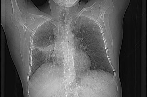
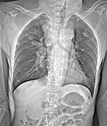
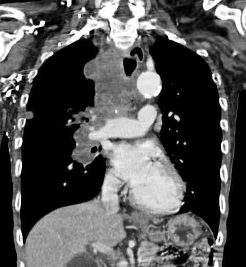
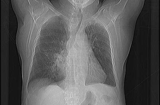
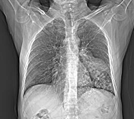
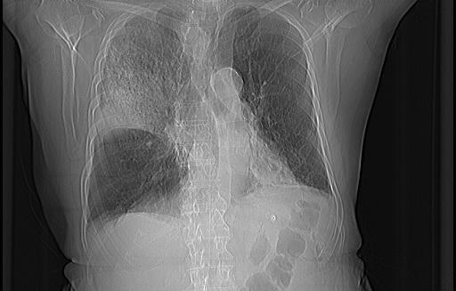
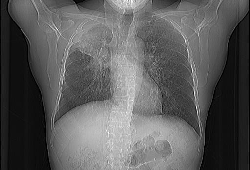
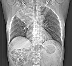
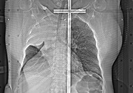
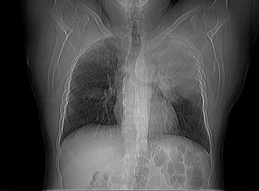
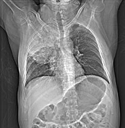
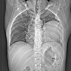
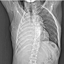
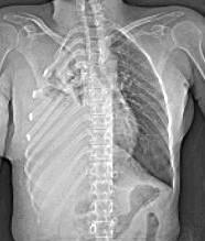
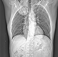
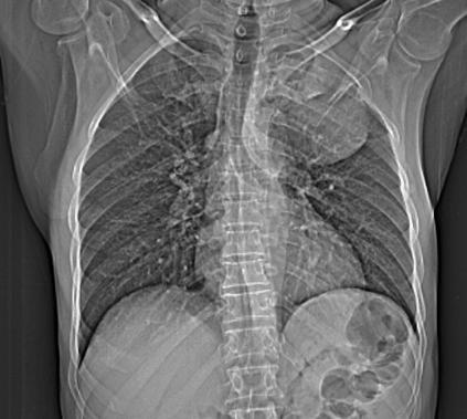
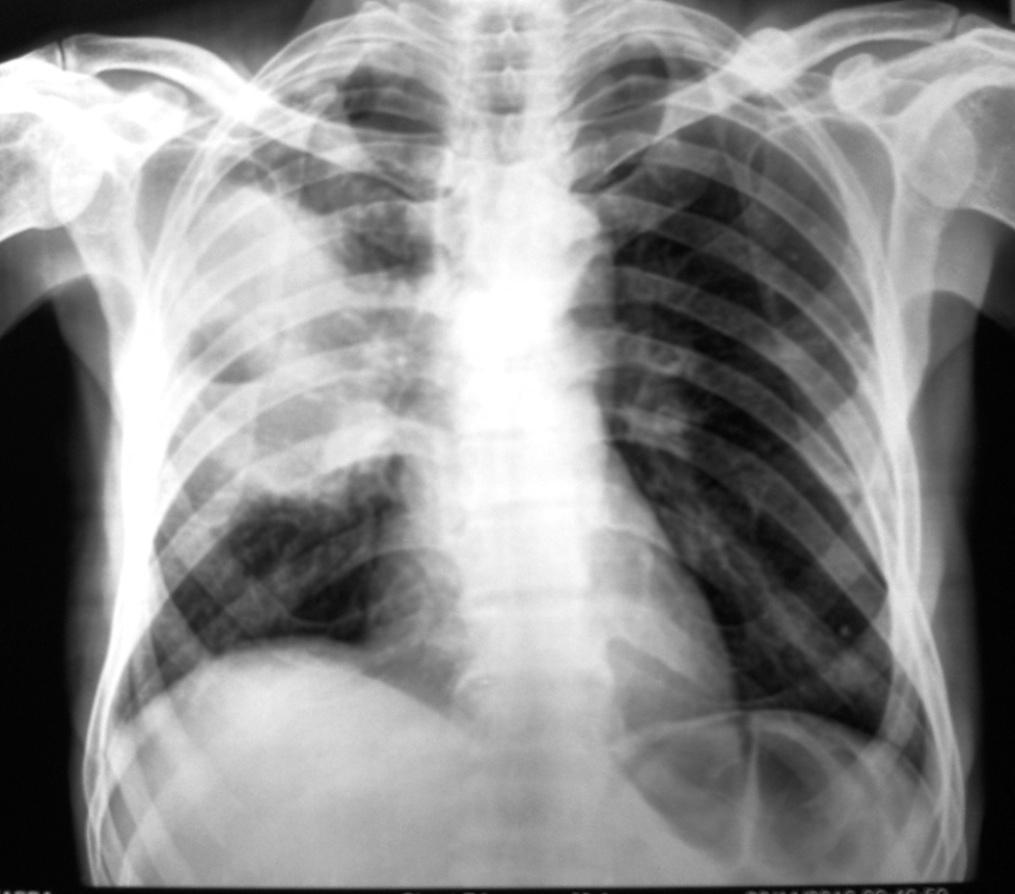
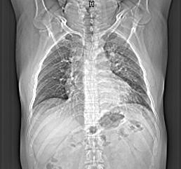
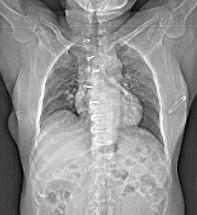
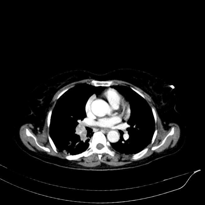
17 LEGENDS: Fig 1 a,b,c : chest images show solitary pulmonary nodule/mass Fig 2 a-d : chest images show hilar and perihilar opacities. Fig 3 a,b : chest images show non resolving consolidation Fig 4 a,b,c : chest images show partial atelectasis Fig 5 a,b : chest images show complete atelectasis Fig 6 a,b,c : chest images show lobar consolidation/ mass Fig 7a,b : chest radiographs show large plural effusion Fig 8: a,b : chest images show apical mass Fig 9: chest image shows hilar adenopathy Fig 10: chest image shows peripheral mass Fig11: chest image shows cavitary mass Fig 12 a,b : retrocardiac mass on CT not seen on radiograph Fig13: a,b : solitary pulmonary opacity seen on CT scan only Dr Madhusudan C."Spectrum of Radiological Findings in Bronchogenic Carcinoma A Retrospective Study. IOSR Journal of Dental and Medical Sciences (IOSR-JDMS), vol. 17, no. 1, 2018, pp DOI: / Page
Histopathological and CT Imaging Correlation of Various Primary Lung Carcinoma
 IOSR Journal of Dental and Medical Sciences (IOSR-JDMS) e-issn: 2279-0853, p-issn: 2279-0861.Volume 15, Issue 3 Ver. VII (Mar. 2016), PP 104-110 www.iosrjournals.org Histopathological and CT Imaging Correlation
PULMONARY TUBERCULOSIS RADIOLOGY
 PULMONARY TUBERCULOSIS RADIOLOGY RADIOLOGICAL MODALITIES Medical radiophotography Radiography Fluoroscopy Linear (conventional) tomography Computed tomography Pulmonary angiography, bronchography Ultrasonography,
Case Scenario 1. The patient agreed to a CT guided biopsy of the left upper lobe mass. This was performed and confirmed non-small cell carcinoma.
 Case Scenario 1 An 89 year old male patient presented with a progressive cough for approximately six weeks for which he received approximately three rounds of antibiotic therapy without response. A chest
TB Radiology for Nurses Garold O. Minns, MD
 TB Nurse Case Management Salina, Kansas March 31-April 1, 2010 TB Radiology for Nurses Garold O. Minns, MD April 1, 2010 TB Radiology for Nurses Highway Patrol Training Center Salina, KS April 1, 2010
Chest Radiology Interpretation: Findings of Tuberculosis
 Chest Radiology Interpretation: Findings of Tuberculosis Get out your laptops, smart phones or other devices pollev.com/chestradiology Case #1 1 Plombage Pneumonia Cancer 2 Reading the TB CXR Be systematic!
Role of CT imaging to evaluate solitary pulmonary nodule with extrapulmonary neoplasms
 Original Research Article Role of CT imaging to evaluate solitary pulmonary nodule with extrapulmonary neoplasms Anand Vachhani 1, Shashvat Modia 1*, Varun Garasia 1, Deepak Bhimani 1, C. Raychaudhuri
Do you want to be an excellent Radiologist? - Focus on the thoracic aorta on lateral chest image!!!
 The lateral chest radiograph: Challenging area around the thoracic aorta!!! Do you want to be an excellent Radiologist? - Focus on the thoracic aorta on lateral chest image!!! Dong Yoon Han 1, So Youn
Common things are common, but not always the answer
 Kevin Conroy, Joe Mackenzie, Stephen Cowie kevin.conroy@nhs.net Respiratory Dept, Darlington Memorial Hospital, Darlington, UK. Common things are common, but not always the answer Case report Cite as:
An Introduction to Radiology for TB Nurses
 An Introduction to Radiology for TB Nurses Garold O. Minns, MD September 14, 2017 TB Nurse Case Management September 12 14, 2017 EXCELLENCE EXPERTISE INNOVATION Garold O. Minns, MD has the following disclosures
An Update: Lung Cancer
 An Update: Lung Cancer Andy Barlow Consultant in Respiratory Medicine Lead Clinician for Lung Cancer (West Herts Hospitals NHS Trust) Lead for EBUS-Harefield Hospital (RB&HFT) Summary Lung cancer epidemiology
Chief Complain. For chemotherapy
 Chief Complain For chemotherapy Present Illness 93.12 Progressive weakness of R t arm for 1 year X-ray: peneative lesion over right proximal humorous Bone scan: multiple increased intake Biopsy of distal
Radiological staging of lung cancer. Shukri Loutfi,MD,FRCR Consultant Thoracic Radiologist KAMC-Riyadh
 Radiological staging of lung cancer Shukri Loutfi,MD,FRCR Consultant Thoracic Radiologist KAMC-Riyadh Bronchogenic Carcinoma Accounts for 14% of new cancer diagnoses in 2012. Estimated to kill ~150,000
TB Intensive Houston, Texas
 TB Intensive Houston, Texas October 15-17, 17 2013 Diagnosis of TB: Radiology Rosa M Estrada-Y-Martin, MD MSc FCCP October 16, 2013 Rosa M Estrada-Y-Martin, MD MSc FCCP, has the following disclosures to
Lung Cancer Staging: The Revised TNM Classification
 Norwegian Society of Thoracic Imaging Oslo, October 2011 Lung Cancer Staging: The Revised TNM Classification Sujal R Desai King s College Hospital, London Lung Cancer The Scale of the Problem Leading cause
Undergraduate Teaching
 Prof. James F Meaney Undergraduate Teaching Chest X-Ray Understanding the normal anatomical by reference to cross sectional imaging Radiology? It s FUN! Cryptic puzzle Sudoku (Minecraft?) It s completely
Right infrahilar nodule
 Right infrahilar nodule Search Infrahilar nodule Nov 9, 2015.. CT chest showed a right infrahilar mass 3.5 2.5 cm along with multiple bilateral lung nodules of size 9 to 11 mm. Bronchoscopy. Jun 13, 2015.
Computed Tomography of Normal Adrenal Glands in Indian Population
 IOSR Journal of Dental and Medical Sciences (IOSR-JDMS) e-issn: 2279-0853, p-issn: 2279-0861.Volume 17, Issue 01 Ver. V January. (2018), PP 26-30 www.iosrjournals.org Computed Tomography of Normal Adrenal
Lung Cancer Imaging. Terence Z. Wong, MD,PhD. Department of Radiology Duke University Medical Center Durham, NC 9/9/09
 Lung Cancer Imaging Terence Z. Wong, MD,PhD Department of Radiology Duke University Medical Center Durham, NC 9/9/09 Acknowledgements Edward F. Patz, Jr., MD Jenny Hoang, MD Ellen L. Jones, MD, PhD Lung
Pulmonary Nodules & Masses
 Pulmonary Nodules & Masses A Diagnostic Approach Heber MacMahon The University of Chicago Department of Radiology Disclosure Information Consultant for Riverain Technology Minor equity in Hologic Royalties
Atypical Presentations of lung cancers
 8 Original Article Atypical Presentations of lung cancers PVV Bharadwaj, VV Ramana Reddy, Ganeswar Behera, JV Praveen, DSS Sowjanya, B Krishna Prithvi Abstract: Aim: The aim of this study was to examine
Diagnosis and Staging of Non-Small Cell Lung Cancer Carlos Eduardo Oliveira Baleeiro, MD. November 18, 2017
 Diagnosis and Staging of Non-Small Cell Lung Cancer Carlos Eduardo Oliveira Baleeiro, MD November 18, 2017 Disclosures I do not have a financial interest/arrangement or affiliation with one or more organizations
Radiology Pathology Conference
 Radiology Pathology Conference Sharlin Johnykutty,, MD, Cytopathology Fellow Sara Majewski, MD, Radiology Resident Friday, August 28, 2009 Presentation material is for education purposes only. All rights
THE BENEFITS OF BIG DATA
 THE BENEFITS OF BIG DATA Disclosures I am a named inventor on a number of patents and patent applications relating to the evaluation of pulmonary nodules on CT scans of the chest which are owned by Cornell
Validation of the T descriptor in the new 8th TNM classification for non-small cell lung cancer
 Original Article Validation of the T descriptor in the new 8th TNM classification for non-small cell lung cancer Hee Suk Jung 1, Jin Gu Lee 2, Chang Young Lee 2, Dae Joon Kim 2, Kyung Young Chung 2 1 Department
Excavated pulmonary nodule: steps to diagnosis?
 Excavated pulmonary nodule: steps to diagnosis? Poster No.: C-1044 Congress: ECR 2014 Type: Authors: Keywords: DOI: Educational Exhibit W. Mnari, M. MAATOUK, A. Zrig, B. Hmida, M. GOLLI; Monastir/ TN Metastases,
Use of Integrated PET CT in the Clinical Staging of Non Small Cell Lung Cancer
 November 2010 Use of Integrated PET CT in the Clinical Staging of Non Small Cell Lung Cancer Laura Myers, Harvard Medical School, Year III Clinical Presentation 79yo woman with cough productive of green
How to Analyse Difficult Chest CT
 How to Analyse Difficult Chest CT Complex diseases are:- - Large lesion - Unusual or atypical pattern - Multiple discordant findings Diffuse diseases are:- - Numerous findings in both sides 3 basic steps
Nanda Horeweg, Carlijn M. van der Aalst, Erik Thunnissen, Kristiaan Nackaerts, Carla Weenink, Harry J.M. Groen, Jan-Willem J.
 Characteristics of lung cancers detected in the randomized NELSON lung cancer screening trial Nanda Horeweg, Carlijn M. van der Aalst, Erik Thunnissen, Kristiaan Nackaerts, Carla Weenink, Harry J.M. Groen,
Approach to Pulmonary Nodules
 Approach to Pulmonary Nodules Edwin Jackson, Jr., DO Assistant Professor-Clinical Director, James Early Detection Clinic Department of Internal Medicine Division of Pulmonary, Allergy, Critical Care and
Diagnosis of TB: Radiology David Finlay, MD
 TB Intensive Tyler, Texas June 2-4, 2010 Diagnosis of TB: Radiology David Finlay, MD June 3, 2010 2stages stages- Tuberculosis 1. primary infection 2. reactivation, or post primary disease 2 1 Primary
Small Pulmonary Nodules: Our Preliminary Experience in Volumetric Analysis of Doubling Times
 Small Pulmonary Nodules: Our Preliminary Experience in Volumetric Analysis of Doubling Times Andrea Borghesi, MD Davide Farina, MD Roberto Maroldi, MD Department of Radiology University of Brescia Brescia,
Rodney C Richie MD FACP FCCP DBIM Texas Life and EMSI
 Rodney C Richie MD FACP FCCP DBIM Texas Life and EMSI Pulmonary Nodules Well-circumscribed, radiographic opacities measuring 3 cm in diameter Surrounded by aerated lung Not associated with atelectesis
A Case of Pediatric Plasma Cell Granuloma
 August 2001 A Case of Pediatric Plasma Cell Granuloma Nii Tetteh, Harvard Medical School Year IV Our Patient 8 year old male with history of recurrent left lower lobe and lingular pneumonias since 1994.
September 2014 Imaging Case of the Month. Michael B. Gotway, MD. Department of Radiology Mayo Clinic Arizona Scottsdale, AZ
 September 2014 Imaging Case of the Month Michael B. Gotway, MD Department of Radiology Mayo Clinic Arizona Scottsdale, AZ Clinical History: A 57-year-old non-smoking woman presented to her physician as
Slide 1. Slide 2. Slide 3. Investigation and management of lung cancer Robert Rintoul. Epidemiology. Risk factors/aetiology
 Slide 1 Investigation and management of lung cancer Robert Rintoul Department of Thoracic Oncology Papworth Hospital Slide 2 Epidemiology Second most common cancer in the UK (after breast). 38 000 new
X-Rays. Prepared by Prof.Dr. Magda Hassab Allah Assist.lecturer Marwa Al Hady
 X-Rays Prepared by Prof.Dr. Magda Hassab Allah Assist.lecturer Marwa Al Hady CHEST X-RAYS Normal Chest X-ray Comments on chest X ray includes examination of 1- Bony cage (ribs,clavicles &vertebral column
Lung. 10/24/13 Chest X-ray: 2.9 cm mass like density in the inferior lingular segment worrisome for neoplasm. Malignancy cannot be excluded.
 Lung Case Scenario 1 A 54 year white male presents with a recent abnormal CT of the chest. The patient has a history of melanoma, kidney, and prostate cancers. 10/24/13 Chest X-ray: 2.9 cm mass like density
Chest XRay interpretation INTERPRETATIONS Identifications: Name & Date Technical evaluation Basic Interpretations
 Chest XRay interpretation INTERPRETATIONS Identifications: Name & Date Technical evaluation Basic Interpretations TECHNICAL EVALUATION 1. Projection: AP/PA view To differentiate between AP & PA films,
OBJECTIVES. Solitary Solid Spiculated Nodule. What would you do next? Case Based Discussion: State of the Art Management of Lung Nodules.
 Organ Imaging : September 25 2015 OBJECTIVES Case Based Discussion: State of the Art Management of Lung Nodules Dr. Elsie T. Nguyen Dr. Kazuhiro Yasufuku 1. To review guidelines for follow up and management
David E. Griffith, MD has the following disclosures to make:
 Diagnosis of TB: Radiology David E. Griffith, MD March 13, 2015 TB for Pulmonologist March 13, 2015 Phoenix, AZ EXCELLENCE EXPERTISE INNOVATION David E. Griffith, MD has the following disclosures to make:
Lung Cancer - Suspected
 Lung Cancer - Suspected Shared Decision Making Lung Cancer: http://www.enhertsccg.nhs.uk/ Patient presents with abnormal CXR Lung cancer - clinical presentation History and Examination Incidental finding
Steering Committee. Waiting on photo. Paul A. Bunn, Jr., MD Kavita Garg, MD Kim Geisinger, MD Fred R. Hirsch, Gregory Riely, MD, PhD.
 Steering Committee Paul A. Bunn, Jr., MD Kavita Garg, MD Kim Geisinger, MD Fred R. Hirsch, Gregory Riely, MD, PhD MD, PhD Waiting on photo Paul Van Schil, MD, PhD William D. Travis, MD Ming-Sound Tsao,
Learning Objectives. 1. Identify which patients meet criteria for annual lung cancer screening
 Disclosure I, Taylor Rowlett, DO NOT have a financial interest /arrangement or affiliation with one or more organizations that could be perceived as a real or apparent conflict of interest in the context
Thoracic CT pattern in lung cancer: correlation of CT and pathologic diagnosis
 19 th Congress of APSR PG of Lung Cancer (ESAP): Update of Lung Cancer Thoracic CT pattern in lung cancer: correlation of CT and pathologic diagnosis Kazuma Kishi, M.D. Department of Respiratory Medicine,
Bronchial syndrome. Atelectasis Draining bronchus Bronchiectasis
 Bronchial syndrome Atelectasis Draining bronchus Bronchiectasis Etienne Leroy Terquem Pierre L Her SPI / ISP Soutien Pneumologique International / International Support for Pulmonology Atelectasis Consequence
Radiological Aspects of Pulmonary Tuberculosis in Immunocompetent Hosts
 Nov 2003 Radiological Aspects of Pulmonary Tuberculosis in Immunocompetent Hosts Josh Rempell, Harvard Medical School Year III Tuberculosis: the captain of all (wo)men of death Overall, one third of the
American College of Radiology ACR Appropriateness Criteria
 American College of Radiology ACR Criteria Radiologic Management of Thoracic Nodules and Masses Variant 1: Middle-aged patient (35 60 years old) with an incidental 1.5-cm lung nodule. The lesion was smooth.
The tumor, node, metastasis (TNM) staging system of lung
 staging system of lung") ORIGINAL ARTICLE Peripheral Direct Adjacent Lobe Invasion Non-small Cell Lung Cancer Has a Similar Survival to That of Parietal Pleural Invasion T3 Disease Hao-Xian Yang, MD, PhD,* Xue Hou, MD, Peng Lin,
ORIGINAL ARTICLE Peripheral Direct Adjacent Lobe Invasion Non-small Cell Lung Cancer Has a Similar Survival to That of Parietal Pleural Invasion T3 Disease Hao-Xian Yang, MD, PhD,* Xue Hou, MD, Peng Lin,
North of Scotland Cancer Network Clinical Management Guideline for Non Small Cell Lung Cancer
 THIS DOCUMENT IS North of Scotland Cancer Network Clinical Management Guideline for Non Small Cell Lung Cancer [Based on WOSCAN NSCLC CMG with further extensive consultation within NOSCAN] UNCONTROLLED
CT Diagnosis of Pulmonary Wegener s Granulomatosis: A Case Report and Review of Literature
 CASE REPORT JIACM 2008; 9(4): 321-5 CT Diagnosis of Pulmonary Wegener s Granulomatosis: A Case Report and Review of Literature Shibani Mehra*, Shailendra Aggarwal Abstract The diagnosis of Wegener s granulomatosis
PET/CT in lung cancer
 PET/CT in lung cancer Andrei Šamarin North Estonia Medical Centre 3 rd Baltic Congress of Radiology 08.10.2010 Imaging in lung cancer Why do we need PET/CT? CT is routine imaging modality for staging of
Thoracic Sarcoidosis Imaging Updated: Jul 19, 2013
 Thoracic Sarcoidosis Imaging Updated: Jul 19, 2013 Overview Radiography Computed Tomography Magnetic Resonance Imaging Nuclear Imaging Show All Multimedia Library References Overview For patients with
Lung Cancer Diagnosis for Primary Care
 Lung Cancer Diagnosis for Primary Care Daniel Nader, DO, FCCP Cancer Treatment Center of America Case 1 In which of the following situations would the U.S. Preventive Services Task Force (USPSTF) recommend
objectives Pitfalls and Pearls in PET/CT imaging Kevin Robinson, DO Assistant Professor Department of Radiology Michigan State University
 objectives Pitfalls and Pearls in PET/CT imaging Kevin Robinson, DO Assistant Professor Department of Radiology Michigan State University To determine the regions of physiologic activity To understand
The solitary pulmonary nodule: Assessing the success of predicting malignancy
 The solitary pulmonary nodule: Assessing the success of predicting malignancy Poster No.: C-0829 Congress: ECR 2010 Type: Scientific Exhibit Topic: Chest Authors: R. W. K. Lindsay, J. Foster, K. McManus;
Bronchioloalveolar Carcinoma Mimicking DILD:
 Bronchioloalveolar Carcinoma Mimicking DILD: A Case Report 1 Ju Young Lee, M.D., In Jae Lee, M.D., Dong Gyu Kim, M.D. 2, Soo Kee Min, M.D. 3, Min-Jeong Kim, M.D., Sung Il Hwang, M.D., Yul Lee, M.D., Sang
Educational Objectives. Managing Lung Cancer From the Solitary Pulmonary Nodule to Complex Cases: A Multidisciplinary Approach.
 Managing Lung Cancer From the Solitary Pulmonary Nodule to Complex Cases: A Multidisciplinary Approach Robert A. Meguid, MD, MPH, FACS Assistant Professor of Cardiothoracic Surgery Surgical Director, Surgical
Lung Cancer Screening: To Screen or Not to Screen?
 Lung Cancer Screening: To Screen or Not to Screen? Lorriana Leard, MD Co-Director of UCSF Lung Cancer Screening Program Vice Chief of Clinical Activities UCSF Pulmonary, Critical Care, Allergy & Sleep
The right middle lobe is the smallest lobe in the lung, and
 ORIGINAL ARTICLE The Impact of Superior Mediastinal Lymph Node Metastases on Prognosis in Non-small Cell Lung Cancer Located in the Right Middle Lobe Yukinori Sakao, MD, PhD,* Sakae Okumura, MD,* Mun Mingyon,
Seventh Edition of the Cancer Staging Manual and Stage Grouping of Lung Cancer. Quick Reference Chart and Diagrams
 CHEST Special Features Seventh Edition of the Cancer Staging Manual and Stage Grouping of Lung Cancer Quick Reference Chart and Diagrams Omar Lababede, MD ; Moulay Meziane, MD ; and Thomas Rice, MD, FCCP
Computed Tomography (CT) Scan Features of Pulmonary Drug-Resistant Tuberculosis in Non-HIV-Infected Patients
 Scan Features of Pulmonary Drug-Resistant Tuberculosis in Non-HIV-Infected Patients") Cronicon OPEN ACCESS EC BACTERIOLOGY AND VIROLOGY Research Article Computed Tomography (CT) Scan Features of Pulmonary Drug-Resistant Tuberculosis in Non-HIV-Infected Patients Ehsan Shahverdi 1 *, Ashkan
Cronicon OPEN ACCESS EC BACTERIOLOGY AND VIROLOGY Research Article Computed Tomography (CT) Scan Features of Pulmonary Drug-Resistant Tuberculosis in Non-HIV-Infected Patients Ehsan Shahverdi 1 *, Ashkan
Surgical indications: Non-malignant pulmonary diseases. Punnarerk Thongcharoen
 Surgical indications: Non-malignant pulmonary diseases Punnarerk Thongcharoen Non-malignant Malignant as a pathological term: Cancer Non-malignant = not cancer Malignant as an adjective: Disposed to cause
Bronchogenic Carcinoma
 A 55-year-old construction worker has smoked 2 packs of ciggarettes daily for the past 25 years. He notes swelling in his upper extremity & face, along with dilated veins in this region. What is the most
1/25/13 Right partial nephrectomy followed by completion right radical nephrectomy.
 History and Physical Case Scenario 1 45 year old white male presents with complaints of nausea, weight loss, and back pain. A CT of the chest, abdomen and pelvis was done on 12/8/12 that revealed a 12
Isolated anthracosis: benign but neglected cause of bronchial stenosis and obstruction
 Isolated anthracosis: benign but neglected cause of bronchial stenosis and obstruction Poster No.: C-0143 Congress: ECR 2013 Type: Scientific Exhibit Authors: S. Kahkouee, R. Pourghorban, M. Bitarafan,
Diagnostic Correlation of Findings of Multidetector Computed Tomography and Fine Needle Aspiration Cytology in Lung Masses
 RESEARCH ARTICLE Diagnostic Correlation of Findings 10.5005/jp-journals-10057-0004 of MDCT and FNAC in Lung Masses Diagnostic Correlation of Findings of Multidetector Computed Tomography and Fine Needle
CT Screening for Lung Cancer for High Risk Patients
 CT Screening for Lung Cancer for High Risk Patients The recently published National Lung Cancer Screening Trial (NLST) showed that low-dose CT screening for lung cancer reduces mortality in high-risk patients
Diagnostic challenge: Sclerosing Hemangioma of the Lung. Department of Medicine, Division of Pulmonary and Critical Care, Lincoln Medical and
 Diagnostic challenge: Sclerosing Hemangioma of the Lung. S. Arias M.D, R. Loganathan M.D, FCCP Department of Medicine, Division of Pulmonary and Critical Care, Lincoln Medical and Mental Health Center/Weill
Signs in Chest Radiology
 Signs in Chest Radiology Jonathan H. Chung, MD Disclosures No pertinent disclosures Jonathan H. Chung, MD Assistant Professor Institute t of fadvanced d Biomedical Imaging National Jewish Health Denver,
The Various Methods to Biopsy the Lung PROF SHITRIT DAVID HEAD, PULMONARY DEPARTMENT MEIR MEDICAL CENTER, ISRAEL
 The Various Methods to Biopsy the Lung PROF SHITRIT DAVID HEAD, PULMONARY DEPARTMENT MEIR MEDICAL CENTER, ISRAEL Conflict of Interest This presentation is supported by AstraZeneca Two main steps before
PET CT for Staging Lung Cancer
 PET CT for Staging Lung Cancer Rohit Kochhar Consultant Radiologist Disclosures Neither I nor my immediate family members have financial relationships with commercial organizations that may have a direct
Boot Camp Case Scenarios
 Boot Camp Case Scenarios Case Scenario 1 Patient is a 69-year-old white female. She presents with dyspnea on exertion, cough, and right rib pain. Patient is a smoker. 9/21/12 CT Chest FINDINGS: There is
PULMONARY NODULES AND MASSES : DIAGNOSTIC APPROACH AND NEW MANAGEMENT GUIDELINES. https://tinyurl.com/hmpn2018
 PULMONARY NODULES AND MASSES : DIAGNOSTIC APPROACH AND NEW MANAGEMENT GUIDELINES Heber MacMahon MB, BCh Department of Radiology The University of Chicago https://tinyurl.com/hmpn2018 Disclosures Consultant
Endobronchial Ultrasound in the Diagnosis & Staging of Lung Cancer
 Endobronchial Ultrasound in the Diagnosis & Staging of Lung Cancer Dr Richard Booton PhD FRCP Lead Lung Cancer Clinician, Consultant Respiratory Physician & Speciality Director Manchester University NHS
Bronchial carcinosarcoma
 Bronchial carcinosarcoma Carolina Carcano 1*, Edward Savage 2, Maria Julia Diacovo 3, Jacobo Kirsch 1 1. Division of Radiology, Cleveland Clinic Florida, Weston, Fl, USA 2. Department of Thoracic and Cardiovascular
Low-dose CT Lung Cancer Screening Guidelines for Pulmonary Nodules Management Version 2
 Low-dose CT Lung Cancer Screening Guidelines for Pulmonary Nodules Management Version 2 The Committee for Management of CT-screening-detected Pulmonary Nodules 2009-2011 The Japanese Society of CT Screening
JMSCR Vol 04 Issue 10 Page October 2016
 www.jmscr.igmpublication.org Impact Factor 5.244 Index Copernicus Value: 83.27 ISSN (e)-2347-176x ISSN (p) 2455-0450 DOI: http://dx.doi.org/10.18535/jmscr/v4i10.08 Ultrasound and CT Evaluation of Pleural
Follow Up Chest X-Ray Post- Pneumonia
 IOSR Journal of Dental and Medical Sciences (IOSR-JDMS) e-issn: 227-83, p-issn: 227-861.Volume 17, Issue 8 Ver. 13 (August. 218), PP 6-6 www.iosrjournals.org Follow Up Chest X-Ray Post- Pneumonia Dr. CleofinaFurtado,
Guidelines for the Management of Pulmonary Nodules Detected by Low-dose CT Lung Cancer Screening
 Guidelines for the Management of Pulmonary Nodules Detected by Low-dose CT Lung Cancer Screening 1. Introduction In January 2005, the Committee for Preparation of Clinical Practice Guidelines for the Management
Pictorial essay of unusual radiologic manifestations of pulmonary and airway metastasis at initial presentation of lung cancer
 Pictorial essay of unusual radiologic manifestations of pulmonary and airway metastasis at initial presentation of lung cancer Poster No.: C-2297 Congress: ECR 2012 Type: Educational Exhibit Authors: Y.
Pediatric TB Intensive Houston, Texas October 14, 2013
 Pediatric TB Intensive Houston, Texas October 14, 2013 Radiologic Presentation of Childhood TB Susan D. John, MD, FACR October 14, 2013 Disclosures I have no disclosures or conflicts of interest to report
A Chronology of Advancements in the Diagnosing of Lung Nodules
 November 17, 2017 A Chronology of Advancements in the Diagnosing of Lung Nodules Presenter: Daniel P. Harley, MD, MSB, FACS Surgical Director of the Angelos Center for Lung Diseases 1 Pulmonary Nodules
Synchronous Triple Primary Lung Cancers: A Case Report
 Case Report Thoracic Imaging http://dx.doi.org/10.3348/kjr.2014.15.5.646 pissn 1229-6929 eissn 2005-8330 Korean J Radiol 2014;15(5):646-650 Synchronous Triple Primary Lung Cancers: A Case Report Hyun Jung
Atlas of the Vasculitic Syndromes
 CHAPTER e40 Atlas of the Vasculitic Syndromes Carol A. Langford Anthony S. Fauci Diagnosis of the vasculitic syndromes is usually based upon characteristic histologic or arteriographic findings in a patient
Positron Emission Tomography in Lung Cancer
 May 19, 2003 Positron Emission Tomography in Lung Cancer Andrew Wang, HMS III Patient DD 53 y/o gentleman presented with worsening dyspnea on exertion for the past two months 30 pack-year smoking Hx and
Pediatric TB Intensive Houston, Texas
 Pediatric TB Intensive Houston, Texas November 13, 2009 Radiographic Manifestations of Pediatric TB Susan D. John, MD, FACR November 13, 2009 Radiologic Presentation of Childhood TB Susan D. John, MD,
Adam J. Hansen, MD UHC Thoracic Surgery
 Adam J. Hansen, MD UHC Thoracic Surgery Sometimes seen on Chest X-ray (CXR) Common incidental findings on computed tomography (CT) chest and abdomen done for other reasons Most lung cancers discovered
Stage I synchronous multiple primary non-small cell lung cancer: CT findings and the effect of TNM staging with the 7th and 8th editions on prognosis
 Original Article Stage I synchronous multiple primary non-small cell lung cancer: CT findings and the effect of TNM staging with the 7th and 8th editions on prognosis Jingxu Li, Xinguan Yang, Tingting
Prof. Dr. NAGUI M. ABDELWAHAB,M.D.; MARYSE Y. AWADALLAH, M.D. AYA M. BASSAM, Ms.C.
 Role of Whole-body Diffusion MR in Detection of Metastatic lesions Prof. Dr. NAGUI M. ABDELWAHAB,M.D.; MARYSE Y. AWADALLAH, M.D. AYA M. BASSAM, Ms.C. Cancer is a potentially life-threatening disease,
GUIDELINES FOR CANCER IMAGING Lung Cancer
 GUIDELINES FOR CANCER IMAGING Lung Cancer Greater Manchester and Cheshire Cancer Network Cancer Imaging Cross-Cutting Group April 2010 1 INTRODUCTION This document is intended as a ready reference for
Interpretation of Chest Radiographs Paul Christensen, MD 10/21/09. Diagnostic Evaluation. Medical Evaluation & CXR Interpretation.
 Diagnostic Evaluation Medical Evaluation & CXR Interpretation University of Michigan TB Consultant Washtenaw County Medical history Physical examination Testing for TB exposure (previously covered) Radiologic
I9 COMPLETION INSTRUCTIONS
 The I9 Form is completed for each screening exam at T0, T1, and T2. At T0 (baseline), the I9 documents comparison review of the baseline screen (C2 Form) with any historical images available. At T1 and
The 8th Edition of the TNM Classification for Lung Cancer Background, Innovations and Implications for Clinical Practice
 The 8th Edition of the TNM Classification for Lung Cancer Background, Innovations and Implications for Clinical Practice University of Torino Lecture 28th June 2017 Torino, Italy Ramón Rami-Porta Thoracic
An Image Repository for Chest CT
 An Image Repository for Chest CT Francesco Frajoli for the Chest CT in Antibody Deficiency Group An Image Repository for Chest CT he Chest CT in Antibody Deficiency Group is an international and interdisciplinary
GUIDELINES FOR PULMONARY NODULE MANAGEMENT : RECENT CHANGES AND UPDATES
 Venice 2017 GUIDELINES FOR PULMONARY NODULE MANAGEMENT : RECENT CHANGES AND UPDATES Heber MacMahon MB, BCh Department of Radiology The University of Chicago Disclosures Consultant for Riverain Medical
Monophasic Synovial Carcinoma of knee joint- A Case Report and Review of Literature
 IOSR Journal of Dental and Medical Sciences (IOSR-JDMS) e-issn: 2279-0853, p-issn: 2279-0861.Volume 17, Issue 3 Ver.5 March. (2018), PP 13-17 www.iosrjournals.org Monophasic Synovial Carcinoma of knee
Los Angeles Radiological Society 62 nd Annual Midwinter Radiology Conference January 31, 2010
 Los Angeles Radiological Society 62 nd Annual Midwinter Radiology Conference January 31, 2010 Self Assessment Module on Nuclear Medicine and PET/CT Case Review FDG PET/CT IN LYMPHOMA AND MELANOMA Submitted
Lung Cancer Risk Associated With New Solid Nodules in the National Lung Screening Trial
 Cardiopulmonary Imaging Original Research Pinsky et al. Lung Cancer Risk Associated With New Nodules Cardiopulmonary Imaging Original Research Paul F. Pinsky 1 David S. Gierada 2 P. Hrudaya Nath 3 Reginald
LUNG NODULES: MODERN MANAGEMENT STRATEGIES
 Department of Radiology LUNG NODULES: MODERN MANAGEMENT STRATEGIES Christian J. Herold M.D. Department of Biomedical Imaging and Image-guided Therapy Medical University of Vienna Vienna, Austria Pulmonary