Cross sectional imaging of Intracranial cystic lesions Abdel Razek A
|
|
|
- Gladys Lambert
- 5 years ago
- Views:
Transcription
1 Cross sectional imaging of Intracranial cystic lesions Abdel Razek A Department of Radiology. Mansoura Faculty of Medicine, Mansoura. Egypt. arazek@mans.edu.eg
2 Introduction Intracranial cystic lesions can present a diagnostic challenge. These lesions include benign tumors, malignant tumors and other lesions. Differentiation between good, bad and ugly cystic lesions is essential for treatment planning.
3 Purpose 1- To illustrate the causes of intracranial cystic lesions. 2- To review the typical and atypical MR appearance of intracranial cystic lesions. 3- To describe the role of diffusion weighted imaging in the differential diagnosis of intracranial cystic lesions.
4 Cystic lesions Variant: Virchow Robin space Cavum Septum Pellucidum Pineal cyst Rathke s cleft cyst Neuroepithelial cyst Interhemispheric cyst Developmental: Epidermoid Dermoid Arachnoid cyst Neuroeneteric cyst Colloid cyst Others: Cystic Neoplasm Hydatid cyst Leptomeningeal cyst
5 Methods of examination MR imaging DWI MRS Dynamic MR CT Metrimzinde


6 Cavum septum pellucidium The septum pellucidum consists of two thin sheets of white matter surrounded by gray matter. The persistence of this septum after birth is called cavum septi pellucidum
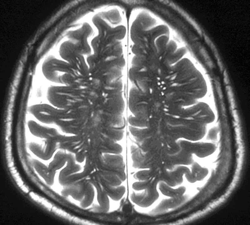
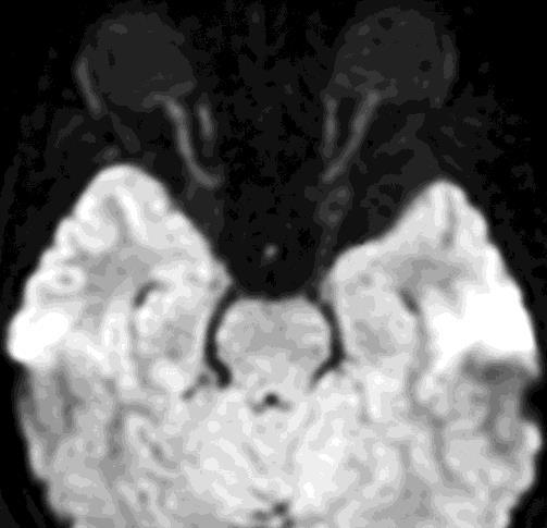
7 Enlarged Virtchow Robins Spaces (VRS) VRS are pial-lined interstitial fluid-filled structures that accompany penetrating arteries and veins Normal variant. Appear as smoothly demarcated fluid-filled cysts, of variable size. May be isolated or occur in clusters in basal ganglia. They are isointense to CSF on all sequences. No enhancement, no focal mass effect, no restricted diffusion on DWI. Etat Crible- cluster of very large VR spaces-look ugly, but are benign.

8 Dilated Pervascular Spaces
9 Pineal cyst seen in up to 10% of cases at routine imaging and in 20% 40% of cases at autopsy unilocular fluidfilled mass within the pineal gland. SI varies with cyst contents. Rim or nodular calcium (25%). Rim or nodular enhancement. T1WI: ~60% are slightly hyperintense to CSF. Most do not appear hypointense on FLAIR images, and 60% enhance.

10 Pineal Cyst

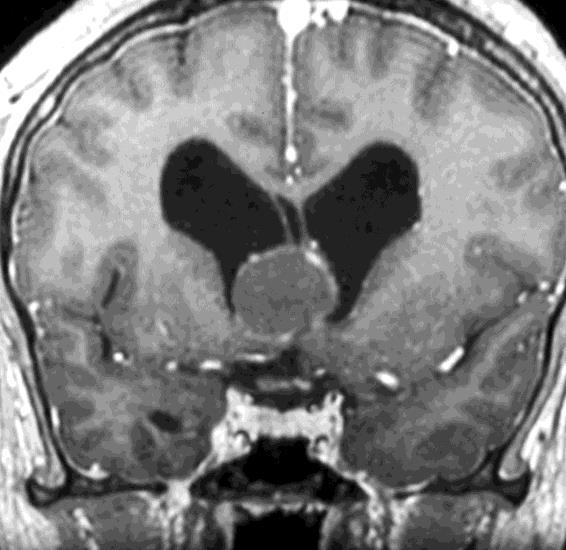
11 Interhemispheric cyst Commonly seen in patient with corpus callosum agenesis and other midline defects. The origin of the interhemispheric cyst in agenesis of the corpus callosum is controversial.


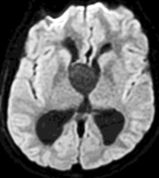
12 Neuro-epithelial Cyst Neuroepithelial cyst similar to ependymal cyst Follow CSF signal on MR Oval or round ranging from a few mm to 2 cm in size Minimal mass effect
13 Ependymal cyst Rare, benign, ependymal-lined neuroepithelial cysts that can occur anywhere in the central nervous system. They may seen in subarachnoid spaces, brainstem, cerebellum, and spinal cord. Intrinsically benign, but may cause problems secondary to mass effect or obstruction of CSF circulation MRI shows intraparenchymal, fluid filled space occupying lesions usually located close to a ventricle. They are thin walled, follow CSF signal on all sequences (although occasionallymay be hyperintense relative to CSF due to high protein content), do not enhance and do not have restricted diffusion.
14 Rathke s cleft cyst Arise from remnants of embryonic Rathke cleft. Usually an incidental finding imaging clue is a nonenhancing noncalcified intra- and/or suprasellar cyst with an intracystic non enhancing mural nodule. T1WI: hyperintense (50%), hypointense (50%). T2WI: hyperintense (70%), iso- hypointense (30%). If within an otherwise normal pituitary gland, no clinical significance If in suprasellar location may impinge on optic chiasm
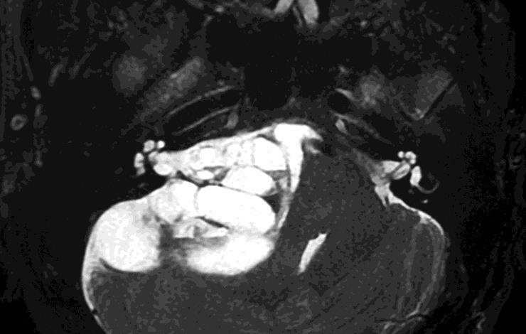
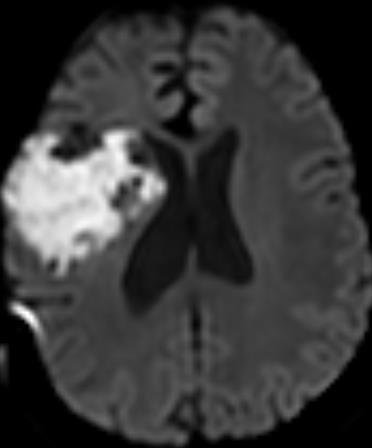
15 Epidermoid Epidermoid cysts comprise 0.2% 1.8% of primary intracranial tumors and are four to nine times as common as dermoid cysts. Site: cerebellopontine angle cistern (40% 50%),4 th ventricle (17%), sellar regions (10% 15%) All are located off the midline. The best diagnostic clue is a CSF-like mass that insinuates within cisterns, encasing adjacent nerves and vessels. On CT scans, most epidermoid cysts are well-defined hypoattenuated masses that resemble CSF and do not enhance. Calcification is present in 10% 25% of cases. Most epidermoid cysts are isointense or slightly hyperintense to CSF on T1 & T2WI.
16 They do not suppress completely on FLAIR images and have restricted diffusion (show high signal intensity) on DWI Most epidermoid cysts do not enhance Rare white epidermoids have high protein content and may appear hyperattenuated on CT scans. show reversed signal intensity on MR images, with high SI on T1- and low SI on T2WI
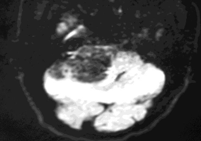
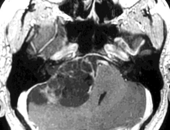
17 Epidermoid cyst
18 Epidermoid cyst
19 Dermoid On CT have lower attenuation then a epidermoid ( HU) Contain lipid material from sebaceous and apocrine sweat glands Dystrophic calcifications Layering: fat fluid level MR:T1: hyperintense fat T2: low signal with loculations & inhomogenous Chemical Shift artifact The fat signal suppresses on STIR or other fat suppression sequences Rupture: Fat/csf levels in subarachnoid spaces and ventricles Ventriculomegally Chemical meningitis.

20 Dermoid cyst
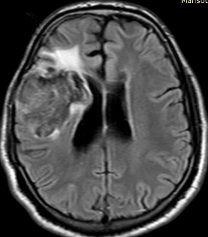
21 Arachnoid cyst Most commonly seen in middle cranial fossa (60%), suprasellar cistern & posterior fossa (10%). The best diagnostic clue is a sharply demarcated extraaxial cyst with scalloping of the adjacent. The classic arachnoid cyst has no identifiable internal architecture and does not enhance. The cyst typically has the same signal intensity as CSF at all sequences. Benign unless so large they are causing mass effect on adjacent structures

22 Arachnoid cyst
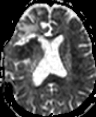
23 Colloid cyst 15% 20% of intraventricular masses Size: cm. The mean size is 1.5 cm Often more easily identified on non contrast enhanced CT-round sharply marginated hyperattenuating mass in roof of third ventricle near foramen of Monroe T1: hyperintense in 2/3 of cases. May be very subtle T2: isointense to brain may peripheral rim enhamcnet. Dangerous lesion: may cause sudden ventricular obstruction and death

24 Colloid cyst
25 Neuroeneteric cyst (NEC) Develop from persistent neuroenetric canal Lesions in midline, anterior to brainstem. Diagnostic clue is a round and/or lobulated, nonenhancing, slightly hyperintense mass anterior to medulla. High SI on T1 & T2WI due to its protein content. Hyperintense on FLAIR with restricted diffusion Rarely show rim enhancement.


26 Leptomeingeal cysts After a skull fracture associated with a dural tear, meninges and brain tissue may herniate into a diastatic fracture site. This prevents osteoblastic migration and healing and can result in a growing fracture or leptomeningeal cyst. Leptomeningeal cyst


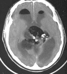
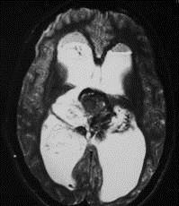
27 Hydatid cyst Appears as unilocular cyst w/thick wall, unilocular mother cyst with multiple vesicles arranged along its wall, or multiple daughter cysts filling the maternal cyst. A B


28 Cyst associated with benign extra axial tumor Large non neoplastic cysts seen with extraaxial tumors such as meningioma, schwannoma, craniopharyngioma It has SI similar to CSF at all pulse sequnces 2-10% of adult & 25% of pediatric meningiomas.

29 Cystic tumor
30 Craniopharyngioma Craniopharyngiomas arise from remnant of Rathke s pouch and typically demonstrate calcification in children Approximately 90% have nodular, globular, or rim enhancement. The presence of solid enhancing nodules in the cyst wall also favors the diagnosis of craniopharyngioma. The rare noncalcified cystic nonenhancing craniopharyngioma is more commonnly seenin adults than in children.
31 Conclusion 1) MR imaging is essential for diagnosis and characterization of intracranial cystic lesions as well as differentiating malignant from benign lesions. 2) Advanced MR imaging techniques helps in refining diagnosis of intracranial cystic lesions. This information is essential for treatment planning
32 References Ajtai B, Bertelson JA. Imaging of Intracranial Cysts. Continuum 2016;22: Taillibert S, Le Rhun E, Chamberlain MC. Intracranial cystic lesions: a review. Curr Neurol Neurosci Rep 2014;14:481. Osborn A, Preece M. Intracranial Cysts: Radiologic-Pathologic Correlation and Imaging Approach. Radiology 2006;239: Kwee RM, Kwee TC. Virchow- Robin spaces at MR imaging. Radiographics. 2007;27: Hu X, Hu C, Fang X, et al. Intraparenchymal epidermoid cysts in the brain: diagnostic value of MR diffusion-weighted imaging. Clin Radiol 2008;63: Park KJ, Kang SH, Chae YS, et al. Supratentorial arachnoid cyst located in the brain parenchyma: case report. Neurosurgery 2011;68:E Brown JY, Morokoff AP, Mitchell PJ, et al. Unusual imaging appearance of an intracranial dermoid cyst. AJNR 2001;22: Rao G, Anderson R, Feldstein NA, Brockmeyer DL. Expansion of Chang,Song, Kim, et al. In Vivo Single Voxel Proton MR Spectroscopy in Intracranial Cystic Masses. AJNR 1998;19:

33
Benign brain lesions
 Benign brain lesions Diagnostic and Interventional Radiology Hung-Wen Kao Department of Radiology, Tri-Service General Hospital, National Defense Medical Center Computed tomography Hounsfield unit (HU)
S Alandete, M Meseguer, CR Poyatos, D Uceda, E de la Via, J Sales, J Vilar. H.U. Dr Peset, Valencia (Spain)
") S Alandete, M Meseguer, CR Poyatos, D Uceda, E de la Via, J Sales, J Vilar. H.U. Dr Peset, Valencia (Spain) Introduction Cystic lesions are usually a common finding in clinical practice and you can find
S Alandete, M Meseguer, CR Poyatos, D Uceda, E de la Via, J Sales, J Vilar. H.U. Dr Peset, Valencia (Spain) Introduction Cystic lesions are usually a common finding in clinical practice and you can find
Differential diagnosis of intracranial cystic lesions.
 Differential diagnosis of intracranial cystic lesions. Poster No.: C-0215 Congress: ECR 2015 Type: Educational Exhibit Authors: S. P. G. Alandete, M. A. Meseguer, E. De la Via, D. Uceda, C. Poyatos; Valencia/ES
Pediatric CNS Tumors. Disclosures. Acknowledgements. Introduction. Introduction. Posterior Fossa Tumors. Whitney Finke, MD
 Pediatric CNS Tumors Disclosures Whitney Finke, MD Neuroradiology Fellow PGY-6 University of Utah Health Sciences Center Salt Lake City, Utah None Acknowledgements Introduction Nicholas A. Koontz, MD Luke
Brain Tumors. Medulloblastoma. Pilocytic astrocytoma: Ahmed Koriesh, MD. Pathological finding
 NeuroPathology Page 8 Brain Tumors Pathological finding Pseudorosette Rosenthal fibers Rosettes Wet Keratin Psammoma bodies Fried egg Tumor Ependymoma, SEGA Pilocytic astrocytoma Medulloblastoma Craniopharyngioma
NEURO IMAGING 2. Dr. Said Huwaijah Chairman of radiology Dep, Damascus Univercity
 NEURO IMAGING 2 Dr. Said Huwaijah Chairman of radiology Dep, Damascus Univercity I. EPIDURAL HEMATOMA (EDH) LOCATION Seventy to seventy-five percent occur in temporoparietal region. CAUSE Most likely caused
Essentials of Clinical MR, 2 nd edition. 51. Primary Neoplasms
 51. Primary Neoplasms As with spinal central canal neoplasms in other regions, those of the lumbar spine may be classified as extradural, intradural extramedullary, and medullary. If an extradural lesion
PITUITARY PARASELLAR LESIONS. Kim Learned, MD
 PITUITARY PARASELLAR LESIONS Kim Learned, MD DIFFERENTIALS Pituitary Sella Clivus, Sphenoid Sinus Suprasellar Optic chiasm, Hypothalamus, Circle of Willis Parasellar Cavernous Sinus Case 1 17 YEAR-OLD
Complex Hydrocephalus
 2012 Hydrocephalus Association Conference Washington, DC - June 27-July1, 2012 Complex Hydrocephalus Marion L. Walker, MD Professor of Neurosurgery & Pediatrics Primary Children s Medical Center University
Imaging The Turkish Saddle. Russell Goodman, HMS III Dr. Gillian Lieberman
 Imaging The Turkish Saddle Russell Goodman, HMS III Dr. Gillian Lieberman Learning Objectives Review the anatomy of the sellar region Discuss the differential diagnosis of sellar masses Discuss typical
Posterior fossa tumors: clues to differential diagnosis with case-based review
 Posterior fossa tumors: clues to differential diagnosis with case-based review Poster No.: C-0323 Congress: ECR 2017 Type: Educational Exhibit Authors: H. A. Aboughalia, M. Abdelhady; Doha/QA Keywords:
Cerebro-vascular stroke
 Cerebro-vascular stroke CT Terminology Hypodense lesion = lesion of lower density than the normal brain tissue Hyperdense lesion = lesion of higher density than normal brain tissue Isodense lesion = lesion
Cysts Arachnoid Cyst (also called Leptomeningeal Cyst)
") Cysts This article was provided to us by David Schiff, MD, Co-Director of the Neuro-Oncology Center and Professor of Neurology, Neurosurgery, and Medicine at the University of Virginia, Charlottesville.
Cysts This article was provided to us by David Schiff, MD, Co-Director of the Neuro-Oncology Center and Professor of Neurology, Neurosurgery, and Medicine at the University of Virginia, Charlottesville.
Table of Contents: SKULL AND BRAIN. Scalp, Skull. Anatomically Based Differentials. Skull Normal Variants. Scalp Mass, Child.
 Table of Contents: SKULL AND BRAIN Scalp, Skull Skull Normal Variants Scalp Mass, Child Scalp Mass, Adult Congenital Anomalies of Skull Base Sellar/Parasellar Mass With Skull Base Invasion "Hair on End"
Meninges and Ventricles
 Meninges and Ventricles Irene Yu, class of 2019 LEARNING OBJECTIVES Describe the meningeal layers, the dural infolds, and the spaces they create. Name the contents of the subarachnoid space. Describe the
CNS TUMORS. D r. Ali Eltayb ( U. of Omdurman. I ). M. Path (U. of Alexandria)
. M. Path (U. of Alexandria)") CNS TUMORS D r. Ali Eltayb ( U. of Omdurman. I ). M. Path (U. of Alexandria) CNS TUMORS The annual incidence of intracranial tumors of the CNS ISmore than intraspinal tumors May be Primary or Secondary
CNS TUMORS D r. Ali Eltayb ( U. of Omdurman. I ). M. Path (U. of Alexandria) CNS TUMORS The annual incidence of intracranial tumors of the CNS ISmore than intraspinal tumors May be Primary or Secondary
Pure Intracavernous Sinus Epidermoid Cyst: Diffusion-Weighted (DW) and Constructive Interference in Steady State (CISS) Images 1
 and Constructive Interference in Steady State (CISS) Images 1") Pure Intracavernous Sinus Epidermoid Cyst: Diffusion-Weighted (DW) and Constructive Interference in Steady State (CISS) Images 1 Suk Jin Park, M.D., In Kyu Yu, M.D., Min Sun Kim, M.D., Hyeon Mi Yoo, M.D.,
Pure Intracavernous Sinus Epidermoid Cyst: Diffusion-Weighted (DW) and Constructive Interference in Steady State (CISS) Images 1 Suk Jin Park, M.D., In Kyu Yu, M.D., Min Sun Kim, M.D., Hyeon Mi Yoo, M.D.,
Attenuation value in HU From -500 To HU From -10 To HU From 60 To 90 HU. From 200 HU and above
 Brain Imaging Common CT attenuation values Structure Air Fat Water Brain tissue Recent hematoma Calcifications Bone Brain edema and infarction Normal liver parenchyma Attenuation value in HU From -500
Intracranial arachnoid cysts: radiological study of the incidental, the symptomatic and the complicated.
 Intracranial arachnoid cysts: radiological study of the incidental, the symptomatic and the complicated. Poster No.: C-1092 Congress: ECR 2015 Type: Educational Exhibit Authors: C. Ospina Moreno, I. Montejo
Adult Brain Tumours: an approach based on imaging findings
 Adult Brain Tumours: an approach based on imaging findings Robert J Sevick, MD, FRCPC, FACR Professor, Radiology and Clinical Neurosciences Cumming School of Medicine University of Calgary Learning objectives:
intracranial anomalies
 Chapter 5: Fetal Central Nervous System 84 intracranial anomalies Hydrocephaly Dilatation of ventricular system secondary to an increase in the amount of CSF. Effects of hydrocephalus include flattening
The central nervous system
 Sectc.qxd 29/06/99 09:42 Page 81 Section C The central nervous system CNS haemorrhage Subarachnoid haemorrhage Cerebral infarction Brain atrophy Ring enhancing lesions MRI of the pituitary Multiple sclerosis
Supplementary Appendix
 Supplementary Appendix This appendix has been provided by the authors to give readers additional information about their work. Supplement to: Vernooij MW, Ikram MA, Tanghe HL, et al. Incidental findings
Metastasis. 57 year old with progressive Headache and Right Sided Visual Loss
 Metastasis 1% of sellar/parasellar masses Usually occurs with known primary Can involve third ventricle, hypothalamus, infundibular stalk May be both supra-, intrasellar 57 year old with progressive Headache
Primary Central Nervous System Lymphoma with Lateral Ventricle Involvement
 The Open Medical Imaging Journal, 2012, 6, 103-107 103 Open Access Primary Central Nervous System Lymphoma with Lateral Ventricle Involvement Yumi Oie 1,*, Kazuhiro Murayama 1, Shinya Nagahisa 2, Masato
EXPERT DIFFERENTIAL DIAGNOSIS:
 EXPERT DIFFERENTIAL DIAGNOSIS: Sellar Region Anne G. Osborn, M.D. DISCLOSURE: Published RSNA 2008 SELLA, PITUITARY: Normal Gross, 3T Anatomy SELLA, PITUITARY: Anatomically-Based Differential Diagnoses
General: Brain tumors are lesions that have mass effect distorting the normal tissue and often result in increased intracranial pressure.
 1 Lecture Objectives Know the histologic features of the most common tumors of the CNS. Know the differences in behavior of the different tumor types. Be aware of the treatment modalities in the various
CT & MRI Evaluation of Brain Tumour & Tumour like Conditions
 CT & MRI Evaluation of Brain Tumour & Tumour like Conditions Dr. Anjana Trivedi 1, Dr. Jay Thakkar 2, Dr. Maulik Jethva 3, Dr. Ishita Virda 4 1 M.D. Radiology, Professor and Head, P.D.U. Medical College
Laurie A. Loevner, MD
 Laurie A. Loevner, MD Chief, Division of Neuroradiology UPHS Professor of Radiology, Otorhinolaryngology: Head & Neck Surgery, Neurosurgery, and Ophthalmology University of Pennsylvania Health System Disclosures
Head CT Scan Interpretation: A Five-Step Approach to Seeing Inside the Head Lawrence B. Stack, MD
 Head CT Scan Interpretation: A Five-Step Approach to Seeing Inside the Head Lawrence B. Stack, MD Five Step Approach 1. Adequate study 2. Bone windows 3. Ventricles 4. Quadrigeminal cistern 5. Parenchyma
Case Studies in Sella/Parasellar Region. Child thirsty, increased urination. Imaging. Suprasellar Germ Cell Tumor (Germinoma) No Disclosures
 No Disclosures") Case Studies in Sella/Parasellar Region No Disclosures 2018 Head and Neck Imaging Conference Child thirsty, increased urination Suprasellar Germ Cell Tumor (Germinoma) Midline Pineal >> Suprasellar > Other
Case Studies in Sella/Parasellar Region No Disclosures 2018 Head and Neck Imaging Conference Child thirsty, increased urination Suprasellar Germ Cell Tumor (Germinoma) Midline Pineal >> Suprasellar > Other
Dr. T. Venkat Kishan Asst. Prof Department of Radiodiagnosis
 Dr. T. Venkat Kishan Asst. Prof Department of Radiodiagnosis Schwannomas (also called neurinomas or neurilemmomas) constitute the most common primary cranial nerve tumors. They are benign slow-growing
MRI Findings Of An Atypical Cystic Meningioma A Rare Case
 ISPUB.COM The Internet Journal of Radiology Volume 14 Number 1 MRI Findings Of An Atypical Cystic Meningioma A Rare Case D Saxena, P Rout, K Pavan, B Philip Citation D Saxena, P Rout, K Pavan, B Philip.
RINGS N THINGS: Imaging Patterns in Differential Diagnosis. Anne G. Osborn, M.D.
 RINGS N THINGS: Imaging Patterns in Differential Diagnosis Anne G. Osborn, M.D. ExpDDxs: Intra-axial (Parenchymal) Lesions Ring-enhancing lesions, solitary 1 Ring-enhancing lesion crossing corpus callosum
MRI Evaluation of Intracranial Cystic Lesions
 Original Research Article. MRI Evaluation of Intracranial Cystic Lesions Naveen Kumar. S 1, Vidyadhara Rani. P 2 1Assistant Professor, Department of Radiology, 2Associate Professor, Department of Pathology,
Year 2003 Paper two: Questions supplied by Tricia
 question 43 A 42-year-old man presents with a two-year history of increasing right facial numbness. He has a history of intermittent unsteadiness, mild hearing loss and vertigo but has otherwise been well.
Interesting Cases from Liver Tumor Board. Jeffrey C. Weinreb, M.D.,FACR Yale University School of Medicine
 Interesting Cases from Liver Tumor Board Jeffrey C. Weinreb, M.D.,FACR Yale University School of Medicine jeffrey.weinreb@yale.edu Common Liver Diseases Hemangioma Cyst FNH Focal Fat/Sparing THID Non-Cirrhotic
Tumors of the Central Nervous System
 Tumors of the Central Nervous System 1 Financial Disclosures I have NO SIGNIFICANT FINANCIAL, GENERAL, OR OBLIGATION INTERESTS TO REPORT Introduction General: Brain tumors are lesions that have mass effect
A case of posterior fossa dermoid
 University Journal of Surgery and Surgical Specialties ISSN 2455-2860 Volume 2 Issue 4 2016 A case of posterior fossa dermoid SURESH BABU THANGAVEL Department of Neuro Surgery, MADRAS MEDICAL COLLEGE AND
Imaging the Spinal Cord & Intradural Disease
 Department of Radiology University of California San Diego Imaging the Spinal Cord & Intradural Disease John R. Hesselink, M.D. Spinal Cord Diseases Tumors Syringohydromyelia Trauma Ischemia / Infarction
RING ENCHANCING LESION BY M.S. HEMHNATH
 RING ENCHANCING LESION BY M.S. HEMHNATH A 21 YRS FEMALE CAME WITH H/O HEADACHE AND SEIZURE FOR THE PAST ONE MONTH. NO OTHER FOCAL NEUROLOGICAL DEFICIT. DIFFERENTIAL DIAGNOSIS For this case are Neurocysticerosis
DISCLOSURES LEARNING OBJECTIVES WE WILL NOT DISCUSS. CSB: Birdseye View MESSAGE NAVIGATING THE SELLA AND CENTRAL SKULL BASE
 NAVIGATING THE SELLA AND CENTRAL SKULL BASE Christopher P. Hess, M.D., Ph.D. DISCLOSURES Research Support, General Electric SLIDES: http://www.radiology.ucsf.edu/research/meetings/rsna LEARNING OBJECTIVES
Brain Meninges, Ventricles and CSF
 Brain Meninges, Ventricles and CSF Lecture Objectives Describe the arrangement of the meninges and their relationship to brain and spinal cord. Explain the occurrence of epidural, subdural and subarachnoid
Astroblastoma: Radiologic-Pathologic Correlation and Distinction from Ependymoma
 AJNR Am J Neuroradiol 23:243 247, February 2002 Case Report Astroblastoma: Radiologic-Pathologic Correlation and Distinction from Ependymoma John D. Port, Daniel J. Brat, Peter C. Burger, and Martin G.
Case 7391 Intraventricular Lesion
 Case 7391 Intraventricular Lesion Bastos Lima P1, Marques C1, Cabrita F2, Barbosa M2, Rebelo O3, Rio F1. 1Neuroradiology, 2Neurosurgery, 3Neuropathology, Coimbra University Hospitals, Portugal. University
Neuroanatomy. Assistant Professor of Anatomy Faculty of Medicine The University of Jordan Dr Maha ELBeltagy
 Neuroanatomy Dr. Maha ELBeltagy Assistant Professor of Anatomy Faculty of Medicine The University of Jordan 2018 Development of the Central Nervous System Development of the nervous system Development
RADIOLOGY TEACHING CONFERENCE
 RADIOLOGY TEACHING CONFERENCE John Athas, MD Monica Tadros, MD Columbia University, College of Physicians & Surgeons Department of Otolaryngology- Head & Neck Surgery September 27, 2007 CT SCAN IMAGING
Disclosure. + Outline. Case-based approach to neurological emergencies that might present to the ED
 Kathleen R. Fink, MD University of Washington 5 th Nordic Emergency Radiology Course May 21, 2015 Disclosure My spouse receives research salary support from: Bracco BayerHealthcare Guerbet Outline Case-based
Part II - Revising the sellar and parasellar region: differential diagnosis of a sellar region mass
 Part II - Revising the sellar and parasellar region: differential diagnosis of a sellar region mass Poster No.: C-1390 Congress: ECR 2015 Type: Educational Exhibit Authors: I. Candelaria, C. Figueira,
Lipomas of the head and neck
 Lipomas of the head and neck Poster No.: C-1737 Congress: ECR 2010 Type: Educational Exhibit Topic: Head and Neck Authors: G. J. McNeill, S. Hamilton; Dublin/IE Keywords: Lipoma, Head and Neck, Intracranial
Craniopharyngioma. Michael Gottschalk, MD,PhD University of California San Diego Rady Children s Hospital
 Craniopharyngioma Michael Gottschalk, MD,PhD University of California San Diego Rady Children s Hospital Objectives Incidence Clinical Presentation Treatment Options Perioperative concerns Long-term endocrine
Supra- and infratentorial brain tumors from childhood to maternity
 Supra- and infratentorial brain tumors from childhood to maternity What to expect? I am going to show you the characteristic imaging findings of following tumors: Thierry A.G.M. Huisman, MD, FICIS, EQNR
JMSCR Vol 05 Issue 08 Page August 2017
 www.jmscr.igmpublication.org Impact Factor 5.84 Index Copernicus Value: 83.27 ISSN (e)-2347-176x ISSN (p) 2455-0450 DOI: https://dx.doi.org/10.18535/jmscr/v5i8.19 Magnetic Resonance Imaging in Evaluation
Where Has My Vision Gone? Evaluation of Sellar Lesions. Caleb Stowell,, HMS III Gillian Lieberman, MD November 2008
 Where Has My Vision Gone? Evaluation of Sellar Lesions Caleb Stowell,, HMS III Gillian Lieberman, MD November 2008 Objectives Present a case highlighting the clinical presentation and evaluation of a sellar
Lesions of the Pineal Region: A Practical Approach
 Lesions of the Pineal Region: A Practical Approach Poster No.: C-0937 Congress: ECR 2013 Type: Educational Exhibit Authors: C. Calles Blanco, J. A. Guzman de Villoria ; Madrid/ES, Madrid, Ma/ES Keywords:
Case Studies in CPA/IAC
 Outline Case Studies in CPA/IAC Atul K Mallik MD PhD Department of Radiology and Imaging Sciences University of Utah Health Sciences Center Salt Lake City, Utah, USA Case based review of cerebellopontine
Radiological Appearance of Extra-axial CNS Hemangioma
 Chin J Radiol 2002; 27: 183-190 183 Radiological Appearance of Extra-axial CNS Hemangioma MING-SHIANG YANG CLAYTON CHI-CHANG CHEN WEN-HSIEN CHEN HAO-CHUN HUNG SAN-KAN LEE Department of Radiology, Taichung
Masses of the Corpus Callosum
 Masses of the Corpus Callosum Kesav Raghavan, HMS Year III Dr. Agenda Corpus Callosum Development and Anatomy Our Patient: Clinical Presentation Differential Diagnosis of Masses in the Corpus Callosum
IMAGING OF INTRACRANIAL INFECTIONS
 IMAGING OF INTRACRANIAL INFECTIONS Dr Carolina Kachramanoglou LYSHOLM DEPARTMENT OF NEURORADIOLOGY NATIONAL HOSPITAL FOR NEUROLOGY AND NEUROSURGERY Plan Introduce MR sequences that are useful in the diagnosis
Imaging of Hearing Loss
 Contemporary Imaging of Sensorineural Hearing Loss Imaging of Hearing Loss Discussion Outline (SNHL) Imaging Approaches Anatomic Relationships Lesions: SNHL KL Salzman, MD University of Utah School of
Infratentorial and Intraparenchymal Subependymoma in the Cerebellum: Case Report
 Case Report Neuroimaging and Head and Neck http://dx.doi.org/10.3348/kjr.2014.15.1.151 pissn 1229-6929 eissn 2005-8330 Korean J Radiol 2014;15(1):151-155 Infratentorial and Intraparenchymal Subependymoma
Neurenteric (NE) cysts, also called enterogenous cysts, are
 cysts, also called enterogenous cysts, are") ORIGINAL RESEARCH M.T. Preece A.G. Osborn S.S. Chin J.G. Smirniotopoulos Intracranial Neurenteric Cysts: Imaging and Pathology Spectrum BACKGROUND AND PURPOSE: Intracranial neurenteric (NE) cysts are rare
ORIGINAL RESEARCH M.T. Preece A.G. Osborn S.S. Chin J.G. Smirniotopoulos Intracranial Neurenteric Cysts: Imaging and Pathology Spectrum BACKGROUND AND PURPOSE: Intracranial neurenteric (NE) cysts are rare
Essentials of Clinical MR, 2 nd edition. 65. Benign Hepatic Masses
 65. Benign Hepatic Masses Pulse sequences acquired for abdominal MRI typically consist of fast acquisition schemes such as single-shot turbo spin echo (i.e. HASTE) and gradient echo schemes such as FLASH
Intrasphenoidal Rathke's Cleft Cyst: Case presentation and review of the literature
 Romanian Neurosurgery Volume XXX Number 4 2016 October - December Article Intrasphenoidal Rathke's Cleft Cyst: Case presentation and review of the literature Umit Kocaman, Muhammet Bahadir Yilmaz, Hakan
2. Subarachnoid Hemorrhage
 Causes: 2. Subarachnoid Hemorrhage A. Saccular (berry) aneurysm - Is the most frequent cause of clinically significant subarachnoid hemorrhage is rupture of a saccular (berry) aneurysm. B. Vascular malformation
Original Research Article
 Original Research Article Study Determining Correlation between Histopathological Diagnosis and MRI Findings of Posterior Fossa Tumors Srinivasarao S. Gummadidala 1, B. Jyothi 2 1 Assistant Professor,
HEAD AND NECK IMAGING. James Chen (MS IV)
") HEAD AND NECK IMAGING James Chen (MS IV) Anatomy Course Johns Hopkins School of Medicine Sept. 27, 2011 OBJECTIVES Introduce cross sectional imaging of head and neck Computed tomography (CT) Review head
HEAD AND NECK IMAGING James Chen (MS IV) Anatomy Course Johns Hopkins School of Medicine Sept. 27, 2011 OBJECTIVES Introduce cross sectional imaging of head and neck Computed tomography (CT) Review head
Role of imaging in RCC. Ultrasonography. Solid lesion. Cystic RCC. Solid RCC 31/08/60. From Diagnosis to Treatment: the Radiologist Perspective
 Role of imaging in RCC From Diagnosis to Treatment: the Radiologist Perspective Diagnosis Staging Follow up Imaging modalities Limitations and pitfalls Duangkamon Prapruttam, MD Department of Therapeutic
Structural and functional imaging for the characterization of CNS lymphomas
 Structural and functional imaging for the characterization of CNS lymphomas Cristina Besada Introduction A few decades ago, Primary Central Nervous System Lymphoma (PCNSL) was considered as an extremely
Meningeal thickening in MRI: from signs to etiologies
 Meningeal thickening in MRI: from signs to etiologies Poster No.: C-1979 Congress: ECR 2016 Type: Educational Exhibit Authors: A. Hssine, N. Mallat, M. Limeme, H. Zaghouani, S. Majdoub, H. Amara, D. Bakir,
An enterogenous cyst with atypical pathological findings and chemical meningitis
 DOI 10.1186/s40064-016-3677-0 CASE STUDY Open Access An enterogenous cyst with atypical pathological findings and chemical meningitis Lu Wang 1, Xiaona Chang 2, Chao Fu 1, Weidong Yu 1 and Xiaoxuan Fang
Index. aneurysm, 92 carotid occlusion, 94 ICA stenosis, 95 intracranial, 92 MCA, 94
 A ADC. See Apparent diffusion coefficient (ADC) Aneurysm cerebral artery aneurysm, 93 CT scan, 93 gadolinium, 93 Angiography, 13 Anoxic brain injury, 25 Apparent diffusion coefficient (ADC), 7 Arachnoid
Role of Diffusion weighted Imaging in the Evaluation of Intracranial Tumors
 IOSR Journal of Dental and Medical Sciences (IOSR-JDMS) e-issn: 2279-0853, p-issn: 2279-0861.Volume 15, Issue 12 Ver. IX (December. 2016), PP 99-104 www.iosrjournals.org Role of Diffusion weighted Imaging
IMAGING OF A CASE OF SPINAL MENINGIOMA- A CASE REPORT
 IMAGING OF A CASE OF SPINAL MENINGIOMA- A CASE REPORT Ramneet Wadi 1, Anil Kumar Shukla 2, Seetha Pramila V. V 3, Sabyasachi Basu 4, Sonam Sanjay 5 1Postgraduate Student, Department of Radiodiagnosis,
Spectrum of benign intracranial cystic lesions
 Spectrum of benign intracranial cystic lesions Poster No.: C-1945 Congress: ECR 2012 Type: Educational Exhibit Authors: B. Alami, A. L. M. Youssef, O. Addou, M. Jaffal, N. Sqali, M. Boubou, M. Mustapha,
TABLES. Imaging Modalities Evidence Tables Table 1 Computed Tomography (CT) Imaging. Conclusions. Author (Year) Classification Process/Evid ence Class
 Imaging. Conclusions. Author (Year) Classification Process/Evid ence Class") TABLES Imaging Modalities Evidence Tables Table 1 Computed Tomography (CT) Imaging Author Clark (1986) 9 Reformatted sagittal images in the differential diagnosis meningiomas and adenomas with suprasellar
TABLES Imaging Modalities Evidence Tables Table 1 Computed Tomography (CT) Imaging Author Clark (1986) 9 Reformatted sagittal images in the differential diagnosis meningiomas and adenomas with suprasellar
Peter Canoll MD. PhD.
 Tumors of the Nervous System Peter Canoll MD. PhD. What I want to cover What are the most common types of brain tumors? Who gets them? How do they ypresent? What do they look like? How do they behave?
Disclosures. Posterior Fossa Masses. I m from the Government. and I here to help! Differential Diagnosis
 Posterior Fossa Masses Differential Diagnosis James G. Smirniotopoulos, M.D. Radiology, Neurology, Biomedical Informatics Uniformed Services University Bethesda, Maryland http://rad.usuhs.edu http://medpix.usuhs.edu
In-Training Examination for Diagnostic Radiology Residents Rationales
 28th Annual In-Training Examination for Diagnostic Radiology Residents Rationales Sponsored by: Commission on Education Committee on Residency Training in Diagnostic Radiology February 3, 2005 The American
Ventricles, CSF & Meninges. Steven McLoon Department of Neuroscience University of Minnesota
 Ventricles, CSF & Meninges Steven McLoon Department of Neuroscience University of Minnesota 1 Coffee Hour Thursday (Sept 14) 8:30-9:30am Surdyk s Café in Northrop Auditorium Stop by for a minute or an
Intracranial Lesions: MRI Signs for Localization
 Intracranial Lesions: MRI Signs for Localization Poster No.: C-1574 Congress: ECR 2017 Type: Educational Exhibit Authors: M. Cucos, A. Puiu, S. Manole ; Cluj-Napoca/RO, Cluj napoca/ RO Keywords: Cerebrospinal
NEURORADIOLOGY-NEUROPATHOLOGY CONFERENCE
 THE UNIVERSITY OF NORTH CAROLINA at CHAPEL HILL SEPTEMBER 2013 NEURORADIOLOGY-NEUROPATHOLOGY CONFERENCE Claudia da Costa Leite, MD, PhD Thomas Bouldin, MD CASE 1 6 y-o female with headaches and vomiting
Cavum velum interpositum cyst causing symptomatic trapped ventricle: A case report
 Cavum velum interpositum cyst causing symptomatic trapped ventricle: A case report Poster No: R-0286 Congress: 2014 CSM Type: Scientific Exhibit Authors: T Singh, S Dupre; NAMBOUR/AU Keywords: CNS, Neuroradiology
Put your thinking cap on: An illustrative review of the imaging features of central nervous system lipomas
 Put your thinking cap on: An illustrative review of the imaging features of central nervous system lipomas Poster No.: C-0248 Congress: ECR 2016 Type: Educational Exhibit Authors: C. Azzopardi, C. Cannataci,
A Guide to the Radiologic Evaluation of Extra-Axial Hemorrhage
 July 2013 A Guide to the Radiologic Evaluation of Extra-Axial Hemorrhage John Dickson, Harvard Medical School Year III Agenda 1. Define extra-axial hemorrhage and introduce its subtypes 2. Review coup
INTRACRANIAL ARACHNOID CYSTS: CLASSIFICATION AND MANAGEMENT. G. Tamburrini, Rome
 INTRACRANIAL ARACHNOID CYSTS: CLASSIFICATION AND MANAGEMENT G. Tamburrini, Rome Incidence 2% of occasional neuroradiological findings From clinical studies (1960 s): 0.4-1% of intracranial space occupying
Tumors of the Nervous System
 Tumors of the Nervous System Peter Canoll MD. PhD. What I want to cover What are the most common types of brain tumors? Who gets them? How do they present? What do they look like? How do they behave? 1
NEURORADIOLOGY DIL part 3
 NEURORADIOLOGY DIL part 3 Bleeds and hemorrhages K. Agyem MD, G. Hall MD, D. Palathinkal MD, Alexandre Menard March/April 2015 OVERVIEW Introduction to Neuroimaging - DIL part 1 Basic Brain Anatomy - DIL
Spinal Neoplasms. First Things First!! Localize the Lesion!! Ependymomas. Common Intramedullary Lesions
 Acta Radiológica Portuguesa, Vol.XXIII, nº 90, pág. 101-114, Abr.-Jun., 2011 Spinal Neoplasms Bruno A Policeni University of Iowa Hospitals and Clinics Assistant Professor of Radiology Disclosure of Commercial
Supratentorial Gangliocytoma Mimicking Extra-axial Tumor: A Report of Two Cases
 Supratentorial Gangliocytoma Mimicking Extra-axial Tumor: A Report of Two Cases Ho Sung Kim, MD 1 Ho Kyu Lee, MD 1 Ae Kyung Jeong, MD 1 Ji Hoon Shin, MD 1 Choong Gon Choi, MD 1 Shin Kwang Khang, MD 2 We
DelMarVa-DC Regional Cancer Registrar s Educational Meeting. Doordan Conference Center Anne Arundel Medical Center Annapolis, MD
 DelMarVa-DC Regional Cancer Registrar s Educational Meeting Doordan Conference Center Anne Arundel Medical Center Annapolis, MD TNM Transition Updates & News from SEER Peggy Adamo, RHIT, CTR NCI SEER adamom@mail.nih.gov
Common and unusual CT and MRI manifestations of pancreatic adenocarcinoma: a pictorial review
 Review Article Common and unusual CT and MRI manifestations of pancreatic adenocarcinoma: a pictorial review Min-Jie Yang, Su Li, Yong-Guang Liu, Na Jiao, Jing-Shan Gong Department of Radiology, Shenzhen
MR Evaluation of Hydrocephalus
 591 MR Evaluation of Hydrocephalus Taher EI Gammal 1 Marshall B. Allen, Jr. 2 Betty Sue Brooks 1 Edward K. Mark2 An analysis of sagittal T1-weighted MR studies was performed in 23 patients with hydrocephalus,
MR Imaging of Intracranial Fluid Levels
 695 MR Imaging of Intracranial Fluid Levels James J. Abrahams 1 Mika Lidov Carlos Artiles Six patients with seven intracranial fluid levels were evaluated with both CT and MR at 1.5 T. A surgical diagnosis
An Introduction to Imaging the Brain. Dr Amy Davis
 An Introduction to Imaging the Brain Dr Amy Davis Common reasons for imaging: Clinical scenarios: - Trauma (NICE guidelines) - Stroke - Tumours - Seizure - Neurological degeneration memory, motor dysfunction,
Value of MRI in Characterizing Adnexal Masses
 The Journal of Obstetrics and Gynecology of India (July August 2015) 65(4):259 266 DOI 10.1007/s13224-015-0730-9 PHOTO ESSAY Value of MRI in Characterizing Adnexal Masses Alpana Karnik 1 Raina Anil Tembey
Unknown Cases. Financial Disclosures & Disclaimers. Unknown Cases Case #2. Unknown Cases Case #1. Differentials in Pediatric Brain Imaging
 Financial Disclosures & Disclaimers Differentials in Pediatric Brain Imaging William T. O Brien, Sr., D.O. Program Director, Diagnostic Radiology Residency, DGMC Associate Clinical Professor of Radiology
MRI imaging in meningeal diseases
 Original article MRI imaging in meningeal diseases 1Dr. Narendrakumar M Shah, 2 Dr Vaishali D M 1Associate professor, Department of Radiodiagnosis, SDM Medical college, Dharwad 2Consultant radiologist,
RADIOANATOMY OF SELLA TURCICA
 RADIOANATOMY OF SELLA TURCICA O.BAKKACHA, H.MALAJATI, M.RHISSASSI, H. BENCHAABOUNE, N.CHAKIR, My R. EL HASSANI,M.JIDDANE Department of Neuroradiology specialties Hospital. Rabat Objective: New imaging
Radiology of hypothalamic lesions: A pictorial essay depicting characteristic hypothalamic pathologies
 Radiology of hypothalamic lesions: A pictorial essay depicting characteristic hypothalamic pathologies Poster No.: C-2713 Congress: ECR 2010 Type: Scientific Exhibit Topic: Neuro Authors: A. J. B. Baxi,