Department of Histology histologi.usu.ac.id Medicine Faculty of USU 2009
|
|
|
- Abigail Long
- 5 years ago
- Views:
Transcription

1 Eye Department of Histology histologi.usu.ac.id Medicine Faculty of USU 2009
2 Eye Anatomy External (Accesory) 1.Eyelids (palpebrae) 2.Conjunctiva 3.Glands and ducts Internal (Bulb of Eye) 3 Tunics 1. Fibrous: 1. Cornea 2. Sclera 2. Vascular: 1. Choroid, 2. Ciliary body, 3. Iris 3. Sensory: Retina 1. pigmented layer 2. neural layers


3 Bulb of Eye Fibrous Tunic Vascular Tunic Sensory Tunic
4 Sensory Tunic Vascular Tunic Fibrous Tunic Fascial Sheath (Capsule of Tenon)
5 Tunica Fibrosa Sclera: White and opaque covers the posterior 5/6 of the orb Cornea: Colorless and transparent covers the anterior 1/6 of the orb.
6 Sclera A tough fibrous connective tissue + 1 mm thick posteriorly, thinning at the equator, thickening near its junctions with the cornea Consists of interlacing type I collagen bundles alternating with networks of elastic fibers Nearly devoid of blood vessels Cells: Fibroblast and melanocytes Tendons of the extraocular muscles insert into the surface layer of the sclera, which is enveloped by the capsule of Tenon capsule of Tenon A fascial sheath that covers the optic nerve and the orb as far anteriorly as the ciliary region Separates the orb from the periorbital fat Episclera: a thin layer of loose connective tissue that is connected to capsula Tenon
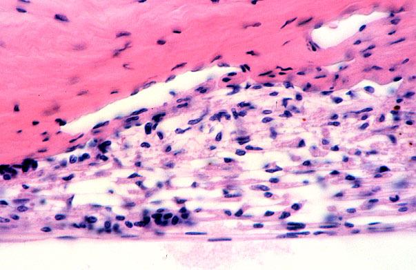
7 Cornea Transparent, avascular, and highly innervated anterior portion of the fibrous tunic that bulges out anteriorly of the orb Thicker than sclera Five layers: Corneal epithelium Bowman s membrane Stroma Descemet s membrane Corneal endothelium

8 Corneal epithelium Corneal epithelium Stratified squamous nonkeratinized ep 5 7 layers of cells Larger superficial cells have microvilli and exhibit zonulae occludentes Interdigitation; junctions: desmosome Have usual array of organelles and intermediate filaments Mitotic figures: mostly near the periphery of the cornea Turn over rate: 7 days Cells can migrate to injured regions Innervated by numeous free nerve endings Sensory nerve fibers from trigeminal ganglion Sympathetic nerve fibers from superior cervical ganglio
9 Bowman s membrane Lies immediately deep to the corneal epithelium Fibrillar lamina, composed of type I collagen fibers arranged in random fashion Synthesized by corneal epithelium and stroma Sensory nerve fibers pass bowman s membrane to enter and terminate in the epithelium

 Limbus Trabecular meshwork Canal")
10 Stroma The thickest layer of the cornea (90%) Transparent Composed of collagen (mostly type I) that are arranged in lamella in parallel to one another Thin elastic fibers, interspersed with collagen fibers Ground substances: (mostly) chondroitin sulfate and keratan sulfate Cells: Fibroblasts Lymphocytes and neutrophil (inflammation condition) Limbus Trabecular meshwork Canal of schlemm


11
12 Descemet s membrane Thick basement membrane interposed between the stroma and endothelium Thin and homogenous in younger becomes thicker and has cross-striations and hexagonal fiber patterns in older adults
13 Corneal endothelium Posterior surface of the cornea; facing anterior chamber Simple squamous epithelium Exhibit numerous pinocytotic vesicles Their membrane have sodium pumps Functions: Responsible for protein synthesis necessary for secreting and maintaining Descemet s membrane Keeping relatively dehydrated maintaining the corneal refractive quality by preventing influx of aqueous humor into stroma
14 Tunica Vasculosa Middle tunic of the eye Is composed of: Choroid Ciliary body Ciliary body Iris
15 Choroid Well vascularized, pigmented layer of the posterior wall Consists of 3 layers Bruch s membrane Choriocapillaries Fenestrated capillaries are responsible providing nutrients to the retina Choroidal stroma Consists of large arteries and veins surrounded by collagen and elastic fibers, fibroblasts, smooth muscles, neurons o/t ANS, and melanocytes Is composed of loose connective tissue The black color is due to the myriad of melanocytes The choroid is separated from retina by Bruch s membrane 1 4 µm thick membrane composed of elastic fibers in the central region and sandwhiched on both sides by collagen fibers. The outer aspect of each collagen fiber layer is covered by basal lamina that belongs to capillaries on one side and the pigment epithelium of the retina on the other side


16 Ciliary Body Ciliary Body Wedge-shaped extension o/t choroid: rings the inner wall o/t eye a/t level o/t lens occupies the space between the ora serrata o/t retina and the iris Surface Sclera: sclerocorneal junction Vitreous body Medial surface projects toward lens: ciliary process Is composed of loose connective tissue containing elastic fibers, blood vessels, and melanocytes
17 Ciliary body Inner surface is lined by pars ciliaris o/t retina that is composed of 2 cell layers: Outer cell layer, w/ faces the lumen o/t orb, is a nonpigmented columnar epithelium Inner cell layer is a pigmented simple columnar epithelium Ciliary process Anterior one third o/t ciliary body Radiate out f/ central core of connective tissue containing fenestrated capillaries Are covered by the same epithelials as ciliary body Nonpigmented layer has many interdigitations and infolding forming aqueous humor that provides nutrients and oxygen for lens n cornea Fiber of Zonula fibers radiate f/ ciliary process to insert into lens capsule suspensory ligaments o/t lens and macromolecule barrier
18 Flow of Aqueous Humor
19
20 Ciliary Body Bulk o/t ciliary body is composed of 3 bundles of smooth muscle (ciliary muscle) 1 bundles stretches the choroid altering the canal schlemm for drainage o/t aqueous humor 2 bundles Attached a/t scleral spur Contraction is mediaterd CN III stretch the choroid body Reducing tensions o/t zonulae lens become thicker and more convex accomodation
21 Iris The anteriormost extension o/t choroid, lies between posterior and anterior chamber; covering the lens excep pupil Anterior surface consists of 2 concentric rings: Pupillary zone Ciliary zone; wider Is irregular Is covered by incomplete layer of pigmented cells and fibroblast Stroma: Poorly vascularized Loose connective tissue: fibroblast and melanocytes Posterior surface: Smooth; covered by two layers of retinal epithelium Heavily pigmented block the light from passing through the iris except pupil Muscle Dilator pupillae; myoepithelial in nature, extension of epithelial cells, innervated by sympathetic nerve, dilates the pupil Sphincter pupillae muscle; smooth muscle, alter diameter of pupil, innervated by CN III (parasympathetic nerve), constricts the pupil Melanocytes block the light from passing through the iris except pupil Imparts the eye color High dark Low blue
22
23 Lens Flexible, biconvex, transparent disc consist of lens capsule, subcapsular epithelium, and lens fiber Lens capsule Basal lamina Type IV collagen + glycoprotein Covers the epithelial and envelops the entire lens Subcapsular epithelium Only on the anterior surface Single layer of cuboidal cells but becoming columnar in the vicinity o/t equator; communicate each other via gap juntions, interdigitation Apices of the cells interdigitate with lens fibers Lens fiber 2000 long cells Compose the bulk o/t lens The cells of subcapsular epithelium give rise to highly differentiated and hexagonal cells (lens fiber) which lose nuclei and organelles and continue elongating; a process called maturation Hexagonal cells are filled with crystallin, lensprotein, increase the refractory index
24
25 Vitreous Body Transparent, refractive gel that fills the vitreous cavity behind the lens Is composed of water (99%), electrolytes, collagen fibers, hyaluronic acid Cells: macrophages and hyalocytes at the periphery o/t vitreous body Hyaloid canal Narrow channel that was occupied by the hyaloid artery in the fetus From the posterior lens to optic disk
26 Neural Tunic (Retina) Innermost tunic; neural portion; Consists of 2 zones Light sensitive sone (pars optica): 2/3 posterior Light non-sensitive zone (pars ciliaris and iridica): 1/3 anterior The scalloped border is ora serrata Consists of 2 layers (light sensitive zone): Outer pigmented layer Inner retinal layer (is composed of 9 distinct layers) Optic disc On the posterior wall o/t orb Is the exit site o/t optic nerve Contains no photoreceptor cells blind spot


27

28 Neural Tunic (Retina) Macula lutea (Yellow spot) 2.5 mm lateral to optic disk Fovea centralis: An oval depression in the center of yellow spot Greatest of visual acuity Contains only cones which are packed tightly as the other layers o/t retina are pushed aside
29


30 Pigment Epithelium (RPE) Pigment Epithelium (RPE)Cuboidal to columnar cells; basal nuclei Basal are attached to Bruch s membrane Mitochondria; invaginations transport Lateral Blood-retina barrier Apical Microvilli and sleeve-like structures that surround and isolate the photoreceptor Abundance Melanin granules Residual bodies Functions Blood-retina barrier Absorb light Preventing reflection from the tunics Phagocytose spent membranous Esterifying vit A derivatives
31 Rods and Cones Rods Activated in dim light only Elongated cells oriented parallel to one another but perpendicular to the retina Are composed of an outer segment, an inner segment, a nuclear region, a synaptic region Outer segment o/t rod Dendritic end; longer in rods th/ in cones Flattened membranous lamellae oriented perpendicular to its long axis Each lamella represents an invaginations o/t plasmalemma Detachment of plasmalemma form a disk Disk is composed of 2 membranes containing rhodopsin Disk migrate to apical end and shed into the sheaths o/t pigment cells and they ll be phagocytosed Inner segment o/t rod separated f/ outer segment by connecting stalk Abundant mitochondria and cytoplasmic granules necessary for production energy for visual process Protein produced in the inner segment migrate to outer segment Cones Are activated in bright light Elongated cells 3 types of cones; different variety of iodopsin sensitivity to red, green, and blue The structure is similar to that of rods with a few exceptions: outer segments, the disk, protein location in outer segment, sensitivity to light and color, and pigment recycling.
32 External (outer) limiting membrane A region of zonulae adherens between Muller cells and photoreceptors Outer nuclear layer Occupied by nuclei o/t rods and cones Outer plexiform layer Axodendritic synapses btw photoreceptor cells and dendrites of bipolar and horizontal cells Two types of synapses: Flat Invaginated: triad a dendrite of a bipolar cells and a dendrite from each of two horizontal cells Ribbon-like lamellae (synaptic ribbon) Neurotransmitter Inner Nuclear layer Occupied by nuclei of bipolar, horizontal, amacrine, and Muller cells Inner plexiform layer The processes of amacrine, bipolar, and ganglion cells Axodendritic synapses Axons of bipolar cells and dendrites of ganglion cells and amacrine cells Two types of synapses: flat and invaginated Dyad: axon of bipolar cell and two dendrites of either amacrine cells or ganglion cells or one dendrite from each two different cells Synapted ribbon Ganglion cell layer Cell bodies of ganglion cells Optic nerve fiber layer Unmyelinated axons of ganglion cells Inner limiting membrane Basal lamina o/t Muller cells
33 Accesory Structure o/t eye 1. Eyelid (palpebra) 2. Conjuctiva 3. Lacrimal apparatus (Glands and ducts)
")

34 Eyelids Fold of skins Are supported by a framework of tarsal plate The margin contain eyelashes arranged in rows of 3 or 4 w/out arrector pilli muscle External surface: str squamous ep of skin Sweat glands, fine hairs, and sebaceous glands of skin Glands of Moll (modified sweat glands) form a spiral before opening into the eyelash follicles Modified sebaceous glands Meibomian glands located in the tarsus of each lid and open on the free edge of the lids Glands of Zeis are associated w/ eyelashes and secrete theirproduct into eyelash follicles Internal surface: palpebral conjunctiva


35 A transparent mucous membrane palpebral conjunctiva: lines the inner surface o/t eyelids Bulbar conjunctiva: covers the sclera is composed of a stratified columnar ep that contains goblet cells Basal lamina Lamina propria composed of loose connective tissue Secretions o/t goblet cells is a part of tear film continues as stratified squamous corneal ep at corneoscleral junction and is devoid of goblet cells Conjuctiva


36 Lacrimal apparatus Lacrimal gland secretes tears Serous, tubuloalveolar gland Myoepithelial cells surround acini Lacrimal canaliculi Lacrimal canaliculi join into a common conduit to lacrimal sac Stratified squamous ep. Lacrimal sac Is a dilated superior portion of nasolacrimal duct Pseudostratified ciliated columnar ep Nasolacrimal duct Inferior continuation o/t lacrimal sac Pseudostratified ciliated columnar ep Carries the lacrimal fluid into inferior meatus located in the floor of nasal cavity
Histology of the Eye
 Histology of the Eye Objectives By the end of this lecture, the student should be able to describe: The general structure of the eye. The microscopic structure of:»cornea.»retina. EYE BULB Three coats
XUE HUI Department of Histology& Embryology, Basic Medicine College of Jilin University
 SENSE ORGAN XUE HUI Department of Histology& Embryology, Basic Medicine College of Jilin University EYE fibrous globe lens photosensitive cells a system of cells and nerves concentric layers the sclera
Unit VIII Problem 8 Anatomy: Orbit and Eyeball
 Unit VIII Problem 8 Anatomy: Orbit and Eyeball - The bony orbit: it is protecting our eyeball and resembling a pyramid: With a base directed: anterolaterally. And an apex directed: posteromedially. Notes:
The Special Senses: Part A
 PowerPoint Lecture Slides prepared by Janice Meeking, Mount Royal College CHAPTER 15 The Special Senses: Part A Warm Up What is the function of the eyeball? List any structures of the eyeball that you
4/22/16. Eye. External Anatomy of Eye. Accessory Structures. Bio 40B Dr. Kandula
 Eye Bio 40B Dr. Kandula External Anatomy of Eye Accessory Structures l Eyebrows l Levator Palpebrae Superioris - opens eye l Eyelashes l Ciliary glands modified sweat glands l Small sebaceous glands l
The Orbit. The Orbit OCULAR ANATOMY AND DISSECTION 9/25/2014. The eye is a 23 mm organ...how difficult can this be? Openings in the orbit
 The eye is a 23 mm organ...how difficult can this be? OCULAR ANATOMY AND DISSECTION JEFFREY M. GAMBLE, OD COLUMBIA EYE CONSULTANTS OPTOMETRY & UNIVERSITY OF MISSOURI DEPARTMENT OF OPHTHALMOLOGY CLINICAL
THE EYE: RETINA AND GLOBE
 Neuroanatomy Suzanne Stensaas February 24, 2011, 10:00-12:00 p.m. Reading: Waxman Ch. 15. Your histology and gross anatomy books should be useful. Reading: Histology of the Eye from any histology book
Special Senses: The Eye
 Unit 4 Special Senses: The Eye ESSENTIALS OF HUMAN ANATOMY & PHYSIOLOGY The Senses General senses of touch Temperature Pressure Pain Special senses Smell Taste Sight Hearing Equilibrium The Eye and Vision
Ocular Anatomy for the Paraoptometric
 Ocular Anatomy for the Paraoptometric Minnesota Optometric Association Paraoptometric CE Friday September 30, 2016 Lindsay A. Sicks, OD, FAAO Assistant Professor, Illinois College of Optometry lsicks@ico.edu
The Eye. The Orbit. The EYE What a Trip!!! - The Anterior Segment 5/12/2015. Jill J Luebbert, CPOT, ABOC
 The EYE What a Trip!!! - The Anterior Segment Jill J Luebbert, CPOT, ABOC The Eye The Orbit Bony socket containing the eye and most of its accessory organs consisting of 7 bones 1 The Seven Bones of the
Special Senses PART A
 8 Special Senses PART A PowerPoint Lecture Slide Presentation by Jerry L. Cook, Sam Houston University ESSENTIALS OF HUMAN ANATOMY & PHYSIOLOGY EIGHTH EDITION ELAINE N. MARIEB The Senses General senses
Taste buds Gustatory cells extend taste hairs through a narrow taste pore
 The Special Senses Objectives Describe the sensory organs of smell, and olfaction. Identify the accessory and internal structures of the eye, and explain their function. Explain how light stimulates the
Vision I. Steven McLoon Department of Neuroscience University of Minnesota
 Vision I Steven McLoon Department of Neuroscience University of Minnesota 1 Eye Cornea Sclera Conjunctiva 2 Eye The conjunctiva lines the inner surface of the eyelids and outer surface of the sclera. 3
THE SPECIAL SENSES. Introduction Vision
 THE SPECIAL SENSES Introduction Vision RECEPTORS Structures designed to respond to stimuli Variable complexity RECEPTORS: GENERAL PROPERTIES Transducers Receptor Potential Generator Potential RECEPTORS
Sense of Vision. Chapter 8. The Eye and Vision. The Eye Orbit. Eyebrows, Eyelids, Eyelashes. Accessory Organs 5/3/2016.
 Sense of Vision Chapter 8 Special Senses The Eye and Vision 70 percent of all sensory receptors are in the eyes Each eye has over 1 million nerve fibers Protection for the eye Most of the eye is enclosed
1 Eyelids. Lacrimal Apparatus. Orbital Region. 3 The Orbit. The Eye
 1 1 Eyelids Orbital Region 2 Lacrimal Apparatus 3 The Orbit 4 The Eye 2 Eyelids The eyelids protect the eye from injury and excessive light by their closure. The upper eyelid is larger and more mobile
Medical School Histology Basics. VIBS 289 lab. Eye
 Medical School Histology Basics VIBS 289 lab Eye Larry Johnson Texas A&M University Aqueous humor OUTLINE OVERVIEW CELLULAR STRUCTURES THROUGH WHICH LIGHT PASSES A. CORNEA B. LENS C. RETINA STRUCTURES
02/03/2014. Average Length: 23mm (Infant ~16mm) Approximately the size of a quarter Volume: ~5mL
 Approximately the size of a quarter Volume: ~5mL") Identify the anatomy of the eye. Explain the basic physiology of the parts of the eye. Briefly discuss various surgeries related to different parts of the anatomy. Average Length: 23mm (Infant ~16mm) Approximately
Identify the anatomy of the eye. Explain the basic physiology of the parts of the eye. Briefly discuss various surgeries related to different parts of the anatomy. Average Length: 23mm (Infant ~16mm) Approximately
The Nervous System: General and Special Senses Pearson Education, Inc.
 18 The Nervous System: General and Special Senses Introduction Sensory information arrives at the CNS Information is picked up by sensory receptors Sensory receptors are the interface between the nervous
ACTIVITIES. Complete Diagrams PNS 18 and 19 Complete PNS 23 Worksheet 3 #1 only Complete PNS 24 Practice Quiz
 ACTIVITIES Complete Diagrams PNS 18 and 19 Complete PNS 23 Worksheet 3 #1 only Complete PNS 24 Practice Quiz THE SPECIAL SENSES Introduction Vision RECEPTORS Structures designed to respond to stimuli Variable
SPECIAL SENSES PART I: OLFACTION & GUSTATION
 SPECIAL SENSES PART I: OLFACTION & GUSTATION 5 Special Senses Olfaction Gustation Vision Equilibrium Hearing Olfactory Nerves Extend through cribriform plate into nasal cavity on both sides of nasal septum
n Corneal epithelium is derived from surface ectoderm n Composed of stratified squamous epith. n 5% of total corneal thickness (50-90micro m thick)
") Cornea overview Dr. Sarita Tuladhar MD, Ophthalmology Gandaki Medical College Embryology CORNEA: n Corneal epithelium is derived from surface ectoderm n Corneal stroma, descement memb, bowman s layer,
Cornea overview Dr. Sarita Tuladhar MD, Ophthalmology Gandaki Medical College Embryology CORNEA: n Corneal epithelium is derived from surface ectoderm n Corneal stroma, descement memb, bowman s layer,
Bony orbit Roof The orbital plate of the frontal bone Lateral wall: the zygomatic bone and the greater wing of the sphenoid
 Bony orbit Roof: Formed by: The orbital plate of the frontal bone, which separates the orbital cavity from the anterior cranial fossa and the frontal lobe of the cerebral hemisphere Lateral wall: Formed
o A cushion of fat surrounds most of the eye
 Name Period SPECIAL SENSES The Senses of touch o Temperature o Pressure o Pain o Smell o Taste o Sight o Hearing o Equilibrium The Eye and Vision are in the eyes has over a o Most of the eye is enclosed
Chapter 17, Part 1! The Special Senses! SECTION 17-1! Olfaction, the sense of smell, involves olfactory receptors responding to chemical stimuli!
 Chapter 17, Part 1! The Special Senses! SECTION 17-1! Olfaction, the sense of smell, involves olfactory receptors responding to chemical stimuli! 2! 1! Olfaction A Chemical Sensation! Olfactory epithelium!
INTRODUCTION: ****************************************************************************************************
 BIOLOGY 211: HUMAN ANATOMY & PHYSIOLOGY **************************************************************************************************** EYES AND VISION ****************************************************************************************************
The sebaceous glands (glands of Zeis) open directly into the eyelash follicles, ciliary glands (glands of Moll) are modified sweat glands that open
 open directly into the eyelash follicles, ciliary glands (glands of Moll) are modified sweat glands that open") The Orbital Region The orbits are a pair of bony cavities that contain the eyeballs; their associated muscles, nerves, vessels, and fat; and most of the lacrimal apparatus upper eyelid is larger and more
The Orbital Region The orbits are a pair of bony cavities that contain the eyeballs; their associated muscles, nerves, vessels, and fat; and most of the lacrimal apparatus upper eyelid is larger and more
Basic Histology. By Mrs. Bailey
 Basic Histology By Mrs. Bailey Primary Tissues 1. Epithelial Tissue 2. Connective Tissue 3. Muscle Tissue 4. Nervous Tissue Very cellular Supported by underlying connective tissue Epithelial & connective
o A cushion of fat surrounds most of the eye
 Name Period SPECIAL SENSES The Senses General senses of touch o Temperature o Pressure o Pain Special senses o Smell o Taste o Sight o Hearing o Equilibrium The Eye and Vision 70 percent of all sensory
Chapter 15 Taste, Smell and Vision
 Chapter 15 Taste, Smell and Vision The special senses are so named because they are associated with specific areas of the cortex. Touch is a general sense, so it s not included with the special senses.
Sensory system. Dr. Carmen E. Rexach Anatomy 35 Mt San Antonio College
 Sensory system Dr. Carmen E. Rexach Anatomy 35 Mt San Antonio College Sensory receptors Detect stimuli Classified by structure Origin Distribution Modality Structural Classification naked nerve endings
Special Senses: Vision
 ighapmlre24pg223_230 5/12/04 2:27 PM Page 223 impos03 302:bjighapmL:ighapmLrevshts:layouts: NAME LAB TIME/DATE Special Senses: Vision REVIEW SHEET exercise 24 Anatomy of the Eye 1. Name five accessory
let's continue talking about the eye,
 Eye is mainly composed of 3 layers: External layer, which called The Sclera which is a hard connective tissue that gives the eye its round shape. Extension of the sclera into the front is the cornea, which
Tissues. tissue = many cells w/ same structure and function. cell shape aids its function tissue shape aids its function
 Tissues tissue = many cells w/ same structure and function cell shape aids its function tissue shape aids its function Histology = study of tissues 4 types of tissues Epithelial coverings contact openings
1 Anatomy of the Eyeball
 1 Anatomy of the Eyeball 1 Anatomy of the Eyeball ANATOMY Eyeball Tunics of Eyeball Segments of the Eye Blood Supply of Eyeball Blood Supply of Retina Blood Supply of Uvea Nerve Supply of Eyeball DEVELOPMENT
The orbit-2. Dr. Heba Kalbouneh Assistant Professor of Anatomy and Histology
 The orbit-2 Dr. Heba Kalbouneh Assistant Professor of Anatomy and Histology Eyelids The eyelids (act like the curtains) protect the eye from injury and excessive light by their closure The upper eyelid
Eye. iris. pupil. ciliary body
 Eye iris pupil ciliary body Eyeball Anterior segment Posterior segment Eyeball Procesus ciliares Corpus ciliare Pars caeca retinae Sclera Iris Cornea Eyebulb wall Tunica externa (fibrosa) Posterior segment
Unit I Problem 9 Histology: Basic Tissues of The Body
 Unit I Problem 9 Histology: Basic Tissues of The Body - What is the difference between cytology and histology? Cytology: it is the study of the structure and functions of cells and their contents. Histology:
The Tissue Level of Organization
 Tissue The Tissue Level of Organization Chapter 3 Definition an aggregation of cells in which each cooperates with all others in the performance of a given function Examples of general functions Movement
A. cells that perform related functions and are similar in structure. B. extracellular material - made by cells and secreted into interstitial space
 I. tissue components A. cells that perform related functions and are similar in structure B. extracellular material - made by cells and secreted into interstitial space II. tissue types A. epithelium (e.)
Tissues are: group of similar or identical cells that share a common function. used to build organs
 Tissues: Four classes Epithelium Connective Muscle Nervous Tissues are: group of similar or identical cells that share a common function. used to build organs Overview: Epithelial o Line body cavities
Test Bank for Medical Surgical Nursing An Integrated Approach 3rd Edition by White
 Test Bank for Medical Surgical Nursing An Integrated Approach 3rd Edition by White Link full download : http://testbankair.com/download/test-bank-for-medical-surgical-nursing-anintegrated-approach-3rd-edition-by-white/
Lecture Overview. Marieb s Human Anatomy and Physiology. Chapter 4 Tissues: The Living Fabric Epithelial Tissues Lecture 9. Introduction to Tissues
 Marieb s Human Anatomy and Physiology Marieb Hoehn Chapter 4 Tissues: The Living Fabric Epithelial Tissues Lecture 9 Lecture Overview Introduction to Tissues Epithelial Tissues Location General characteristics
Dr Narmeen S. Ahmad. Lab 1
 Dr Narmeen S. Ahmad Lab 1 1 Tissues are groups of cells with a common structure (form) and function (job). There are (4) types of tissue: 1. Epithelial 2. Connective 3. Muscle 4. Nervous 2 Epithelial cells
GNK485 The eye and related structures. Prof MC Bosman 2012
 GNK485 The eye and related structures Prof MC Bosman 2012 Surface anatomy Bony orbit Eyeball and Lacrimal apparatus Extra-ocular muscles Movements of the eye Innervation Arterial supply and venous drainage
1. Anatomy of the Eye, Ocular Adnexa and Visual Pathway (26-34 Items)
") B. Ocular/Visual Biology - 90 Items (21%) "Ocular/Visual Biology" covers the fundamental knowledge and scientific principles that support the application of these principles in the prevention, diagnosis,
B. Ocular/Visual Biology - 90 Items (21%) "Ocular/Visual Biology" covers the fundamental knowledge and scientific principles that support the application of these principles in the prevention, diagnosis,
Image Formation and Phototransduction. By Dr. Abdelaziz Hussein Lecturer of Physiology
 Image Formation and Phototransduction By Dr. Abdelaziz Hussein Lecturer of Physiology Vision Vision is a complex process through which an image of the external environment is formed on the photosensitive
20-20,000 Hertz range of human hearing
 20-20,000 Hertz range of human hearing accommodation automatic adjustment in focal length of the lens of the eye; changing the shape of the lens aqueous humor Watery fluid in the anterior chambers of the
Head: Special Senses. Taste Smell Vision Hearing/Balance
 Head: Special Senses Taste Smell Vision Hearing/Balance TASTE: how does it work? Taste buds on tongue on fungiform papillae ( mushroom-like projections) Each bud contains several cell types in microvilli
Lecture Overview. Chapter 4 Epithelial Tissues Lecture 9. Introduction to Tissues. Epithelial Tissues. Glandular Epithelium
 Visual Anatomy & Physiology First Edition Martini & Ober Chapter 4 Lecture 9 Lecture Overview Introduction to Tissues Location General characteristics Functions Classification Glandular Epithelium 2 Where
Chapter 7, Section 1 Review Questions. Directions: Place the letter of the best definition next to each key term. Name PER Date
 Name PER Date Chapter 7, Section 1 Review Questions Directions: Place the letter of the best definition next to each key term. A. the middle layer of the wall of the eye B. the structure between the choroid
Epithelium. Four primary tissue types:
 Epithelium Four primary tissue types: Epithelial (covering) Connective (support) Nervous (control) Muscular (movement) Smooth muscle Cardiac muscle Skeletal muscle 1 Epithelial Tissue Features Epithelial
The Senses. Chapter 10 7/8/11. Introduction
 Chapter 10 The Senses Introduction A. Sensory receptors detect changes in the environment and stimulate neurons to send nerve impulses to the brain. B. A sensation is formed based on the sensory input.
Copyright 2009 Pearson Education, Inc.
 Outline Nervous System Sensory Systems I. II. III. IV. V. VI. Biol 105 Lecture 11 Chapter 9 Senses Sensory receptors Touch Vision Hearing and balance Smell Senses Sensory receptor cells Sensory receptors
Tissues Chapter 5...Tissue - a group or mass of similar cells working together to perform certain common functions
 Tissues Chapter 5...Tissue - a group or mass of similar cells working together to perform certain common functions There are 4 major types of tissue Epithelial Connective Muscle Nervous 1. Epithelial Tissue
Epithelial Lecture Test Questions
 Epithelial Lecture Test Questions 1. Which of the following free surfaces lack(s) epithelia: a. lung alveoli (air sacs) b. hard palate c. joint cavities d. abdominal cavity e. salivary gland ducts 2. Which
Around The Globe in 60 Minutes
 Around The Globe in 60 Minutes Around the GLOBE in Sixty Minutes Basic Ocular Anatomy, Examination, and Diagnostic Techniques Introduction Focusing on canine and feline ocular anatomy and basic examination
The Special Senses. Smell, taste, vision, hearing and equilibrium Housed in complex sensory organs
 The Special Senses Smell, taste, vision, hearing and equilibrium Housed in complex sensory organs Chemical Senses Interaction of molecules with receptor cells Olfaction (smell) and gustation (taste) Both
Bony orbit. Lateral wall: Formed by : the zygomatic bone and the greater wing of the sphenoid
 Bony orbit Roof: Formed by: The orbital plate of the frontal bone, which separates the orbital cavity from the anterior cranial fossa and the frontal lobe of the cerebral hemisphere Lateral wall: Formed
Lec #2 histology. Bronchioles:
 Lec #2 histology. Last lecture we talked about the upper respiratory tract histology, this one is about the lower part histology. We will discuss the histology of: -bronchioles -respiratory bronchioles
Chapter 1: Cells and Tissues
 Chapter 1: Cells and Tissues Cells and Tissues Carry out all chemical activities needed to sustain life Cells are the building blocks of all living things Tissues are groups of cells that are similar in
Vision is the most dominant sense, about 70% of all sensory receptors in the body are in the eyes Accessory Structures of the eye : Eyelashes :
 Sight By Jess Kapp Vision is the most dominant sense, about 70% of all sensory receptors in the body are in the eyes Accessory Structures of the eye : Eyelashes : Protect eye from debris and bacteria Eyebrows
Surgical Anatomy Ear and Eye. Presenters: Dr. Jim Hurrell and Dr. Dennis McCurnin
 Surgical Anatomy Ear and Eye Presenters: Dr. Jim Hurrell and Dr. Dennis McCurnin A Warm Welcome from My Faculty TEAM and Me!!! 2 The Pledge of Allegiance 3 The Senses 4 Hearing 3 Layers of Ear EXTERNAL
Histology review. Histology. Slides. Epithelial tissue. Another example - kidney. Simple cuboidal epithelium. What to look for
 Histology review Histology What to look for Histology Practical = 50 pts Some slides set up on scopes (~10) Some Powerpoint pictures on the projector Questions I will ask: What kind of tissue? General
Eye Movements. Geometry of the Orbit. Extraocular Muscles
 Eye Movements Geometry of the Orbit The eye (oculus) is located in the anterior aspect of the orbit: the equator of the eye (defined by a coronal plane passing through its middle) lies at the margin of
Histology = the study of tissues. Tissue = a complex of cells that have a common function
 { EPITHELIAL TISSUE Histology = the study of tissues Tissue = a complex of cells that have a common function The Four Primary Tissue Types: Epithelium (epithelial tissue) covers body surfaces, lines body
Chapter(2):the lid page (1) THE LID
:the lid page (1) THE LID") Chapter(2):the lid page (1) THE LID Anatomy of the lid: * Check movie anatomy of the lid model The eyelids are two movable muco-cutaneous folds which protect the eye on closure. The are joined temporary
Chapter(2):the lid page (1) THE LID Anatomy of the lid: * Check movie anatomy of the lid model The eyelids are two movable muco-cutaneous folds which protect the eye on closure. The are joined temporary
Study of different tissues Abnormal cells and tissues can be compared to normal tissues to identify disease, such as cancer Being able to know and
 CHAPTER 4 Study of different tissues Abnormal cells and tissues can be compared to normal tissues to identify disease, such as cancer Being able to know and recognize normal tissues under the microscope
is the clear, transparent part at the front of the eye. It allows light to enter the eye and it also refracts (focuses) the light onto the retina.
 the light onto the retina.") Senses- Vision Light is a small part (1/70th) of the total electromagnetic (EM) spectrum. The EM band extends from radio waves at one extreme to x-rays at the other. The eye detects light and converts
Senses- Vision Light is a small part (1/70th) of the total electromagnetic (EM) spectrum. The EM band extends from radio waves at one extreme to x-rays at the other. The eye detects light and converts
Body Tissues Pearson Education, Inc.
 Body Tissues Tissues Groups of cells with similar structure and function Four primary types: Epithelial tissue (epithelium).1 Connective tissue.2 Muscle tissue.3 Nervous tissue.4 Epithelial Tissues Locations:
Lab Animal Tissue. LEARNING OBJECTIVES: To understand the relationship between the structure and function of different animal tissues
 Name: Bio A.P. PURPOSE: HYPOTHESIS: NONE Lab Animal Tissue BACKGROUND: In animals, groups of closely related cells specialized to perform the same function are called tissues. There are four general classes
The Visual System. Retinal Anatomy Dr. Casagrande February 2, Phone: Office: T2302 MCN
 The Visual System Retinal Anatomy Dr. Casagrande February 2, 2004 Phone: 343-4538 Email: vivien.casagrande@mcmail.vanderbilt.edu Office: T2302 MCN Reading assignments and Good Web Sites Chapter 2 in Tovée,
Tissue: The Living Fabric: Part A
 PowerPoint Lecture Slides prepared by Janice Meeking, Mount Royal College C H A P T E R 4 Tissue: The Living Fabric: Part A Tissues Groups of cells similar in structure and function Types of tissues Epithelial
Tissue Outline (chapter 4) Tissues group of cells that perform structural and roles. List the 4 types:
 Tissues group of cells that perform structural and roles. List the 4 types:") Tissue Outline (chapter 4) Tissues group of cells that perform structural and roles. List the 4 types: 1. 2. 3. 4. I. Epithelial Tissue covers all the surfaces, inside & out. Are the major tissues of,
Tissue Outline (chapter 4) Tissues group of cells that perform structural and roles. List the 4 types: 1. 2. 3. 4. I. Epithelial Tissue covers all the surfaces, inside & out. Are the major tissues of,
Tissues organs system organism. pg151
 Histology is the study of tissues A TISSUE is a group of cells, usually of one kind, & their intercellular substance (e.g. intercellular matrix in animal) which are linked together & perform a particular
Anatomy Chapter 4 Tissues
 4 Principle Tissue Types Epithelial tissue Covering and lining Glandular Connective tissue Highly variable Most abundant tissue type Muscular tissue 3 major types Produce force through contraction Nervous
Integumentary System. Integumentary System
 1. General aspects a. The integumentary system consists of several organs major organ of the system is the skin other organs are relatively small and they can be considered as specialized structures of
Histology. Study of body tissues
 Histology Study of body tissues 2 Introduction to Body Tissues 1. Composed of specialized cells of similar structure and perform a common function 2. Four major types (4 Cs) a. Epithelial - Cover b. Connective
Tissues. How do cells form tissues?
 Tissues How do cells form tissues? Using cell junctions Tissues Epithelial tissue Connective tissue Muscle tissue Nervous tissue Epithelial Tissue Closely packed cells in continuous sheets connected by
Tissues. tissue = many cells w/ same structure and function. cell shape aids function tissue shape aids function. Histology = study of tissues
 Tissues tissue = many cells w/ same structure and function cell shape aids function tissue shape aids function Histology = study of tissues 4 types of tissues Epithelial coverings contact openings Connective
HISTOLOGY OF THE RESPIRATORY SYSTEM I. Introduction A. The respiratory system provides for gas exchange between the environment and the blood. B.
 HISTOLOGY OF THE RESPIRATORY SYSTEM I. Introduction A. The respiratory system provides for gas exchange between the environment and the blood. B. The human respiratory system may be subdivided into two
Bi 121 Lab OLFACTION. olfactory bulb, olfactory nerve (=cranial nerve I), olfactory foramina, olfactory epithelium
, olfactory foramina, olfactory epithelium") Bi 121 Lab Week 9: THE SPECIAL SENSES The special senses include smell, taste, vision, hearing, and balance. In this laboratory exercise, we will look at many of the structures that provide for these senses.
Bi 121 Lab Week 9: THE SPECIAL SENSES The special senses include smell, taste, vision, hearing, and balance. In this laboratory exercise, we will look at many of the structures that provide for these senses.
Ocular Anatomy & Physiology. Learning Objectives: Let s get oriented first. 3 Major Layers (Tunics) of EYE. Topics to be covered: FIBROUS TUNIC
 of EYE. Topics to be covered: FIBROUS TUNIC") Lecturer: Ocular Anatomy & Physiology M. Patrick COLEMAN, ABOC, COT Kerrville, TX Learning Objectives: 1. Correctly identify ocular structures around or within the eye 2. List the key functions of various
Lecturer: Ocular Anatomy & Physiology M. Patrick COLEMAN, ABOC, COT Kerrville, TX Learning Objectives: 1. Correctly identify ocular structures around or within the eye 2. List the key functions of various
The white of the eye and the part that maintains its shape is know n as the:
 Scrub In The white of the eye and the part that maintains its shape is know n as the: a. Cornea b. Pupil c. Retina d. Sclera The structure that is found in the ear and contains the organ of hearing is
EPITHELIUM 3/12/2018 د. درويش بدران د. ماهر الحديدي د.امجد الشطرات و احسان العمري
 EPITHELIUM 1 2 3 1- SIMPLE SQUAMOUS EPITHELIUM It is a single layer of flat cells that resembles a tiled floor when viewed from apical surface; centrally located nucleus that is flattened and oval or spherical
Essentials of Anatomy and Physiology, 9e (Marieb) Chapter 3 Cells and Tissues. Short Answer. Figure 3.1
 Chapter 3 Cells and Tissues. Short Answer. Figure 3.1") Essentials of Anatomy and Physiology, 9e (Marieb) Chapter 3 Cells and Tissues Short Answer Figure 3.1 Using Figure 3.1, match the following: 1) The illustration of simple cuboidal epithelium is. Answer:
Essentials of Anatomy and Physiology, 9e (Marieb) Chapter 3 Cells and Tissues Short Answer Figure 3.1 Using Figure 3.1, match the following: 1) The illustration of simple cuboidal epithelium is. Answer:
Epithelia will be discussed according to the following scheme: Type Number of layers Shape Line drawing. Squamous Cuboidal Columnar
 Epithelia Epithelia will be discussed according to the following scheme: Type Number of layers Shape Line drawing Simple Squamous Cuboidal Columnar Covering and Lining epithelium Pseudostratified Stratified
Special Senses. Accessory Structures of the Eye. The Eye and Vision. Accessory Structures of the Eye. Accessory Structures of the Eye
 8 PART A Special Senses PowerPoint Lecture Slide Presentation by Jerry L. Cook, Sam Houston University ESSENTIALS OF HUMAN ANATOMY & PHYSIOLOGY EIGHTH EDITION ELAINE N. MARIEB The Senses General senses
Biology. Dr. Khalida Ibrahim
 Dr. Khalida Ibrahim Biology Histology: Histology: is the study of the tissues of the body. Tissue: group of similar cells combined to perform a common function. The human body is composed of only 4 basic
Tissues 10/21/2016. Epithelial Tissue
 Tissues This is a generalized cell diagram. It shows the anatomy of a cell, but most cells do not actually look like this. Cells can have a wide variety of shapes and sizes, depending on their function.
Essential questions. What are the structures of the sensory system? 3.03 Remember the structures of the sensory system 2
 Essential questions What are the structures of the sensory system? 3.03 Remember the structures of the sensory system 2 The Senses Eyes Sight Ears Hearing Nose Smell Tongue Taste Skin Touch 3.03 Remember
B. Classification of epithelium: by number of cell layers present and by shape of the superficial cell layers.
 I. Introduction - tissue: group of cells that are closely associated, similar in structure and function, and perform a common or related function. - four primary tissues: epithelial tissue, connective
Lesson 9A Tissues in Animals
 Lesson 9A Tissues in Animals Levels of Organization in the Human Body Similar types of cells Different types of tissues Different organs Many organ systems cell tissue organ organ system organism Levels
Skin. Kristine Krafts, M.D.
 Skin Kristine Krafts, M.D. Skin Lecture Objectives Describe the functions of skin. Describe the structure, location and function of the cell types found in epidermis: keratinocytes, melanocytes, Langerhans
Most abundant and widely distributed tissues in the body Binds, support, and strengthen body tissues, protect and insulate internal organ, serve as
 Connective tissue Most abundant and widely distributed tissues in the body Binds, support, and strengthen body tissues, protect and insulate internal organ, serve as major transport system, compartmentalizes
CHAPTER 05 Histology: EPITHELIUM
 BIO 211: ANATOMY & PHYSIOLOGY I 1 CHAPTER 05 Histology: EPITHELIUM Part 01: Brief Introduction Part 02: Survey of Types Dr. Lawrence G. G. Altman www.lawrencegaltman.com Some illustrations are courtesy
TISSUES TYPES. CHAPTER 05 Histology: EPITHELIUM BIO 211: ANATOMY & PHYSIOLOGY I. HISTOLOGY = the study of tissues
 BIO 211: ANATOMY & PHYSIOLOGY I 1 CHAPTER 05 Histology: EPITHELIUM Part 01: Brief Introduction Part 02: Survey of Types Dr. Lawrence G. G. Altman www.lawrencegaltman.com Some illustrations are courtesy
Anatomy Ch 6: Integumentary System
 Anatomy Ch 6: Integumentary System Introduction: A. Organs are body structures composed of two or more different tissues. B. The skin and its accessory organs make up the integumentary system. Types of