Bony orbit. Lateral wall: Formed by : the zygomatic bone and the greater wing of the sphenoid
|
|
|
- Karin Chase
- 5 years ago
- Views:
Transcription


1 Bony orbit Roof: Formed by: The orbital plate of the frontal bone, which separates the orbital cavity from the anterior cranial fossa and the frontal lobe of the cerebral hemisphere Lateral wall: Formed by : the zygomatic bone and the greater wing of the sphenoid


2 Medial wall: Formed from before backward by: The frontal process of the maxilla The lacrimal bone The orbital plate of the ethmoid (which separates the orbital cavity from the ethmoid sinuses) The body of the sphenoid Floor :Formed by: the orbital plate of the maxilla, which separates the orbital cavity from the maxillary sinus
: It")


3 Openings Into the Orbital Cavity 1-Supraorbital notch (Foramen): It transmits the supraorbital nerve and blood vessels 2-Infraorbital groove and canal: Situated they transmit the infraorbital nerve (a continuation of the maxillary nerve) and blood vessels.


4 3-Inferior orbital fissure: Located posteriorly between the maxilla and the greater wing of the sphenoid it communicates with the pterygopalatine fossa. It transmits 1-the maxillary nerve and its zygomatic branch 2-the inferior ophthalmic vein and sympathetic nerves. 4-Nasolacrimal canal: Located anteriorly on the medial wall; it communicates with the inferior meatus of the nose It transmits the nasolacrimal duct.


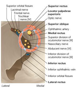
5 5-Superior orbital fissure: Located posteriorly between the greater and lesser wings of the sphenoid it communicates with the middle cranial fossa. It transmits the lacrimal nerve the frontal nerve the trochlear nerve the oculomotor nerve (upper and lower divisions) the abducent nerve, the nasociliary nerve the superior ophthalmic vein. 6-Optic canal: Located posteriorly in the lesser wing of the sphenoid it communicates with the middle cranial fossa. It transmits the optic nerve and the ophthalmic artery

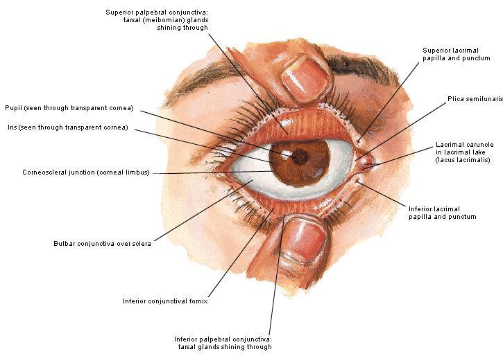
6 The Orbital Region The orbits are a pair of bony cavities that contain the eyeballs Eyelids The eyelids (they act like the curtains) protect the eye from injury and excessive light by their closure The upper eyelid is larger and more mobile than the lower because of its attachment to the levator palpebrae superioris The upper and lower eyelids meet each other at the medial and lateral angles. The palpebral fissure is the elliptical opening between the eyelids The palpebral fissure is the entrance into the conjunctival sac
 open directly into the eyelash follicles.")
 Contains the palpebral part of")
7 structure of the eye lids 1-skin:thin and can be easily become oedematous (with fluid or blood) Contains The sebaceous glands (glands of Zeis) open directly into the eyelash follicles. The ciliary glands (glands of Moll) are modified sweat glands that open separately between adjacent lashes 2- Superficial fascia: ( remember we said earlier No fat) Contains the palpebral part of orbicularis occuli


8 3- Palpebral fascia The framework of the eyelids is formed by a fibrous sheet, the orbital septum The orbital septum is attached to the periosteum at the orbital margins. The orbital septum is thickened at the margins of the lids to form the superior and inferior TARSAL PLATES. The lateral ends of the tarsal plates are attached by a band, the lateral palpebral ligament, the orbital margin. The medial ends of the plates are attached by a band, the medial palpebral ligament, to the lacrimal bone The tarsal glands are embedded in the posterior surface of the tarsal plates


9 4-The conjunctiva is a thin mucous membrane that lines the eyelids It is reflected at the superior and inferior fornices onto the anterior surface of the eyeball The upper lateral part of the superior fornix is pierced by the ducts of the lacrimal gland The conjunctiva thus forms a potential space, the conjunctival sac, which is open at the palpebral fissure.
10


![cervical ganglion Loss of oculomotor nerve [III] function results in complete ptosis or drooping of](/docs-images/84/90676776/images/11-7.jpg "the superior eyelid, whereas loss of sympathetic innervation to the superior tarsal muscle results")
11 Movements of the Eyelids The position of the eyelids at rest depends on the tone of : 1-The orbicularis oculi 2-The levator palpebrae superioris muscles and the position of the eyeball. The eyelids are closed by : 1-The contraction of the orbicularis oculi and 2-The relaxation of the levator palpebrae superioris muscles The eye is opened by: THE LEVATOR PALPEBRAE SUPERIORIS Raising the upper lid the superior tarsal muscle which is part of the levator palpebrae superioris, helps maintain eyelid elevation and are innervated by postganglionic sympathetic fibers from the superior cervical ganglion Loss of oculomotor nerve [III] function results in complete ptosis or drooping of the superior eyelid, whereas loss of sympathetic innervation to the superior tarsal muscle results in partial ptosis


12 Horner's syndrome Horner's syndrome is caused by a lesion in the sympathetic trunk in the neck that results in sympathetic dysfunction. It is characterized by three typical features: 1-Pupillary constriction due to paralysis of the dilator pupillae muscle; 2-Partial ptosis (drooping of the upper eyelid) due to paralysis of the superior tarsal muscle of the levator palpebrae superioris; 3-Absence of sweating on the ipsilateral side of the face and the neck due to absence of innervation of the sweat glands.


13 Lacrimal Gland The lacrimal gland consists of: 1-a large orbital part 2- a small palpebral part which are continuous with each other around the lateral edge of the aponeurosis of the levator palpebrae superioris. It is situated above the eyeball in the anterior and upper part of the orbit posterior to the orbital septum The gland opens into the lateral part of the superior fornix of the conjunctiva by 12 ducts.
14


15 Lacrimal Ducts The tears circulate across the cornea and accumulate in the lacus lacrimalis. From here the tears enter the canaliculi lacrimales through the puncta lacrimalis. The canaliculi lacrimales pass medially and open into the lacrimal sac which lies in the lacrimal groove behind the medial palpebral ligament and is the upper blind end of the nasolacrimal duct. The nasolacrimal duct is about 0.5 in. (1.3 cm) long and emerges from the lower end of the lacrimal sac The duct descends downward, backward, and laterally in a bony canal and opens into the inferior meatus of the nose.


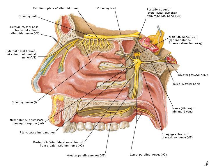
16 5-They then pass into its zygomatic branch and the zygomaticotemp oral nerve 6-They reach the lacrimal gland within the lacrimal nerve 2-The preganglionic fibers reach the pterygopalatine ganglion (sphenopalatine ganglion) via the nervus intermedius and its great petrosal branch 4-joins the maxillary nerve. 1-The parasympathetic secretomotor nerve supply is derived from the lacrimal nucleus of the facial nerve 3-via the nerve of the pterygoid canal. The postganglionic fibers leave the ganglion


17 5-finally the lacrimal nerve 4- via the zygomatic nerve, the zygomaticotempor al nerve 4-the maxillary nerve the maxillary nerve 2-travels in the deep petrosal nerve 3-then in the nerve of the pterygoid canal, 1-The sympathetic postganglionic nerve supply is from the internal carotid plexus
18


19 Nerves of the Orbit Optic Nerve The optic nerve enters the orbit from the middle cranial fossa by passing through the optic canal It is accompanied by the ophthalmic artery, which lies on its lower lateral side. The nerve is surrounded by sheaths of pia mater, arachnoid mater, and dura mater It runs forward and laterally within the cone of the recti muscles and pierces the sclera at a point medial to the posterior pole of the eyeball Remember that the meninges fuse with the sclera so that the subarachnoid space with its contained cerebrospinal fluid extends forward from the middle cranial fossa, around the optic nerve, and through the optic canal, as far as the eyeball. A rise in pressure of the cerebrospinal fluid within the cranial cavity therefore is transmitted to the back of the eyeball.


20 Lacrimal Nerve arises from the ophthalmic division of the trigeminal nerve It enters the orbit through the upper part of the superior orbital fissure passes forward along the upper border of the lateral rectus muscle It is joined by a branch of the zygomaticotemporal nerve, which later leaves it to enter the lacrimal gland


21 Frontal Nerve The frontal nerve arises from the ophthalmic division of the trigeminal nerve It enters the orbit through the upper part of the superior orbital fissure and passes forward on the upper surface of the levator palpebrae superioris beneath the roof of the orbit It divides into the supratrochlear and supraorbital nerves that wind around the upper margin of the orbital cavity to supply the skin of the forehead; the supraorbital nerve also supplies the mucous membrane of the frontal air sinus.
22 Trochlear Nerve The trochlear nerve enters the orbit through the upper part of the superior orbital fissure It runs forward and supplies the superior oblique muscle


23 Oculomotor Nerve The superior ramus of the oculomotor nerve enters the orbit through the lower part of the superior orbital fissure It supplies the superior rectus muscle then pierces it, and supplies the levator palpebrae superioris muscle The inferior ramus of the oculomotor nerve enters the orbit in a similar manner and supplies the inferior rectus, the medial rectus, and the inferior oblique muscles. The nerve to the inferior oblique gives off a branch that passes to the ciliary ganglion and carries parasympathetic fibers to the sphincter pupillae and the ciliary muscle SO4 LR6


24 Nasociliary Nerve The nasociliary nerve arises from the ophthalmic division of the trigeminal nerve. It enters the orbit through the lower part of the superior orbital fissure It crosses above the optic nerve, runs forward along the upper margin of the medial rectus muscle, and ends by dividing into the anterior ethmoidal and infratrochlear nerves Branches of the Nasociliary Nerve 1-The communicating branch to the ciliary ganglion is a sensory nerve. The sensory fibers from the eyeball pass to the ciliary ganglion via the short ciliary nerves without interruption, and then join the nasociliary nerve by means of the communicating branch. 2-The long ciliary nerves, two or three in number, arise from the nasociliary nerve as it crosses the optic nerve They contain sympathetic fibers for the dilator pupillae muscle. The nerves pass forward with the short ciliary nerves and pierce the sclera of the eyeball. They continue forward between the sclera and the choroid to reach the iris.


25 3-The posterior ethmoidal nerve supplies the ethmoidal and sphenoidal air sinuses 4-The infratrochlear nerve supplies the skin of the medial part of the upper eyelid and the adjacent part of the nose 5-The anterior ethmoidal nerve passes through the anterior ethmoidal foramen After supplying an area of mucous membrane in the nasal cavity, it appears on the face as the external nasal nerve at the lower border of the nasal bone, and supplies the skin of the nose down as far as the tip


26 The Sixth Cranial nerve ABDUCENT NERVE The abducent nerve enters the orbit through the lower part of the superior orbital fissure It supplies the lateral rectus muscle


27 Ciliary Ganglion Is a parasympathetic ganglion About the size of a pinhead and situated in the posterior part of the orbit. It receives its preganglionic parasympathetic fibers from the oculomotor nerve via the nerve to the inferior oblique muscle The postganglionic fibers leave the ganglion in the short ciliary nerves, which enter the back of the eyeball and supply the sphincter pupillae and the ciliary muscle. It receives its sympathetic fibers from the internal carotid sympathetic plexus in the orbit and run through the ganglion without interruption.


 The ciliary arteries The lacrimal artery to the lacrimal gland The")
28 Ophthalmic Artery is a branch of the internal carotid artery It enters the orbit through the optic canal with the optic nerve It runs forward and crosses the optic nerve to reach the medial wall of the orbit. It gives off numerous branches, which accompany the nerves in the orbital cavity Branches of the Ophthalmic Artery The central artery of the retina is a small branch that pierces the meningeal sheaths of the optic nerve to gain entrance to the nerve It runs in the substance of the optic nerve and enters the eyeball at the center of the optic disc. Here, it divides into branches, which may be studied in a patient through an ophthalmoscope The muscular branches (of the ophthalmic artery) The ciliary arteries The lacrimal artery to the lacrimal gland The supratrochlear and supraorbital arteries are distributed to the skin of the forehead


29 Ophthalmic Veins The superior ophthalmic vein communicates in front with the facial vein The inferior ophthalmic vein communicates through the inferior orbital fissure with the pterygoid venous plexus. Both veins pass backward through the superior orbital fissure and drain into the cavernous sinus.


30 MUSCLES OF THE EYE There are two groups of muscles within the orbit: 1-extrinsic muscles of eyeball (extra-ocular muscles) involved in movements of the eyeball or raising upper eyelids; 2-intrinsic muscles within the eyeball, which control the shape of the lens and size of the pupil. The extrinsic muscles include THE LEVATOR PALPEBRAE SUPERIORIS SUPERIOR RECTUS INFERIOR RECTUS MEDIAL RECTUS LATERAL RECTUS SUPERIOR OBLIQUE INFERIOR OBLIQUE The intrinsic muscles include THE CILIARY MUSCLE THE SPHINCTER PUPILLAE THE DILATOR PUPILLAE 7muscles 6 muscles 4 recti muscles 2 oblique muscles + 1 levator palpebrae superioris Superior Inferior Lateral medial Superior inferior


 external rotation-rotating the upper part of the pupil laterally (or towards")
31 Extrinsic muscles Of the seven muscles in the extrinsic group of muscles, one raises the eyelids, while the other six move the eyeball itself The movements of the eyeball, in three dimensions are: elevation-moving the pupil superiorly depression-moving the pupil inferiorly abduction-moving the pupil laterally adduction-moving the pupil medially internal rotation-rotating the upper part of the pupil medially (or towards the nose) external rotation-rotating the upper part of the pupil laterally (or towards the temple


32 1-LEVATOR PALPEBRAE SUPERIORIS Origin:Lesser wing of sphenoid anterior to optic canal Insertion:Anterior surface of tarsal plate; a few fibers to skin and superior conjunctival fornix Nerve supply: Oculomotor nerve /superior branch Actions:Elevation of upper eyelid 2-SUPERIOR RECTUS Origin:Superior part of common tendinous ring Isertion:Anterior half of eyeball superiorly Nerve supply:oculomotor nerve /superior branch Function: Elevation, adduction, medial rotation of eyeball 3-INFERIOR RECTUS Origin:Inferior part of common tendinous ring Insertion:Anterior half of eyeball inferiorly Nerve supply:oculomotor nerve /inferior branch ACTION:Depression, adduction, lateral rotation of eyeball


33 4-MEDIAL RECTUS Origin:Medial part of common tendinous ring Insertion:Anterior half of eyeball medially Nerve supply:oculomotor nerve /inferior branch Action:Adduction of eyeball 5-Lateral rectus Origin:Lateral part of common tendinous ring Insertion:Anterior half of eyeball laterally Nerve supply:abducent nerve [VI] Action: Abduction of eyeball 6-Superior oblique Origin:Body of sphenoid, superior and medial to optic canal Insertion:Outer posterior quadrant of eyeball Nerve supply:trochlear nerve Action:Depression, abduction, medial rotation of eyeball

34 7-INFERIOR OBLIQUE Origin:Medial floor of orbit posterior to rim; maxilla lateral to nasolacrimal groove Insertion:Outer posterior quadrant of eyeball Nerve supply:oculomotor nerve /inferior branch Action:Elevation, abduction, lateral rotation of eyeball
35
36
 or lower (inferior rectus) the cornea the superior and")

 or raise (inferior oblique) the cornea.")
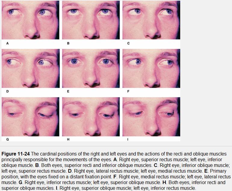
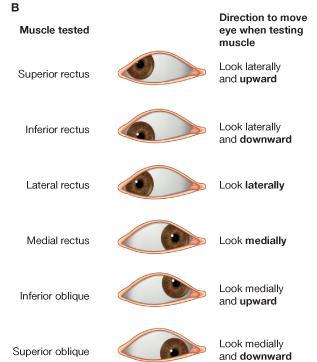
37 The origins of the superior and inferior recti are situated about 23 medialآ to their insertions, and, therefore, when the patient is asked to turn the cornea laterally, these muscles are placed in the optimum position to raise (superior rectus) or lower (inferior rectus) the cornea the superior and inferior oblique muscles can be tested. The pulley of the superior oblique and the origin of the inferior oblique muscles lie medial and anterior to their insertions. The physician tests the action of these muscles by asking the patient first to look medially, thus placing these muscles in the optimum position to lower (superior oblique) or raise (inferior oblique) the cornea. Because the lateral and medial recti are simply placed relative to the eyeball, asking the patient to turn his or her cornea directly laterally tests the lateral rectus and turning the cornea directly medially tests the medial rectus
38


39 Coats of the Eyeball 1- OUTER FIBROUS COAT is made up of : 1-Posterior opaque part 2-THE SCLERA the dense white part 1- THE CORNEA the anterior transparent part The Sclera The sclera is composed of dense fibrous tissue and is white. Posteriorly, it is pierced by the optic nerve and is fused with the dural sheath of that nerve The sclera is also pierced by the ciliary arteries and nerves and their associated veins. The sclera is directly continuous in front with the cornea at the corneoscleral junction, or limbus


40 The Cornea The transparent cornea is largely responsible for the refraction of the light entering the eye It is in contact posteriorly with the aqueous humor. Blood Supply The cornea is avascular and devoid of lymphatic drainage It is nourished by diffusion from the aqueous humor and from the capillaries at its edge. Nerve Supply Long ciliary nerves from the ophthalmic division of the trigeminal nerve Function of the Cornea The cornea is the most important refractive medium of the eye.


 which is")
41 2-MIDDLE VASCULAR COAT THE VASCULAR COAT CONSISTS OF: FROM BEHIND FORWARD 1- THE CHOROID 2-THE CILIARY BODY 3-THE IRIS. 1-THE CHOROID The choroid is a black vascular membrane deep to the sclera 2-THE CILIARY BODY The ciliary body is continuous posteriorly with the choroid, and anteriorly it lies behind the peripheral margin of the iris Contains the ciliary muscle (the main muscle of accomodation) which is connected to the suspensory ligaments of the lens

42 The ciliary muscle Nerve supply: The ciliary muscle is supplied by the parasympathetic fibers from the oculomotor nerve. After synapsing in the ciliary ganglion, the postganglionic fibers pass forward to the eyeball in the short ciliary nerves. Action: Contraction of the ciliary muscle, This relieves the tension in the suspensory ligament, and the elastic lens becomes more convex. This increases the refractive power of the lens.


43 The Iris and Pupil is a thin, contractile, pigmented diaphragm with a centre a aperture The pupil It is suspended in the aqueous humor between the cornea and the lens. The periphery of the iris is attached to the anterior surface of the ciliary body. It divides the space between the lens and the cornea into an anterior and a posterior chamber. The muscle fibers of the iris are involuntary and consist of circular and radiating fibers. The circular fibers form the sphincter pupillae Nerve supply: The sphincter pupillae is supplied by parasympathetic fibers from the oculomotor nerve. After synapsing in the ciliary ganglion, the postganglionic fibers pass forward to the eyeball in the short ciliary nerves. The radial fibers form the dilator pupillae is supplied by sympathetic fibers, which pass forward to the eyeball in the long ciliary nerves. Action: The sphincter pupillae constricts the pupil in the presence of bright light and during accommodation. The dilator pupillae dilates the pupil in the presence of light of low intensity or in the presence of excessive sympathetic activity such as occurs in fright


44 3-Nervous Coat: The Retina The retina consists of : 1-AN OUTER PIGMENTED LAYER 2-AN INNER NERVOUS LAYER. Its outer surface is in contact with the choroid, and its inner surface is in contact with the vitreous body At the center of the posterior part of the retina is an oval, yellowish area, the macula lutea, which is the area of the retina for the most distinct vision. It has a central depression, the fovea centralis

45 Contents of the Eyeball The contents of the eyeball consist of: 1-THE AQUEOUS HUMOR 2-THE VITREOUS BODY 3-THE LENS Aqueous Humor is a clear fluid that fills the anterior and posterior chambers of the eyeball Obstruction to the draining of the aqueous humor results in a rise in intraocular pressure called glaucoma.

46 Vitreous Body The vitreous body fills the eyeball behind the lens and is a transparent gel. The hyaloid canal is a narrow channel that runs through the vitreous body from the optic disc to the posterior surface of the lens; in the fetus, it is filled by the hyaloid artery, which disappears before birth. The function of the vitreous body is to contribute slightly to the magnifying power of the eye. It supports the posterior surface of the lens and assists in holding the neural part of the retina against the pigmented part of the retina.

.")
47 The Lens The lens is a transparent, biconvex structure enclosed in a transparent capsule. It is situated behind the iris and in front of the vitreous body and is encircled by the ciliary processes. Accommodation of the Eye To accommodate the eye for close objects, the ciliary muscle contracts and pulls the ciliary body forward and inward so that the radiating fibers of the suspensory ligament are relaxed. This allows the elastic lens to assume a more globular shape. With advancing age, the lens becomes denser and less elastic, and, as a result, the ability to accommodate is lessened (presbyopia). This disability can be overcome by the use of an additional lens in the form of glasses to assist the eye in focusing on nearby objects. Constriction of the Pupil During Accommodation of the Eye To ensure that the light rays pass through the central part of the lens so spherical aberration is diminished during accommodation for near objects, the sphincter pupillae muscle contracts so the pupil becomes smaller Convergence of the Eyes During Accommodation of the Lens In humans, the retinae of both eyes focus on only one set of objects (single binocular vision). When an object moves from a distance toward an individual, the eyes converge so that a single object, not two, is seen. Convergence of the eyes results from the coordinated contraction of the medial rectus muscles
Bony orbit Roof The orbital plate of the frontal bone Lateral wall: the zygomatic bone and the greater wing of the sphenoid
 Bony orbit Roof: Formed by: The orbital plate of the frontal bone, which separates the orbital cavity from the anterior cranial fossa and the frontal lobe of the cerebral hemisphere Lateral wall: Formed
The sebaceous glands (glands of Zeis) open directly into the eyelash follicles, ciliary glands (glands of Moll) are modified sweat glands that open
 open directly into the eyelash follicles, ciliary glands (glands of Moll) are modified sweat glands that open") The Orbital Region The orbits are a pair of bony cavities that contain the eyeballs; their associated muscles, nerves, vessels, and fat; and most of the lacrimal apparatus upper eyelid is larger and more
The Orbital Region The orbits are a pair of bony cavities that contain the eyeballs; their associated muscles, nerves, vessels, and fat; and most of the lacrimal apparatus upper eyelid is larger and more
1 Eyelids. Lacrimal Apparatus. Orbital Region. 3 The Orbit. The Eye
 1 1 Eyelids Orbital Region 2 Lacrimal Apparatus 3 The Orbit 4 The Eye 2 Eyelids The eyelids protect the eye from injury and excessive light by their closure. The upper eyelid is larger and more mobile
The orbit-2. Dr. Heba Kalbouneh Assistant Professor of Anatomy and Histology
 The orbit-2 Dr. Heba Kalbouneh Assistant Professor of Anatomy and Histology Eyelids The eyelids (act like the curtains) protect the eye from injury and excessive light by their closure The upper eyelid
The orbit-1. Dr. Heba Kalbouneh Assistant Professor of Anatomy and Histology
 The orbit-1 Dr. Heba Kalbouneh Assistant Professor of Anatomy and Histology Orbital plate of frontal bone Orbital plate of ethmoid bone Lesser wing of sphenoid Greater wing of sphenoid Lacrimal bone Orbital
Unit VIII Problem 8 Anatomy: Orbit and Eyeball
 Unit VIII Problem 8 Anatomy: Orbit and Eyeball - The bony orbit: it is protecting our eyeball and resembling a pyramid: With a base directed: anterolaterally. And an apex directed: posteromedially. Notes:
Maxilla, ORBIT and infratemporal fossa. Neophytos C Demetriades MD, DDS, MSc Associate professor European University of Cyprus School of Medicine
 Maxilla, ORBIT and infratemporal fossa Neophytos C Demetriades MD, DDS, MSc Associate professor European University of Cyprus School of Medicine MAXILLA Superior, middle, and inferior meatus Frontal sinus
213: HUMAN FUNCTIONAL ANATOMY: PRACTICAL CLASS 12 Cranial cavity, eye and orbit
 213: HUMAN FUNCTIONAL ANATOMY: PRACTICAL CLASS 12 Cranial cavity, eye and orbit OSTEOLOGY Identify the bones which comprise the walls of the orbit: maxilla, zygomatic, ethmoid, lachrymal, frontal, and
MAXILLA, ORBIT & PTERYGOPALATINE FOSSA. Neophytos C Demetriades MD, DDS, MSc Associate professor European University of Cyprus School of Medicine
 MAXILLA, ORBIT & PTERYGOPALATINE FOSSA Neophytos C Demetriades MD, DDS, MSc Associate professor European University of Cyprus School of Medicine Maxilla MAXILLA Superior, middle, and inferior meatus Frontal
4/22/16. Eye. External Anatomy of Eye. Accessory Structures. Bio 40B Dr. Kandula
 Eye Bio 40B Dr. Kandula External Anatomy of Eye Accessory Structures l Eyebrows l Levator Palpebrae Superioris - opens eye l Eyelashes l Ciliary glands modified sweat glands l Small sebaceous glands l
GNK485 The eye and related structures. Prof MC Bosman 2012
 GNK485 The eye and related structures Prof MC Bosman 2012 Surface anatomy Bony orbit Eyeball and Lacrimal apparatus Extra-ocular muscles Movements of the eye Innervation Arterial supply and venous drainage
The Special Senses: Part A
 PowerPoint Lecture Slides prepared by Janice Meeking, Mount Royal College CHAPTER 15 The Special Senses: Part A Warm Up What is the function of the eyeball? List any structures of the eyeball that you
Ocular Anatomy for the Paraoptometric
 Ocular Anatomy for the Paraoptometric Minnesota Optometric Association Paraoptometric CE Friday September 30, 2016 Lindsay A. Sicks, OD, FAAO Assistant Professor, Illinois College of Optometry lsicks@ico.edu
The Orbit. The Orbit OCULAR ANATOMY AND DISSECTION 9/25/2014. The eye is a 23 mm organ...how difficult can this be? Openings in the orbit
 The eye is a 23 mm organ...how difficult can this be? OCULAR ANATOMY AND DISSECTION JEFFREY M. GAMBLE, OD COLUMBIA EYE CONSULTANTS OPTOMETRY & UNIVERSITY OF MISSOURI DEPARTMENT OF OPHTHALMOLOGY CLINICAL
Sense of Vision. Chapter 8. The Eye and Vision. The Eye Orbit. Eyebrows, Eyelids, Eyelashes. Accessory Organs 5/3/2016.
 Sense of Vision Chapter 8 Special Senses The Eye and Vision 70 percent of all sensory receptors are in the eyes Each eye has over 1 million nerve fibers Protection for the eye Most of the eye is enclosed
Bony orbit. Sup. Med. Inf. Lat. frontal bone. frontal process of maxilla. zygomatic process of maxilla zygomatic bone
 Orbit 解剖學科鄭授德 本教材之圖片取自於 1. Gray s Anatomy for Students, 3rd ed., 2015, by Drake, Vogl, and Mitchell 2. Clinically Oriented Anatomy, 7th ed., 2014, by Moore, Dalley, and Agur 3. Anatomy, an Essential Textbook,
REVIEW OF HEAD AND NECK CRANIAL NERVES AND EVERYTHING ELSE
 REVIEW OF HEAD AND NECK CRANIAL NERVES AND EVERYTHING ELSE OLFACTORY NERVE CN I ANTERIOR CRANIAL FOSSA CRISTA GALLI OF ETHMOID OLFACTORY FORAMINA IN CRIBIFORM PLATE OF ETHMOID BONE CN I OLFACTORY NERVE
3-Deep fascia: is absent (except over the parotid gland & buccopharngeal fascia covering the buccinator muscle)
") The Face 1-Skin of the Face The skin of the face is: Elastic Vascular (bleed profusely however heal rapidly) Rich in sweat and sebaceous glands (can cause acne in adults) It is connected to the underlying
The Face 1-Skin of the Face The skin of the face is: Elastic Vascular (bleed profusely however heal rapidly) Rich in sweat and sebaceous glands (can cause acne in adults) It is connected to the underlying
Omran Saeed. Luma Taweel. Mohammad Almohtaseb. 1 P a g e
 2 Omran Saeed Luma Taweel Mohammad Almohtaseb 1 P a g e I didn t include all the photos in this sheet in order to keep it as small as possible so if you need more clarification please refer to slides In
mistake ;slides in bold but you still have to go back to our slides to see the figure, tables and some scheme
 Khozama jehad : I am doing my best and I am sorry for any unintended mistake ;slides in bold but you still have to go back to our slides to see the figure, tables and some scheme The Orbit, Orbital Contents
Face. Definition: The area between the two ears and from the chin to the eye brows. The muscles of the face
 Face Definition: The area between the two ears and from the chin to the eye brows. The muscles of the face The muscle of facial expression (include the muscle of the face and the scalp). All are derived
Chapter(2):the lid page (1) THE LID
:the lid page (1) THE LID") Chapter(2):the lid page (1) THE LID Anatomy of the lid: * Check movie anatomy of the lid model The eyelids are two movable muco-cutaneous folds which protect the eye on closure. The are joined temporary
Chapter(2):the lid page (1) THE LID Anatomy of the lid: * Check movie anatomy of the lid model The eyelids are two movable muco-cutaneous folds which protect the eye on closure. The are joined temporary
Lecture 10 Orbit and control of eye movements
 Lecture 10 Orbit and control of eye movements Overview of structures in the orbit (Moore pp 899, Netter Plate 1) The orbit contains the eye, from which the optic nerve exits into the cranial cavity optic
Infratemporal fossa: Tikrit University college of Dentistry Dr.Ban I.S. head & neck Anatomy 2 nd y.
 Infratemporal fossa: This is a space lying beneath the base of the skull between the lateral wall of the pharynx and the ramus of the mandible. It is also referred to as the parapharyngeal or lateral pharyngeal
Dr.Ban I.S. head & neck anatomy 2 nd y جامعة تكريت كلية طب االسنان مادة التشريح املرحلة الثانية أ.م.د. بان امساعيل صديق 6102/6102
 جامعة تكريت كلية طب االسنان مادة التشريح املرحلة الثانية أ.م.د. بان امساعيل صديق 6102/6102 Pterygopalatine fossa: The pterygopalatine fossa is a cone-shaped depression, It is located between the maxilla,
PTERYGOPALATINE FOSSA
 PTERYGOPALATINE FOSSA Outline Anatomical Structure and Boundaries Foramina and Communications with other spaces and cavities Contents Pterygopalatine Ganglion Especial emphasis on certain arteries and
Mohammad Hisham Al-Mohtaseb. Lina Mansour. Reyad Jabiri. 0 P a g e
 2 Mohammad Hisham Al-Mohtaseb Lina Mansour Reyad Jabiri 0 P a g e This is only correction for the last year sheet according to our record. If you already studied this sheet just read the yellow notes which
Eye Movements. Geometry of the Orbit. Extraocular Muscles
 Eye Movements Geometry of the Orbit The eye (oculus) is located in the anterior aspect of the orbit: the equator of the eye (defined by a coronal plane passing through its middle) lies at the margin of
Special Senses: The Eye
 Unit 4 Special Senses: The Eye ESSENTIALS OF HUMAN ANATOMY & PHYSIOLOGY The Senses General senses of touch Temperature Pressure Pain Special senses Smell Taste Sight Hearing Equilibrium The Eye and Vision
THE SPECIAL SENSES. Introduction Vision
 THE SPECIAL SENSES Introduction Vision RECEPTORS Structures designed to respond to stimuli Variable complexity RECEPTORS: GENERAL PROPERTIES Transducers Receptor Potential Generator Potential RECEPTORS
INTRODUCTION: ****************************************************************************************************
 BIOLOGY 211: HUMAN ANATOMY & PHYSIOLOGY **************************************************************************************************** EYES AND VISION ****************************************************************************************************
Anatomic Relations Summary. Done by: Sohayyla Yasin Dababseh
 Anatomic Relations Summary Done by: Sohayyla Yasin Dababseh Anatomic Relations Lecture 1 Part-1 - The medial wall of the nose is the septum. - The vestibule lies directly inside the nostrils (Nares). -
Tikrit University collage of dentistry Dr.Ban I.S. head & neck anatomy 2 nd y. Lec [5] / Temporal fossa :
![Tikrit University collage of dentistry Dr.Ban I.S. head & neck anatomy 2 nd y. Lec [5] / Temporal fossa :](/thumbs/88/115294566.jpg "Tikrit University collage of dentistry Dr.Ban I.S. head & neck anatomy 2 nd y. Lec [5] / Temporal fossa :") Lec [5] / Temporal fossa : Borders of the Temporal Fossa: Superior: Superior temporal line. Inferior: gap between zygomatic arch and infratemporal crest of sphenoid bone. Anterior: Frontal process of the
Lec [5] / Temporal fossa : Borders of the Temporal Fossa: Superior: Superior temporal line. Inferior: gap between zygomatic arch and infratemporal crest of sphenoid bone. Anterior: Frontal process of the
Special Senses PART A
 8 Special Senses PART A PowerPoint Lecture Slide Presentation by Jerry L. Cook, Sam Houston University ESSENTIALS OF HUMAN ANATOMY & PHYSIOLOGY EIGHTH EDITION ELAINE N. MARIEB The Senses General senses
Muscles of the Eyeball (Extra Ocular Muscles) Prof. Dr. Imran Qureshi
 Prof. Dr. Imran Qureshi") Muscles of the Eyeball (Extra Ocular Muscles) Prof. Dr. Imran Qureshi There are six extrinsic muscles of the eyeball, namely the (S), Medial (M), (I), & Lateral (L) recti, and (SO) and (IO) Obliques. In
Muscles of the Eyeball (Extra Ocular Muscles) Prof. Dr. Imran Qureshi There are six extrinsic muscles of the eyeball, namely the (S), Medial (M), (I), & Lateral (L) recti, and (SO) and (IO) Obliques. In
Superior View of the Skull (Norma Verticalis) Anteriorly the frontal bone articulates with the two parietal bones AT THE CORONAL SUTURE
 Anteriorly the frontal bone articulates with the two parietal bones AT THE CORONAL SUTURE") Superior View of the Skull (Norma Verticalis) Anteriorly the frontal bone articulates with the two parietal bones AT THE CORONAL SUTURE 1 The two parietal bones articulate in the midline AT THE SAGITTAL
Superior View of the Skull (Norma Verticalis) Anteriorly the frontal bone articulates with the two parietal bones AT THE CORONAL SUTURE 1 The two parietal bones articulate in the midline AT THE SAGITTAL
Tikrit University College of Dentistry Dr.Ban I.S. head & neck anatomy 2 nd y.
 Lec [3]/The scalp The scalp extends from the supraorbital margins anteriorly to the nuchal lines at the back of the skull and down to the temporal lines at the sides. The forehead, from eyebrows to hairline,
The Eye. The Orbit. The EYE What a Trip!!! - The Anterior Segment 5/12/2015. Jill J Luebbert, CPOT, ABOC
 The EYE What a Trip!!! - The Anterior Segment Jill J Luebbert, CPOT, ABOC The Eye The Orbit Bony socket containing the eye and most of its accessory organs consisting of 7 bones 1 The Seven Bones of the
Trigeminal Nerve Worksheets, Distributions Page 1
 Trigeminal Nerve Worksheet #1 Distribution by Nerve Dr. Darren Hoffmann Dental Gross Anatomy, Spring 2013 We have drawn out each of the branches of CN V in lecture and you have an idea now for their basic
Special Senses: Vision
 ighapmlre24pg223_230 5/12/04 2:27 PM Page 223 impos03 302:bjighapmL:ighapmLrevshts:layouts: NAME LAB TIME/DATE Special Senses: Vision REVIEW SHEET exercise 24 Anatomy of the Eye 1. Name five accessory
02/03/2014. Average Length: 23mm (Infant ~16mm) Approximately the size of a quarter Volume: ~5mL
 Approximately the size of a quarter Volume: ~5mL") Identify the anatomy of the eye. Explain the basic physiology of the parts of the eye. Briefly discuss various surgeries related to different parts of the anatomy. Average Length: 23mm (Infant ~16mm) Approximately
Identify the anatomy of the eye. Explain the basic physiology of the parts of the eye. Briefly discuss various surgeries related to different parts of the anatomy. Average Length: 23mm (Infant ~16mm) Approximately
Dr.Ban I.S. head & neck anatomy 2 nd y. جامعة تكريت كلية طب االسنان املرحلة الثانية أ.م.د. بان امساعيل صديق 6102/6102
 جامعة تكريت كلية طب االسنان التشريح مادة املرحلة الثانية أ.م.د. بان امساعيل صديق 6102/6102 Parotid region The part of the face in front of the ear and below the zygomatic arch is the parotid region. The
The Nervous System: General and Special Senses Pearson Education, Inc.
 18 The Nervous System: General and Special Senses Introduction Sensory information arrives at the CNS Information is picked up by sensory receptors Sensory receptors are the interface between the nervous
Taste buds Gustatory cells extend taste hairs through a narrow taste pore
 The Special Senses Objectives Describe the sensory organs of smell, and olfaction. Identify the accessory and internal structures of the eye, and explain their function. Explain how light stimulates the
Lec [8]: Mandibular nerve:
![Lec [8]: Mandibular nerve:](/thumbs/94/121295776.jpg "Lec [8]: Mandibular nerve:") Lec [8]: Mandibular nerve: The mandibular branch from the trigeminal ganglion lies in the middle cranial fossa lateral to the cavernous sinus. With the motor root of the trigeminal nerve [motor roots lies
Lec [8]: Mandibular nerve: The mandibular branch from the trigeminal ganglion lies in the middle cranial fossa lateral to the cavernous sinus. With the motor root of the trigeminal nerve [motor roots lies
ACTIVITIES. Complete Diagrams PNS 18 and 19 Complete PNS 23 Worksheet 3 #1 only Complete PNS 24 Practice Quiz
 ACTIVITIES Complete Diagrams PNS 18 and 19 Complete PNS 23 Worksheet 3 #1 only Complete PNS 24 Practice Quiz THE SPECIAL SENSES Introduction Vision RECEPTORS Structures designed to respond to stimuli Variable
Head and Face Anatomy
 Head and Face Anatomy Epicranial region The Scalp The soft tissue that covers the vault of skull. Extends from supraorbital margin to superior nuchal line. Layers of the scalp S C A L P = skin = connective
Dr. Sami Zaqout, IUG Medical School
 The skull The skull is composed of several separate bones united at immobile joints called sutures. Exceptions? Frontal bone Occipital bone Vault Cranium Sphenoid bone Zygomatic bones Base Ethmoid bone
Anatomy images for MSS practical exam- 2019
 Anatomy images for MSS practical exam- 2019 Ilium Ischium Pubis Acetabulaum Iliac crest Iliac tubercle ASIS (muscle and ligament attached) AIIS (muscle attached) PSIS PIIS Ischial spine Ischial tuberosity
Temporal region. temporal & infratemporal fossae. Zhou Hong Ying Dept. of Anatomy
 Temporal region temporal & infratemporal fossae Zhou Hong Ying Dept. of Anatomy Temporal region is divided by zygomatic arch into temporal & infratemporal fossae. Temporal Fossa Infratemporal fossa Temporal
HEAD AND NECK ANATOMY PRACTICE QUESTIONS
 HEAD AND NECK ANATOMY PRACTICE QUESTIONS 1. A patient complains that he has lost sensation on his face and that the skin of his face feels numb. The physician tests tactile acuity by touching the forehead
The Ear The ear consists of : 1-THE EXTERNAL EAR 2-THE MIDDLE EAR, OR TYMPANIC CAVITY 3-THE INTERNAL EAR, OR LABYRINTH 1-THE EXTERNAL EAR.
 The Ear The ear consists of : 1-THE EXTERNAL EAR 2-THE MIDDLE EAR, OR TYMPANIC CAVITY 3-THE INTERNAL EAR, OR LABYRINTH 1-THE EXTERNAL EAR Made of A-AURICLE B-EXTERNAL AUDITORY MEATUS A-AURICLE It consists
The Organs of Special Senses
 8 The Organs of Special Senses Special senses are those other than touch, pain, temperature, and proprioception. Vision, hearing, and equilibrium are the special senses discussed in this chapter. The Eye
SPECIAL SENSES PART I: OLFACTION & GUSTATION
 SPECIAL SENSES PART I: OLFACTION & GUSTATION 5 Special Senses Olfaction Gustation Vision Equilibrium Hearing Olfactory Nerves Extend through cribriform plate into nasal cavity on both sides of nasal septum
The Skull and Temporomandibular joint II Prof. Abdulameer Al-Nuaimi. E. mail:
 The Skull and Temporomandibular joint II Prof. Abdulameer Al-Nuaimi E-mail: a.al-nuaimi@sheffield.ac.uk E. mail: abdulameerh@yahoo.com Temporal fossa The temporal fossa is a depression on the temporal
THE EYE: RETINA AND GLOBE
 Neuroanatomy Suzanne Stensaas February 24, 2011, 10:00-12:00 p.m. Reading: Waxman Ch. 15. Your histology and gross anatomy books should be useful. Reading: Histology of the Eye from any histology book
Anatomy for ophthalmic anaesthesia
 British Journal of Anaesthesia 1995; 75: 80 87 REVIEW ARTICLES Anatomy for ophthalmic anaesthesia R. W. JOHNSON Study of the anatomy of the orbit, its contents and surrounding structures allows the anaesthetist
Ocular Anatomy & Physiology. Learning Objectives: Let s get oriented first. 3 Major Layers (Tunics) of EYE. Topics to be covered: FIBROUS TUNIC
 of EYE. Topics to be covered: FIBROUS TUNIC") Lecturer: Ocular Anatomy & Physiology M. Patrick COLEMAN, ABOC, COT Kerrville, TX Learning Objectives: 1. Correctly identify ocular structures around or within the eye 2. List the key functions of various
Lecturer: Ocular Anatomy & Physiology M. Patrick COLEMAN, ABOC, COT Kerrville, TX Learning Objectives: 1. Correctly identify ocular structures around or within the eye 2. List the key functions of various
Head: Special Senses. Taste Smell Vision Hearing/Balance
 Head: Special Senses Taste Smell Vision Hearing/Balance TASTE: how does it work? Taste buds on tongue on fungiform papillae ( mushroom-like projections) Each bud contains several cell types in microvilli
Cranial Cavity REFERENCES: OBJECTIVES OSTEOLOGY. Stephen A. Gudas, PT, PhD
 Stephen A. Gudas, PT, PhD Cranial Cavity REFERENCES: Moore and Agur, Essential Clinical Anatomy (ECA), 3rd ed., pp. 496 498; 500 507; 512 514 Grant s Atlas 12 th ed., Figs 7.6; 7.19 7.30. Grant s Dissector
Trigeminal Nerve (V)
") Trigeminal Nerve (V) Lecture Objectives Discuss briefly how the face is developed. Follow up the course of trigeminal nerve from its point of central connections, exit and down to its target areas. Describe
Trigeminal Nerve (V) Lecture Objectives Discuss briefly how the face is developed. Follow up the course of trigeminal nerve from its point of central connections, exit and down to its target areas. Describe
20-20,000 Hertz range of human hearing
 20-20,000 Hertz range of human hearing accommodation automatic adjustment in focal length of the lens of the eye; changing the shape of the lens aqueous humor Watery fluid in the anterior chambers of the
Brain ميهاربا لض اف دمح ا د The Meninges 1- Dura Mater of the Brain endosteal layer does not extend meningeal layer falx cerebri tentorium cerebelli
 .احمد د فاضل ابراهيم Lecture 15 Brain The Meninges Three protective membranes or meninges surround the brain in the skull: the dura mater, the arachnoid mater, and the pia mater 1- Dura Mater of the Brain
Major Anatomic Components of the Orbit
 Major Anatomic Components of the Orbit 1. Osseous Framework 2. Globe 3. Optic nerve and sheath 4. Extraocular muscles Bony Orbit Seven Bones Frontal bone Zygomatic bone Maxillary bone Ethmoid bone Sphenoid
The SCALP. Prof. Dr. Muhammad Imran Qureshi
 The SCALP By Prof. Dr. Muhammad Imran Qureshi The SCALP includes FIVE layers external to the Calvaria. These are: S: Skin & Superficial Fascia C: Connective Tissue A: Aponeurosis (Epicranial) L: Loose
THIEME. Scalp and Superficial Temporal Region
 CHAPTER 2 Scalp and Superficial Temporal Region Scalp Learning Objectives At the end of the dissection of the scalp, you should be able to identify, understand and correlate the clinical aspects: Layers
The Pharynx. Dr. Nabil Khouri MD. MSc, Ph.D
 The Pharynx Dr. Nabil Khouri MD. MSc, Ph.D Introduction The pharynx is the Musculo-fascial halfcylinder that links the oral and nasal cavities in the head to the larynx and esophagus in the neck Common
*in general the blood supply of the nose comes from branches of the internal and external carotid arteries.
 In the previous lecture we talked about the anatomy of the nasal cavity, today we will talk about its blood supply, venous drainage, innervations, and finally about the paranasal sinuses. When we describe
Parotid Gland, Temporomandibular Joint and Infratemporal Fossa
 M1 - Anatomy Parotid Gland, Temporomandibular Joint and Infratemporal Fossa Jeff Dupree Sanger 9-057 jldupree@vcu.edu Parotid gland: wraps around the mandible positioned between the mandible and the sphenoid
Temporal fossa Infratemporal fossa Pterygopalatine fossa Terminal branches of external carotid artery Pterygoid venous plexus
 Outline of content Temporal fossa Infratemporal fossa Pterygopalatine fossa Terminal branches of external carotid artery Pterygoid venous plexus Boundary Content Communication Mandibular division of trigeminal
Vision I. Steven McLoon Department of Neuroscience University of Minnesota
 Vision I Steven McLoon Department of Neuroscience University of Minnesota 1 Eye Cornea Sclera Conjunctiva 2 Eye The conjunctiva lines the inner surface of the eyelids and outer surface of the sclera. 3
Bones Ethmoid bone Inferior nasal concha Lacrimal bone Maxilla Nasal bone Palatine bone Vomer Zygomatic bone Mandible
 splanchnocranium - Consists of part of skull that is derived from branchial arches - The facial bones are the bones of the anterior and lower human skull Bones Ethmoid bone Inferior nasal concha Lacrimal
External Occipital Protuberance
 Osteology Exterior Skull Frontal Bone Glabella Superciliary Arch Supraorbital Notch/Foramen Nasion (junction w/ Nasal bone) Frontal/Metopic Suture (usually absent in adult, b/w ossification centers of
Unit 18: Cranial Cavity and Contents
 Unit 18: Cranial Cavity and Contents Dissection Instructions: The calvaria is to be removed without damage to the dura mater which is attached to the inner surface of the calvaria. Cut through the outer
human anatomy 2016 lecture fifteen Dr meethak ali ahmed neurosurgeon
 Cranial Nerves Organization of the Cranial Nerves The cranial nerves are named as follows: I. Olfactory II. Optic III. Oculomotor IV. Trochlear V. Trigeminal VI. Abducent VII. Facial VIII. Vestibulocochlear
Dr.Ban I.S. head & neck anatomy 2 nd y. جامعة تكريت كلية طب االسنان املرحلة الثانية
 جامعة تكريت كلية طب االسنان التشريح مادة املرحلة الثانية أ.م.د. بان امساعيل صديق 6102-6102 1 The Palate The palate forms the roof of the mouth and the floor of the nasal cavity. It is divided into two
The white of the eye and the part that maintains its shape is know n as the:
 Scrub In The white of the eye and the part that maintains its shape is know n as the: a. Cornea b. Pupil c. Retina d. Sclera The structure that is found in the ear and contains the organ of hearing is
Veins of the Face and the Neck
 Veins of the Face and the Neck Facial Vein The facial vein is formed at the medial angle of the eye by the union of the supraorbital and supratrochlear veins. connected through the ophthalmic veins with
C h a p t e r PowerPoint Lecture Slides prepared by Jason LaPres North Harris College Houston, Texas
 C h a p t e r 15 The Nervous System: The Brain and Cranial Nerves PowerPoint Lecture Slides prepared by Jason LaPres North Harris College Houston, Texas Copyright 2009 Pearson Education, Inc., publishing
Rashed Al-Jomard. Alanood Bostanji
 Anatomy #2 The Orbit & Cranial Nerve III, IV, VI Rashed AlJomard Alanood Bostanji 1 P a g e The Orbit & Cranial nerves III,IV&VI ** Some notes about the last lec & first MM : Lens :: "just clarify for
Cranial nerves.
 Cranial nerves eaglezhyxzy@163.com Key Points of Learning Name Components Passing through Peripheral distribution Central connection Function Cranial nerves Ⅰ olfactory Ⅱ optic Ⅲ occulomotor Ⅳ trochlear
Bisection of Head & Nasal Cavity 頭部對切以及鼻腔. 解剖學科馮琮涵副教授 分機
 Bisection of Head & Nasal Cavity 頭部對切以及鼻腔 解剖學科馮琮涵副教授 分機 3250 E-mail: thfong@tmu.edu.tw Outline: The structure of nose The concha and meatus in nasal cavity The openings of paranasal sinuses Canals, foramens
VISUAL REFLEXES. B. The oculomotor nucleus, Edinger-Westphal nucleus, and oculomotor nerve at level of the superior colliculus.
 Neuroanatomy Suzanne Stensaas February 24, 2011, 10:00-12:00 p.m. Reading: Waxman Ch. 15 HyperBrain: Ch 7 with quizzes and or Lab 7 videotape http://www-medlib.med.utah.edu/kw/hyperbrain/anim/reflex.html
o A cushion of fat surrounds most of the eye
 Name Period SPECIAL SENSES The Senses of touch o Temperature o Pressure o Pain o Smell o Taste o Sight o Hearing o Equilibrium The Eye and Vision are in the eyes has over a o Most of the eye is enclosed
The dura is sensitive to stretching, which produces the sensation of headache.
 Dural Nerve Supply Branches of the trigeminal, vagus, and first three cervical nerves and branches from the sympathetic system pass to the dura. Numerous sensory endings are in the dura. The dura is sensitive
TRANSVERSE SECTION PLANE Scalp 2. Cranium. 13. Superior sagittal sinus
 TRANSVERSE SECTION PLANE 1 1. Scalp 2. Cranium 3. Superior sagittal sinus 4. Dura mater 5. Falx cerebri 6. Frontal lobes of the cerebrum 7. Middle meningeal artery 8. Cortex, grey matter 9. Cerebral vessels
o A cushion of fat surrounds most of the eye
 Name Period SPECIAL SENSES The Senses General senses of touch o Temperature o Pressure o Pain Special senses o Smell o Taste o Sight o Hearing o Equilibrium The Eye and Vision 70 percent of all sensory
Trigeminal nerve. Slide in bold and please go back to see the pictures, if I skipped any part of record that because it wasn t clear to me
 Trigeminal nerve Slide in bold and please go back to see the pictures, if I skipped any part of record that because it wasn t clear to me Hala nsour 2/26/2018 P a g e 1 this lecture contain two topics
Vision is the most dominant sense, about 70% of all sensory receptors in the body are in the eyes Accessory Structures of the eye : Eyelashes :
 Sight By Jess Kapp Vision is the most dominant sense, about 70% of all sensory receptors in the body are in the eyes Accessory Structures of the eye : Eyelashes : Protect eye from debris and bacteria Eyebrows
Introduction to Head and Neck Anatomy
 Introduction to Head and Neck Anatomy Nervous Tissue Controls and integrates all body activities within limits that maintain life Three basic functions 1. sensing changes with sensory receptors 2. interpreting
Scrub In. What is the function of vitreous humor? What does the pupil do when exposed to bright light? a. Maintain eye shape and provide color vision
 Scrub In What is the function of vitreous humor? a. Maintain eye shape and provide color vision b. Maintain eye shape and refract light rays c. Provide night vision and color vision d. Provide night vision
1 Anatomy of the Eyeball
 1 Anatomy of the Eyeball 1 Anatomy of the Eyeball ANATOMY Eyeball Tunics of Eyeball Segments of the Eye Blood Supply of Eyeball Blood Supply of Retina Blood Supply of Uvea Nerve Supply of Eyeball DEVELOPMENT
The Senses. Chapter 10 7/8/11. Introduction
 Chapter 10 The Senses Introduction A. Sensory receptors detect changes in the environment and stimulate neurons to send nerve impulses to the brain. B. A sensation is formed based on the sensory input.
Dr.Ban I.S. head & neck anatomy 2 nd y. جامعة تكريت كلية طب االسنان مادة التشريح املرحلة الثانية أ.م.د. بان امساعيل صديق 6102/6102
 جامعة تكريت كلية طب االسنان مادة التشريح املرحلة الثانية أ.م.د. بان امساعيل صديق 6102/6102 The scalp The scalp extends from the supraorbital margins anteriorly to the nuchal lines at the back of the skull
Introduction to Local Anesthesia and Review of Anatomy
 5-Sep Introduction and Anatomy Review 12-Sep Neurophysiology and Pain 19-Sep Physiology and Pharmacology part 1 26-Sep Physiology and Pharmacology part 2 Introduction to Local Anesthesia and Review of