The Eye. The Orbit. The EYE What a Trip!!! - The Anterior Segment 5/12/2015. Jill J Luebbert, CPOT, ABOC
|
|
|
- Randell McLaughlin
- 5 years ago
- Views:
Transcription


1 The EYE What a Trip!!! - The Anterior Segment Jill J Luebbert, CPOT, ABOC The Eye The Orbit Bony socket containing the eye and most of its accessory organs consisting of 7 bones 1
2 The Seven Bones of the Orbit Maxillary largest in inferior wall Frontal largest in superior wall Zygomatic thickest in lateral wall Ethmoid largest medial wall & thinnest Lacrimal Palatine Sphenoid Maxillary Frontal Zygomatic Ethmoid Palatine Lacrimal Sphenoid First three located at front Last ones form the apex in the back Bones are fused together at lines called sutures Eight openings called foramen Some bones have air spaces called sinuses External Structures Eyelids Palpebrae (three functions): REPLENISH and distribute the tear film evenly across the cornea (front surface of the eye) PUMP the tears through the lacrimal sac for drainage from the eye and in this way regulates the amount of tear fluid in the eye PROTECT from light and objects 2


3 Reflex blinking Forceful closure Blepharospasm Tightly shut and cannot be opened Palpebral aperture - fissure Approximately 10 mm wide Canthus Point which upper & lower lids meet Laterial Closer to ear Medial Closer to bridge of nose Ptosis Ectropian Entropian 3
4 Eyelids Seven layers of tissue Skin Subcutaneous areolar layer Orbicularis oculi muscle Submuscular areolar layer Levator palpebrae superioris Tarsal plate Meibomian glands Skin layer Outer most layer Thinnest skin of the entire body Contains sweat glands and hair Nasal portion smoother & greasier Begins at gray line Subcutaneous areolar layer Loose connective tissue Contains no fat Demonstrates edema (puffy) 4
5 Orbicularis Oculi Runs the entire length of the eyelid Responsible for lid closure Controlled by Cranial Nerve VII Submuscular areolar Loose connective tissue Contains majority of nervous and circulatory supply Levator palpebrae superioris Occupies the entire width of lid Is the major muscle responsible for lid opening 5

")
6 Tarsal Plate Located in upper & lower lid Made of dense & elastic connective tissue Contains Meibomian glands Produces oil layer of tears Conjunctiva Mucous membrane Begins at gray line Continous across the globe Contains goblet cells to produce mucus layer of tears Contains glands of Krause and Wolfring Produce water layer of tears Hordeolum (Sty) Chalazion 6
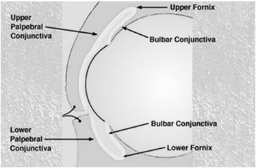
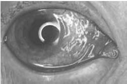
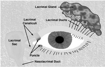
7 Conjunctiva Palpebral Bulbar Fornices Conjunctivitis Blepharitis Lacrimal System 7

8 Tear Film Three Concentric Tunics Fibrous Vascular Nervous Fibrous Tunic Sclera The white of the eye Provides a rigid protective shell for the intraocular contents while allowing for variations in intraocular pressure Covers 5/6ths of the Fibrous Tunic Cornea Is Avascular 8

9 Cornea Epithelium Continous with conjunctiva Prevent water from entering the eye Contain nerve endings Bare nerve endings account for corneal sensitivity Quickly regenerates Bowman s Membrane Tough membrane Scars easily 9
10 Stroma 90% of cornea Comprised of collagen similar to sclera Increase risk of scarring when damaged Descemet s membrane Very thin Does not regenerate when damaged Endothelium Single thickness layer of cells Born with 500,000 cells Does not regenerate cells throughout life Keeps water out of cornea Transports nutrients from aqueous humor to cornea 10
11 Anterior Chamber The area inside the eye, behind the cornea and in front of the iris Filled with aqueous humor (produced by the ciliary body) Iris Colored portion of the eye- amount of melanin (brown pigment) Divider anterior chamber and posterior chamber - has three layers Stroma Sphincter muscle Dilator muscle Stroma Contains pigment cells Sphincter Muscle (constrictor) Autonomic (nonvolutary) Nervous system Parasympathetic (normal) system Weaker of the two muscles Knocked out with cycloglegic drugs 11

12 Non-pigmented Epithelium Contains dilator muscle Autonomic (nonvoluntary) Nervous System Sympathetic :flight or flight Knocked out with Mydratic drugs The Journey Continues Thank you! 12
13 The EYE What a Trip!!! The Posterior Segment Jill J Luebbert, CPOT, ABOC The Eye A Quick Review of the Anterior Segment 13
14 The Orbit Bony socket containing the eye and most of its accessory organs consisting of 7 bones External Structures Eyelids Palpebrae (three functions): Replenish and distribute the tear film evenly across the cornea (front surface of the eye) Pumps the tears through the lacrimal sac for drainage from the eye and in this way regulates the amount of tear fluid in the eye Protection from light and objects Reflex blinking Blepharospasm Palpebral aperture Ptosis Ectropian Entropian 14
15 Hordeolum (Sty) Chalazion Conjunctiva Palpebral Bulbar Fornices Conjunctivitis Blepharitis 15
16 Lacrimal System Tear Film Cornea 16

17 Any Questions? Let s continue on Posterior Chamber Posterior Chamber The area inside the eye and behind the iris 17
18 Vascular tunic Iris Ciliary body Choroid Iris Colored portion of the eye- amount of melanin (brown pigment) Divider anterior chamber and posterior chamber - has three layers Stroma Sphincter muscle Dilator muscle 18


19 Crystalline Lens Nucleus Cortex Capsule Accommodation Cataract Cataract Cilary Body Located behind iris, inside sclera Anteriorly begins at scleral spur Posteriorly ends where joins retina Cilary muscle Responsible for shape of ciliary body Makes up the bulk of ciliary body Stroma Contains blood vessels and ciliary processes Produce aqueous humor 19


20 Ciliary Muscle Makes up the bulk of the ciliary body Responsible for the triangular shape Responsible for accommodation Controlled by parasympathetic system Can be paralyzed with cycloplegic drugs Accommodation 20
21 Stroma Contains blood vessels and ciliary process Production of aqueous humor Provides nutrients for lens and cornea Carries aways wastes Choroid Lies between sclera and retina Network of blood vessels, vascular support, supplies oxygen and nutrients Pigment cells (melanocytes) scattered throughout Four layers Hallers Sattlers Choriocapillaris Bruchs Membrane Four Layers Haller s layer Sattlers layer Arterioles and venules Generally considered as one group Choriocapillaries Smaller 21

22 Bruch s Membrane Thin connective tissue Site of RPE detachments Nervous Tunic Retina Retina Converts light energy into electrical impulses Consists of 10 layers Covers 65% of the interior surface of the eye 22
 External limiting membrane Outer nuclear layer Outer plexiform layer Inner nuclear layerr Inner plexiform layerr Ganglion cell layer Nerve fiber layer Internal limiting")
23 Retina Begins at Ora Serrata Generally divided into two general sections Outer layer (RPE) Inner layer (Retina) 10 layers Retina Pigment Epithilium (RPE) Neural Retina (rods and cones) External limiting membrane Outer nuclear layer Outer plexiform layer Inner nuclear layerr Inner plexiform layerr Ganglion cell layer Nerve fiber layer Internal limiting membrane 23
24 Outer layer RPE Regenerates chemicals used to convert light to electrical energy (Vitamin A) Transports nutrients to retina Phagocytize ( eat ) cellular debris Failure to do this is retinitis pigmentosa Begins with night blindness and leads to total blindness Prevents intraocular reflection of stray light Inner layer Neural Retina Nerve cells contain Axon (transmitters) Dendrite (receivers) Communication between cells Synapse Small gap between cells Neurotranmitters The chemical released by axons 24
 Used for motion and vision in low illumination Peripheral Vision Contain")

 Optic Disc")
25 Inner Segments Photoreceptors: Rods and Cones Rods Tall cylindrical cells (60 mm by 2 mm) Used for motion and vision in low illumination Peripheral Vision Contain Rhodopsin Larger number Are located primarily in the peripheral retina Cones Short cone shaped cells Responsible for pattern detection and fine details Less than rods Cones are located more centrally Fovea Centralis contains only cones Visual pigments sensitive to blue, green and red light (color discrimination) Optic Disc Macula 25


26 Vision with Macular Degeneration Cable Network Continues 126 million photoreceptors transmit 1 million optic nerve fibers Continues through the visual pathway The Jouney Continues Visual Pathway 26
27 Thank You!! Enjoy the other courses and activities at Optometry s Meeting
The Orbit. The Orbit OCULAR ANATOMY AND DISSECTION 9/25/2014. The eye is a 23 mm organ...how difficult can this be? Openings in the orbit
 The eye is a 23 mm organ...how difficult can this be? OCULAR ANATOMY AND DISSECTION JEFFREY M. GAMBLE, OD COLUMBIA EYE CONSULTANTS OPTOMETRY & UNIVERSITY OF MISSOURI DEPARTMENT OF OPHTHALMOLOGY CLINICAL
Ocular Anatomy for the Paraoptometric
 Ocular Anatomy for the Paraoptometric Minnesota Optometric Association Paraoptometric CE Friday September 30, 2016 Lindsay A. Sicks, OD, FAAO Assistant Professor, Illinois College of Optometry lsicks@ico.edu
4/22/16. Eye. External Anatomy of Eye. Accessory Structures. Bio 40B Dr. Kandula
 Eye Bio 40B Dr. Kandula External Anatomy of Eye Accessory Structures l Eyebrows l Levator Palpebrae Superioris - opens eye l Eyelashes l Ciliary glands modified sweat glands l Small sebaceous glands l
Histology of the Eye
 Histology of the Eye Objectives By the end of this lecture, the student should be able to describe: The general structure of the eye. The microscopic structure of:»cornea.»retina. EYE BULB Three coats
The Special Senses: Part A
 PowerPoint Lecture Slides prepared by Janice Meeking, Mount Royal College CHAPTER 15 The Special Senses: Part A Warm Up What is the function of the eyeball? List any structures of the eyeball that you
Special Senses: The Eye
 Unit 4 Special Senses: The Eye ESSENTIALS OF HUMAN ANATOMY & PHYSIOLOGY The Senses General senses of touch Temperature Pressure Pain Special senses Smell Taste Sight Hearing Equilibrium The Eye and Vision
Unit VIII Problem 8 Anatomy: Orbit and Eyeball
 Unit VIII Problem 8 Anatomy: Orbit and Eyeball - The bony orbit: it is protecting our eyeball and resembling a pyramid: With a base directed: anterolaterally. And an apex directed: posteromedially. Notes:
Sense of Vision. Chapter 8. The Eye and Vision. The Eye Orbit. Eyebrows, Eyelids, Eyelashes. Accessory Organs 5/3/2016.
 Sense of Vision Chapter 8 Special Senses The Eye and Vision 70 percent of all sensory receptors are in the eyes Each eye has over 1 million nerve fibers Protection for the eye Most of the eye is enclosed
Vision I. Steven McLoon Department of Neuroscience University of Minnesota
 Vision I Steven McLoon Department of Neuroscience University of Minnesota 1 Eye Cornea Sclera Conjunctiva 2 Eye The conjunctiva lines the inner surface of the eyelids and outer surface of the sclera. 3
Special Senses PART A
 8 Special Senses PART A PowerPoint Lecture Slide Presentation by Jerry L. Cook, Sam Houston University ESSENTIALS OF HUMAN ANATOMY & PHYSIOLOGY EIGHTH EDITION ELAINE N. MARIEB The Senses General senses
Bony orbit Roof The orbital plate of the frontal bone Lateral wall: the zygomatic bone and the greater wing of the sphenoid
 Bony orbit Roof: Formed by: The orbital plate of the frontal bone, which separates the orbital cavity from the anterior cranial fossa and the frontal lobe of the cerebral hemisphere Lateral wall: Formed
02/03/2014. Average Length: 23mm (Infant ~16mm) Approximately the size of a quarter Volume: ~5mL
 Approximately the size of a quarter Volume: ~5mL") Identify the anatomy of the eye. Explain the basic physiology of the parts of the eye. Briefly discuss various surgeries related to different parts of the anatomy. Average Length: 23mm (Infant ~16mm) Approximately
Identify the anatomy of the eye. Explain the basic physiology of the parts of the eye. Briefly discuss various surgeries related to different parts of the anatomy. Average Length: 23mm (Infant ~16mm) Approximately
1 Eyelids. Lacrimal Apparatus. Orbital Region. 3 The Orbit. The Eye
 1 1 Eyelids Orbital Region 2 Lacrimal Apparatus 3 The Orbit 4 The Eye 2 Eyelids The eyelids protect the eye from injury and excessive light by their closure. The upper eyelid is larger and more mobile
GNK485 The eye and related structures. Prof MC Bosman 2012
 GNK485 The eye and related structures Prof MC Bosman 2012 Surface anatomy Bony orbit Eyeball and Lacrimal apparatus Extra-ocular muscles Movements of the eye Innervation Arterial supply and venous drainage
THE SPECIAL SENSES. Introduction Vision
 THE SPECIAL SENSES Introduction Vision RECEPTORS Structures designed to respond to stimuli Variable complexity RECEPTORS: GENERAL PROPERTIES Transducers Receptor Potential Generator Potential RECEPTORS
THE EYE: RETINA AND GLOBE
 Neuroanatomy Suzanne Stensaas February 24, 2011, 10:00-12:00 p.m. Reading: Waxman Ch. 15. Your histology and gross anatomy books should be useful. Reading: Histology of the Eye from any histology book
XUE HUI Department of Histology& Embryology, Basic Medicine College of Jilin University
 SENSE ORGAN XUE HUI Department of Histology& Embryology, Basic Medicine College of Jilin University EYE fibrous globe lens photosensitive cells a system of cells and nerves concentric layers the sclera
o A cushion of fat surrounds most of the eye
 Name Period SPECIAL SENSES The Senses of touch o Temperature o Pressure o Pain o Smell o Taste o Sight o Hearing o Equilibrium The Eye and Vision are in the eyes has over a o Most of the eye is enclosed
Vision is the most dominant sense, about 70% of all sensory receptors in the body are in the eyes Accessory Structures of the eye : Eyelashes :
 Sight By Jess Kapp Vision is the most dominant sense, about 70% of all sensory receptors in the body are in the eyes Accessory Structures of the eye : Eyelashes : Protect eye from debris and bacteria Eyebrows
Special Senses: Vision
 ighapmlre24pg223_230 5/12/04 2:27 PM Page 223 impos03 302:bjighapmL:ighapmLrevshts:layouts: NAME LAB TIME/DATE Special Senses: Vision REVIEW SHEET exercise 24 Anatomy of the Eye 1. Name five accessory
The sebaceous glands (glands of Zeis) open directly into the eyelash follicles, ciliary glands (glands of Moll) are modified sweat glands that open
 open directly into the eyelash follicles, ciliary glands (glands of Moll) are modified sweat glands that open") The Orbital Region The orbits are a pair of bony cavities that contain the eyeballs; their associated muscles, nerves, vessels, and fat; and most of the lacrimal apparatus upper eyelid is larger and more
The Orbital Region The orbits are a pair of bony cavities that contain the eyeballs; their associated muscles, nerves, vessels, and fat; and most of the lacrimal apparatus upper eyelid is larger and more
The orbit-2. Dr. Heba Kalbouneh Assistant Professor of Anatomy and Histology
 The orbit-2 Dr. Heba Kalbouneh Assistant Professor of Anatomy and Histology Eyelids The eyelids (act like the curtains) protect the eye from injury and excessive light by their closure The upper eyelid
ACTIVITIES. Complete Diagrams PNS 18 and 19 Complete PNS 23 Worksheet 3 #1 only Complete PNS 24 Practice Quiz
 ACTIVITIES Complete Diagrams PNS 18 and 19 Complete PNS 23 Worksheet 3 #1 only Complete PNS 24 Practice Quiz THE SPECIAL SENSES Introduction Vision RECEPTORS Structures designed to respond to stimuli Variable
Taste buds Gustatory cells extend taste hairs through a narrow taste pore
 The Special Senses Objectives Describe the sensory organs of smell, and olfaction. Identify the accessory and internal structures of the eye, and explain their function. Explain how light stimulates the
Head: Special Senses. Taste Smell Vision Hearing/Balance
 Head: Special Senses Taste Smell Vision Hearing/Balance TASTE: how does it work? Taste buds on tongue on fungiform papillae ( mushroom-like projections) Each bud contains several cell types in microvilli
INTRODUCTION: ****************************************************************************************************
 BIOLOGY 211: HUMAN ANATOMY & PHYSIOLOGY **************************************************************************************************** EYES AND VISION ****************************************************************************************************
SPECIAL SENSES PART I: OLFACTION & GUSTATION
 SPECIAL SENSES PART I: OLFACTION & GUSTATION 5 Special Senses Olfaction Gustation Vision Equilibrium Hearing Olfactory Nerves Extend through cribriform plate into nasal cavity on both sides of nasal septum
Around The Globe in 60 Minutes
 Around The Globe in 60 Minutes Around the GLOBE in Sixty Minutes Basic Ocular Anatomy, Examination, and Diagnostic Techniques Introduction Focusing on canine and feline ocular anatomy and basic examination
213: HUMAN FUNCTIONAL ANATOMY: PRACTICAL CLASS 12 Cranial cavity, eye and orbit
 213: HUMAN FUNCTIONAL ANATOMY: PRACTICAL CLASS 12 Cranial cavity, eye and orbit OSTEOLOGY Identify the bones which comprise the walls of the orbit: maxilla, zygomatic, ethmoid, lachrymal, frontal, and
The Nervous System: General and Special Senses Pearson Education, Inc.
 18 The Nervous System: General and Special Senses Introduction Sensory information arrives at the CNS Information is picked up by sensory receptors Sensory receptors are the interface between the nervous
Ocular Anatomy & Physiology. Learning Objectives: Let s get oriented first. 3 Major Layers (Tunics) of EYE. Topics to be covered: FIBROUS TUNIC
 of EYE. Topics to be covered: FIBROUS TUNIC") Lecturer: Ocular Anatomy & Physiology M. Patrick COLEMAN, ABOC, COT Kerrville, TX Learning Objectives: 1. Correctly identify ocular structures around or within the eye 2. List the key functions of various
Lecturer: Ocular Anatomy & Physiology M. Patrick COLEMAN, ABOC, COT Kerrville, TX Learning Objectives: 1. Correctly identify ocular structures around or within the eye 2. List the key functions of various
The Senses. Chapter 10 7/8/11. Introduction
 Chapter 10 The Senses Introduction A. Sensory receptors detect changes in the environment and stimulate neurons to send nerve impulses to the brain. B. A sensation is formed based on the sensory input.
20-20,000 Hertz range of human hearing
 20-20,000 Hertz range of human hearing accommodation automatic adjustment in focal length of the lens of the eye; changing the shape of the lens aqueous humor Watery fluid in the anterior chambers of the
Chapter 7, Section 1 Review Questions. Directions: Place the letter of the best definition next to each key term. Name PER Date
 Name PER Date Chapter 7, Section 1 Review Questions Directions: Place the letter of the best definition next to each key term. A. the middle layer of the wall of the eye B. the structure between the choroid
o A cushion of fat surrounds most of the eye
 Name Period SPECIAL SENSES The Senses General senses of touch o Temperature o Pressure o Pain Special senses o Smell o Taste o Sight o Hearing o Equilibrium The Eye and Vision 70 percent of all sensory
Bony orbit. Lateral wall: Formed by : the zygomatic bone and the greater wing of the sphenoid
 Bony orbit Roof: Formed by: The orbital plate of the frontal bone, which separates the orbital cavity from the anterior cranial fossa and the frontal lobe of the cerebral hemisphere Lateral wall: Formed
The orbit-1. Dr. Heba Kalbouneh Assistant Professor of Anatomy and Histology
 The orbit-1 Dr. Heba Kalbouneh Assistant Professor of Anatomy and Histology Orbital plate of frontal bone Orbital plate of ethmoid bone Lesser wing of sphenoid Greater wing of sphenoid Lacrimal bone Orbital
Chapter(2):the lid page (1) THE LID
:the lid page (1) THE LID") Chapter(2):the lid page (1) THE LID Anatomy of the lid: * Check movie anatomy of the lid model The eyelids are two movable muco-cutaneous folds which protect the eye on closure. The are joined temporary
Chapter(2):the lid page (1) THE LID Anatomy of the lid: * Check movie anatomy of the lid model The eyelids are two movable muco-cutaneous folds which protect the eye on closure. The are joined temporary
1. Anatomy of the Eye, Ocular Adnexa and Visual Pathway (26-34 Items)
") B. Ocular/Visual Biology - 90 Items (21%) "Ocular/Visual Biology" covers the fundamental knowledge and scientific principles that support the application of these principles in the prevention, diagnosis,
B. Ocular/Visual Biology - 90 Items (21%) "Ocular/Visual Biology" covers the fundamental knowledge and scientific principles that support the application of these principles in the prevention, diagnosis,
Test Bank for Medical Surgical Nursing An Integrated Approach 3rd Edition by White
 Test Bank for Medical Surgical Nursing An Integrated Approach 3rd Edition by White Link full download : http://testbankair.com/download/test-bank-for-medical-surgical-nursing-anintegrated-approach-3rd-edition-by-white/
Department of Histology histologi.usu.ac.id Medicine Faculty of USU 2009
 Eye Department of Histology histologi.usu.ac.id Medicine Faculty of USU zulham@usu.ac.id 2009 Eye Anatomy External (Accesory) 1.Eyelids (palpebrae) 2.Conjunctiva 3.Glands and ducts Internal (Bulb of Eye)
Chapter 15 Taste, Smell and Vision
 Chapter 15 Taste, Smell and Vision The special senses are so named because they are associated with specific areas of the cortex. Touch is a general sense, so it s not included with the special senses.
Chapter 17, Part 1! The Special Senses! SECTION 17-1! Olfaction, the sense of smell, involves olfactory receptors responding to chemical stimuli!
 Chapter 17, Part 1! The Special Senses! SECTION 17-1! Olfaction, the sense of smell, involves olfactory receptors responding to chemical stimuli! 2! 1! Olfaction A Chemical Sensation! Olfactory epithelium!
The white of the eye and the part that maintains its shape is know n as the:
 Scrub In The white of the eye and the part that maintains its shape is know n as the: a. Cornea b. Pupil c. Retina d. Sclera The structure that is found in the ear and contains the organ of hearing is
SPECIAL SENSES. Anatomy & Physiology
 SPECIAL SENSES Anatomy & Physiology BELL WORK: DEFINE LACRIMAL ACHROMATIC OTOSCOPE TENNITIS VERTIGO STANDARD 25) Define key terms associated with vision disorders, ear disorders, nose disorders, and mouth
The Sense Organs 10/13/2016. The Human Eye. 1. Sclera 2. Choroid 3. Retina. The eye is made up of three layers:
 The human body gathers information from the outside world by using the five senses of: The Sense Organs 12.3 Sight Hearing Taste Smell Touch This information is essential in helping the body maintain homeostasis.
Medical School Histology Basics. VIBS 289 lab. Eye
 Medical School Histology Basics VIBS 289 lab Eye Larry Johnson Texas A&M University Aqueous humor OUTLINE OVERVIEW CELLULAR STRUCTURES THROUGH WHICH LIGHT PASSES A. CORNEA B. LENS C. RETINA STRUCTURES
let's continue talking about the eye,
 Eye is mainly composed of 3 layers: External layer, which called The Sclera which is a hard connective tissue that gives the eye its round shape. Extension of the sclera into the front is the cornea, which
Surgical Anatomy Ear and Eye. Presenters: Dr. Jim Hurrell and Dr. Dennis McCurnin
 Surgical Anatomy Ear and Eye Presenters: Dr. Jim Hurrell and Dr. Dennis McCurnin A Warm Welcome from My Faculty TEAM and Me!!! 2 The Pledge of Allegiance 3 The Senses 4 Hearing 3 Layers of Ear EXTERNAL
Scrub In. What is the function of vitreous humor? What does the pupil do when exposed to bright light? a. Maintain eye shape and provide color vision
 Scrub In What is the function of vitreous humor? a. Maintain eye shape and provide color vision b. Maintain eye shape and refract light rays c. Provide night vision and color vision d. Provide night vision
is the clear, transparent part at the front of the eye. It allows light to enter the eye and it also refracts (focuses) the light onto the retina.
 the light onto the retina.") Senses- Vision Light is a small part (1/70th) of the total electromagnetic (EM) spectrum. The EM band extends from radio waves at one extreme to x-rays at the other. The eye detects light and converts
Senses- Vision Light is a small part (1/70th) of the total electromagnetic (EM) spectrum. The EM band extends from radio waves at one extreme to x-rays at the other. The eye detects light and converts
The Special Senses. Chapter 17
 The Special Senses Chapter 17 Objective Describe the structure of vertebrate sensory organs and relate structure to function in vertebrate sensory systems. The 5 Special Senses 1. Olfaction 2. Gustation
Sensory system. Dr. Carmen E. Rexach Anatomy 35 Mt San Antonio College
 Sensory system Dr. Carmen E. Rexach Anatomy 35 Mt San Antonio College Sensory receptors Detect stimuli Classified by structure Origin Distribution Modality Structural Classification naked nerve endings
Copyright 2009 Pearson Education, Inc.
 Outline Nervous System Sensory Systems I. II. III. IV. V. VI. Biol 105 Lecture 11 Chapter 9 Senses Sensory receptors Touch Vision Hearing and balance Smell Senses Sensory receptor cells Sensory receptors
Image Formation and Phototransduction. By Dr. Abdelaziz Hussein Lecturer of Physiology
 Image Formation and Phototransduction By Dr. Abdelaziz Hussein Lecturer of Physiology Vision Vision is a complex process through which an image of the external environment is formed on the photosensitive
Special Senses. Accessory Structures of the Eye. The Eye and Vision. Accessory Structures of the Eye. Accessory Structures of the Eye
 8 PART A Special Senses PowerPoint Lecture Slide Presentation by Jerry L. Cook, Sam Houston University ESSENTIALS OF HUMAN ANATOMY & PHYSIOLOGY EIGHTH EDITION ELAINE N. MARIEB The Senses General senses
Maxilla, ORBIT and infratemporal fossa. Neophytos C Demetriades MD, DDS, MSc Associate professor European University of Cyprus School of Medicine
 Maxilla, ORBIT and infratemporal fossa Neophytos C Demetriades MD, DDS, MSc Associate professor European University of Cyprus School of Medicine MAXILLA Superior, middle, and inferior meatus Frontal sinus
Frequently Asked Questions about General Ophthalmology:
 1. Normal Eye Structure The eye is a slightly asymmetrical globe, about an inch in diameter. The parts of the eye include: Cornea (a clear dome over the iris), Iris (the pigmented part); Pupil (the black
ASSESSING THE EYES. Structures. Eyelids Extraocularmuscles Eyelashes Lacrimal glands: Lacrimal ducts Cornea Conjunctiva Sclera Pupils Iris.
 ASSESSING THE EYES Structures External Eyelids Extraocularmuscles Eyelashes Lacrimal glands: Lacrimal ducts Cornea Conjunctiva Sclera Pupils Iris 1 2 Structures Internal Optic disc Physiological cup Retinal
Essential questions. What are the structures of the sensory system? 3.03 Remember the structures of the sensory system 2
 Essential questions What are the structures of the sensory system? 3.03 Remember the structures of the sensory system 2 The Senses Eyes Sight Ears Hearing Nose Smell Tongue Taste Skin Touch 3.03 Remember
The Special Senses. Smell, taste, vision, hearing and equilibrium Housed in complex sensory organs
 The Special Senses Smell, taste, vision, hearing and equilibrium Housed in complex sensory organs Chemical Senses Interaction of molecules with receptor cells Olfaction (smell) and gustation (taste) Both
Essentials of Human Anatomy & Physiology. Chapter 8. Special Senses. Slides Lecture Slides in PowerPoint by Jerry L.
 Essentials of Human Anatomy & Physiology Elaine N. Marieb Seventh Edition Chapter 8 Special Senses Slides 8.1 8.19 Lecture Slides in PowerPoint by Jerry L. Cook Special Senses Title Somatosensation Essential
LECTURE # 3 EYECARE REVIEW FOR PRIMARY CARE PHYSICIANS METHODS: OBJECTIVES 1/15/2016 BACKGROUND
 LECTURE # 3 EYECARE REVIEW: PART I FOR PRIMARY PHYSICIANS STEVE BUTZON, O.D. EYECARE REVIEW FOR PRIMARY CARE PHYSICIANS Steve Butzon, O.D. Member Director IDOC President of W.S.O.S. Sbutzon@gmail.com BACKGROUND
OUR EYES & HOW WE SEE
 OUR EYES & HOW WE SEE UNDERSTAND MORE ABOUT OUR EYES & HOW WE SEE Our Eyes & How We See The eye is our visual gateway to the world. Within it, an array of delicate components labour away to give us the
REVIEW OF HEAD AND NECK CRANIAL NERVES AND EVERYTHING ELSE
 REVIEW OF HEAD AND NECK CRANIAL NERVES AND EVERYTHING ELSE OLFACTORY NERVE CN I ANTERIOR CRANIAL FOSSA CRISTA GALLI OF ETHMOID OLFACTORY FORAMINA IN CRIBIFORM PLATE OF ETHMOID BONE CN I OLFACTORY NERVE
MAXILLA, ORBIT & PTERYGOPALATINE FOSSA. Neophytos C Demetriades MD, DDS, MSc Associate professor European University of Cyprus School of Medicine
 MAXILLA, ORBIT & PTERYGOPALATINE FOSSA Neophytos C Demetriades MD, DDS, MSc Associate professor European University of Cyprus School of Medicine Maxilla MAXILLA Superior, middle, and inferior meatus Frontal
Face. Definition: The area between the two ears and from the chin to the eye brows. The muscles of the face
 Face Definition: The area between the two ears and from the chin to the eye brows. The muscles of the face The muscle of facial expression (include the muscle of the face and the scalp). All are derived
Introduction. Senses our perception of what is out there 2 groups. General senses Special senses
 Introduction Senses our perception of what is out there 2 groups General senses Special senses Central Processing and Adaptation Adaptation the loss of sensitivity after continuous stimulation Tonic receptors
Eye Movements. Geometry of the Orbit. Extraocular Muscles
 Eye Movements Geometry of the Orbit The eye (oculus) is located in the anterior aspect of the orbit: the equator of the eye (defined by a coronal plane passing through its middle) lies at the margin of
Special Senses. Chapter 17
 Special Senses Chapter 17 Overview of Special Senses Special senses: Sense of smell.olfaction. Sense of taste.gustation. Sense of sight.vision. Sense of hearing and balance.auditory and equilibrium. Visual
4. Which letter in figure 9.1 points to the fovea centralis? Ans: b
 Chapter 9: The Sensory System 1. Proprioceptors are involved in the sense of A) pain. B) temperature. C) pressure. D) movement of limbs. 2. Which are chemoreceptors? A) taste B) olfactory C) proprioceptors
THE SPECIAL SENSES (1) THE CHEMICAL SENSES: TASTE (GUSTATION) AND SMELL (OLFACTION)
 THE CHEMICAL SENSES: TASTE (GUSTATION) AND SMELL (OLFACTION)") THE SPECIAL SENSES Senses allow the body to maintain homeostasis by constantly receiving information regarding internal and external environmental changes. There are many ways we sense things, but there
THE SPECIAL SENSES Senses allow the body to maintain homeostasis by constantly receiving information regarding internal and external environmental changes. There are many ways we sense things, but there
The Senses Help to maintain homeostasis General senses receptors located throughout the body
 The Senses Help to maintain homeostasis General senses receptors located throughout the body Within the skin, organs & joints Sense of touch Special senses receptors in the head Sight Smell Taste Hearing
Unit 8: The Special Senses
 Unit 8: The Special Senses I. The Senses A. General senses of touch 1. Temperature 2. Pressure 3. Pain B. Special senses 1. Smell 2. Taste 3. Sight 4. Hearing 5. Equilibrium II. The Eye and Vision A. 70%
Nervous System Integumentary System Skeletal System Muscular System Circulatory System
 Nervous System Integumentary System Skeletal System Muscular System Circulatory System Respiratory System Digestive System Excretory System Endocrine System Reproductive System Lymphatic/Immune Systems
By Darlene Jones, Nurse. May 2017
 By Darlene Jones, Nurse May 2017 Disclosure of potential conflict of interest Darlene Jones, Nurse I have no conflict of interest Course objectives Become familiar with the different pathologies in ophthalmology
The Organs of Special Senses
 8 The Organs of Special Senses Special senses are those other than touch, pain, temperature, and proprioception. Vision, hearing, and equilibrium are the special senses discussed in this chapter. The Eye
Senses and Sense Organs
 Senses and Sense Organs SENSORY SYSTEMS Human experience is effected by both internal and external stimuli. Humans are able to distinguish among many different types of stimuli by means of a highly developed
Rashed Al-Jomard. Alanood Bostanji
 Anatomy #2 The Orbit & Cranial Nerve III, IV, VI Rashed AlJomard Alanood Bostanji 1 P a g e The Orbit & Cranial nerves III,IV&VI ** Some notes about the last lec & first MM : Lens :: "just clarify for
Disclosure Ocular Anatomy and Motility
 Disclosure Ocular Anatomy and Motility Jenean Carlton BA, ABOC, NCLC President, Carlton & Associates, LLC Carlton and Associates, LLC provides communications and educational materials for the optical industry
Presentation On SENSATION. Prof- Mrs.Kuldeep Kaur
 Presentation On SENSATION Prof- Mrs.Kuldeep Kaur INTRODUCTION:- Sensation is a specialty area within Psychology that works at understanding how are senses work and how we perceive stimuli in the environment.
Biology. Slide 1 of 49. End Show. Copyright Pearson Prentice Hall
 Biology 1 of 49 2 of 49 Sensory Receptors Neurons that react directly to stimuli from the environment are called sensory receptors. Sensory receptors react to stimuli by sending impulses to other neurons
[CHAPTER 12: THE NERVOUS SYSTEM] [ANSWER KEY]
![[CHAPTER 12: THE NERVOUS SYSTEM] [ANSWER KEY]](/thumbs/71/65616244.jpg "[CHAPTER 12: THE NERVOUS SYSTEM] [ANSWER KEY]") WORDBANK: Cholinesterase Dopamine Axon Choroid layer Cochlea Incus Action Potential Cataract Cornea Astigmatism Dendrite Malleus Alzheimer s Disease Central Excitatory Response Fovea Centralis Acetylcholine
WORDBANK: Cholinesterase Dopamine Axon Choroid layer Cochlea Incus Action Potential Cataract Cornea Astigmatism Dendrite Malleus Alzheimer s Disease Central Excitatory Response Fovea Centralis Acetylcholine
Lecture 10 Orbit and control of eye movements
 Lecture 10 Orbit and control of eye movements Overview of structures in the orbit (Moore pp 899, Netter Plate 1) The orbit contains the eye, from which the optic nerve exits into the cranial cavity optic
Unit 8 - The Special Senses 1
 Unit 8 - The Special Senses 1 I. Unit 8: The Special Senses A. The Senses 1. General senses a) Light touch (1) Meissner's corpuscles b) Temperature c) Pressure (1) Pacinian corpuscles; also called lamellar
Introduction to Health Care & Careers. Chapter 27. Answers to Checkpoint and Review Questions
 Introduction to Health Care & Careers Chapter 27 Answers to Checkpoint and Review Questions Checkpoints 1. The nervous system can be divided functionally into two divisions. Which division is under a person
Bony orbit. Sup. Med. Inf. Lat. frontal bone. frontal process of maxilla. zygomatic process of maxilla zygomatic bone
 Orbit 解剖學科鄭授德 本教材之圖片取自於 1. Gray s Anatomy for Students, 3rd ed., 2015, by Drake, Vogl, and Mitchell 2. Clinically Oriented Anatomy, 7th ed., 2014, by Moore, Dalley, and Agur 3. Anatomy, an Essential Textbook,
Special Senses. Unit 6.7 (6 th Edition) Chapter 7.7 (7 th Edition)
 Chapter 7.7 (7 th Edition)") Special Senses Unit 6.7 (6 th Edition) Chapter 7.7 (7 th Edition) 1 Learning Objectives Identify the five special senses. Identify the four general senses. Trace the pathway of light rays as they pass
Special Senses Unit 6.7 (6 th Edition) Chapter 7.7 (7 th Edition) 1 Learning Objectives Identify the five special senses. Identify the four general senses. Trace the pathway of light rays as they pass
Chap Senses. 1. Give an example of something a general sensory receptor would detect.
 Carl Christensen, PhD Chap. 17 - Senses Bio. 2304 Human Anatomy 1. Give an example of something a general sensory receptor would detect. 2. Classification of Sensory Receptors a. mechanoreceptors b. thermoreceptors
Neuroanatomy, Text and Atlas (J. H. Martin), 3 rd Edition Chapter 7, The Visual System, pp ,
, 3 rd Edition Chapter 7, The Visual System, pp ,") Normal CNS, Special Senses, Head and Neck TOPIC: FACULTY: LECTURE: READING: RETINA and CENTRAL VISUAL PATHWAYS P. Hitchcock, Ph.D. Department Cell and Developmental Biology Kellogg Eye Center Friday, 20
Normal CNS, Special Senses, Head and Neck TOPIC: FACULTY: LECTURE: READING: RETINA and CENTRAL VISUAL PATHWAYS P. Hitchcock, Ph.D. Department Cell and Developmental Biology Kellogg Eye Center Friday, 20
The Visual System. Retinal Anatomy Dr. Casagrande February 2, Phone: Office: T2302 MCN
 The Visual System Retinal Anatomy Dr. Casagrande February 2, 2004 Phone: 343-4538 Email: vivien.casagrande@mcmail.vanderbilt.edu Office: T2302 MCN Reading assignments and Good Web Sites Chapter 2 in Tovée,
Chapter 18. The Nervous System. General and Special Senses. Lecture Presentation by Steven Bassett Southeast Community College
 Chapter 18 The Nervous System General and Special Senses Lecture Presentation by Steven Bassett Southeast Community College Introduction Every plasmalemma functions as a receptor for the cell Plasmalemma
Annette Sims, MD, Ophthalmologist next Tuesday! Hooray!!
 BI 358 Lecture 18 Annette Sims, MD, Ophthalmologist next Tuesday! Hooray!! I. Announcements Quiz 5 returned at end of lecture. Eye Dissection & Vision lab next Tuesday > Lecture by Dr. Sims! Final Quiz
Annette Sims, MD, Ophthalmologist next Tuesday! Hooray!!
 BI 358 Lecture 18 Annette Sims, MD, Ophthalmologist next Tuesday! Hooray!! I. Announcements Quiz 5 returned at end of lecture. Eye Dissection & Vision lab next Tuesday > Lecture by Dr. Sims! Final Quiz
This lab activity is aligned with Visible Body s Human Anatomy Atlas app.
 1 This lab activity is aligned with Visible Body s Human Anatomy Atlas app. Learn more at visiblebody.com/professors We've split our Cranial Nerves lab activity into two parts. Part 1 is pre-lab exercises
Glaucoma. Cornea. Iris
 Glaucoma Introduction Glaucoma is a group of eye diseases that can lead to blindness if not treated. Openangle glaucoma, the most common form of glaucoma, affects about 3 million Americans. Half of those
Activity 1: Anatomy of the Eye and Ear Lab
 Activity 1: Anatomy of the Eye and Ear Lab 1. Launch the view! Launch Human Anatomy Atlas. Navigate to Quizzes/Lab Activities, find the Eye and Ear Lab section. Launch Augmented Reality mode and scan the
Essentials of Human Anatomy and Physiology, 11e (Marieb) Chapter 8 Special Senses. 8.1 Multiple Choice Part I Questions
 Chapter 8 Special Senses. 8.1 Multiple Choice Part I Questions") Essentials of Human Anatomy and Physiology, 11e (Marieb) Chapter 8 Special Senses 8.1 Multiple Choice Part I Questions Using Figure 8.1, identify the following: 1) The auricle (pinna) is indicated by.
Essentials of Human Anatomy and Physiology, 11e (Marieb) Chapter 8 Special Senses 8.1 Multiple Choice Part I Questions Using Figure 8.1, identify the following: 1) The auricle (pinna) is indicated by.