Descriptive Cytology. Agenda. Overview. Limitations. Advantages 6/3/2014
|
|
|
- Sharlene Conley
- 6 years ago
- Views:
Transcription


1 Descriptive Cytology Brian W. Smith, DVM, DACVP JPC, Veterinary Pathology Service Silver Spring, Maryland Shannon Lacy Jeremy Bearss Sarah Hale Don Meuten Edward Stevens Bruce Williams Descriptive Veterinary Pathology C.L. Davis DVM Foundation Agenda Overview Overview: Advantages Limitations Techniques Interpretation Description Case examples Histology Touch imprint Fine-needle aspirate Advantages Limitations Fast Cheap Relatively non invasive Anesthesia not needed for cutaneous masses Basic interpretation can be performed in clinic Establish the diagnosis (or differential diagnosis) Inform the treatment plan Better than histopathology for some lesions Samples are not always representative Cannot assess architecture of the lesion May not be able to exclude malignancy Cytologist s skill and experience 1

 Tumors that exfoliate (round cell, epithelial, PNST) Non inflammatory / non")

 Implications Board exam")

2 Indications Indications: When Cytology is Great Lymphadenopathy Bone marrow evaluation Blood smear Effusions, fluids Vaginal smears Infectious agents Examination for metastasis Indications: When Cytology is Okay Cutaneous/subcutaneous mass Cooperative neoplasms Easily distinguishable tumors (mast cell tumors) Tumors that exfoliate (round cell, epithelial, PNST) Non inflammatory / non neoplastic lesions (sialocele, hematoma, cysts) Organomegaly Abdominal mass Pulmonary, BAL Conjunctival/vitreous/aqueous cytology Indications: When Cytology is of Limited Value Uncooperative poorly exfoliative neoplasms Mammary gland hyperplasia/neoplasia Some organ aspirates, especially blind Spleen Liver In a vacuum (must be part of a bigger diagnostic plan) Implications Board exam preparation should focus on first two lists on preceding slides Need to communicate the limitations of cytology to clients/contributors Agenda Overview: Advantages Limitations Techniques Interpretation Description Case examples 2

 Adequate staining Examination with a clean, functioning")
 Touch imprint Scraping")


3 Techniques Specimen management affects accuracy of observation Representative specimen Proper application to glass slide Sample handling (no formalin!) Adequate staining Examination with a clean, functioning microscope Techniques Fine needle biopsy (FNB) / fine needle aspiration biopsy (FNAB) Touch imprint Scraping Regardless of sampling technique, dry wipe the surface of the glass slides to remove invisible glass particles that interfere with spreading FNAB / FNB FNAB / FNB Most common, easiest to obtain 20 or 22 gauge needle, 1 or 1.5 inch inch spinal needle for deeper organs, mouth; leave stylet in place to protect from contamination +/ attached to a 6 or 12 cc syringe Retract plunger slightly (1/2 to 1 cc vacuum) Practice makes perfect! FNAB / FNB Squash Prep FNAB / FNB Squash Prep 3

 Scraping Requires cut surface of lesion Blot dry and scrape gently with scalpel blade")
 and cytospin")


4 Touch Imprint Perform on biopsy (or autopsy) specimen Permits immediate evaluation of a biopsy Provides a second means of tissue evaluation Valuable instructional tool Technique: blot dry on gauze or paper towel until tacky Gently blot, don t wipe, on slide Make several, vary pressure Touch Imprint Aggressively debride ulcerated surfaces first Otherwise, true etiology obscured by contaminants (debris, bacteria, inflammation) Scraping Requires cut surface of lesion Blot dry and scrape gently with scalpel blade Smear onto glass slide Useful for mesenchymal tumors Fluids Place immediately into an EDTA tube to prevent clot formation Handle fluid with a plasma like consistency as you would a blood smear When possible, make direct, centrifuged (buffy coat) and cytospin preps Refractometer determined TS (protein) needed for all pleural & peritoneal fluids to correctly classify the fluid Helpful Hints Good preparation is the key to success Avoid formalin fumes Impression smear: touch, don t wipe! Prepare thin smears (monolayer) Don t apply pressurewhen preparing smears Put liquid samples into EDTA tubes ASAP No fluid samples to JPC, but we welcome the cytology from them Air dry slides well before shipping or staining Always include anamnesis Let dry completely first Follow stain instructions Staining 4


5 Diff Quick (Modified Wright Giemsa) Great cytoplasmic detail Progressive staining ability Mast cell granules may not stain well Frequent use contamination Blood smears Complex technique Wright Stain New Methylene Blue Excellent nuclear detail Don t over interpret nuclear features of malignancy! Does not stain erythrocytes Allows identification of reticulocytes No alcohol fixation = better for lipid rich samples Dog Giemsa Mast cell granules (esp. in cats) Infectious organisms Bacteria Fungi Myxosporea Apicomplexa 5


6 Papanicolaou Fluid cytology Vaginal swabs Nuclei: black Keratin containing cells: yellow Superficial epithelium: pink Parabasal cells: green/blue Metaplastic cells: green and pink Complex technique Other Stains Gram stains Acid fast stains Immunocytochemistry Can be done as counterstains Coverslip! Coverslip! Needed to focus at 40x objective Can use a drop of oil on top of coverslip if you need to go to 100x Short cut = dry coverslip on top of slide No Coverslip / Coverslip Coverslip AND Oil! 6


 Oil rarely")


7 Common Technical Problems Diagnostic material wiped off while trying to remove oil! Inadequate drying before fixation Poor cell preservation Rolled keratinocytes Formalin exposure Hypocellularity Too thick Agenda Overview: Advantages Limitations Techniques Interpretation Description Case examples Interpretation Meet your new friends Interpretation Scan entire slide at low magnification Ensure adequate staining Find areas of cellularity to examine on high magnification (40x) Oil rarely needed! Always begin by assessing specimen quality and cellularity Slide Quality & Cellular Preservation Blue green Hazy Cells Formalin exposure Avoid exposure to fumes during preparation Ship in separate container Wet prep slides Ensure adequate drying time Use hair dryer if in a hurry Inadequately fixed specimens Fix before shipping Slides with lots of lipid/wax won t stain well 7


8 Freeze Artifact Staining Cellularity Quality Not enough Just right Too much Most spindle cell tumor cytologies are paucicellular Cellularity Contaminants 8


9 Rolled Keratinocytes Glove Powder Keratin AND Glove Powder Stain Precipitate Stain Contaminant Ultrasound Gel 9


10 Name & Significance? Bacteria / Yeast Simonsiella sp. oral contaminant Diagnostic Tree Mass Inflammatory Noninflammatory Neutrophils Eosinophils Mixed Nonneoplastic Neoplasia Hyperplasia Healthy Cyst Round Degenerate Hematoma Epithelial Sialocele Mesenchymal Inflammation Cytologic Evaluation of a Lump YES Various species Purulent inf. Abscess Fungi? YES Bacteria? NO >90% neuts? NO NO >90% macs? Mycobacterium Nocardia Actinomyces YES YES Bacteria? NO First question to ask, Are there neutrophils? If yes, then lesion is inflammatory If no, then lesion is a cyst or hyperplasia/neoplasm YES NO Chronic infection Immune mediated Foreign body Resolving infection Histoplasma Blastomyces Sporothrix Pythium Aspergillus NO Fungi? YES 10


 & Smaller Histoplasma")


11 Inflammatory Lesions Next question is: Do neutrophils predominate? If yes, consider: Abscess Bacterial infection Foreign body Is etiology evident? RBC (7 μm) & Smaller Histoplasma Toxoplasma Leishmania Trypanosoma Malassezia Sporothrix Microbial Etiologies Neutrophil (15 μm) & Up Cryptococcus Blastomyces Rhinosporidium Coccidioides Hyphae Aspergillus Mucor Parasites Case Study Adult cat Swelling over bridge of the nose Fine needle aspirate What s the first question? Inflammatory or neoplastic? Inflammatory cell types? Neutrophils and macrophages primarily Cause? Cryptococcus neoformans Cryptococcus neoformans Macrophages contain oval organisms with a non staining capsule 11


12 Six year old dog Are there neutrophils? Ulcer on foot Labored breathing Thoracic radiographs: Interstitial pneumonia Aspiration of foot lesion Transtracheal wash Blastomycosis Organisms = blue extracellular blobs larger than neutrophils Multinucleated giant cell suggests fungal infection. Intracellular blue yeasts Pyogranulomatous Inflammation Blastomycosis Blastomycosis 100 X Histoplasmosis Numerous small oval organisms Intracellular (intrahistiocytic) Eccentric nucleus surrounded by a halo Histoplasma capsulatum 12


 and")


,")
13 10 X Often don t stain and appear as ghosts or tracks Fungal Hyphae 20 X 100 X Fungal Hyphae Nasal mass or osteolysis Bone Lung Stomach Eye, especially in horses Fungal Keratitis GMS Leishmania sp. Often present in large numbers Diagnostic key: small rod shaped kinetoplast near the nucleus in each organism Histoplasma capsulatum lacks a kinetoplast Toxoplasma gondii organisms in macrophages (above) and free tachyzoites (right) in a tracheobronchial wash from a cat Toxoplasma sp. Elongated; banana shaped Low numbers Hard to find! Malassezia pachydermatis Small budding yeasts shaped like footprints or peanuts Small numbers in ear swabs from clinically normal dogs and cats When in large numbers (>10 per HPF), they are likely to be significant Also involved in some exudative skin conditions of dogs, as a primary or a secondary etiologic agent 13


")


14 Equine Transtracheal Wash Curschmann s spirals Diagnosis? Eosinophils Allergic bronchitis Mast cells Foamy macrophages Lymphocytes Eosinophils If >20% of nucleated cells, suspect: Parasite Immune mediated/allergic Neoplasia (mast cell tumor, lymphoma) Collagenolytic disorders (cats and horses) Eosinophils on endometrial cytology in horses is associated with pneumovagina and pneumouterus Eosinophils do not always have brilliant red granules, especially if stained with new methylene blue Feline Eosinophils have rust brown granules Diagnostic Tree Mass Inflammatory Noninflammatory Neutrophils Eosinophils Mixed Nonneoplastic Neoplasia Hyperplasia Healthy Cyst Round Degenerate Hematoma Epithelial Sialocele Mesenchymal Non inflammatory / Non neoplastic Lesions Non inflammatory / Non neoplastic Lesions If there are few to no neutrophils: Cyst Seroma/Hygroma Hematoma Sialocoele Neoplasm 14


15 Keratinizing Cyst or Neoplasm Follicular cysts: Epidermal inclusion cyst Infundibular cyst Isthmus cyst Panfollicular cyst Follicular neoplasms: Infundibular keratinizing acanthoma Trichoepithelioma Pilomatricoma Keratinizing Cyst or Neoplasm Common in dogs Lined by stratified squamous epithelium Filled with squames and keratin debris Rectangular cholesterol crystals are common Keratinizing Crystalinization of cholesterol from dead cells squamous May look like a vaginal preparation because of the epithelial keratinized cells squames and amorphous cellular debris Neutrophils Cholesterol crystals Amorphous debris Few neutrophils Squames Hematoma vs. Hemorrhage Platelets= blood contamination Erythrophagocytosis, erythrocyte aging, hemosiderin and xanthochromia = hematoma Cholesterol crystals Hematoma vs. Hemorrhage Hematoma vs. Hemorrhage Erythrophagocytosis + hemosiderin pigment Platelets indicate blood contamination, rather than hematoma (or hemoabdomen/hemot horax, etc). 15


")

16 Sialocele Windrows Streams of blue mucinous material Rowing of erythrocytes Hematoidin crystals Cholestasis Diagnostic Tree Mass Inflammatory Noninflammatory Neutrophils Eosinophils Mixed Nonneoplastic Neoplasia Hyperplasia Healthy Cyst Round Degenerate Hematoma Epithelial Sialocele Mesenchymal Neoplasms Assess cellularity & slide quality Determine there are no (few) neutrophils Determine if cells are individual or in clusters Cell shape Nuclear shape Features of malignancy 16
 ** A few mesenchymal neoplasms are exfoliative (e.")
17 Neoplasms Assess cellularity & slide quality Determine there are no (few) neutrophils Determine if cells are individual or in clusters Cell shape Nuclear shape Features of malignancy Neoplasms Type Cellularity Arrangement Shape Round cell High Individual Round Epithelial High Clusters Polygonal* Mesenchymal Low** Individual Spindle * Some epithelial cells spindle (e.g. trichoblastoma) ** A few mesenchymal neoplasms are exfoliative (e.g. PNST) Round cell vs. epithelial vs. mesenchymal Cytologic Features of Malignancy Hypercellularity (decreased cohesiveness) Pleomorphism (anisocytosis, anisokaryosis) High/variable N:C ratio Multinucleation Kew word: variability Karyomegaly Mitoses (+/ bizarre) Nuclear molding (rapid cell growth) Coarse nuclear chromatin pattern Large, angular, or variable nucleoli (anisonucleolosis) Cytologic Features of Malignancy Hypercellularity (decreased cohesiveness) Pleomorphism (anisocytosis, anisokaryosis) High/variable N:C ratio Multinucleation Karyomegaly Mitoses (+/ bizarre) Nuclear molding (rapid cell growth) Coarse nuclear chromatin pattern Large, angular, or variable nucleoli (anisonucleolosis) Nuclear features more reliable than others for diagnosis of malignancy Cytologic Features of Malignancy At least 3 nuclear criteria of malignancy should be present for a confident diagnosis of malignancy. Unless you know the biologic behavior of the tumor in question e.g. if it is an oral melanoma you don t need any of the above 17
 gland")


 The erythrocytes & neutrophils in")


 Usually many cells are present")
18 Benign Epithelial Tumors Malignant Epithelial Tumors Mammary adenoma in a bitch Uniform nuclear size & consistent N:C ratio Perianal (hepatoid) gland adenoma in a dog Both of these are perfect examples of cell-to-cell adhesion: sheets and uniformity of cells and nuclei with distinct cell borders! Malignant Mesenchymal Tumor Diagnostic Tree Fine needle aspirate from the lung of a dog with histiocytic sarcoma Note the large binucleate cell with massive nuclei and multiple, irregular shaped nucleoli. Neutrophils Inflammatory Eosinophils Mixed Mass Noninflammatory Nonneoplastic Neoplasia Hyperplasia Note phagocytized erythrocytes (a Healthy Cyst Round feature of malignant histiocytic tumors) The erythrocytes & neutrophils in the Degenerate Hematoma Epithelial field provide an indication of the size of the tumor cell. Sialocele Mesenchymal Round Cell Tumors Round Cell Tumors Lymphoma Mast cell tumor Plasmacytoma Histiocytoma Transmissible venereal tumor +/ Melanoma Cells are usually individual Sometimes close together and appear to be in small aggregates (mimic epithelial tumors) Usually many cells are present Round cells with round nuclei Distinct cytoplasmic borders Cells may be well differentiated (mast cell tumors) 18


19 11 year old Labrador Retriever Variability of Mast Cell Staining Fine needle aspirate from a mass on the lip Numerous large round cells with round nuclei Diagnosis? Variable numbers of metachromatic (purple) granules typical of neoplastic mast cells Mast cell granules do not always stain with Diff Quik Urine from a male dog Neoplasms With Cytoplasmic Granules Mast cell tumor Melanoma Giemsa can be technically difficult Toluidine Blue Immunocytochemistry Neoplasms With Cytoplasmic Granules Mast cell tumor Round cell Purple granules Variable numbers of granules From many to none Neoplasms With Cytoplasmic Granules Mast cell tumor Melanoma Black, green, blackish green granules Variable numbers of granules Pleomorphic cells Round Polygonal Spindle Mixture of shapes 19


20 Melanoma Often difficult to classify as round, epithelial or spindle Amelanotic Fontana-Masson Bring what you know to the cytology specimen FNA, left ear of a 2-year-old female dog FNA, left ear of a 2-year-old female dog Inflammatory or neoplastic? Predominant nucleated cell type? Primary nucleated cell neutrophil, so non-inflammatory Round to oval nuclei, fairly uniform, moderate amount of cytoplasm Round cell tumor = MCT, lymphoma, histiocytoma, TVT, PCT Most likely: Histiocytoma based on species, age, location and cytology Transmissible venereal tumor Fine needle aspirate from a mass on the penis of a dog Uniform population of round cells with variable nuclear size, coarse chromatin pattern and discrete cytoplasmic vacuoles. TVT: round cells, more cytoplasm than in lymphoma, look like histiocytoma Location is KEY! The large cell is a non-keratinized squame from the penile or preputial mucosa. Diagnosis? 20


")

21 Skin, Oral, GI If in bone = multiple myeloma Round cell tumor, eccentric nuclei, abundant blue cytoplasm, pale Golgi zone, variable nuclear size, multinucleate cells Plasma Cell Tumor PCT: Most are behaviorally benign; aggressive tumors infrequently occur Diagnosis? Plasma cells resemble osteoblasts!! Malignant PCT Lymph Node Cytology Indications: Lymphadenomegaly Suspected metastasis Normal size = normal node = do not aspirate! Lymph Node Cytology Technique Popliteal and prescapular are preferred sites to sample in generalized lymphadenopathy Avoid the center of very large lymph nodes (aim tangentially) FNB preferred over FNAB (less blood contamination) Be very gentle with smears immature lymphoid cells are fragile Avoid formalin like the plague Lymphadenomegaly Hyperplasia (reactive) antigenic stimulation Inflammation lymphadenitis Primary neoplasia lymphoma Metastatic neoplasia 21


 Metastatic neoplasia Lymph Node Aspirate----Interpretation?")
, and Mott cell (large arrowhead). FNA from an enlarged LN Interpretation?")
22 Reactive Lymph Node (Hyperplasia) Predominantly small lymphocytes Increase in plasma cells (5 20%) Medium and large lymphocytes increased, but still <20% Parameter Small Lymphocytes Lymphoblasts RBC diameter X 2 3 X Neutrophil diameter Smaller Larger Other Lymphoma Lymphoblasts >50% Few to no plasma cells or other cells Normal node (don t aspirate!) 75 90% small lymphocytes, dense chromatin Can look like small cell lymphoma Lymphadenitis (inflammatory cells +/ cause) Metastatic neoplasia Lymph Node Aspirate----Interpretation? Reactive Lymph Node Plasma cells (small arrows), lymphoblast (large arrow), small lymphocytes (small arrowhead), and Mott cell (large arrowhead). FNA from an enlarged LN Interpretation? Lymphoma Lymphoblasts predominate (arrowheads) & small lymphocytes (arrow) are decreased. Note lymphoglandular bodies that resemble platelets. 22


23 Lymphoma Increased lymphoblasts (>50% of cells); decreased small lymphocytes; rare plasma cells May have numerous lymphoglandular bodies (cytoplasmic remnants) Presence of mitotic figures variable Lymphoglandular Bodies Small blue droplets in the background that are fragments of cytoplasm and common in lymphoid hyperplasia and neoplasia. 100 X oil Fine Needle Aspirate of a Lymph Node Interpretation? Non diagnostic Specimen / Not representative of lesion Aspirate contains only fat Lymph node was missed Must re aspirate Small lymphocytes are 75 90% of nucleated cells Slightly larger than canine erythrocyte & smaller than neutrophil Normal lymph nodes: DON T aspirate Normal lymph nodes: Lymphoblasts: < 20% (2 in this photo) Larger than neutrophil Often up to 4x size of a RBC Chromatin less dark Nucleoli often visible More cytoplasm (blue) Dense chromatin Plasma cell 23


 with few lymphocytes")


, erythrocytes and")

24 Lymphadenitis Suppurative: Increased neutrophils, which may be degenerate if bacteria are in the node Eosinophilic Granulomatous : Increased macrophages Specific etiologic agents may/may not be present Granulomatous lymphadenitis: Macrophages (arrows) & neutrophils (arrowheads) with few lymphocytes Blastomyces dermatididis: Organisms are deep blue, with no capsule, broad-based budding, and size of neutrophils. Lymph Node Aspirate----Interpretation? Metastatic Carcinoma Clusters of large malignant epithelial cells (large arrows) resembling macrophages, small lymphocytes (large arrowheads), erythrocytes and vacuolated macrophage (small arrowhead). Aspirate of submandibular lymph node with metastatic fibrosarcoma. Note spindle shaped fibroblasts (arrows) and small lymphocyte (arrowhead). 24


25 Lymph Node Metastasis Metastatic mast cell tumor in lymph node: Mast cells are indicated by arrows and small lymphocytes by arrowheads. Metastasis of any malignant tumor is a possibility Epithelial cells Easy to confuse with macrophages or accidental aspiration of submandibular salivary gland Cytology is as accurate as histopathology in predicting presence or absence of metastasis Mast Cell Tumor Metastasis to Lymph Node Rare individual mast cells: Reactive hyperplasia (e.g. draining allergic inflammation) 2 to 3 aggregates of 2 or more mast cells: Possible metastasis 4 or more aggregates of 2 mast cells; or 2 or more aggregates of more than 4 mast cells: Probable metastasis 6 or more aggregates of more than 3 mast cells; or 5% mast cells in specimen: Almost certain metastasis Beware of false negatives Metastatic mast cell tumor stained with Diff-Quik: Poorly staining mast cells indicated by large arrows; plasma cell by large arrowhead; small lymphocytes by small arrow. E Aspirate of same node but now stained w/ traditional Wright s stain. Lymph node aspirate from dog: Almost all cells are macrophages (large arrows) filled with clear, non-staining organisms (small arrows). A few small lymphocytes are present (arrowhead). 25
 & neutrophils (small arrow).")


26 Same lymph node as previous stained w/ acid-fast stain. Mycobacteria are pink & intracellular (macrophages-large arrows). Few small lymphocytes (arrowhead) & neutrophils (small arrow). Summary: Round Cell Tumors Mast cell tumor granules; eosinophils Histiocytoma young dog, abundant pale blue cytoplasm; kidney shaped nuclei TVT location; looks like histiocytoma Lymphoma scant cytoplasm; blast cells Plasma cell eccentric nuclei, Golgi zone, abundant cytoplasm If in doubt, excisional biopsy at least you ruled out inflammation and ruled in round cell tumor Diagnostic Tree Epithelial Cell Tumors Inflammatory Neutrophils Eosinophils Healthy Degenerate Mixed Mass Noninflammatory Nonneoplastic Cyst Hematoma Neoplasia Hyperplasia Round Epithelial Adenoma vs. carcinoma Sebaceous gland tumors Mammary neoplasms Prostatic neoplasms Nasal tumors Transitional (urothelial) cell tumors Perianal neoplasms Basaloid epithelial tumors Sialocele Mesenchymal Cells in sheets or clusters Usually many cells present Cytoplasmic borders usually distinct Often large cells w/ abundant cytoplasm May show signs of differentiation Epithelial Tumors Above. Some of the clusters forming acini Uniform cells & nuclei = benign Adenoma: Uniform cells. Acinar formation on far right. Mass on the Prepuce of a Dog Benign or malignant? Squamous cell carcinoma Note neutrophils 26
 Hormonally dependent May regress after castration;")


27 Diagnosis? Circumanal adenoma/perianal/hepatoid adenoma (not anal sac adenocarcinoma) Hormonally dependent May regress after castration; castration may prevent recurrence Female dogs Adenocarcinoma of the Apocrine Gland of the Anal Sac Not uncommon w/ hypercalcemia This tumor was small but had already advanced into the pelvic inlet and metastasized Adenocarcinoma of the Apocrine Gland of the Anal Sac Cells from this tumor cells often look bland and benign Knowing the biologic behavior of certain tumors is a better predictor of malignancy than looking at cytology or histology in this case These tumors tend to produce naked nuclei Adenocarcinoma of the Apocrine Gland of the Anal Sac Inflammatory or Neoplastic? Neutrophils? Shape of cells and nuclei? Variability? Abdominal Metastasis 27


28 Transitional Cell Carcinoma Diagnostic Tree Mass Inflammatory Noninflammatory Neutrophils Eosinophils Mixed Nonneoplastic Neoplasia Hyperplasia Healthy Cyst Round Degenerate Hematoma Epithelial Sialocele Mesenchymal Mesenchymal (Spindle) Cell Tumors oma vs. sarcoma Fibroma / Fibrosarcoma Osteoma / Osteosarcoma Hemangioma / Hemangiosarcoma Peripheral nerve sheath tumor (hemangiopericytoma) Exfoliate poorly in FNAs and imprints; few cells present Exceptions: PNST, osteosarcoma, feline vaccine site sarcomas Usually elongated nuclei Cytoplasmic tails (spindle cells) Usually individual cells but sometimes clusters with intercellular matrix Active fibroblasts resemble malignant mesenchymal cells Mesenchymal Tumors Mesenchymal Tumor Fine needle aspirate from a firm mass in the skin of a dog. Fusiform cells, suggesting either mesenchymal tumor or granulation tissue Diagnosis: Peripheral nerve sheath tumor (hemangiopericytoma) made with histopathology evaluation. Mesenchymal lesions should be submitted for histopathology as active fibroblasts are difficult to differentiate from neoplastic fibroblasts and may even have features that suggest malignancy. Ten cm diameter, non ulcerated mass; hind leg of dog History of a mass is key Mesenchymal tumor is more likely here than granulation tissue Cell variability indicates malignancy Recommend histopathology (incisional vs. excisional biopsy) 28


29 Cytology vs. Histopathology: Diagnosis? Mesenchymal Tumors Don t worry about the exact name Grading schemes DO NOT differentiate based on the name: fibrosarcoma, hemangiopericytoma, neurofibrosarcoma, peripheral nerve sheath tumor, poorly differentiated sarcoma etc. Grading schemes are based on histopathology: degree of differentiation, necrosis, and mitotic index (some use other criteria) Canine Soft Tissue Sarcomas Canine Soft Tissue Sarcomas Prognostic Factors for Cutaneous and Subcutaneous Soft Tissue Sarcomas in Dogs, Vet Pathol 48(1):73 84 (2011) Canine Soft Tissue Sarcomas Canine Soft Tissue Sarcomas 29


30 Canine Soft Tissue Sarcomas Mitotic index, independent of grade, provides important prognostic information (>9 per 10 hpf = reduced survival) Grade is more informative regarding likelihood of recurrence No clear consensus on STS grade vs. survival Complete margins predict nonrecurrence Wide or radical excision lowest recurrence rate, but relationship to survival not yet proven so not sure if necessary Radiograph of a Cat Intramedullary pin Mass Bone lysis femur is gone! Differential diagnosis? Osteomyelitis Osteosarcoma Other neoplasm Plan: Aspirate for cytology and culture Cat Cat This is not osteomyelitis, as there are no neutrophils Culture not needed Osteosarcoma most likely Lytic Bone Lesion From a Dog Neutrophils? Are cells individualized or organized? Shape of cells? Shape of nuclei? Variability? Any other diagnostic clues? Diagnosis: Osteosarcoma Keys to diagnosis: 1. LOCATION: bone with lysis 2. Morphology: spindle cells 3. Product: note the pink material in both specimens; this is osteoid (very useful diagnostically) 30


31 Lytic Bone Lesion Differential Dx and how do you decide? Lytic bone lesion Differentials? Osteosarcoma vs. Multiple myeloma Differentiate by knowing the characteristics of each tumor Osteosarcoma vs. Multiple Myeloma 6-year-old cat with mass at the base of the neck. Fine needle aspirate OSTEOSARCOMA Lame, thin, sick One bone, one lesion Giant breeds (dog) Characteristic locations Radiographic pattern: Osteolysis and osteoproduction Normocalcemia & protein Osteoid; variability; giant cells MULTIPLE MYELOMA Lame, thin, sick Multiple bones; multiple lesions per bone Radiographs: Multiple foci of osteolysis; discrete lytic areas 10% are hypercalcemic Monoclonal gammopathy Round cells with a distinct Golgi zone Diagnosis: Injection site sarcoma; fibrosarcoma with giant cells; giant cell tumor of soft parts Keys to diagnosis: cat, anatomic location, cell appearance Agenda Overview: Advantages Limitations Techniques Interpretation Description Case examples 31
32 Cytology Description Every good cytology description should include the following: Quality Cellularity (usually only nucleated cells) Predominant cell type description (shape, arrangement, nuclei, cytoplasm, cell borders, features of malignancy) Important organism description (including SIZE) Other cell types present Background (fluid, peripheral blood, matrix) Contaminants IMPRESSION! Example This fair quality, moderately cellular cytologic specimen is composed of numerous monomorphic round cells with distinct cell borders, abundant pink cytoplasm which is filled with small purple granules, and round, centrally placed purple nuclei with finely stippled chromatin and distinct nucleoli. Rare mitotic figures are observed. These are admixed with moderate numbers of eosinophils and rare spindle cells on a background of pale blue proteinaceous fluid. Impression: Mast cell tumor. Differences with Histo Description Color descriptors (pink vs. eosinophilic) Mitotic figures (presence vs. rate) More detail on description of cellular features (chromatin patterns, etc.) Bone marrow description Every good bone marrow description should include the following: Quality Cellularity Hematopoietic to adipose ratio M:E ratio Proliferating pool vs. maturation/storage pool Megakaryocyte numbers Iron storage pool Organisms IMPRESSION! Example This excellent quality, densely cellular bone marrow specimen is composed of roughly equal proportions of adipose tissue and hematopoietic tissue. M:E ratio is approximately 1.5:1. Roughly 80% of the myeloid cells are metamyelocytes, bands and segmenters (maturation pool), and 20% are within the proliferative pool. Roughly 90% of the erythroid cells are rubricytes and metarubricytes (maturational pool), and the remaining 10% are within the proliferative pool. There are adequate numbers of immature and mature megakaryocytes, and numerous hemosiderin laden macrophages (iron storage pool). Impression: Normal bone marrow. Blood smear description Every good blood smear description should include the following: Quality Cellularity WBC differential Platelet evaluation RBC evaluation Organisms IMPRESSION! 32

 Submitted FNAs Neutrophils?")


33 Delilah Cytology Case Studies What is your diagnosis? 10 month old Dalmatian Hairless, round, raised cutaneous mass on lower back along spine Growing steadily (1.5 cm at submission) Submitted FNAs Neutrophils? Are cells individualized or organized? Shape of cells? Shape of nuclei? Variability? Type Cellularity Arrangement Shape Round cell High Individual Round Epithelial High Clusters Polygonal* Mesenchymal Low** Individual Spindle


 Typically")

34 Diagnosis? Delilah Noah s ArkiveF10978 Canine cutaneous histiocytoma Benign dermal neoplasms Langerhans cell origin (histiocyte) Typically grow rapidly and spontaneously regress Noah s ArkiveF11806 vetmedicine.about.com 34


35 vet.uga.edu Ozzy Got it? 8 yo MC DMH cat Cutaneous mass on right side of rib cage noticed by owner 1.5 weeks prior 1.5 x 1.0 cm, freely moveable, firm Not bothering Pt, but doesn t want it to be touched Submitted FNAs Type Cellularity Arrangement Shape Round cell High Individual Round Epithelial High Clusters Polygonal* Mesenchymal Low** Individual Spindle Diagnosis? 35


36 Ozzy Feline mast cell tumor Usually have a more benign course than canine counterparts Features of malignancy in cats: High mitotic rate Pleomorphic or poorly differentiated phenotype Multiplicity of tumors Need histopathology to assess the above Switch 8 yo animal? no genus, species, breed! Subcutaneous mass over dorsal shoulder blades, first noticed 6 8 months previously grew into 3 4 cm, irregularly shaped, firm nodular mass Submitted FNABs Diagnosis? 36


37 Switch Numerous foamy macrophages, few multinucleated giant cells, rare spindle cells Background of abundant amorphous debris Recommend biopsy for definitive diagnosis Biopsy submitted 2 weeks later Did not appear attached to deep tissues at surgery


38 Diagnosis?


39 Switch Feline vaccine associated sarcoma Can be any type Incidental mast cell tumor Always submit anamnesis with cytology cases Melody 10 yo FS Shetland sheepdog 3 cm diameter, firm, moveable, lobulated dermal mass on ventral neck Erythematous, non alopecic, nonpainful Decreased appetite for 1 week Submitted FNA


40 Type Cellularity Arrangement Shape Round cell High Individual Round Epithelial High Clusters Polygonal* Mesenchymal Low** Individual Spindle Diagnosis? Melody Cytologic Dx: Spindle cell neoplasm, favor peripheral nerve sheath tumor Mesenchymal neoplasm Soft tissue sarcoma Can be locally aggressive Metastasis rare Recommend biopsy for definitive diagnosis Submitted excisional Bx 1 month later


 Benign")


41 Melody Cytologic Dx: Spindle cell neoplasm, favor peripheral nerve sheath tumor Histologic Dx: Trichoblastoma ( basal cell tumor ) Benign epithelial neoplasm Originates from primitive hair germ Occurs most often on head & neck Cells may be cuboidal to spindle shaped


 Arlo 9yo MC beagle Dermal mass on medial aspect of R stifle, 5 cm diameter w/ small hole in the skin with")
42 Melody Spindle cell does not equal mesenchymal Most mesenchymal neoplasms exfoliate poorly PNST is an exception May be impossible on cytology alone to distinguish, especially in a highly cellular sample Neoplasms Type Cellularity Arrangement Shape Round cell High Individual Round Epithelial High Clusters Polygonal* Mesenchymal Low** Individual Spindle * Some epithelial cells spindle (e.g. trichoblastoma) ** A few mesenchymal neoplasms are exfoliative (e.g. PNST) Arlo 9yo MC beagle Dermal mass on medial aspect of R stifle, 5 cm diameter w/ small hole in the skin with the underlying encapsulated mass sticking out Ddx: lymphoma, MCT, other Submitted FNA and Bx! Diagnosis? 42


; slightly moveable, firm,")
43 Arlo Peripheral nerve sheath tumor Can be locally aggressive Difficult to completely excise Usually shelled out Prone to recur Rarely metastasizes Igor 7 yo M GSD Cutaneous mass: 2 cm, round, raised, wellcircumscribed overlying dorsal midline (sacral region); slightly moveable, firm, nonpainful Submitted FNA 43


44 Igor Keratinizing cyst or neoplasm with mixed histiocytic and neutrophilic inflammation Ddx: Follicular cyst Infundibular keratinizing acanthoma Trichoepithelioma Other adnexal neoplasm Diagnosis? Tibo 5 yo M Belgian malinois USSSD 4 5 cm diameter, soft, ulcerated mass on lateral aspect of R hind leg, proximal to tarsus, near site of previous MCT excision (July 2010) Mass tripled in size in one day per handler In house FNA: neutrophils Convenia Abx Ddx: neoplastic disease or sebaceous cyst with suppurative inflammation


45 Tibo Mixed inflammation Typical of a foreign body reaction The foreign body may well be keratin! Biopsy recommended for definitive dx Diagnosis?

 Inflammatory leukogram, slightly")


46 Mufasa 9 yo male boxer Significant recent weight loss over past 3 weeks (BCS 1.5/6) Inflammatory leukogram, slightly decreased albumin Renal & liver parameters WNL, UA WNL Mufasa Small ulcer on tip of tongue CXR, AXR WNL Abdominal US WNL Slight increase in size of both popliteal LNs Submitted LN aspirates


47 Diagnosis? Mufasa Reactive lymph node suggesting response to antigenic stimulation We don t usually see the cause of antigenic stimulation Scout Dog with generalized lymphadenopathy Submitted LN FNAs


48 Diagnosis? Scout Mass in perineum of an adult bull terrier bitch Lymphoma The aspirate at right contains.. Neutrophils? What else? Mass in perineum of adult bull terrier bitch Mass on ear of an 18 month old dog First step.neutrophils? What is the primary nucleated cell? Round cells with round nuclei & absence of cytoplasmic granules. Cells resemble those found in TVT, but do not have discrete cytoplasmic vacuoles and do not contain large nucleoli. The lesion is located on the ear, NOT the genitalia or mucous membrane! Mast cell tumor Round cells with central nuclei and metachromatic intracytoplasmic granules Diagnosis? 48


 Deep seated focal mass in neck of adult dog Neutrophils? Cellularity?")
49 Mass on ear of an 18 month old dog Canine cutaneous histiocytoma Mass on ventral neck of an adult dog Common benign skin tumor of young dogs; often regresses spontaneously. Tumor cells are round with pale blue cytoplasm. Sometimes reniform (kidney shaped) nuclei. Resemble cells of TVT, but do not possess discrete cytoplasmic vacuoles and do not contain large nucleoli. Inflammatory? What is the primary nucleated cell? Mass on ventral neck of an adult dog Fine needle aspirate from lytic lesion shown below right in radius of a dog What is your diagnosis? Osteosarcoma Malignant melanoma Large cells with multiple, green/black intracytoplasmic granules. Similar granules from ruptured cells are scattered throughout background. Naked nuclei are from ruptured tumor cells Multiple myeloma Lytic foci in bone; increased serum protein; monoclonal gammopathy; Bence-Jones proteinuria; and hypercalcemia (10% of cases; poor prognosis) Deep seated focal mass in neck of adult dog Neutrophils? Cellularity? Individual or organized cohesive cells? Shape of cells? Shape of nuclei? Variability? Chromatin character? Nucleoli? 49
. Occasional acini (above right).")


50 Deep seated focal mass upper neck region dog Fine needle aspirate: lump on the face of a dog Predominant cell type? Thyroid carcinoma Clusters of epithelial cells associated w/ pink secretory material (above left). Occasional acini (above right). Some cells have bright turquoise granules; probably tyrosinase and is a feature of thyroid tumors. Although tumor appears benign cytologically, thyroid carcinomas in dogs are usually highly malignant. Other cell types? Interpretation? Fine needle aspirate: lump on the face of a dog Aspirate of a mass from the nose of a horse Interpretation? Granulomatous inflammation with numerous neutrophils and moderate numbers of macrophages. No etiology is evident, but suspect higher order bacteria or fungus. Aspirate of mass from nose of horse Needle aspirate from face of a dog Round cell tumor Differentials: Histiocytoma Plasma cell tumor TVT Lymphoma Mast cell tumor Numerous macrophages and neutrophils (pyogranulomatous inflammation) with intra- and extracellular septate fungal hyphae My interpretation: Plasma cell tumor eccentric nuclei; perinuclear hoff; variation in cellular/nuclear size; chromatin pattern; no reniform nuclei 50


51 Aspirate from mass on dorsal neck of cat Aspirate from mass on dorsal neck of cat Densely cellular with clusters (rafts) of uniform-sized & shaped epithelial cells Scant deep blue cytoplasm; spindloid features in some cells suggestive of basal cell layer Interpretation? Epithelial cell proliferation or neoplasm Cell features and arrangement suggestive of feline basal cell tumor; occasionally, these tumors contain melanin pigment Abdominal fluid from a horse Interpretation? Abdominal fluid from a horse Numerous degenerate neutrophils & macrophages with intracellular bacteria Note toxic changes in PMNs Septic exudate with bacteria Abdominal aspirate from dog with ascites Abdominal aspirate from dog with ascites Few small clusters & some individualized epithelial cells Cells are molded or occasionally wrap each other Large variation in cellular and nuclear size and shape Very prominent, large nucleoli. Interpretation? Malignant neoplasm, consistent w/ carcinoma 51


52 Aspirate: Skin mass on neck; older adult dog Aspirate: Skin mass on neck; older adult dog Interpretation? Interpretation: Basal cell tumor / Trichoblastoma Dog digit: Skin impression Dog digit: Skin impression Interpretation? Interpretation: Hyperkeratosis w/ Malassezia yeasts Aspirate: Fluctuant mass, upper neck, adult dog Aspirate: Fluctuant mass, upper neck, adult dog Interpretation? Interpretation: Salivary mucocele 52


53 Aspirate: Hard mass over tibia adult dog Aspirate: Hard mass over tibia adult dog Interpretation? Interpretation: Sarcoma, favor osteosarcoma Aspirate: Skin mass on head young dog Aspirate: Skin mass on head young dog Interpretation? Interpretation: Cutaneous histiocytoma; reniform nucleus & lymphocytes? Aspirate: Sub Q mass on limb of older dog Aspirate: Sub Q mass on limb of older dog Interpretation? Interpretation: Spindle cell neoplasm, favor PNST 53


")


54 Aspirate: Mass dorsal lumbosacrum, male dog Aspirate: Mass dorsal lumbosacrum male dog Interpretation? Interpretation: Hepatoid (perianal) adenoma Aspirate: Sub Q mass thorax older female dog Aspirate: Sub Q mass thorax older female dog Interpretation? Interpretation: Lipoma Aspirate: Nodular skin mass male working dog Aspirate: Nodular skin mass male working dog Interpretation? Interpretation: Infundibular keratinizing acanthoma 54

55 55
Diagnostic Cytology of Cancer Cases
 Diagnostic Cytology of Cancer Cases Somporn Techangamsuwan Companion Animal Cancer Research Unit (CAC-RU) Department of Pathology, Faculty of Veterinary Science, Chulalongkorn University 1 Tumor or Non-tumor
Submission of samples. Cytology of Lumps and Bumps. Evaluation of samples. Use caution interpreting. Criteria of malignancy.
 Submission of samples Cytology of Lumps and Bumps Paul Avery VMD, PhD, DACVP paul.avery@colostate.edu Air dry only No wet fixation using formalin or ethanol Stain 1-2 on-site to evaluate quality Send all
Almost any suspected tumor can be aspirated easily and safely. Some masses are more risky to aspirate including:
 DOES THIS PATIENT HAVE CANCER? USING IN-HOUSE CYTOLOGY TO HELP YOU MAKE THIS DIAGNOSIS. Joyce Obradovich, DVM, Diplomate, ACVIM (Oncology) Animal Cancer & Imaging Center, Canton, Michigan Almost every
Proceeding of the SEVC Southern European Veterinary Conference
 www.ivis.org Proceeding of the SEVC Southern European Veterinary Conference Oct. 17-19, 2008 Barcelona, Spain http://www.sevc.info Reprinted in the IVIS website with the permission of the SEVC www.ivis.org
Table of Contents. Preface xi. Acknowledgments xiii. Part I Overview of the Diagnostic Process 1. 1 Overview of Grading and Staging 3
 Table of Contents Preface xi Acknowledgments xiii Part I Overview of the Diagnostic Process 1 1 Overview of Grading and Staging 3 Identification of the process 3 Identification of tumor types 5 Grading
Cytology of NeoPlasia
 PEEr reviewed Cytology of NeoPlasia An Essential Component of Diagnosis Anne Barger, DVM, MS, Diplomate ACVP Cytology is a quick, easy, and inexpensive diagnostic tool. It is commonly used for the diagnosis
Cytology in Veterinary Practice: Sample Collection, Slide Preparation and Interpretation Guidelines
 Cytology in Veterinary Practice: Sample Collection, Slide Preparation and Interpretation Guidelines ASPIRATION OF A MASS A. Rick Alleman, DVM, PhD, DABVP, DACVP Lighthouse Veterinary Consultants, LLC Gainesville,
Proudly Presents: CLINICAL PATHOLOGY
 Chicago Veterinary Medical Association Shaping the Future of Veterinary Medicine - Promoting the Human-Animal Bond Proudly Presents: CLINICAL PATHOLOGY With: ANNE BARGER DVM, MS, DACVP April 13, 2016 Chicago
HISTOPATHOLOGY. Shannon Martinson
 HISTOPATHOLOGY Shannon Martinson March 2013 Case #1 History: 8 year old beagle Neck pain for the past couple of weeks Paresis, followed by paralysis developed over the past few days Gross Description courtesy
VETERINARY HEMATOLOGY ATLAS OF COMMON DOMESTIC AND NON-DOMESTIC SPECIES COPYRIGHTED MATERIAL SECOND EDITION
 VETERINARY HEMATOLOGY ATLAS OF COMMON DOMESTIC AND NON-DOMESTIC SPECIES SECOND EDITION COPYRIGHTED MATERIAL CHAPTER ONE HEMATOPOIESIS GENERAL FEATURES All blood cells have a finite life span, but in normal
Cytology of Inflammatory Cutaneous lesions in the Dog and Cat
 Cytology of Inflammatory Cutaneous lesions in the Dog and Cat Rick L. Cowell, DVM, MS, MRCVS, Diplomate ACVP Clinical Pathologist IDEXX Laboratories Inc A. Cytologic Patterns of Inflammation: 1. Neutrophilic
A Look at the Lymphoid System Rose Raskin, DVM, PhD, DACVP Purdue University West Lafayette, IN
 A Look at the Lymphoid System Rose Raskin, DVM, PhD, DACVP Purdue University West Lafayette, IN The lymphoid organs commonly biopsied include the peripheral and internal lymph nodes, spleen, and occasionally
Principles of Surgical Oncology. Winnie Achilles Tierklinik Hollabrunn Lastenstrasse Hollabrunn
 Principles of Surgical Oncology Winnie Achilles Tierklinik Hollabrunn Lastenstrasse 2 2020 Hollabrunn boexi@gmx.de The first surgery provides the best chance for a cure in an animal with a tumor Clinical
Cytology of Neoplasms that Occur on the Limbs Rick Alleman, DVM, PhD, DABVP, DACVP
 Cytology of Neoplasms that Occur on the Limbs Rick Alleman, DVM, PhD, DABVP, DACVP I. Introduction The purpose of this material is to provide information that may be useful in the identification of tumors
A case of giant cell tumour of soft parts in a horse Francesco Cian 1, Sarah Whiteoak 2, Jennifer Stewart 1
 A case of giant cell tumour of soft parts in a horse Francesco Cian 1, Sarah Whiteoak 2, Jennifer Stewart 1 1 Animal Health Trust, Newmarket, UK 2 608 Equine and Farm Vets, Rowington, UK Signalment: Horse,
Histiocytic Neoplasms of the Dog and Cat
 Histiocytic Neoplasms of the Dog and Cat V.E. Valli DVM Histiocytic and Dendritic Cell Populations Both lineages are bone marrow derived. Macrophages are part of the innate immune system that are phagocytic
Salivary Gland Cytology
 Salivary Gland Cytology Diagnostic challenges and potential pitfalls Tarik M. Elsheikh, MD Professor and Medical Director Anatomic Pathology Cleveland Clinic FNA Salivary Gland Lesions Indications Distinguish
LGM International, Inc.
 Liqui-PREP TM Cytology Atlas Preface The following pictures are examples with descriptions of cytology slides processed with the Liqui-PREP TM System.. The descriptions are reviewed by Pathologists. It
Dr. Issraa Ali Hussein
 CLINICAL 09888888;rCYTOLOGY Dr. Issraa Ali Hussein objectives Define diagnostic cytology (clinical cytology). Explain the differences between histopathology and cytopathology. Recognize the methods for
EPIDEMIOLOGICAL AND MORPHOLOGICAL CHARACTERISTICS OF CUTANEOUS ROUND CELL TUMORS DIAGNOSED USING ASPIRATIVE CYTOLOGY IN DOGS
 Scientific Works. Series C. Veterinary Medicine. Vol. LXIII (1) ISSN 2065-1295; ISSN 2343-9394 (CD-ROM); ISSN 2067-3663 (Online); ISSN-L 2065-1295 Abstract EPIDEMIOLOGICAL AND MORPHOLOGICAL CHARACTERISTICS
Pathology of the Hematopoietic System. Case studies
 Pathology of the Hematopoietic System Case studies Shannon Martinson, September 2015 Signalment: 9 yr-old MC cat Case Study 1 History: Cat had been anorexic and developed bleeding in the eyes Physical
Introduction. 23 rd Annual Seminar in Pathology. FLUIDS, Part 1. Pittsburgh, PA Gladwyn Leiman UVMMC, VT
 23 rd Annual Seminar in Pathology Pittsburgh, PA Gladwyn Leiman UVMMC, VT FLUIDS, Part 1 "Blue walls", Claudia Hansen, 2009 Introduction o Challenging to everyone o Almost any benign or malignant process
Disorders of Cell Growth & Neoplasia. Histopathology Lab
 Disorders of Cell Growth & Neoplasia Histopathology Lab Paul Hanna April 2010 Case #84 Clinical History: 5 yr-old, West Highland White terrier. skin mass from axillary region. has been present for the
CYTOLOGY OF THE LIVER
 CYTOLOGY OF THE LIVER Maxey L. Wellman, DVM, PhD, DACVP (Clinical Pathology) Professor, Department of Veterinary Biosciences, College of Veterinary Medicine, The Ohio State University, Columbus, OH, USA
CINtec p16 INK4a Staining Atlas
 CINtec p16 INK4a Staining Atlas Rating Rating Positive The rating positive will be assigned if the p16 INK4a -stained slide shows a continuous staining of cells of the basal and parabasal cell layers of
SESSION 1: GENERAL (BASIC) PATHOLOGY CONCEPTS Thursday, October 16, :30am - 11:30am FACULTY COPY
 PATHOLOGY CONCEPTS Thursday, October 16, :30am - 11:30am FACULTY COPY") SESSION 1: GENERAL (BASIC) PATHOLOGY CONCEPTS Thursday, October 16, 2008 9:30am - 11:30am FACULTY COPY GOAL: Describe the basic morphologic (structural) changes which occur in various pathologic conditions.
SESSION 1: GENERAL (BASIC) PATHOLOGY CONCEPTS Thursday, October 16, 2008 9:30am - 11:30am FACULTY COPY GOAL: Describe the basic morphologic (structural) changes which occur in various pathologic conditions.
Stating the obvious. The Whats, Wheres, and Hows of Treating Companion Animal Cancer. Lethargic. Steven Suter VMD PhD ACVIM (Oncology)
") The Whats, Wheres, and Hows of Treating Companion Animal Cancer Steven Suter VMD PhD ACVIM (Oncology) Stating the obvious Animals with cancer usually don t come to you with a diagnosis or a treatment plan.
The Whats, Wheres, and Hows of Treating Companion Animal Cancer Steven Suter VMD PhD ACVIM (Oncology) Stating the obvious Animals with cancer usually don t come to you with a diagnosis or a treatment plan.
FNA of Thyroid. Toward a Uniform Terminology With Management Guidelines. NCI NCI Thyroid FNA State of the Science Conference
 FNA of Thyroid NCI NCI Thyroid FNA State of the Science Conference Toward a Uniform Terminology With Management Guidelines Thyroid Thyroid FNA Cytomorphology NCI Thyroid FNA State of the Science Conference
Salivary Glands 3/7/2017
 Salivary Glands 3/7/2017 Goals and objectives Focus on the entities unique to H&N Common board type facts Information for your future practice Salivary Glands Salivary Glands Major gland. Paratid. Submandibular.
Canine Histiocytic Disorders DR. MEREDITH GAUTHIER, DVM DACVIM (ONCOLOGY) OCTOBER 29, 2015
 OCTOBER 29, 2015") Canine Histiocytic Disorders DR. MEREDITH GAUTHIER, DVM DACVIM (ONCOLOGY) OCTOBER 29, 2015 Canine Histiocytes! Cells derived from CD34+ stem cells and blood monocytes! Macrophages! Dendritic cells (DC)!
Canine Histiocytic Disorders DR. MEREDITH GAUTHIER, DVM DACVIM (ONCOLOGY) OCTOBER 29, 2015 Canine Histiocytes! Cells derived from CD34+ stem cells and blood monocytes! Macrophages! Dendritic cells (DC)!
Normal Morphology. Anatomic Considerations. Normal Urothelial Histology and Cytology
 1 Normal Morphology Anatomic Considerations The urinary tract can be divided into three regions: the kidney; the calyces, pelves and ureters (upper collecting system or upper tract); and the bladder and
EDUCATIONAL COMMENTARY DISTINGUISHING MORPHOLOGIC LOOK-ALIKES
 EDUCATIONAL COMMENTARY DISTINGUISHING MORPHOLOGIC LOOK-ALIKES Educational commentary is provided through our affiliation with the American Society for Clinical Pathology (ASCP). To obtain FREE CME/CMLE
Case year female. Routine Pap smear
 Case 1 57 year female Routine Pap smear Diagnosis? 1. Atypical glandular cells of unknown significance (AGUS) 2. Endocervical AIS 3. Endocervical adenocarcinoma 4. Endometrial adenocarcinoma 5. Adenocarcinoma
HEMATOLOGY: BLOOD SMEAR REVIEW IN THE EMERGENCY SETTING
 HEMATOLOGY: BLOOD SMEAR REVIEW IN THE EMERGENCY SETTING Deanna Schaefer, DVM, MS, MT(ASCP), Dipl ACVP Assistant Professor, Veterinary Clinical Pathology, University of Tennessee The main objective of this
Canine Cutaneous Melanoma
 Canine Cutaneous Melanoma By Elizabeth Downing Clinical Advisor: Dr. Angharad Waite, VMD Basic Science Advisor: Dr. Cheryl Balkman, DVM, DACVIM Senior Seminar Paper Cornell University College of Veterinary
2015 Descriptive Vet Path Course. Histo Exam #3 KEY
 2015 Descriptive Vet Path Course Histo Exam #3 KEY Test 3, Slide 1 Tissue from a guinea pig. MORPHOLOGIC DIAGNOSIS: Heart: Multifocally and randomly (1 pt), within the left and right ventricular myocardium
FNA OF SALIVARY GLANDS: A PRACTICAL APPROACH
 FNA OF SALIVARY GLANDS: A PRACTICAL APPROACH FNA of Salivary Glands: Challenges Wide range of neoplastic and non-neoplastic lesions Cytological overlap between the different benign and malignant tumors
Respiratory Tract Cytology
 Respiratory Tract Cytology 40 th European Congress of Cytology Liverpool, UK Momin T. Siddiqui M.D. Professor of Pathology and Laboratory Medicine Director of Cytopathology Emory University Hospital, Atlanta,
Participants Identification No. % Evaluation. Mitotic figure Educational Erythrocyte precursor, abnormal 1 0.
 Cell Identification Mitotic figure 212 99.5 Educational Erythrocyte precursor, abnormal BMD-02 The arrowed cell is a mitotic figure. It was correctly identified by 99.5% of the participants. A cell containing
Presentation material is for education purposes only. All rights reserved URMC Radiology Page 1 of 98
 Presentation material is for education purposes only. All rights reserved. 2011 URMC Radiology Page 1 of 98 Radiology / Pathology Conference February 2011 Brooke Koltz, Cytopathology Resident Presentation
Prelab #4 BLOOD; BONE MARROW; RESPIRATORY; INTEGUEMENT Page 1
 Prelab #4 BLOOD; BONE MARROW; RESPIRATORY; INTEGUEMENT Page 1 Blood Slide 101 This a classic slide of blood cells using a Wright stain. Inspect red blood cells and their appearance. Note the approximate
FORELIMB SWEAT GLAND ADENOCARCINOMA IN A CAT
 I: 2047-2051 ISSN: 2277 4998 FORELIMB SWEAT GLAND ADENOCARCINOMA IN A CAT ABEDI G 1, HESARAKI S 2, ASGHARI A 1* 1: Department of Clinical Science, Science and Research branch, Islamic Azad University,
Pathology of the Hematopoietic System GROSS/HISTO LAB
 Pathology of the Hematopoietic System GROSS/HISTO LAB Paul Hanna (thanks to Dr s Aburto, Martinson & Fenton) Fall 2014 Slide 1 Spleen from a Beaver Give a morphologic diagnosis and possible etiology &
Salivary gland tumor cytologic and histologic correlation: Algorithmic and risk stratification based approaches
 Salivary gland tumor cytologic and histologic correlation: Algorithmic and risk stratification based approaches Christopher C. Griffith, MD, PhD Raja R. Seethala, MD 1. Salivary gland tumor cytology: A
Cellular Pathology. Histopathology Lab #2 (web) Paul Hanna Jan 2018
 Paul Hanna Jan 2018") Cellular Pathology Histopathology Lab #2 (web) Paul Hanna Jan 2018 Slide #91 Clinical History: a necropsy was performed on an aged cat the gross pathological changes included: widespread subcutaneous edema
Cellular Pathology Histopathology Lab #2 (web) Paul Hanna Jan 2018 Slide #91 Clinical History: a necropsy was performed on an aged cat the gross pathological changes included: widespread subcutaneous edema
Gross appearance of nodular hyperplasia in material obtained from suprapubic prostatectomy. Note the multinodular appearance and the admixture of
 Tiền liệt tuyến Tiền liệt tuyến Gross appearance of nodular hyperplasia in material obtained from suprapubic prostatectomy. Note the multinodular appearance and the admixture of solid and microcystic areas.
Histopathology: skin pathology
 Histopathology: skin pathology These presentations are to help you identify, and to test yourself on identifying, basic histopathological features. They do not contain the additional factual information
LYMPH GLAND. By : Group 1
 LYMPH GLAND By : Group 1 ANATOMY LYMPH NODE Lymphatic Organs Red bone marrow Thymus gland Lymph nodes Lymph nodules Spleen Primary organs Secondary organs Lymph Nodes Firm, smooth-surfaced, bean-shaped
Desmoplastic Melanoma R/O BCC. Clinical Information. 74 y.o. man with lesion on left side of neck r/o BCC
 R/O BCC Sabine Kohler, M.D. Professor of Pathology and Dermatology Dermatopathology Service Stanford University School of Medicine Clinical Information 74 y.o. man with lesion on left side of neck r/o
Cytology for the Endocrinologist. Nicole Massoll M.D
 Cytology for the Endocrinologist Nicole Massoll M.D Objectives Discuss slide preperation Definitions of adequacy ROSE (Rapid On-Site Evaluation) Thyroid Cytology Adequacy Nicole Massoll M.D. University
Cytoplasmic changes Nuclear changes
 The presence of infection in the female genital tract may procure certain cellular changes in the epithelium. Such changes are seen in nucleus and cytoplasm surrounding the nucleus. Cytoplasmic changes
Neoplasia 2018 Lecture 2. Dr Heyam Awad MD, FRCPath
 Neoplasia 2018 Lecture 2 Dr Heyam Awad MD, FRCPath ILOS 1. List the differences between benign and malignant tumors. 2. Recognize the histological features of malignancy. 3. Define dysplasia and understand
Objectives. Atypical Glandular Cells. Atypical Endocervical Cells. Reactive Endocervical Cells
 2013 California Society of Pathologists 66 th Annual Meeting San Francisco, CA Atypical Glandular Cells to Early Invasive Adenocarcinoma: Cervical Cytology and Histology Christina S. Kong, MD Associate
PLEOMORPHIC ADENOMA ( BENIGN MIXED TUMOR )
") ( BENIGN MIXED TUMOR ) Grossly, the tumor is freely movable, solid, sometimes lobulated and occasionally cystic. If recurrent, multinodular masses are common. Histologically, within a fibrous capsule,
( BENIGN MIXED TUMOR ) Grossly, the tumor is freely movable, solid, sometimes lobulated and occasionally cystic. If recurrent, multinodular masses are common. Histologically, within a fibrous capsule,
EDUCATIONAL COMMENTARY BLOOD CELL IDENTIFICATION
 EDUCATIONAL COMMENTARY BLOOD CELL IDENTIFICATION Educational commentary is provided through our affiliation with the American Society for Clinical Pathology (ASCP). To obtain FREE CME/CMLE credits click
SCOPE OF PRACTICE PGY-5
 Recognize normal cytomorphology of cells derived from the respiratory, gastrointestinal, and genitourinary tracts, and body fluid (Cerebrospinal fluid, pleural and peritoneal fluid) Recognize normal cytomorphology
External Neoplasms in Goats: A Clinicopathological Study on Five Types. Abu-Seida, A.M and Kawkab, A. Ahmed
 External Neoplasms in Goats: A Clinicopathological Study on Five Types By Abu-Seida, A.M and Kawkab, A. Ahmed Introduction Introduction Neoplasia is occasionally diagnosed in goats. A survey of 800000
EDUCATIONAL COMMENTARY MORPHOLOGIC ABNORMALITIES IN LEUKOCYTES
 EDUCATIONAL COMMENTARY MORPHOLOGIC ABNORMALITIES IN LEUKOCYTES Educational commentary is provided through our affiliation with the American Society for Clinical Pathology (ASCP). To obtain FREE CME/CMLE
Prepared By Jocelyn Palao and Layla Faqih
 Prepared By Jocelyn Palao and Layla Faqih The structure of the suspected atypical cell should always be compared to the structure of other similar, benign, cells which are present in the smears. The diagnosis
Clinical Cytology of Companion Animals: Part 2. Cytology of subcutaneous swellings, skin tumours and skin lesions
 GENERAL COMMISSIONED PAPER Clinical Cytology of Companion Animals: Part 2. Cytology of subcutaneous swellings, skin tumours and skin lesions E. Teske (1) INTRODUCTION Subcutaneous swellings, skin tumours,
From Morphology to Molecular Pathology: A Practical Approach for Cytopathologists Part 1-Cytomorphology. Songlin Zhang, MD, PhD LSUHSC-Shreveport
 From Morphology to Molecular Pathology: A Practical Approach for Cytopathologists Part 1-Cytomorphology Songlin Zhang, MD, PhD LSUHSC-Shreveport I have no Conflict of Interest. FNA on Lymphoproliferative
Pathology of the Hematopoietic System - Lab.
 Pathology of the Hematopoietic System - Lab http://people.upei.ca/smartinson/ Shannon Martinson, September 2015 Case #1 Signalment: 96 kg gilt History: Pig from minimal disease herd. Sudden death Case
EDUCATIONAL COMMENTARY DIFFERENTIATING IMMATURE PERIPHERAL BLOOD CELLS
 Educational commentary is provided through our affiliation with the American Society for Clinical Pathology (ASCP). To obtain FREE CME/CMLE credits click on Continuing Education on the left side of the
CELL AND TISSUE INJURY COURSE-II PATHOLOGY LABORATORY
 CELL AND TISSUE INJURY COURSE-II PATHOLOGY LABORATORY PATHOLOGY of INFECTIOUS DISEASES MICROSCOPY Rengin Ahıskalı Macroscopy samples are shown in the macroscopy presentations of the first two courses.
Case Scenario 1: Thyroid
 Case Scenario 1: Thyroid History and Physical Patient is an otherwise healthy 80 year old female with the complaint of a neck mass first noticed two weeks ago. The mass has increased in size and is palpable.
African Trypanosomes
 African Trypanosomes Giemsa-stained blood smear of African trypanosomes viewed under the 100X objective lens. The block arrows denote trypomastigote forms of the African trypanosomes found within the blood
Charalampos Attipa, Rachel Hampel, Simon L. Priestnall, Kate English Pathobiology and Population Sciences, Royal Veterinary College, Hatfield, UK
 SUBCUTANEOUS MASS OVER THE SCAPULA IN A CAT Charalampos Attipa, Rachel Hampel, Simon L. Priestnall, Kate English Pathobiology and Population Sciences, Royal Veterinary College, Hatfield, UK Signalment:
Cytyc Corporation - Case Presentation Archive - March 2002
 FirstCyte Ductal Lavage History: 68 Year Old Female Gail Index: Unknown Clinical History: Negative Mammogram in 1995 6 yrs. later presents with bloody nipple discharge Subsequent suspicious mammogram Suspicious
number Done by Corrected by Doctor Maha Shomaf
 number 16 Done by Waseem Abo-Obeida Corrected by Zeina Assaf Doctor Maha Shomaf MALIGNANT NEOPLASMS The four fundamental features by which benign and malignant tumors can be distinguished are: 1- differentiation
Objectives. Salivary Gland FNA: The Milan System. Role of Salivary Gland FNA 04/26/2018
 Salivary Gland FNA: The Milan System Dr. Jennifer Brainard Section Head Cytopathology Cleveland Clinic Objectives Introduce the Milan System for reporting salivary gland cytopathology Define cytologic
performed to help sway the clinician in what the appropriate diagnosis is, which can substantially alter the treatment of management.
 Hello, I am Maura Polansky at the University of Texas MD Anderson Cancer Center. I am a Physician Assistant in the Department of Gastrointestinal Medical Oncology and the Program Director for Physician
Hematopathology Lab. Third year medical students
 Hematopathology Lab Third year medical students Objectives Identify the lesion Know the specific name of the lesion Know associated disease Know relevant pathologic background Spherocytes: appear small,
What s Your Diagnosis? Signalment: Species: Canine Breed: Golden Retriever Sex: Female (spayed) Date of Birth: 04/01/99
 Date of Birth: 04/01/99") What s Your Diagnosis? Signalment: Species: Canine Breed: Golden Retriever Sex: Female (spayed) Date of Birth: 04/01/99 Presenting Complaint: Acute onset of lethargy Vomited twice (partially digested food)
What s Your Diagnosis? Signalment: Species: Canine Breed: Golden Retriever Sex: Female (spayed) Date of Birth: 04/01/99 Presenting Complaint: Acute onset of lethargy Vomited twice (partially digested food)
Mody. AIS vs. Invasive Adenocarcinoma of the Cervix
 Common Problems in Gynecologic Pathology Michael T. Deavers, M.D. Houston Methodist Hospital, Houston, Texas Common Problems in Gynecologic Pathology Adenocarcinoma in-situ (AIS) of the Cervix vs. Invasive
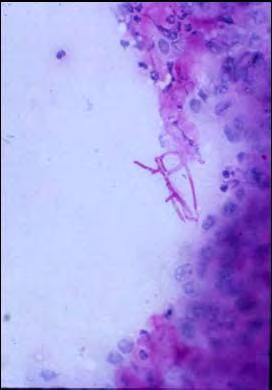
Impression smear from a nasal mass on a 2 year old cat Presented with: one month duration of epistaxis
 Impression smear from a nasal mass on a 2 year old cat Presented with: one month duration of epistaxis Identify the structures neutrophils macrophages x40 Organisms 8 30 micron in size, with variable capsule
Melanoma Case Scenario 1
 Melanoma Case Scenario 1 History and physical 11/5/16 Patient is a single, 48-year-old male in good health who presented to his primary physician for a yearly physical exam during which a 3.4 x 2.8 x 1.5
ACCME/Disclosures. Cribriform Lesions of the Prostate. Case
 Cribriform Lesions of the Prostate Ming Zhou, MD, PhD Departments of Pathology and Urology New York University Langone Medical Center New York, NY Ming.Zhou@NYUMC.ORG ACCME/Disclosures The USCAP requires
Mast Cell Tumors in Dogs
 Mast Cell Tumors in Dogs 803-808-7387 www.gracepets.com These notes are provided to help you understand the diagnosis or possible diagnosis of cancer in your pet. For general information on cancer in pets
Thyroid master class. Thyroid Fine needle aspiration cytology and liquid-based techniques: Hologic and Becton Dickinson
 Thyroid master class Thyroid Fine needle aspiration cytology and liquid-based techniques: Hologic and Becton Dickinson Principle of LBC Collection of cells in liquid medium Immediate fixation Processor-prepared
DOWNLOAD ENTIRE DOCUMENT FROM
 PREVIEW ONLY 1 Atlas on Bethesda system for reporting Thyroid Cytology PREVIEW ONLY 2 OVERVIEW 1. Indications and goal of thyroid FNA 2. Contraindications 3. Procurement of cell sample 4. Staining methods
DETERMINATION OF A LYMPHOID PROCESS
 Chapter 2 Applications of Touch Preparation Cytology to Intraoperative Consultations: Lymph Nodes and Extranodal Tissues for Evaluation of Hematolymphoid Disorders INTRODUCTION As discussed in Chap. 1,
ARIZONA SOCIETY OF PATHOLOGISTS 13 TH APRIL 2013 HEAD AND NECK CYTOPATHOLOGY. F ZAHRA ALY, MD, PhD
 ARIZONA SOCIETY OF PATHOLOGISTS 13 TH APRIL 2013 HEAD AND NECK CYTOPATHOLOGY F ZAHRA ALY, MD, PhD The main areas sites amenable for cytopathology include lymph nodes, thyroid, major salivary glands especially
Melanoma Case Scenario 1
 Melanoma Case Scenario 1 History and physical 11/5/16 Patient is a single, 48-year-old male in good health who presented to his primary physician for a yearly physical exam during which a 3.4 x 2.8 x 1.5
XX. Tumours of the nasal cavity *
 XX. Tumours of the nasal cavity * H. STONZI 1 & B. HAUSER2 Tumours of the nasal cavity are rare in domestic animals, most cases occurring in the dog. Epithelial tumours are the most common type in carnivores
Neoplasms of the Canine, Feline and Lemur Liver:
 Neoplasms of the Canine, Feline and Lemur Liver: Classification and Prognosis Annual Seminar of the French Society of Veterinary Pathology John M. Cullen VMD PhD DACVP North Carolina State University Primary
Epithelia will be discussed according to the following scheme: Type Number of layers Shape Line drawing. Squamous Cuboidal Columnar
 Epithelia Epithelia will be discussed according to the following scheme: Type Number of layers Shape Line drawing Simple Squamous Cuboidal Columnar Covering and Lining epithelium Pseudostratified Stratified
Histopathology: Cervical HPV and neoplasia
 Histopathology: Cervical HPV and neoplasia These presentations are to help you identify basic histopathological features. They do not contain the additional factual information that you need to learn about
A neoplasm is defined as "an abnormal tissue proliferation, which exceeds that of adjacent normal tissue. This proliferation continues even after
 NEOPLASIA Neoplasia is a very important topic in pathology because neoplasms are both common and serious diseases. A neoplasm literally means a new growth, and this term is used interchangeably with a
Blood: Functions. Liquid connective tissue 3 general functions 1. Transportation. 2. Regulation. 3. Protection
 Blood Elements Lecture Objectives List blood components. Classify formed elements of blood. Discuss the scientific basis of the above classification. Describe the basic structure of erythrocytes and criteria
Oncocytic-Appearing Salivary Gland Tumors. Oncocytic, Cystic, Mucinous, and High Grade Salivary Gland Tumors SALIVARY GLAND FNA: PART II
 William C. Faquin, MD, PhD Professor of Pathology Harvard Medical School Director of Head and Neck Pathology Massachusetts Eye and Ear Massachusetts General Hospital SALIVARY GLAND FNA: PART II Oncocytic,
Fellowship in Cytopathology Department of Pathology. All India Institute of Medical Sciences (AIIMS) Jodhpur, Rajasthan, India
 Jodhpur, Rajasthan, India") Fellowship in Cytopathology Department of Pathology All India Institute of Medical Sciences (AIIMS) Jodhpur, Rajasthan, India Syllabus for Fellowship in Cytopathology: FNAC Direct, Guided, EUS Exfoliative
Fellowship in Cytopathology Department of Pathology All India Institute of Medical Sciences (AIIMS) Jodhpur, Rajasthan, India Syllabus for Fellowship in Cytopathology: FNAC Direct, Guided, EUS Exfoliative
Urine Sediment Photomicrographs/Photographs
 Urine Sediment Photomicrographs/Photographs Case History CMP-17 This urine sample is from a 48-year-old male with a 30-year history of diabetes mellitus and new onset renal failure. Laboratory data include:
Basal cell carcinoma 5/28/2011
 Goal of this Presentation A practical approach to the diagnosis of cutaneous carcinomas and their mimics Thaddeus Mully, MD University of California San Francisco To review common non-melanoma skin cancers
Outline 11/2/2017. Pancreatic EUS-FNA general aspects. Cytomorphologic features of solid neoplasms/lesions of the pancreas
 ENDOSCOPIC ULTRASOUND GUIDED-FINE NEEDLE ASPIRATION CYTOLOGY OF PANCREAS Khalid Amin M.D. Assistant Professor Department of Laboratory Medicine and Pathology University of Minnesota Outline Pancreatic
Benign and malignant epithelial lesions: Seborrheic keratosis: A common benign pigmented epidermal tumor occur in middle-aged or older persons more
 Benign and malignant epithelial lesions: Seborrheic keratosis: A common benign pigmented epidermal tumor occur in middle-aged or older persons more common on the trunk; but extremities, head and neck are
This is the second learning component (Learning Component 2) in our first learning module (Learning Module 1). In this component we review a very
 in our first learning module (Learning Module 1). In this component we review a very") This is the second learning component (Learning Component 2) in our first learning module (Learning Module 1). In this component we review a very basic response to injury inflammation. We ll look at examples
This is the second learning component (Learning Component 2) in our first learning module (Learning Module 1). In this component we review a very basic response to injury inflammation. We ll look at examples
E. Staging a WHO (World Health Organization) b TNM (Tumor Node Metastasis)
 b TNM (Tumor Node Metastasis)") I KNOWLEDGE LIST ONCOLOGY Knowledge of neoplasms in animals (dog, cat, pocket pets, horses, cows) A. Clinical signs, History, Physical Exam (PE) B. Work up 1. Diagnostic Imaging a Bone Scan b Thyroid Scan
I KNOWLEDGE LIST ONCOLOGY Knowledge of neoplasms in animals (dog, cat, pocket pets, horses, cows) A. Clinical signs, History, Physical Exam (PE) B. Work up 1. Diagnostic Imaging a Bone Scan b Thyroid Scan
MECHANISMS OF HUMAN DISEASE: LABORATORY SESSION CYTOPATHOLOGY Monday, April 26, 2013 FACULTY COPY
 GOAL: MECHANISMS OF HUMAN DISEASE: LABORATORY SESSION CYTOPATHOLOGY Monday, April 26, 2013 FACULTY COPY 1. Understated the role of cytopathology in the clinical management of the patient and recognize
Benign, Reactive and Inflammatory Lesions of the Breast
 Benign, Reactive and Inflammatory Lesions of the Breast Marilin Rosa, MD Associate Member Section Head of Breast Pathology Department of Anatomic Pathology Program Director, Breast Pathology Fellowship
Salivary Gland Cytology: A Clinical Approach to Diagnosis and Management of Atypical and Suspicious Lesions
 Salivary Gland Cytology: A Clinical Approach to Diagnosis and Management of Atypical and Suspicious Lesions W.C. Faquin, M.D., Ph.D. Massachusetts General Hospital Harvard Medical School, USA Marc Pusztaszeri,