Pathologic Analysis of CNS Surgical Specimens
|
|
|
- Georgina Dean
- 6 years ago
- Views:
Transcription


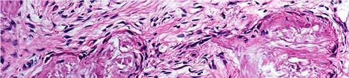


1 2015 Kenneth M. Earle Memorial Neuropathology Review Pathologic Analysis of CNS Surgical Specimens Peter C. Burger, MD Interdisciplinary Quality Control Familiarity with entities Use of diagnostic algorithm Consideration of clinical, radiologic and pathologic features Communication Consultation Education Follow up Intraspinal Lesion In An Adult Immunohistochemistry for EMA

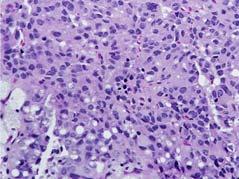
2 Calcifying pseudoneoplasm of the of the neuraxis Interdisciplinary Quality Control Familiarity with entities Use of diagnostic algorithm Consideration of clinical, radiologic and pathologic features Communication Consultation Education Follow up Diagnostic Algorithm Case Is there a lesion? If so, is it neoplastic If neoplastic, what tumor type? What grade? Is pathological diagnosis c/w clinical and radiologic findings? 18 year old woman Headaches, vomiting, blurred vision Obstructive hydrocephalus Cystic pineal region mass Calcified, Cystic Pineal Region Mass Diagnosis?


3 Pineal Region Neoplasms Pineal parenchymal tumors (pineocytoma, of intermediate differentiation, pineoblastoma Papillary tumor of the pineal region Other regional, e.g. pilocytic astrocytoma, ependymoma Normal Tissues Potentially Misinterpreted as Abnormal Normal Pineal Normal Pineal Normal Pineal


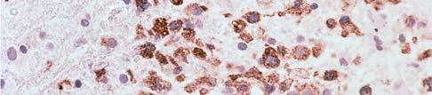
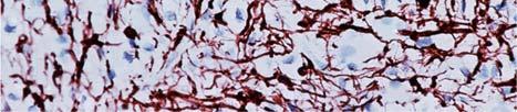
4 Normal Pineal With Brain Sand Normal Pineal Synaptophysin PPTID vs.pineoblastoma Papillary Tumour of the Pineal Region Normal Pineal! Calcified, Cystic Pineal Region Mass Pineal Cyst


")
5 Pineal Cyst Normal pineal gland in pineal cyst Young adults, more often women May be obstructive,with hydrocephalus, HA, vomiting, etc. Often asymptomatic, incidental finding Diagnostic Algorithm Is there a lesion? If so, is it neoplastic If neoplastic, what tumor type? What grade? Is pathological diagnosis c/w clinical and radiologic findings? College Student With T2 Bright Frontal Lobe Lesion: Clinical Diagnosis B.T. Mixed Glioma (Oligoastrocytoma)

")


6 Demyelinating Disease (Multiple Sclerosis) Astrocytoma Grade II Demyelinating Disease Astrocytoma Grade II Oligodendroglioma Like Lesions Oligodendroglioma Diffuse astrocytoma Macrophage rich lesions Clear cell ependymoma Clear cell meningioma Neurocytic tumors Neurocytomas Dysembryoplastic neuroepithelial tumor Rosette forming glioneuronal tumor (of the fourth ventricle)


7 Oligodendroglioma Like Lesions Clear cell ependymoma Clear cell meningioma Astrocytoma Macrophage-rich lesions Pilocytic astrocytoma Neuronal tumors Oligodendroglioma Clear cell ependymoma Clear cell meningioma Demyelinating Disease Histological Features Astrocytoma Neuronal tumors Macrophage-rich lesions Pilocytic astrocytoma Oligodendroglioma White matter localization Sharp border Macrophage infiltrate Lymphocytic infiltrates (some cases) Preserved axons Reactive astrocytes Creutzfeldt cells DemyelinatingDisease Needle Biopsy Specimen DemyelinatingDisease Needle Biopsy Specimen


8 Hypercellularity, Sharp Border Demyelinating Disease Macrophages (CD68) Demyelinating Disease Neurofilament Protein (Preserved Axons) Demyelinating Disease Macrophages and Creutzfeldt Cell Demyelinating Disease Macrophages and Creutzfeldt Cells Demyelinating Disease Creutzfeldt Cell and Granular Mitosis


9 Demyelinating Disease Smear Preparation Demyelinating disease (multiple sclerosis) Consequences of Misdiagnosis of Demyelinating Disease Demyelinating Disease Radiologic Features Psychological trauma ( pain and suffering ) Biopsy, esp. when open, resulting in neurological deficit Attempt at excision after frozen section diagnosis of tumor, resulting in neurological deficit Radiotherapy with radionecrosis Solitary or multiple Any location, but lesions approached surgically usually subependymal, subcortical, or spinal intramedullary Considerable edema in some cases Open ring or horseshoe pattern of CE Trust, but verify! * Normal Cerebellum * Ronald Reagan


10 Diagnostic Algorithm Brain Tumor in an Adult Is there a lesion? If so, is it neoplastic If neoplastic, what tumor type? What grade? Is pathological diagnosis c/w clinical and radiologic findings? Pilocytic Astrocytoma with Vascular Proliferation Brain Tumor in an Adult Radiologic Features Suggesting Low Grade Lesion Pilocytic astrocytoma Skull erosion Lack of mass effect in contrastenhancing lesion Absence of perilesional edema in contrast enhancing lesion Dense calcification Macrocyst


11 Diagnostic Algorithm Is there a lesion? If so, is it neoplastic If neoplastic, what tumor type? What grade? Is pathological diagnosis c/w clinical and radiologic findings? Interdisciplinary Quality Control Familiarity with entities Use of diagnostic algorithm Consideration of clinical, radiologic and pathologic features Communication Consultation Education Follow up Demyelinating Disease Open Ring Configuration Open ring pattern of contrast enhancement Inconsistency: Grade II infiltrating astrocytomas and oligodendrogliomas rarely enhance? glioma

 Sites")


 most")


12 Multiple Sclerosis Multiple Sclerosis Dawson s Fingers fff Cerebral Deep Venous System (Postmortem Venogram ) Sites and Patterns of Contrast Enhancement Dura Focal Neoplasms, e.g. meningioma Non neoplastic lesions, e.g. sarcoidosis; Rosai Dorfman disease Diffuse and bilateral result of intracranial hypotension, with layer of proliferating vessels on inner aspect of the dura Leptomeninges Focal Diffuse (inflammatory lesion; disseminated primary or secondary neoplasm) Parenchyma Ring or rim pattern glioblastoma, primary CNS lymphoma in immunocompromised patients; abscess; some metastases; some infarcts Open ring demyelinating disease Solid ( homogeneous ) most compact; discrete tumors, e.g. pilocytic astrocytoma; ependymoma, sporadic primary CNS lymphoma Cortical, gyriform infarct; inflammatory disease, e.g. herpes simplex encephalitis, radionecrosis Enhancing Edema-Producing Lesion Is this specimen representative? NO! With contrast T2


13 Non-enhancing Region of Edema Is this specimen representative? With contrast FLAIR Interdisciplinary Quality Control Is this specimen representative? Maybe. Familiarity with entities Use of diagnostic algorithm Consideration of clinical, radiologic and pathologic features Communication Consultation Education Follow up Pathologists View of Neurosurgeons Radiologist Clinician Pathologist Communication Unscheduled/informal Tumor board or equivalent Pathologist in OR


14 Interdisciplinary Quality Control Familiarity with entities Use of diagnostic algorithm Consideration of clinical, radiologic and pathologic features Communication Consultation Education Follow up If, as a pathologist, you are asked, or even if only slightly encouraged, to get an outside consultation on one of your cases, the correct response is: A. Ignore it B. Tell him/her you are confident of your impression and a consultation is not necessary C. Sulk, passive aggress and send it with great reluctance and delay D. Of course, I ll send it out today Interdisciplinary Quality Control Familiarity with entities Use of diagnostic algorithm Consideration of clinical, radiologic and pathologic features Communication Consultation Education Follow up Brain Tumor In A 65 year Old Examples


15 Brain Lesions in A 65 Year Old Pre and Post Steroids Sites and Patterns of Contrast Enhancement Dura Focal Neoplasms, e.g. meningioma Non neoplastic lesions, e.g. sarcoidosis; Rosai Dorfman disease Diffuse and bilateral result of intracranial hypotension, with layer of proliferating vessels on inner aspect of the dura Leptomeninges Focal Diffuse (inflammatory lesion; disseminated primary or secondary neoplasm) Parenchyma Ring or rim pattern glioblastoma, primary CNS lymphoma in immunocompromised patients; abscess; some metastases; some infarcts Open ring demyelinating disease Solid ( homogeneous ) most compact; discrete tumors, e.g. pilocytic astrocytoma; ependymoma, sporadic primary CNS lymphoma Cortical, gyriform infarct; inflammatory disease, e.g. herpes simplex encephalitis, radionecrosis Older patient Solid enhancement PCNSL Response to steroids Multiplicity Primary CNS Lymphoma Treated With Steroids


 Oligodendrogliomas (grades")


16 52 year old Man with Intramedullary Spinal Cord Tumor Well circumscribed* Pilocytic astrocytoma PXA Ependymoma Choroid plexus tumors Astroblastoma Ganglion cell tumors Hemangioblastoma Meningioma Infiltrative Diffuse astrocytomas (grades II, III, IV) Oligodendrogliomas (grades II and III) Primary CNS lymphoma?? Medulloblastoma Discrete, Enhancing Intramedullary Tumor * Extent of resection the most important prognostic factor; most contrast enhance Intramedullary Tumors by Degree of Circumscription Discrete, Highly Vascular Intramedullary Tumor Well Circumscribed Intramedullary Tumors Intramedullary Tumor Frozen Section Ependymoma Hemangioblastoma Pilocytic astrocytoma Schwannoma (rare) Paraganglioma (filum)


17 Spinal Intramedullary Tumors Surgical Options?? Diffusely Infiltrative Astrocytoma Diffuse astrocytoma stop (biopsy only) Discrete lesion resect, with risk of producing neurological deficit Intramedullary Tumor With Discrete Border Frozen Intramedullary Tumor Frozen section Oil red O Hemangioblastoma Interdisciplinary Quality Control Familiarity with entities Use of diagnostic algorithm Consideration of clinical, radiologic and pathologic features Communication Consultation Education Follow up
CNS TUMORS. D r. Ali Eltayb ( U. of Omdurman. I ). M. Path (U. of Alexandria)
. M. Path (U. of Alexandria)") CNS TUMORS D r. Ali Eltayb ( U. of Omdurman. I ). M. Path (U. of Alexandria) CNS TUMORS The annual incidence of intracranial tumors of the CNS ISmore than intraspinal tumors May be Primary or Secondary
CNS TUMORS D r. Ali Eltayb ( U. of Omdurman. I ). M. Path (U. of Alexandria) CNS TUMORS The annual incidence of intracranial tumors of the CNS ISmore than intraspinal tumors May be Primary or Secondary
Five Most Common Problems in Surgical Neuropathology
 Five Most Common Problems in Surgical Neuropathology If the brain were so simple that we could understand it, we would be so simple that we couldn t Emerson Pugh What is your greatest difficulty in neuropathology?
Peter Canoll MD. PhD.
 Tumors of the Nervous System Peter Canoll MD. PhD. What I want to cover What are the most common types of brain tumors? Who gets them? How do they ypresent? What do they look like? How do they behave?
Tumors of the Nervous System
 Tumors of the Nervous System Peter Canoll MD. PhD. What I want to cover What are the most common types of brain tumors? Who gets them? How do they present? What do they look like? How do they behave? 1
CNS pathology Third year medical students. Dr Heyam Awad 2018 Lecture 12: CNS tumours 2/3
 CNS pathology Third year medical students Dr Heyam Awad 2018 Lecture 12: CNS tumours 2/3 Pilocytic astrocytoma Relatively benign ( WHO grade 1) Occurs in children and young adults Mostly: in the cerebellum
General: Brain tumors are lesions that have mass effect distorting the normal tissue and often result in increased intracranial pressure.
 1 Lecture Objectives Know the histologic features of the most common tumors of the CNS. Know the differences in behavior of the different tumor types. Be aware of the treatment modalities in the various
Brain Tumors. Medulloblastoma. Pilocytic astrocytoma: Ahmed Koriesh, MD. Pathological finding
 NeuroPathology Page 8 Brain Tumors Pathological finding Pseudorosette Rosenthal fibers Rosettes Wet Keratin Psammoma bodies Fried egg Tumor Ependymoma, SEGA Pilocytic astrocytoma Medulloblastoma Craniopharyngioma
SPECIAL SLIDE SEMINAR CASE 3
 SPECIAL SLIDE SEMINAR CASE 3 Tihana Džombeta, MD Leo Pažanin, MD, PhD Department of Pathology, School of Medicine, University of Zagreb Department of Pathology, Clinical Hospital Centre Sestre milosrdnice
Brain tumors: tumor types
 Brain tumors: tumor types Tumor types There are more than 120 types of brain tumors. Today, most medical institutions use the World Health Organization (WHO) classification system to identify brain tumors.
Imaging the Spinal Cord & Intradural Disease
 Department of Radiology University of California San Diego Imaging the Spinal Cord & Intradural Disease John R. Hesselink, M.D. Spinal Cord Diseases Tumors Syringohydromyelia Trauma Ischemia / Infarction
Supra- and infratentorial brain tumors from childhood to maternity
 Supra- and infratentorial brain tumors from childhood to maternity What to expect? I am going to show you the characteristic imaging findings of following tumors: Thierry A.G.M. Huisman, MD, FICIS, EQNR
SURGICAL MANAGEMENT OF BRAIN TUMORS
 SURGICAL MANAGEMENT OF BRAIN TUMORS LIGIA TATARANU, MD, Ph D NEUROSURGICAL CLINIC, BAGDASAR ARSENI CLINICAL HOSPITAL BUCHAREST, ROMANIA SURGICAL INDICATIONS CONFIRMING HISTOLOGIC DIAGNOSIS REDUCING TUMOR
Tumors of the Central Nervous System
 Tumors of the Central Nervous System 1 Financial Disclosures I have NO SIGNIFICANT FINANCIAL, GENERAL, OR OBLIGATION INTERESTS TO REPORT Introduction General: Brain tumors are lesions that have mass effect
Understanding general brain tumor pathology, Part I: The basics. Craig Horbinski, M.D., Ph.D. Department of Pathology University of Kentucky
 Understanding general brain tumor pathology, Part I: The basics Craig Horbinski, M.D., Ph.D. Department of Pathology University of Kentucky plan of attack what IS a pathologist, anyway? what s so special
Astroblastoma: Radiologic-Pathologic Correlation and Distinction from Ependymoma
 AJNR Am J Neuroradiol 23:243 247, February 2002 Case Report Astroblastoma: Radiologic-Pathologic Correlation and Distinction from Ependymoma John D. Port, Daniel J. Brat, Peter C. Burger, and Martin G.
NEURORADIOLOGY DIL part 5
 NEURORADIOLOGY DIL part 5 Masses and tumors K. Agyem MD, G. Hall MD, D. Palathinkal MD, Alexandre Menard March/April 2015 OVERVIEW Introduction to Neuroimaging - DIL part 1 Basic Brain Anatomy - DIL part
Accuracy of intra-operative rapid diagnosis by Squash smear in CNS lesions An early institutional experience. KK Bansal,
 Accuracy of intra-operative rapid diagnosis by Squash smear in CNS lesions An early institutional experience. KK Bansal, Monika Bansal, Sanjeev Kishore, Anuradha K, Meena H, Dushyant G. Department of Neurosurgery
Histopathological Study and Categorisation of Brain Tumors
 Histopathological Study and Categorisation of Brain Tumors Ruchira Wadhwa 1*, Purvi Patel 2, Hansa Goswami 3 1 Third Year Resident, 2 Assistant Professor, 3 Professor and Head, Department of Pathology,
SUPPLEMENTARY INFORMATION
 VOLUME: 1 ARTICLE NUMBER: 0027 In the format provided by the authors and unedited. Rapid intraoperative histology of unprocessed surgical specimens via fibre-laser-based stimulated Raman scattering microscopy
Pineal region tumors
 Case Series Pineal region tumors Meena Patil, Manjiri Karandikar Abstract The pineal gland is located near the center of the brain, between two hemispheres in between the two thalamic bodies. It is activated
Practical Approach to. Intraoperative Consultation (IOC)
") Texas Society of Pathologists 2013 92 nd Annual Meeting Neuropathology: Intraoperative Frozen Section Consultation for General Surgical Pathologists Gregory N. Fuller, MD, PhD Professor and Chief Neuropathologist
Texas Society of Pathologists 2013 92 nd Annual Meeting Neuropathology: Intraoperative Frozen Section Consultation for General Surgical Pathologists Gregory N. Fuller, MD, PhD Professor and Chief Neuropathologist
Neuroimaging Core Curriculum
 Neuroimaging Core Curriculum Program Content The purpose of the training program is to prepare the physician for the independent practice of neuroimaging. Neuroimaging is the subspecialty of Neurology
Joana Ramalho, MD C. Ryan Miller, MD, PhD
 Joana Ramalho, MD C. Ryan Miller, MD, PhD Case 1 3 month old baby girl Presented with new onset of seizures Newborn. Questionable blurring of the gray-white junction within the right occipital lobe. Findings
Automated Identification of Neoplasia in Diagnostic Imaging text reports
 Automated Identification of Neoplasia in Diagnostic Imaging text reports "This work has been funded in whole or in part with Federal funds from the National Cancer Institute, National Institutes of Health,
Chapter 1 Introduction
 Chapter 1 Introduction Men think epilepsy divine, merely because they do not understand it. But if they called everything divine which they do not understand, why, there would be no end to divine things.
Infections. Meningitis- leptomenings (usually bakterial)
") Neuropathology Neuropathology Cerebral Edema, raised intracranial pressure, Herniatio Malformations, Developmental Diseases Perinatal Brain Injury Trauma Cerebrovascular Diseases Infections Transmissible
Neuropathology Neuropathology Cerebral Edema, raised intracranial pressure, Herniatio Malformations, Developmental Diseases Perinatal Brain Injury Trauma Cerebrovascular Diseases Infections Transmissible
Neuro-oncology Update Andrew Kokkino, MD Medical Director, The Neurosciences Institute at Sacred Heart at Riverbend May 20, 2013
 Neuro-oncology Update 2013 Andrew Kokkino, MD Medical Director, The Neurosciences Institute at Sacred Heart at Riverbend May 20, 2013 Case 1 58 year old man with recent facial droop and HA s Thin, cachectic
Pleomorphic Xanthoastrocytoma
 Pleomorphic Xanthoastrocytoma Christine E. Fuller Keywords Pleomorphic xanthoastrocytoma; Pleomorphic xanthoastrocytoma with anaplastic features 2.1 OVERVIEW Pleomorphic xanthoastrocytoma (PXA) is an uncommon
H Haloes cautions, 57 neurocytomas, perinuclear, 56 Headache blue cell tumors, 147 cautions, 135, 147, 152 clinical history, 132, 144, 148
 Index A ADC. See Apparent diffusion coefficient Adult. See also Supratentorial mass, adult cerebral tumor, 1 headache and ataxia cysts, mural nodules, 118 sporadic tumors, 118 headaches and visual changes,
Adult Brain Tumours: an approach based on imaging findings
 Adult Brain Tumours: an approach based on imaging findings Robert J Sevick, MD, FRCPC, FACR Professor, Radiology and Clinical Neurosciences Cumming School of Medicine University of Calgary Learning objectives:
Site Specific Coding Rules MALIGNANT CENTRAL NERVOUS SYSTEM TUMORS
 Multiple Primary and Histology Site Specific Coding Rules MALIGNANT CENTRAL NERVOUS SYSTEM TUMORS 1 Prerequisites 2 Completion of Multiple Primary and Histology General Coding Rules 3 There are many ways
NEURORADIOLOGY Part I
 NEURORADIOLOGY Part I Vörös Erika University of Szeged Department of Radiology SZEGED DISEASES OF CNS BRAIN Developmental anomalies Cerebrovascular disorders Tumours Inflammatory diseases Trauma DISEASES
I have no conflicts of interest in relation to this presentation. Vogel FS & Burger PC 3/28/2016
 IF THIS IS NOT GLIOBLASTOMA, THEN WHAT IS IT? Murat Gokden, MD Department of Pathology/Neuropathology University of Arkansas for Medical Sciences Little Rock, AR mgokden@uams.edu I have no conflicts of
USCAP Neuropathology. Case No. 3 Elisabeth J. Rushing, MD Armed Forces Institute of Pathology Washington, DC
 USCAP Neuropathology Case No. 3 Elisabeth J. Rushing, MD Armed Forces Institute of Pathology Washington, DC Clinical history The patient is a 9 year-old boy who has had seizures since age 2, at which time
Patients Treated with Leksell Gamma Knife
 Patients Treated with Leksell Gamma Knife 1968-2016 TREATMENTS REPORTED 2016 BY REGION AND INDICATION INDICATION Asia excl. Europe Latin Middle East & Africa North Grand Total Benign Tumors 12283 9778
Case 7391 Intraventricular Lesion
 Case 7391 Intraventricular Lesion Bastos Lima P1, Marques C1, Cabrita F2, Barbosa M2, Rebelo O3, Rio F1. 1Neuroradiology, 2Neurosurgery, 3Neuropathology, Coimbra University Hospitals, Portugal. University
CT & MRI Evaluation of Brain Tumour & Tumour like Conditions
 CT & MRI Evaluation of Brain Tumour & Tumour like Conditions Dr. Anjana Trivedi 1, Dr. Jay Thakkar 2, Dr. Maulik Jethva 3, Dr. Ishita Virda 4 1 M.D. Radiology, Professor and Head, P.D.U. Medical College
Structural and functional imaging for the characterization of CNS lymphomas
 Structural and functional imaging for the characterization of CNS lymphomas Cristina Besada Introduction A few decades ago, Primary Central Nervous System Lymphoma (PCNSL) was considered as an extremely
RINGS N THINGS: Imaging Patterns in Differential Diagnosis. Anne G. Osborn, M.D.
 RINGS N THINGS: Imaging Patterns in Differential Diagnosis Anne G. Osborn, M.D. ExpDDxs: Intra-axial (Parenchymal) Lesions Ring-enhancing lesions, solitary 1 Ring-enhancing lesion crossing corpus callosum
Cerebral Parenchymal Lesions: I. Metastatic Neoplasms
 Chapter 4 Cerebral Parenchymal Lesions: I. Metastatic Neoplasms After one has reasonably ruled out the possibility of a nonneoplastic diagnosis (see Chap. 3), one is left with considering a diagnosis of
Case 9 10/29/2018. CJD (Creutzfeldt -Jakob Disease) CJD (Creutzfeldt -Jakob Disease) CJD (Creutzfeldt -Jakob Disease)
 CJD (Creutzfeldt -Jakob Disease) CJD (Creutzfeldt -Jakob Disease)") CJD (Creutzfeldt -Jakob Disease) Rare fatal neurodegen dz caused by infectious protein Prion (lacks nucleic acid)- causes spongiform changes of the brain and neuronal death. 4 types: scjd- 85% of cases
CJD (Creutzfeldt -Jakob Disease) Rare fatal neurodegen dz caused by infectious protein Prion (lacks nucleic acid)- causes spongiform changes of the brain and neuronal death. 4 types: scjd- 85% of cases
General Identification. Name: 江 X X Age: 29 y/o Gender: Male Height:172cm, Weight: 65kg Date of admission:95/09/27
 General Identification Name: 江 X X Age: 29 y/o Gender: Male Height:172cm, Weight: 65kg Date of admission:95/09/27 Chief Complaint Sudden onset of seizure for several minutes Present illness This 29-year
2. Subependymal giant cell astrocytoma:
 I. Astrocytomas: A. Diffusely infiltrating ( astrocytoma, anaplastic astrocytoma, GBM) B. Localised (pilocytic astrocytoma, pleomorphic xanthoastrocytoma, SGCA) *Grading: Diffuse: 1. Astrocytoma WHO grade
Contents 1 Normal Histology and Commonly Used Stains 2 Basic Pathologic Reactions
 Contents 1 Normal Histology and Commonly Used Stains.... 1 1.1 Cells of the Nervous System... 1 1.1.1 Neurons... 1 1.1.2 Astrocytes.... 2 1.1.3 Oligodendrocytes... 5 1.1.4 Schwann Cells... 5 1.1.5 Ependyma....
Adult Brain and Spinal Cord Tumors
 Adult Brain and Spinal Cord Tumors An adult central nervous system (CNS) tumor is a disease in which abnormal cells form in the tissues of the brain and or the spinal cord. Major Parts of the Brain Anatomy
2 NEUROPATHOLOGY AND MOLECULAR BIOLOGY OF INTRACRANIAL TUMORS
 2 NEUROPATHOLOGY AND MOLECULAR BIOLOGY OF INTRACRANIAL TUMORS Gregory N. Fuller and Kenneth D. Aldape Chapter Overview... 23 Introduction... 24 Cornerstones of Clinical Oncologic Neuropathology... 24 Knowledge
RING ENCHANCING LESION BY M.S. HEMHNATH
 RING ENCHANCING LESION BY M.S. HEMHNATH A 21 YRS FEMALE CAME WITH H/O HEADACHE AND SEIZURE FOR THE PAST ONE MONTH. NO OTHER FOCAL NEUROLOGICAL DEFICIT. DIFFERENTIAL DIAGNOSIS For this case are Neurocysticerosis
An Approach. to Brain. Infection. 37F found down. Disclosures. Approach to CNS Infection. Objectives. Parenchymal. None.
 An Approach Disclosures to Brain None. Infection Jason Shewchuk, MD Clinical Associate Professor Head of Neuroradiology UBC European Course in Neuroradiology 2018 Objectives Following this session the
Q&A. Fabulous Prizes. Collecting Cancer Data:CNS 2/7/12. NAACCR Webinar Series Collecting Cancer Data Central Nervous System
 Collecting Cancer Data Central Nervous System NAACCR 2012 2013 Webinar Series 2/7/2013 Q&A Please submit all questions concerning webinar content through the Q&A panel. Reminder: If you have participants
Case Studies in Sella/Parasellar Region. Child thirsty, increased urination. Imaging. Suprasellar Germ Cell Tumor (Germinoma) No Disclosures
 No Disclosures") Case Studies in Sella/Parasellar Region No Disclosures 2018 Head and Neck Imaging Conference Child thirsty, increased urination Suprasellar Germ Cell Tumor (Germinoma) Midline Pineal >> Suprasellar > Other
Case Studies in Sella/Parasellar Region No Disclosures 2018 Head and Neck Imaging Conference Child thirsty, increased urination Suprasellar Germ Cell Tumor (Germinoma) Midline Pineal >> Suprasellar > Other
Essentials of Clinical MR, 2 nd edition. 51. Primary Neoplasms
 51. Primary Neoplasms As with spinal central canal neoplasms in other regions, those of the lumbar spine may be classified as extradural, intradural extramedullary, and medullary. If an extradural lesion
Rosette-forming glioneuronal tumor in the pineal gland and the third ventricle: a case with radiological and clinical implications
 Case Report Rosette-forming glioneuronal tumor in the pineal gland and the third ventricle: a case with radiological and clinical implications Junqing Xu 1, Yong Yang 1, Ying Liu 1, Mengqi Wei 1, Jing
Spinal cord tumours Luc van den Hauwe et al.
 overview spinal cord tumours L. van den Hauwe 1,2, D. Balériaux 3, J.W. Van Goethem 2, C. Venstermans 2, F. De Belder 2, P.M. Parizel 2 introduction imaging spinal tumour classification spinal cord tumours
Anaplastic Pilocytic Astrocytoma: The fusion of good and bad
 Anaplastic Pilocytic Astrocytoma: The fusion of good and bad Alexandrina Nikova 1, Charalampos-Chrysovalantis Chytoudis-Peroudis 2, Penelope Korkolopoulou 3 and Dimitrios Kanakis 4 Abstract 5 Pilocytic
Examining large groups of cancer patients to identify ways of predicting which therapies cancers might respond to.
 Stratified Medicine Examining large groups of cancer patients to identify ways of predicting which therapies cancers might respond to. Looking in detail at cancer cells and their genetic make up. Permit
TUMORS of nervous system
 TUMORS of nervous system By: Shifaa Alqa qa Done By : Ola Hijjawi CNS tumors : The annual incidence of CNS tumors ranges from 10 to 17 per 100,000 persons for intracranial tumors and 1 to 2 per 100,000
Case #3. USCAP Neuropathology Evening Seminar/Companion Meeting
 Case #3 USCAP Neuropathology Evening Seminar/Companion Meeting Clinical History A 71-year year-old man presented with a 4-4 week history of word finding difficulty. An initial screening head CT followed
Pediatric Spine Tumors (and other masses)
") Pediatric Spine Tumors (and other masses) Francisco A Perez, MD, PhD Assistant Professor Neuroradiology and Pediatric Radiology Seattle Children s Hospital University of Washington, Seattle Commercial
Pediatric Spine Tumors (and other masses) Francisco A Perez, MD, PhD Assistant Professor Neuroradiology and Pediatric Radiology Seattle Children s Hospital University of Washington, Seattle Commercial
Primary Central Nervous System Lymphoma with Lateral Ventricle Involvement
 The Open Medical Imaging Journal, 2012, 6, 103-107 103 Open Access Primary Central Nervous System Lymphoma with Lateral Ventricle Involvement Yumi Oie 1,*, Kazuhiro Murayama 1, Shinya Nagahisa 2, Masato
Masses of the Corpus Callosum
 Masses of the Corpus Callosum Kesav Raghavan, HMS Year III Dr. Agenda Corpus Callosum Development and Anatomy Our Patient: Clinical Presentation Differential Diagnosis of Masses in the Corpus Callosum
We are IntechOpen, the first native scientific publisher of Open Access books. International authors and editors. Our authors are among the TOP 1%
 We are IntechOpen, the first native scientific publisher of Open Access books 3,350 108,000 1.7 M Open access books available International authors and editors Downloads Our authors are among the 151 Countries
Advances In Orbital Neuropathology
 Advances In Orbital Neuropathology Charles G. Eberhart, MD PhD Associate Professor of Pathology, Ophthalmology and Oncology Johns Hopkins University School of Medicine Overview Non-neoplastic lesions Microphthalmos/pseudoglioma
STUDY OFPAEDIATRIC CNS TUMORS IN TERTIARY CARE CENTER
 IJCRR Section: Healthcare Sci. Journal Impact Factor 4.016 Original Article STUDY OFPAEDIATRIC CNS TUMORS IN TERTIARY CARE CENTER Grishma P. Jobanputra Tutor, Department of Pathology, B.J. Medical College,
Oligodendrogliomas & Oligoastrocytomas
 Oligodendrogliomas & Oligoastrocytomas ABOUT THE AMERICAN BRAIN TUMOR ASSOCIATION Founded in 1973, the American Brain Tumor Association (ABTA) was the first national nonprofit organization dedicated solely
AMERICAN BRAIN TUMOR ASSOCIATION. Oligodendroglioma and Oligoastrocytoma
 AMERICAN BRAIN TUMOR ASSOCIATION Oligodendroglioma and Oligoastrocytoma ACKNOWLEDGEMENTS ABOUT THE AMERICAN BRAIN TUMOR ASSOCIATION Founded in 1973, the American Brain Tumor Association (ABTA) was the
Pediatric Brain Tumors: Updates in Treatment and Care
 Pediatric Brain Tumors: Updates in Treatment and Care Writer Classroom Rishi R. Lulla, MD MS Objectives Introduce the common pediatric brain tumors Discuss current treatment strategies for pediatric brain
Epidemiology of Primary Brain and Central Nervous System Tumors in Korea
 www.jkns.or.kr 1.334/jkns.21.48.2.145 J Korean Neurosurg Soc 48 : 145-152, 21 Print ISSN 25-3711 On-line ISSN 1598-7876 Copyright 21 The Korean Neurosurgical Society Clinical Article Epidemiology of Primary
Protocol for management of patients with pineal region tumours v1
 Protocol for management of patients with pineal region tumours v1 West Midlands Cancer Alliance Coversheet for Cancer Alliance Expert Advisory Group Agreed Documentation This sheet is to accompany all
Introduction to Neurosurgical Subspecialties:
 Introduction to Neurosurgical Subspecialties: Tumor and Skull Base Neurosurgery Brian L. Hoh, MD 1 and Gregory J. Zipfel, MD 2 1 University of Florida, 2 Washington University Tumor / Skull Base Neurosurgery
Disclosure. + Outline. Case-based approach to neurological emergencies that might present to the ED
 Kathleen R. Fink, MD University of Washington 5 th Nordic Emergency Radiology Course May 21, 2015 Disclosure My spouse receives research salary support from: Bracco BayerHealthcare Guerbet Outline Case-based
Brain Imaging. IC calcifications. Mamdouh mahfouz MD
 Brain Imaging IC calcifications www.ssregypt.com Mamdouh mahfouz MD mamdouh.m5@gmail.com CT Hyper dense [ more than100 HU ] MRI Low signal in T1 and T2 WIs [non mobile protons] Exceptions Minute calcifications
Adult Central Nervous System Tumors Treatment (PDQ )
") 1 di 20 28/06/2016 11.18 NCBI Bookshelf. A service of the National Library of Medicine, National Institutes of Health. PDQ Cancer Information Summaries [Internet]. Bethesda (MD): National Cancer Institute
1 di 20 28/06/2016 11.18 NCBI Bookshelf. A service of the National Library of Medicine, National Institutes of Health. PDQ Cancer Information Summaries [Internet]. Bethesda (MD): National Cancer Institute
Neurosurgical Management of Brain Tumours. Nicholas Little Neurosurgeon RNSH
 Neurosurgical Management of Brain Tumours Nicholas Little Neurosurgeon RNSH General Most common tumours are metastatic 10x more common than primary Incidence of primary neoplasms is 20 per 100000 per year
Classification of spontaneous brain tumors in rats
 Classification of spontaneous brain tumors in rats Central Nervous System Neoplasms in the Rat (Solleveld HA, et al., 1991) 1. Tumors of Neuroepithelial Tissue A. Astrocytic and oligodendroglial tumors
Disclosures. Posterior Fossa Masses. I m from the Government. and I here to help! Differential Diagnosis
 Posterior Fossa Masses Differential Diagnosis James G. Smirniotopoulos, M.D. Radiology, Neurology, Biomedical Informatics Uniformed Services University Bethesda, Maryland http://rad.usuhs.edu http://medpix.usuhs.edu
IAP XXVI International Congress Slide Seminar 07 (SS07)
") IAP XXVI International Congress Slide Seminar 07 (SS07) Pitfalls in Surgical Neuropathology Case 6 Richard A. Prayson, M.D. Cleveland Clinic Foundation Clinical History 64M S/P resection of pituitary adenoma
IAP XXVI International Congress Slide Seminar 07 (SS07) Pitfalls in Surgical Neuropathology Case 6 Richard A. Prayson, M.D. Cleveland Clinic Foundation Clinical History 64M S/P resection of pituitary adenoma
Protocol for the Examination of Specimens From Patients With Tumors of the Central Nervous System*
 Protocol for the Examination of Specimens From Patients With Tumors of the Central Nervous System* Version: Protocol Posting Date: December 2014 This protocol is NOT required for accreditation purposes
2018 Diagnostic Slide Session Case #8
 2018 Diagnostic Slide Session Case #8 Angela N. Viaene, MacLean P. Nasrallah, and Zissimos Mourelatos Hospital of the University of Pennsylvania AANP June 9, 2018 Disclosures: none Clinical History Healthy,
Histological pattern of central nervous system neoplasms
 ecommons@aku Department of Pathology and Laboratory Medicine Medical College, Pakistan April 2001 Histological pattern of central nervous system neoplasms Z. Ahmed Aga Khan University S. Muzaffar Aga Khan
Oligodendroglioma: imaging findings, radio-pathological correlation and evolution
 Oligodendroglioma: imaging findings, radio-pathological correlation and evolution Poster No.: C-2104 Congress: ECR 2013 Type: Authors: Keywords: DOI: Scientific Exhibit A. Hernandez Castro, M. D. Monedero
In 1988 Dumas-Duport et al. first used
 Copyright 2009, Barrow Neurological Institute Dysembryoplastic Neuroepithelial Tumor: A Review Mark Garrett, MD Jennifer Eschbacher, MD Peter Nakaji, MD Most DNETs are benign, low-grade lesions. However,
Case Report Atypical Presentation of Atypical Teratoid Rhabdoid Tumor in a Child
 Case Reports in Oncological Medicine Volume 2013, Article ID 815923, 4 pages http://dx.doi.org/10.1155/2013/815923 Case Report Atypical Presentation of Atypical Teratoid Rhabdoid Tumor in a Child Y. T.
Benign brain lesions
 Benign brain lesions Diagnostic and Interventional Radiology Hung-Wen Kao Department of Radiology, Tri-Service General Hospital, National Defense Medical Center Computed tomography Hounsfield unit (HU)
1 NORMAL ANATOMY AND HISTOLOGY
 1 NORMAL ANATOMY AND HISTOLOGY OF THE CNS Anatomy 1 Histology 4 ANATOMY Knowledge of nervous system anatomy is essential for success in surgical neuropathology. Familiarity with native cellular elements
MRI OF THE THALAMUS. Mohammed J. Zafar, MD, FAAN Kalamazoo, MI
 1 MRI OF THE THALAMUS Mohammed J. Zafar, MD, FAAN Kalamazoo, MI Objectives: The thalamic nuclei can be involved in a wide variety of conditions. A systematic imaging approach would be useful for narrowing
MALIGNANT GLIOMAS: TREATMENT AND CHALLENGES
 MALIGNANT GLIOMAS: TREATMENT AND CHALLENGES DISCLOSURE No conflicts of interest to disclose Patricia Bruns APRN, CNS Givens Brain Tumor Center Abbott Northwestern Hospital October 12, 2018 OBJECTIVES THEN
Optic Pathway Gliomas, Germinomas, Spinal Cord Tumours. Colin Kennedy March 2015
 Optic Pathway Gliomas, Germinomas, Spinal Cord Tumours Colin Kennedy March 2015 Glioma of the optic chiasm. T1-weighted MRI with gadolinium enhancement, showing intense irregular uptake of contrast. The
MRI imaging in meningeal diseases
 Original article MRI imaging in meningeal diseases 1Dr. Narendrakumar M Shah, 2 Dr Vaishali D M 1Associate professor, Department of Radiodiagnosis, SDM Medical college, Dharwad 2Consultant radiologist,
The central nervous system
 Sectc.qxd 29/06/99 09:42 Page 81 Section C The central nervous system CNS haemorrhage Subarachnoid haemorrhage Cerebral infarction Brain atrophy Ring enhancing lesions MRI of the pituitary Multiple sclerosis
Childhood brain tumours
 Childhood brain tumours Our bodies are made up of billions of cells. Normally, these cells reproduce and repair themselves in a controlled way and do not cause us any problems. If for some reason this
AMERICAN ASSOCIATION OF NEUROPATHOLOGISTS COMPANION SOCIETY MEETING at the 106 th ANNUAL MEETING OF THE USCAP San Antonio, March 4, 2017
 AMERICAN ASSOCIATION OF NEUROPATHOLOGISTS COMPANION SOCIETY MEETING at the 106 th ANNUAL MEETING OF THE USCAP San Antonio, March 4, 2017 SYLLABUS Papillary Tumor of the Pineal Region and the Differential
Clinical, radiological, and histopathological features and prognostic factors of brain tumors in children
 Journal of Physics: Conference Series PAPER OPEN ACCESS Clinical, radiological, and histopathological features and prognostic factors of brain tumors in children To cite this article: M H Siregar et al
Case Report Papillary Tumor of the Pineal Region: MR Signal Intensity Correlated to Histopathology
 Case Reports in Neurological Medicine Volume 2015, Article ID 315095, 4 pages http://dx.doi.org/10.1155/2015/315095 Case Report Papillary Tumor of the Pineal Region: MR Signal Intensity Correlated to Histopathology
Detection of Leptomeningeal CNS Metastases in Children
 Detection of Leptomeningeal CNS Metastases in Children Noah D. Sabin, M.D. Julie H. Harreld M.D. Kathleen J. Helton M.D. Zoltan Patay M.D., Ph.D. St. Jude Children s Research Hospital Memphis, TN Leptomeningeal
This article appeared in a journal published by Elsevier. The attached copy is furnished to the author for internal non-commercial research and
 This article appeared in a journal published by Elsevier. The attached copy is furnished to the author for internal non-commercial research and education use, including for instruction at the authors institution
Neuropathology Evening Session: Case 3
 Neuropathology Evening Session: Case 3 Christine E. Fuller, MD Cincinnati Children s Hospital Medical Center Disclosure of Relevant Financial Relationships USCAP requires that all faculty in a position
MOLECULAR DIAGNOSTICS OF GLIOMAS
 MOLECULAR DIAGNOSTICS OF GLIOMAS Arie Perry, M.D. Director, Neuropathology Division DIFFUSE GLIOMAS Cell types Astrocytomas (A) Oligodendrogliomas (O) Mixed oligoastrocytoma (MOA) Three WHO grades: II,
Clinics in diagnostic imaging (175)
") Singapore Med J 2017; 58(3): 121-125 doi: 10.11622/smedj.2017017 CMEArticle Clinics in diagnostic imaging (175) Vijay Krishnan 1, MD, FRCR, Tze Chwan Lim 1, MBBS, FRCR, Francis Cho Hao Ho 2, MBBS, FRANZCR,
Singapore Med J 2017; 58(3): 121-125 doi: 10.11622/smedj.2017017 CMEArticle Clinics in diagnostic imaging (175) Vijay Krishnan 1, MD, FRCR, Tze Chwan Lim 1, MBBS, FRCR, Francis Cho Hao Ho 2, MBBS, FRANZCR,
Childhood Brain and Spinal Cord Tumors Treatment Overview (PDQ )
") 1 di 8 04/03/2017 07.31 NCBI Bookshelf. A service of the National Library of Medicine, National Institutes of Health. PDQ Cancer Information Summaries [Internet]. Bethesda (MD): National Cancer Institute
1 di 8 04/03/2017 07.31 NCBI Bookshelf. A service of the National Library of Medicine, National Institutes of Health. PDQ Cancer Information Summaries [Internet]. Bethesda (MD): National Cancer Institute
Pediatric CNS Tumors. Disclosures. Acknowledgements. Introduction. Introduction. Posterior Fossa Tumors. Whitney Finke, MD
 Pediatric CNS Tumors Disclosures Whitney Finke, MD Neuroradiology Fellow PGY-6 University of Utah Health Sciences Center Salt Lake City, Utah None Acknowledgements Introduction Nicholas A. Koontz, MD Luke