PRIMARY MUSCULOSKELETAL HYDATID CYST DISEASE. Radiology Department, Military Hospital Mohamed V. Rabat Morocco
|
|
|
- Grant Conley
- 5 years ago
- Views:
Transcription
1 PRIMARY MUSCULOSKELETAL HYDATID CYST DISEASE. A. El Kharras S. Semlali A. Darbi S. Akjouj J. El Fenni A. Hanine M. Benameur D. Bassou Radiology Department, Military Hospital Mohamed V. Rabat Morocco
2 Introduction Hydatid disease is an infestation by the larval stages of Ecchinoccocus Granulosis. It s s the commonest disease in humans caused by helminths. More frequent occurrence in the Mediterranean countries, East Africa, South Africa, Russia and Australia. Hydatid disease more frequently affects the liver and the lungs. Musculoskeletal involvement is seen in only 1-4% of cases.
3 Patients and methods Review a 10 cases (7( 7 men, 3 women) of isolated and primary hydatid cyst in muscle all location combined, diagnosed in our department between 2000 and The mean age of patients was 30 years [20[ years]. Clinical findings: slowly growing mass: : 10 cases Located pain: : 3 cases. Imaging techniques : Ultrasonography (n=8), CT scan (n=6), MRI (n=4). In all cases: hepatic sonography and chest radiography.
4 Results: table I In seven patients hydatid cyst was located in the peripheral muscle (70% 70%) In three cases hydatid cyst interested thoracic muscle (30% 30%). There was no hepatic or pulmonary involvement in those patients.
5 Table I- Musculoskeletal Hydatid cyst: and imaging findings Cases Symptoms Location Imaging Techniques Imaging findings 1 Tumor Left quadricipital muscle US multilocular cystic lesion 2 Tumor- pain Intercostal muscle US CT multilocular cystic lesion 3 Tumor Right knees US- MRI multilocular cystic lesion 4 Tumor Left upper leg US - MRI Tissular lesion 5 Tumor- pain Pelvien muscle CT Tissular lesion 6 Tumor Paravertebral muscle CT Unicystic lesion 7 Tumor- pain Left gluteal and pelvic muscle US - CT multilocular cystic lesion 8 Tumor Intercostal muscle US- CT Unicystic lesion 9 Tumor Posterior left knees US CT - MRI Tissular lesion 10 Tumor Left quadricipital muscle US- MRI multilocular cystic lesion
 lesion in 5 patients. tissular lesion in 3 patients. Fig.")
6 Diagnosis was highly suspected on ultrasonography in 6 patients, showing: unilocular cyst lesion in 1 case and multilocular cyst (fig.1) lesion in 5 patients. tissular lesion in 3 patients. Fig.1: ultrasonography of left quadricipital muscle showed multilocular cystic lesion
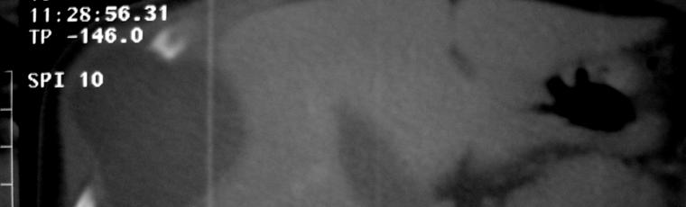
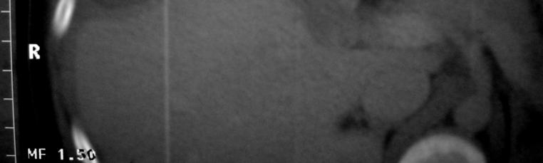
7 CT scan Lesion density: thoracic lesions in 3 patients: 1 unilocular (Fig.2) and 2 multicystics lesions 1 pelvic and tissular mass (Fig.3) gluteal and left iliopsoas: multicystic lesion in 1 patient (Fig.4( Fig.4) bone was affected in 1 patient (femoral head and pubis bone). precise extension and associated lesion.


8 Fig.2: chest CT scan showed intercostal unilocular cystic lesion.

.")
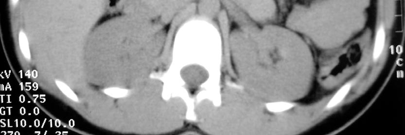
9 Fig.3: pelvien CT scan showed tissular lesion in pelvien muscle with bone lesion (arrow).


10 Fig.4: pelvien CT scan showed left gluteal and pelvien cystic mass without bone lesion.
11 MRI Multilocular cystic lesion ( Fig.with endovesicular daughter cysts and low-signal intensity on T1-weighted MR images was observed in 2 patients. Like-tissular lesion, with intermediary signal on T1- weighted MR images in 2 cases. On T2-weighted images, all cystic lesions were visualized with hight signal intensity.


12 a Fig.5: multilocular hydatid cystic of right knees: a- ultrasonography b- axial T1 weighted MRI c- axial T2 weighted MRI d- frontal T2 weighted MRI b c d


13 T2 T2 T1 Fig.6: sonography and MRI of left upper leg showed liketissular lesion on sonography and heterogeneous cystic lesion on T1 and T2- weighted images.
14 Treatment Radical surgery was performed in all patients, associated with a medical treatment. Recurrence was observed in 3 patients.
15 Discussion Musculoskeletal lesion of cystic echinococcosis was usually occurred as isolated findings and without concomitant hepatic or pulmonary involvement. In our study, musculoskeletal hydatid lesion was isolated. Most cases of muscular hydatid disease have been associated with skeletal lesions. One patient have bone lesion in our study. Immunologic tests are useful in the diagnosis of hepatic hydatidosis.. However, in other series, serological tests was not positive for muscular echinococcosis in all cases
16 Imaging Several patterns of disease have been recognized using various imaging techniques. These include Unilocular cyst, Multilocular lesion Atypical complex or solid lesion. The multilocular lesion with several daughter cysts inside the mother cyst is characteristic, but not pathognomonic of hydatid disease.

17 The CT scan: cystic lesion in soft tissues, described as homogeneous and isodense with liquor, (4 patients) inhomogeneous and hyperdense (2 patients in our study), or abscess- like. Peripheral contrast enhancement was showing for all this lesion. CT scan can showed linear calcification and adjacent bone lesion (1 bone lesion in our study)


18 MRI MRI signal intensity pattern of the hydatid cysts reflects their contents. Production of hydatid fluid stops when they dead. Presence of a bacterial infection, abundant intracystic debris or inflammatory changes may affect the typical cystic morphology, transforming it into complex or solid lesion mimicking a tumor. A T1-weighted low intensity surrounding the cyst and it may help to distinguish hydatid cysts from other lesion.
19 MRI The rim sign on the MR images appears as a low signal intensity surrounding the cyst and it may help to distinguish hydatid cysts from other lesions. On T2-weighted images, all cystic lesions were visualized with hight signal intensity (4 in our series). In our series, diagnosis was easily evocated in MRI images. The endovesicular daughter cysts and calcifications were typically seen in hepatic cystic echinococcosis,, but were the exception in musculoskeletal lesion.
20 Treatment Adjuvant administration of benzimidazole derivatives preoperatively and for about 3 months post-operatively operatively is advocated by some authors. Surgery mast makes with a broad safety margin. Several recurrence can be reported (3 in our series).
21 Conclusion Muscular lesions of cystic echinococcosis are rare even in endemic areas. Several patterns of disease have been recognized using various imaging techniques. However, echinococcosis should be always suspected and bared in mind in the differential diagnosis of cystic lesions in soft tissue. Once the diagnosis is established, the surgeon should consider performing a radical procedure aiming in minimizing the possibility of a recurrence.
Case Report Pulmonary Embolism Originating from a Hepatic Hydatid Cyst Ruptured into the Inferior Vena Cava: CT and MRI Findings
 Case Reports in Radiology Volume 2016, Article ID 3589812, 4 pages http://dx.doi.org/10.1155/2016/3589812 Case Report Pulmonary Embolism Originating from a Hepatic Hydatid Cyst Ruptured into the Inferior
Cardiac Mass in a 15-Year-Old Boy
 Cardiac Mass in a 15-Year-Old Boy Echocardiographic Case Report Hortensia Vuçini Department of Cardiology and Cardiac Surgery UHC Mother Theresa Tirana, Albania October 20, 2007 Case Presentation 15 year-old
HYDATID LIVER DISEASE SPECTRUM OF APPEARANCES
 HYDATID LIVER DISEASE SPECTRUM OF APPEARANCES DR JAIKISHOR JOTHIRAJ 1 ST YEAR POSTGRADUATE MDRD SRMMCH CHENNAI GUIDED BY PROF & HOD DR SRINIVASA MUDALI PROF DR VINAYAGAM.S PROF DR SARAVANAN K.C CASE 1
Biliary cancers: imaging diagnosis. Study of 30 cases
 Biliary cancers: imaging diagnosis. Study of 30 cases N Hammoune, S Semlali, M Eddarai, T. Amil, M Zentar, S. El Kandri,, M Benameur,, S Chaouir. Radiology Department. Mohamed V Military Hospital. Rabat-
Role of imaging in RCC. Ultrasonography. Solid lesion. Cystic RCC. Solid RCC 31/08/60. From Diagnosis to Treatment: the Radiologist Perspective
 Role of imaging in RCC From Diagnosis to Treatment: the Radiologist Perspective Diagnosis Staging Follow up Imaging modalities Limitations and pitfalls Duangkamon Prapruttam, MD Department of Therapeutic
Soft Tissue Tumour & Sarcoma Imaging Guidelines 2012
 Soft Tissue Tumour & Sarcoma Imaging Guidelines 2012 Version Control This is a controlled document please destroy all previous versions on receipt of a new version. Date Approved: March 2011 reissued April
International Journal of Research in Health Sciences ISSN: Available online at: Case Study
 International Journal of Research in Health Sciences ISSN: 2321-7251 Available online at: http://www.ijrhs.org/ Case Study Foreign body granuloma mimicking a soft tissue neoplasm *Rohan Sawant, Abhishek
HOW TO IMAGE AND DESCRIBE CONGENITAL LUNG MALFORMATIONS
 HOW TO IMAGE AND DESCRIBE CONGENITAL LUNG MALFORMATIONS Paul Thacker, MD Assistant Professor Departments of Radiology and Pediatrics Medical University of South Carolina DISCLOSURES I have no relevant
HEPATO-BILIARY IMAGING
 HEPATO-BILIARY IMAGING BY MAMDOUH MAHFOUZ MD PROF.OF RADIOLOGY CAIRO UNIVERSITY mamdouh.m5@gmail.com www.ssregypt.com CT ABDOMEN Indications Patient preparation Patient position Scanogram Fasting 4-6 hours
Primary Intramuscular Hydatid Cyst of the thigh Muscle a Rare Case Report
 IOSR Journal of Dental and Medical Sciences (IOSR-JDMS) e-issn: 2279-0853, p-issn: 2279-0861.Volume 15, Issue 3 Ver. VIII (Mar. 2016), PP 07-11 www.iosrjournals.org Primary Intramuscular Hydatid Cyst of
UPDATES AND SPOTLIGHTS ON SOME HEPATOBILIARY PARASITES
 UPDATES AND SPOTLIGHTS ON SOME HEPATOBILIARY PARASITES Dr. Manar Sobh Azab Associate Professor of Parasitology Faculty of Medicine Mansoura University Parasites are common worldwide and are classified
Congenital Lung Malformations: Radiologic-Pathologic Correlation
 Acta Radiológica Portuguesa, Vol.XVIII, nº 70, pág. 51-60, Abr.-Jun., 2006 Congenital Lung Malformations: Radiologic-Pathologic Correlation Marilyn J. Siegel Mallinckrodt Institute of Radiology, Washington
NEURORADIOLOGY DIL part 5
 NEURORADIOLOGY DIL part 5 Masses and tumors K. Agyem MD, G. Hall MD, D. Palathinkal MD, Alexandre Menard March/April 2015 OVERVIEW Introduction to Neuroimaging - DIL part 1 Basic Brain Anatomy - DIL part
Science & Technologies
 A GIANT LIVER HYDATIDE CYST SIMULTANEOUSLY PERFORATED TO PERITONEAL AND PLEURAL CAVITIES A RARE CASE REPORT. Ivan P. Novakov Department of Special Surgery; Medical University - Plovdiv Abstract. Background.
Cerebro-vascular stroke
 Cerebro-vascular stroke CT Terminology Hypodense lesion = lesion of lower density than the normal brain tissue Hyperdense lesion = lesion of higher density than normal brain tissue Isodense lesion = lesion
Case Report Isolated Retroperitoneal Hydatid Cyst Invading Splenic Hilum
 Case Reports in Surgery, Article ID 303401, 4 pages http://dx.doi.org/10.1155/2014/303401 Case Report Isolated Retroperitoneal Hydatid Cyst Invading Splenic Hilum Safak Ozturk, 1 Mutlu Unver, 1 Burcin
Complicated echinococcal cyst to Biopsy or not to biopsy. V. Rusanov MR Kramer Pulmonary Institute, Rabin medical center
 Complicated echinococcal cyst to Biopsy or not to biopsy V. Rusanov MR Kramer Pulmonary Institute, Rabin medical center Case 1 84 y.o. Male, Iraq descend, past smoker 40 PY Medical History- HTN, Rheumatoid
 INTRAUTERINE DEVICE = IUD INTRAUTERINE DEVICE = IUD CONGENITAL DISORDERS Pyometra = pyometrea is a uterine infection, it is accumulation of purulent material in the uterine cavity. Ultrasound is usually
MRI XR, CT, NM. Principal Modality (2): Case Report # 2. Date accepted: 15 March 2013
: Case Report # 2. Date accepted: 15 March 2013") Radiological Category: Musculoskeletal Principal Modality (1): Principal Modality (2): MRI XR, CT, NM Case Report # 2 Submitted by: Hannah Safia Elamir, D.O. Faculty reviewer: Naga R. Chinapuvvula, M.D.
Radiological Category: Musculoskeletal Principal Modality (1): Principal Modality (2): MRI XR, CT, NM Case Report # 2 Submitted by: Hannah Safia Elamir, D.O. Faculty reviewer: Naga R. Chinapuvvula, M.D.
Imaging in gastric cancer
 Imaging in gastric cancer Gastric cancer remains a deadly disease because of late diagnosis. Adenocarcinoma represents 90% of malignant tumors. Diagnosis is based on endoscopic examination with biopsies.
objectives Pitfalls and Pearls in PET/CT imaging Kevin Robinson, DO Assistant Professor Department of Radiology Michigan State University
 objectives Pitfalls and Pearls in PET/CT imaging Kevin Robinson, DO Assistant Professor Department of Radiology Michigan State University To determine the regions of physiologic activity To understand
Complex Fractures and Hip Dislocations
 IMAGING OF HIP PAIN Patients may present with acute (< 2 weeks) or chronic hip pain. Acute pain may be related or not related to an acute traumatic event such as fall or trauma from a motor vehicle accident.
Dr. T. Venkat Kishan Asst. Prof Department of Radiodiagnosis
 Dr. T. Venkat Kishan Asst. Prof Department of Radiodiagnosis Schwannomas (also called neurinomas or neurilemmomas) constitute the most common primary cranial nerve tumors. They are benign slow-growing
Image Interpretation and Evaluation
 Semester 2 / Commences January 20 Credits Each Course is composed of Modules & Activities. Modules: Image Interpretation Thoracic radiography Abdominal pelvic imaging Musculoskeletal Imaging Neuroimaging
Neckmasses in infancy and childhood: Clinical and radiological classification and imaging approaches M. Mearadji
 Neckmasses in infancy and childhood: Clinical and radiological classification and imaging approaches M. Mearadji International Foundation for Pediatric Imaging Aid Introduction Neck masses are a frequent
Atypical manifestations, complications and pathological correlation of hydatid disease.
 Atypical manifestations, complications and pathological correlation of hydatid disease. Poster No.: C-2501 Congress: ECR 2012 Type: Educational Exhibit Authors: R. Lerma Ortega, D. J. López Ruiz, L. A.
ACG Clinical Guideline: Diagnosis and Management of Focal Liver Lesions
 ACG Clinical Guideline: Diagnosis and Management of Focal Liver Lesions Jorge A. Marrero, MD, 1 Joseph Ahn, MD, FACG, 2 K. Rajender Reddy, MD, FACG 3 1 University of Texas at Southwestern, Dallas, Texas,
MANAGEMENT RECOMMENDATIONS
 1 MANAGEMENT RECOMMENDATIONS 1. Adrenal masses!!!!!!! page 2 2. Liver Masses!!!!!!! page 3 3. Obstetric US Soft Markers for Aneuploidy!! pages 4-6 4. Ovarian and Adnexal Cysts!!!!! pages 7-10 5. Pancreatic
CT & MRI Evaluation of Brain Tumour & Tumour like Conditions
 CT & MRI Evaluation of Brain Tumour & Tumour like Conditions Dr. Anjana Trivedi 1, Dr. Jay Thakkar 2, Dr. Maulik Jethva 3, Dr. Ishita Virda 4 1 M.D. Radiology, Professor and Head, P.D.U. Medical College
Normal Sonographic Anatomy
 hapter 2:The Liver DUNSTAN ABRAHAM Normal Sonographic Anatomy Homogeneous, echogenic texture (Figure 2-1) Measures approximately 15 cm in length and 10 12.5 cm anterior to posterior; measurement taken
Ovarian Lesion Benign vs Malignant?
 Ovarian Lesion Benign vs Malignant? Michele Keenan 1,2 Bernice Dunne 2 Mary Moran 1 Therese Herlihy 1 1. Radiography and Diagnostic Imaging, School of Medicine, University College Dublin, Ireland 2. Midland
PREAMBLE GENERAL DIAGNOSTIC RADIOLOGY
 PREAMBLE The General Diagnostic Radiology category is intended to cover the body of knowledge a practicing board certified Diagnostic Radiologist should know. Since the range of content relevant to the
Liver Cancer (Hepatocellular Carcinoma or HCC) Overview
 Overview") Liver Cancer (Hepatocellular Carcinoma or HCC) Overview Recent advances in liver cancer care seek to address the rising incidence of liver cancer, which has steadily increased over the past three decades.
Liver Cancer (Hepatocellular Carcinoma or HCC) Overview Recent advances in liver cancer care seek to address the rising incidence of liver cancer, which has steadily increased over the past three decades.
CT Findings of Traumatic Posterior Hip Dislocation after Reduction 1
 CT Findings of Traumatic Posterior Hip Dislocation after Reduction 1 Sung Kyoung Moon, M.D., Ji Seon Park, M.D., Wook Jin, M.D. 2, Kyung Nam Ryu, M.D. Purpose: To evaluate the CT images of reduced hips
THE POSTOPERATIVE APPEARANCE OF THE HYDATID CYST SURGERY LIVER ON ULTRASONOGRAPHY FOLLOWING
 HPB Surgery, 1990, Vol. 2, pp. 253-260 Reprints available directly from the publisher Photocopying permitted by license only 1990 Harwood Academic Publishers GmbH Printed in the United Kingdom THE POSTOPERATIVE
Cystic lesions of the liver
 Cystic lesions of the liver Poster No.: C-0408 Congress: ECR 2014 Type: Educational Exhibit Authors: E. Rosado, J. Pereira, S. El Bouchaibi, M. A. A. Bali ; 1 1 2 2 3 3 4 4 Amadora/PT, Lisboa/PT, Bruxelles/BE,
Armed Forces Institute of Pathology.
 Armed Forces Institute of Pathology www.radpath.com Armed Forces Institute of Pathology Breast Disease www.radpath.org Armed Forces Institute of Pathology Interpretation of Breast MRI Leonard M. Glassman
A CASE OF A Huge Submandibular Pleomorphic Adenoma
 ISPUB.COM The Internet Journal of Head and Neck Surgery Volume 4 Number 2 S VERMA Citation S VERMA.. The Internet Journal of Head and Neck Surgery. 2009 Volume 4 Number 2. Abstract Pleomorphic adenoma
MRI Of Locally Recurrent Soft Tissue Tumors Of The Musculoskeletal System
 ISPUB.COM The Internet Journal of Radiology Volume 5 Number 2 MRI Of Locally Recurrent Soft Tissue Tumors Of The Musculoskeletal System C Costelloe, A Yasko, W Murphy, R Kumar, V Lewis, P Lin, R Stafford,
CME Article Clinics in diagnostic imaging (135)
") Medical Education Singapore Med J 2011; 52(5) : 384 CME Article Clinics in diagnostic imaging (135) Pojchamarnwiputh S, Muttarak M, Sriplakich S H 1a 1b 1c 1d Fig. 1 (a) Axial unenhanced; (b & c) delayed
Medical Education Singapore Med J 2011; 52(5) : 384 CME Article Clinics in diagnostic imaging (135) Pojchamarnwiputh S, Muttarak M, Sriplakich S H 1a 1b 1c 1d Fig. 1 (a) Axial unenhanced; (b & c) delayed
1/25/13 Right partial nephrectomy followed by completion right radical nephrectomy.
 History and Physical Case Scenario 1 45 year old white male presents with complaints of nausea, weight loss, and back pain. A CT of the chest, abdomen and pelvis was done on 12/8/12 that revealed a 12
Value of MRI in Characterizing Adnexal Masses
 The Journal of Obstetrics and Gynecology of India (July August 2015) 65(4):259 266 DOI 10.1007/s13224-015-0730-9 PHOTO ESSAY Value of MRI in Characterizing Adnexal Masses Alpana Karnik 1 Raina Anil Tembey
INTERDISCIPLINARY DISCUSSIONS IN LOCALISED RCC DIAGNOSIS AND SURGICAL STRATEGIES FOR ATYPICAL RENAL CYSTIC LESIONS. Maria Cova
 INTERDISCIPLINARY DISCUSSIONS IN LOCALISED RCC DIAGNOSIS AND SURGICAL STRATEGIES FOR ATYPICAL RENAL CYSTIC LESIONS Maria Cova Radiology Department University of Trieste (IT) Eleventh European International
Pediatric TB Intensive Houston, Texas October 14, 2013
 Pediatric TB Intensive Houston, Texas October 14, 2013 Radiologic Presentation of Childhood TB Susan D. John, MD, FACR October 14, 2013 Disclosures I have no disclosures or conflicts of interest to report
Sonography of Pediatric Superficial Lumps and Bumps: Illustrative Examples from Head to Toe
 Sonography of Pediatric Superficial Lumps and umps: Illustrative Examples from Head to Toe nmol Gupta ansal, MD Henrietta Kotlus Rosenberg, MD, FCR, FP Mount Sinai Hospital Icahn School of Medicine at
CASE REPORT PLEOMORPHIC LIPOSARCOMA OF PECTORALIS MAJOR MUSCLE IN ELDERLY MAN- CASE REPORT & REVIEW OF LITERATURE.
 PLEOMORPHIC LIPOSARCOMA OF PECTORALIS MAJOR MUSCLE IN ELDERLY MAN- CASE REPORT & REVIEW OF LITERATURE. M. Madan 1, K. Nischal 2, Sharan Basavaraj. C. J 3. HOW TO CITE THIS ARTICLE: M. Madan, K. Nischal,
Case Report Unusual Presentation of Hydatid Cyst in Breast with Magnetic Resonance Imaging Findings
 Hindawi Case Reports in Medicine Volume 2017, Article ID 6237435, 4 pages https://doi.org/10.1155/2017/6237435 Case Report Unusual Presentation of Hydatid Cyst in Breast with Magnetic Resonance Imaging
TABLES. Imaging Modalities Evidence Tables Table 1 Computed Tomography (CT) Imaging. Conclusions. Author (Year) Classification Process/Evid ence Class
 Imaging. Conclusions. Author (Year) Classification Process/Evid ence Class") TABLES Imaging Modalities Evidence Tables Table 1 Computed Tomography (CT) Imaging Author Clark (1986) 9 Reformatted sagittal images in the differential diagnosis meningiomas and adenomas with suprasellar
TABLES Imaging Modalities Evidence Tables Table 1 Computed Tomography (CT) Imaging Author Clark (1986) 9 Reformatted sagittal images in the differential diagnosis meningiomas and adenomas with suprasellar
Pediatric TB Intensive Houston, Texas
 Pediatric TB Intensive Houston, Texas November 13, 2009 Radiographic Manifestations of Pediatric TB Susan D. John, MD, FACR November 13, 2009 Radiologic Presentation of Childhood TB Susan D. John, MD,
Altought liver (75%) and lung (15%) are the most commonly involved
 and lung (15%) are the most commonly involved") Diagn Interv Radiol 2010; 16:168 174 Turkish Society of Radiology 2010 MAGNETIC RESONANCE IMAGING ORIGINAL ARTICLE Conventional and diffusion-weighted MRI of extrahepatic hydatid cysts Nagihan İnan, Nilay
Diagn Interv Radiol 2010; 16:168 174 Turkish Society of Radiology 2010 MAGNETIC RESONANCE IMAGING ORIGINAL ARTICLE Conventional and diffusion-weighted MRI of extrahepatic hydatid cysts Nagihan İnan, Nilay
Citation American Journal of Surgery, 196(5)
") NAOSITE: Nagasaki University's Ac Title Author(s) Multifocal branch-duct pancreatic i neoplasms Tajima, Yoshitsugu; Kuroki, Tamotsu Amane; Adachi, Tomohiko; Mishima, T Kanematsu, Takashi Citation American
NAOSITE: Nagasaki University's Ac Title Author(s) Multifocal branch-duct pancreatic i neoplasms Tajima, Yoshitsugu; Kuroki, Tamotsu Amane; Adachi, Tomohiko; Mishima, T Kanematsu, Takashi Citation American
Name : 黃 XX Age : 52 Sex : 女 Occupation : 廚房阿姨 Marital status : 已婚
 Name : 黃 XX Age : 52 Sex : 女 Occupation : 廚房阿姨 Marital status : 已婚 Chief Complaint Mild postprandial fullness for 2 months Present Illness This 52 year-old female suffered from intermittent post-prandial
A Rare Cause of Recurrent Vaginal Hydrocele: Herniating Mesenteric Hydatid Cyst
 Iran J Parasitol Tehran University of Medical Sciences Publication http:// tums.ac.ir Open access Journal at http:// ijpa.tums.ac.ir Iranian Society of Parasitology http:// isp.tums.ac.ir Case Report A
Pediatric Retroperitoneal Masses Radiologic-Pathologic Correlation
 Acta Radiológica Portuguesa, Vol.XVIII, nº 70, pág. 61-70, Abr.-Jun., 2006 Pediatric Retroperitoneal Masses Radiologic-Pathologic Correlation Marilyn J. Siegel Mallinckrodt Institute of Radiology, Washington
MUSCULOSKELETAL INFECTIONS IN CHILDREN. Dr Caren Landes Alder Hey Children s NHS Foundation Trust Liverpool
 MUSCULOSKELETAL INFECTIONS IN CHILDREN Dr Caren Landes Alder Hey Children s NHS Foundation Trust Liverpool MUSCULOSKELETAL INFECTIONS Common and uncommon infections Common and uncommon presentations Imaging
Digital tomosynthesis (DT) has been well described as a
 has been well described as a") Case Report The Usefulness of Digital Tomosynthesis (DT) in Assisting in Cases of Doubtful Routine Radiography and/or Computed Tomography (CT) Image. Abstract Digital tomosynthesis is useful in assisting
Case Report The Usefulness of Digital Tomosynthesis (DT) in Assisting in Cases of Doubtful Routine Radiography and/or Computed Tomography (CT) Image. Abstract Digital tomosynthesis is useful in assisting
Chronic pancreatitis mimicking intraductal papillary mucinous neoplasm of the pancreas; Report of tow cases
 Jichi Medical University Journal Chronic pancreatitis mimicking intraductal papillary mucinous neoplasm of the pancreas; Report of tow cases Noritoshi Mizuta, Hiroshi Noda, Nao Kakizawa, Nobuyuki Toyama,
Common Applications for Sonography and Guided Intervention: Shoulder
 Common Applications for Sonography and Guided Intervention: Shoulder Jon A. Jacobson, M.D. Professor of Radiology Director, Division of Musculoskeletal Radiology University of Michigan Disclosures: Consultant:
Osteomyelitis in infancy and childhood: A clinical and diagnostic overview M. Mearadji
 Osteomyelitis in infancy and childhood: A clinical and diagnostic overview M. Mearadji International Foundation for Pediatric Imaging Aid Introduction Osteomyelitis is a relative common disease in infancy
ailgut cyst or retrorectal cystic
 bdominal Imaging Yang et al. MRI of Tailgut Cyst Dal Mo Yang 1 Chul Hi Park 1 Wook Jin 1 Suk Ki Chang 1 Jee Eun Kim 1 Soo Jin Choi 1 Dong Hae Jung 2 Yang DM, Park CH, Jin W, et al. Received June 5, 2004;
Case Discussion Splenic Abscess
 Case Discussion Splenic Abscess Personal Data Gender: male Birth Date: 1928/Mar/06th Allergy: Mefenamic Smoking: 0.5 PPD for 55 years Alcohol: negative (?) 4 Months Ago Abdominal pain: epigastric area
Ultrasound of soft-tissue vascular anomalies
 Ultrasound of soft-tissue vascular anomalies Oscar M. Navarro Associate Professor, University of Toronto Dept. of Diagnostic Imaging, The Hospital for Sick Children Toronto, Canada Declaration of Disclosure
Primary Tumors of Ribs
 Primary Tumors of Ribs Frank E. Schmidt, M.D., and Max J. Trummer, Capt, MC, USN ABSTRACT An analysis of 50 consecutive patients with primary rib tumors operated on at the U.S. Naval Hospital, San Diego,
Essentials of Clinical MR, 2 nd edition. 51. Primary Neoplasms
 51. Primary Neoplasms As with spinal central canal neoplasms in other regions, those of the lumbar spine may be classified as extradural, intradural extramedullary, and medullary. If an extradural lesion
RING ENCHANCING LESION BY M.S. HEMHNATH
 RING ENCHANCING LESION BY M.S. HEMHNATH A 21 YRS FEMALE CAME WITH H/O HEADACHE AND SEIZURE FOR THE PAST ONE MONTH. NO OTHER FOCAL NEUROLOGICAL DEFICIT. DIFFERENTIAL DIAGNOSIS For this case are Neurocysticerosis
Comparative study of high resolusion ultrasonography and magnetic resonance imaging in diagnosing traumatic knee injuries & pathologies
 Original article: Comparative study of high resolusion ultrasonography and magnetic resonance imaging in diagnosing traumatic knee injuries & pathologies Dr. Rakesh Gujjar*, Dr. R. P. Bansal, Dr. Sandeep
The Skeletal System. Support Systems Unit 2
 The Skeletal System Support Systems Unit 2 The Basic Functions of the Skeletal System Hematopoiesis Structure Support Muscle Attachment and Movement Mineral Storage Axial vs. Appendicular The Axial Skeleton
Case Report Treatment of Bifocal Cyst Hydatid Involvement in Right Femur with Teicoplanin Added Bone Cement and Albendazole
 Case Reports in Orthopedics Volume 2015, Article ID 824824, 4 pages http://dx.doi.org/10.1155/2015/824824 Case Report Treatment of Bifocal Cyst Hydatid Involvement in Right Femur with Teicoplanin Added
Ectopic Prostatic Tissue in the Rectum: A Case Report 직장에발생된이소성전립선조직 : 증례보고
 Case Report pissn 1738-2637 / eissn 2288-2928 https://doi.org/10.3348/jksr.2017.76.2.91 Ectopic Prostatic Tissue in the Rectum: A Case Report 직장에발생된이소성전립선조직 : 증례보고 Myung Jin Seol, MD 1, Kyung Hee Noh,
Giant Cell Tumor of the Thoracic Spine Presenting as a Posterior Mediastinal Tumor with Benign Pulmonary Metastases: A Case Report 1
 Giant Cell Tumor of the Thoracic Spine Presenting as a Posterior Mediastinal Tumor with Benign Pulmonary Metastases: A Case Report 1 Tae Hun Kim, M.D., Byung Hak Rho, M.D. 2, Young Eun Bahn, M.D. 2, Won
Retroperitoneal Teratoma Heather Borders, MD
 Retroperitoneal Teratoma Heather Borders, MD 03/04/2012 History Newborn with congenitally diagnosed mass. No other clinical symptoms. Diagnosis Retroperitoneal Teratoma; Immature teratoma, grade 1, with
Radiologic Finding of Failed Percutaneous Vertebroplasty
 Radiologic Finding of Failed Percutaneous Vertebroplasty Liu, Wei Chiang 1, M.D., Sang-Ho Lee 2, M.D., Won Gyu Choi 2, M.D., Dong-Yeob Lee 2, M.D., Sung Suk Paeng 3, M.D., Amy Kwon 4, Ph.D. Department
Tania Gallant MD, FRCPC Internal Medicine Update April
 Tania Gallant MD, FRCPC Internal Medicine Update April 28 2017 Disclosures Honoraria/Ad board: Sanofi-Aventis, Janssen, Merck Frosst, Eli-Lilly, Astra Zeneca, Boehringer-Ingelheim Objectives By the end
Case Report Müllerian Remnant Cyst as a Cause of Acute Abdomen in a Female Patient with Müllerian Agenesis: Radiologic and Pathologic Findings
 Volume 2016, Article ID 6581387, 4 pages http://dx.doi.org/10.1155/2016/6581387 Case Report üllerian Remnant Cyst as a Cause of Acute Abdomen in a Female Patient with üllerian Agenesis: Radiologic and
BEST CASES OF THE AIRP. September 26 October 14, 2011
 BEST CASES OF THE AIRP September 26 October 14, 2011 Musculoskeletal Best Case 59-year-old Caucasian female with a tender, slowly enlarging mass over the left parietal region Intraosseous Hemangioma,
Lung sequestration and Scimitar syndrome
 Lung sequestration and Scimitar syndrome Imaging approaches M. Mearadji International Foundation for Pediatric Imaging Aid Rotterdam, The Netherlands Pulmonary sequestration Pulmonary sequestration (PS)
A Practical Approach to Adnexal Masses
 A Practical Approach to Adnexal Masses Darcy J. Wolfman, MD Section Chief of Genitourinary Imaging American Institute for Radiologic Pathology Clinical Associate Johns Hopkins Community Radiology Division
Ultrasound of the Hip: Anatomy, Pathology, and Procedures
 Ultrasound of the Hip: Anatomy, Pathology, and Procedures Jon A. Jacobson, M.D. Professor of Radiology Director, Division of Musculoskeletal Radiology University of Michigan Outline Hip Joint Native hip
 www.oralradiologists.com CONE BEAM CT REPORT CASE ---- Case Information Referring Doctor: - Patient Name: - Scan Date: December 1, 2015 Patient DOB: - Reason for Exam: - Study Details: icat Flex, 160x160x112
performed to help sway the clinician in what the appropriate diagnosis is, which can substantially alter the treatment of management.
 Hello, I am Maura Polansky at the University of Texas MD Anderson Cancer Center. I am a Physician Assistant in the Department of Gastrointestinal Medical Oncology and the Program Director for Physician
X-Ray Corner. Imaging Approach to Cystic Liver Lesions. Pantongrag-Brown L. Solitary cystic liver lesions. Hepatic simple cyst (Figure 1)
") THAI J 136 Imaging Approach to Cystic Liver Lesions GASTROENTEROL 2013 X-Ray Corner Imaging Approach to Cystic Liver Lesions Pantongrag-Brown L Cystic liver lesions are common findings in daily practice
THAI J 136 Imaging Approach to Cystic Liver Lesions GASTROENTEROL 2013 X-Ray Corner Imaging Approach to Cystic Liver Lesions Pantongrag-Brown L Cystic liver lesions are common findings in daily practice
The Incidental Renal lesion
 The Incidental Renal lesion BACKGROUND Increase in abdominal CT/US in last 15 years Resulted in detection of many (small) renal lesions 50% > 50yrs has at least 1 lesion majority simple cysts Renal lesions
MRI Findings of Giant Cell Tumors of the Spine
 MRI of Spinal Tumors Musculoskeletal Imaging Clinical Observations Jong Won Kwon 1 Hye Won Chung 1,2 Eun Yoon Cho 3 Sung Hwan Hong 4 Sang-Hee Choi 1 Young Cheol Yoon 1 Sang Kyu Yi 1 Kwon JW, Chung HW,
Cierny-Mader classification of chronic osteomyelitis: Preoperative evaluation with cross-sectional imaging
 Cierny-Mader classification of chronic osteomyelitis: Preoperative evaluation with cross-sectional imaging Poster No.: C-590 Congress: ECR 2009 Type: Topic: Educational Exhibit Musculoskeletal Authors:
Manchester Cancer Colorectal Pathway Board: Guidelines for management of colorectal hepatic metastases
 Manchester Cancer Colorectal Pathway Board: Guidelines for management of colorectal hepatic metastases Date: April 2015 Date for review: April 2018 1. Principles The recognised specialist HPB MDT for Greater
Acute pulmonary embolism due to hydatic cysts
 Acute pulmonary embolism due to hydatic cysts Poster No.: C-1301 Congress: ECR 2016 Type: Scientific Exhibit Authors: M. Attia 1, M. Guerfel 2, H. Neji 2, S. Hantous-Zannad 2, I. Keywords: DOI: Baccouche
Thyroid Nodules. Dr. HAKIMI, SpAK Dr. MELDA DELIANA, SpAK Dr. SISKA MAYASARI LUBIS, SpA
 Thyroid Nodules ENDOCRINOLOGY DIVISION ENDOCRINOLOGY DIVISION Dr. HAKIMI, SpAK Dr. MELDA DELIANA, SpAK Dr. SISKA MAYASARI LUBIS, SpA Anatomical Considerations The Thyroid Nodule Congenital anomalies Thyroglossal
The diagnostic criteria of multilocular renal cysts
 Case Report 772 Multilocular Renal Cysts with Renal Cell Carcinoma: Report of Four Cases Chia-Hsi Chen, MD; Cheng-Keng Chuang, MD, PhD; Chun-Te Wu, MD; Kwai-Fong Ng 1, MD; Shuen-Kuei Liao 2, PhD According
Vascular Pattern in Tumours
 Acta Radiologica ISSN: 0001-6926 (Print) (Online) Journal homepage: https://www.tandfonline.com/loi/iaro20 Vascular Pattern in Tumours To cite this article: (1957) Vascular Pattern in Tumours, Acta Radiologica,
Diagnostic value of dynamic contrast-enhanced MRI for unilocular cystic-type
 Diagnostic value of dynamic contrast-enhanced MRI for unilocular cystic-type ameloblastomas with homogeneously bright high signal intensity on T2-weighted or STIR MR images Miki Hisatomi a, Yoshinobu Yanagi
A NEW QUANTITATIVE METHOD TO EVALUATE ADNEXAL TUMORS
 ORIGINAL ARTICLE A NEW QUANTITATIVE METHOD TO EVALUATE ADNEXAL TUMORS Chung-Yuan Lee, Ching-Cheng Tseng, Chen-Bin Wang, Yu-Hsiang Lin, Chun-Hung Chen, Ting-Hung Wun, Ying-Lun Sun, Chih-Jen Tseng* Department
Staging Colorectal Cancer
 Staging Colorectal Cancer CT is recommended as the initial staging scan for colorectal cancer to assess local extent of the disease and to look for metastases to the liver and/or lung Further imaging for
Myxoma of the Vomer Bone
 Myxoma of the Vomer Bone David Besachio 1*, Edward Quigley III 1, Richard Orlandi 2, Hugh Harnsberger 1, Richard Wiggins III 1 1. Department of Radiology, University of Utah, Salt Lake City, USA 2. Department
AACE/ACE Advanced Endocrine Neck Ultrasound Training Course 2016
 AACE/ACE Advanced Endocrine Neck Ultrasound Training Course 2016 This 9mm left inferior nodule should remind us all why we re here! There is no absolute number of images required for documentation
A Very Unusual Case of a Dorsal Heteropagus Twin
 PRG A Very Unusual Case of a Dorsal Heteropagus Twin Nathan David P. Concepcion, MD 1, Bernard F. Laya, DO 1, Eduardo P. Manrique, MD 2 and Faith Caroline D. Bayabos, MD 1 1 Section of Pediatric Radiology,
석회성건염 한양의대재활의학교실 이규훈
 석회성건염 한양의대재활의학교실 이규훈 Definition Calcifying tendinitis Acute or chronically painful condition that is caused by inflammation around calcium deposits located in or around the tendons Vascularized, viable
Abdominal Wall Endometriosis: Clinical Presentation And Imaging Features with Emphasis on Sonography
 Bangladesh J Obstet Gynaecol, 2014; Vol. 29(1) : 3-8 Abdominal Wall Endometriosis: Clinical Presentation And Imaging Features with Emphasis on Sonography QUORRATA EYNUL FORHAD 1, ALI AKBAR BISWAS 2, SK.
Index. Note: Page numbers of article titles are in boldface type.
 Index Note: Page numbers of article titles are in boldface type. A Abdominal injuries clinical presentation of, 23 24 Abdominal trauma evaluation for pediatric surgeon, 59 74 background of, 60 colon and
RADIOLOGY TEACHING CONFERENCE
 RADIOLOGY TEACHING CONFERENCE John Athas, MD Monica Tadros, MD Columbia University, College of Physicians & Surgeons Department of Otolaryngology- Head & Neck Surgery September 27, 2007 CT SCAN IMAGING
PET-CT findings in surgically transposed ovaries
 The British Journal of Radiology, 79 (2006), 110 115 1,2,3 R ZISSIN, MD, 1 U METSER, MD, 1 H LERMAN, MD, 1 G LIEVSHITZ, MD, 4 T SAFRA, MD and 1,3 E EVEN-SAPIR, MD, PhD Department of 1 Nuclear Medicine