A Very Unusual Case of a Dorsal Heteropagus Twin
|
|
|
- Crystal Holmes
- 6 years ago
- Views:
Transcription


1 PRG A Very Unusual Case of a Dorsal Heteropagus Twin Nathan David P. Concepcion, MD 1, Bernard F. Laya, DO 1, Eduardo P. Manrique, MD 2 and Faith Caroline D. Bayabos, MD 1 1 Section of Pediatric Radiology, St. Luke s Medical Center, Philippines 2 Department of Pediatrics, Quirino Memorial Medical Center, Philippines
2 Introduction Conjoined twinning is a rare occurrence Estimated prevalence = 1:40,000 to 2,000,000 Southeast Asia and Africa = 1:14,000 to 25,000 Female : Male ratio = 3:1
3 Nomenclature Diplopagus complete, equal and symmetrical Heteropagus incomplete, unequal and asymmetrical the parasite is smaller and dependent on the host (autosite) parasite is either attached to any portion of the body or even within the host (fetus-in-fetu)
4 Classification according to the most prominent site of union Thoracopagus most common; anterior thoracic conjunction Rachipagus only 2% most common site is at the midportion of the posterior vertebral column above the sacrum


5 Conjoined Twinning thoracopagus rachipagus lumbar rachipagus DIPLOPAGUS HETEROPAGUS Kingston, CA, et al. Radiographics.2001;21: Spencer, R. Teratology.1995;52:

6 Case Report 3 year-old male born with a dorsal mass otherwise asymptomatic Figure 1. Our patient.


7 Dorsal Mass Consists of: immobile lower limb in permanent flexion due to soft tissue fusion supernumerary toes buttocks anus-like orifice phallic structure above the foot A B Figure 2. Our patient with the parasitic rachipagus in lateral (A) and posterior (B) views.
8 Magnetic Resonance Imaging AUTOSITE syrinx at T5 to T8 spinal cord tethering below T8 PARASITE large predominantly fatty mass from T5 to L5 no musculature along the bones blind-ending external orifice in the parasite phallic structure shows no urethra or adjacent testes indiscernible soft tissue and cystic components
 and T2W")


9 Figure 3. T1W (A) and T2W (B) sagittal MR images showing the syrinx and cord tethering. T5 L5 A B
10 Computed Tomography AUTOSITE spina bifida at T6 down to the sacral bones butterfly vertebrae at T11, L1 and hemivertebra at T12 causing levoscoliosis thoracic and abdominal cavities are grossly unremarkable PARASITE malformed vertebrae, pelvic bones, femur, tibia, fibula and supernumerary digits are noted

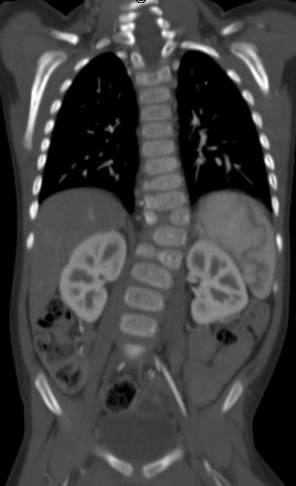
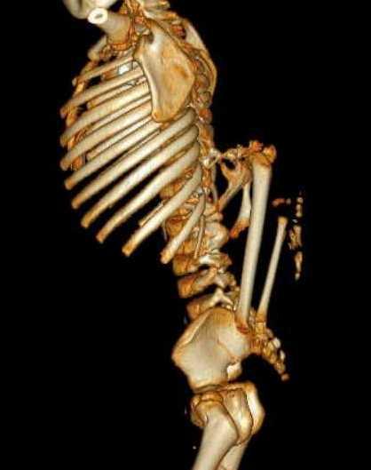
11 Figure 4. Radiograph, coronal and 3D reconstructions of the bones.
12 CT Angiography Blood supply is from a spinal branch of a right intercostal artery from the descending thoracic aorta at the level of T10 vertebral body No discrete draining veins


13 Figure 5. Axial post-contrast CT images and 3D reconstruction of the blood supply.

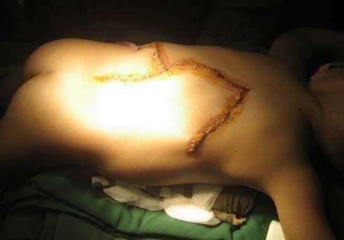
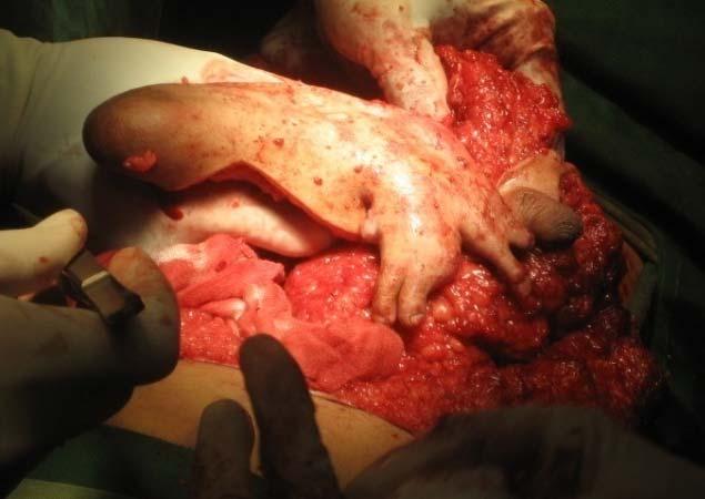
14 During surgery

15 Histopathology Mature elements from skin, bone, cartilage, muscle, fat, brain, respiratory and cervical epithelium were obtained. The phallic structure contained several cysts of epidermal origin all lined by skin epithelium (see below).
16 Discussion Most heteropagus twins are either joined ventrally above the umbilicus (epigastric heteropagus) or dorsally in the vertebral column (rachipagus) 28 rachipagus (dorsal heteropagus) twins have been reported Only 2 are diplopagus, the rest are heteropagus 18 females, 5 males, 5 not reported 24 (86%) has thoracic and/or lumbar connection 9 had meningocele
17 Discussion accurate imaging to define anatomic fusion, vascular and other anomalies and for surgical planning CT and MR of the spine are needed for rachipagus type assessment of the cardiovascular system is recommended in all types no overall mortality or morbidity reported outcome better without shared vital organs prognosis depends on type and extent of union
18 Conclusion Hence, this is a very unusual case of an otherwise asymptomatic boy with a parasitic rachipagus where CT and MRI played a crucial role in the diagnosis, assessment of other anomalies, and for surgical planning and prognostication.
19 References Chadha R, et al. Lumbosacral parasitic rachipagus twin. J Pediatr Surg 2006; 41(1): e45-e48. Ratan SK, et al. Thoracolumbar rachipagus parasite. Pediatr Surg Int 2004; 20(4): Spitz L and EM Kiely. Conjoined Twins. JAMA 2003; 289(10): Kingston CA, et al. Imaging in the Preoperative Assessment of Conjoined Twins. Radiographics 2001; 21: Spencer R, et al. Anencephaly, dorsal hypermelia and duplication of the vertebral column: a rare type of rachipagus conjoined twins. Teratology 1996; 53(4): Spencer R. Rachipagus conjoined twins: they really do occur! Teratology 1995; 52(6):
Case Report Multimodality Imaging in the Assessment of Thoraco-Omphalopagus Conjoined Twin: Lessons to Learn
 Case Reports in Radiology Volume 2012, Article ID 564036, 4 pages doi:10.1155/2012/564036 Case Report Multimodality Imaging in the Assessment of Thoraco-Omphalopagus Conjoined Twin: Lessons to Learn Kanaga
1 Normal Anatomy and Variants
 1 Normal Anatomy and Variants 1.1 Normal Anatomy MR Technique. e standard MR protocol for a routine evaluation of the spine always comprises imaging in sagittal and axial planes, while coronal images are
Ligaments of the vertebral column:
 In the last lecture we started talking about the joints in the vertebral column, and we said that there are two types of joints between adjacent vertebrae: 1. Between the bodies of the vertebrae; which
Scoliosis: Orthopaedic Perspectives
 Scoliosis: Orthopaedic Perspectives Scott B. Rosenfeld, MD Division of Pediatric Orthopaedic Surgery Texas Children s Hospital Page 0 xxx00.#####.ppt 9/23/2012 8:26:24 AM I have no disclosures Disclosures
Ex. 1 :Language of Anatomy
 Collin College BIOL 2401 : Human Anatomy & Physiology Ex. 1 :Language of Anatomy The Anatomical Position Used as a reference point when referring to specific areas of the human body Body erect Head and
Case report: Imaging findings in a butterfly vertebra
 Acta Neurol. Belg., 2011, 111, 344-348 Case report: Imaging findings in a butterfly vertebra Cedric BOULET, Ann SCHIETTECATTE, Johan DE MEY and Michel DE MAESENEER Department of Radiology, UZ Brussel,
The Skeletal System. Parts of the skeletal system. Bones (Skeleton) Joints Cartilages Ligaments
 Joints Cartilages Ligaments") The Skeletal System Parts of the skeletal system Bones (Skeleton) Joints Cartilages Ligaments Functions of the Bones Support Internal framework of the body Protection Skull and vertebrae protect brain
The Skeletal System Parts of the skeletal system Bones (Skeleton) Joints Cartilages Ligaments Functions of the Bones Support Internal framework of the body Protection Skull and vertebrae protect brain
ISUOG Basic Training. Examining Fetal Anatomy from Longitudinal Sections Titia Cohen-Overbeek, The Netherlands
 ISUOG Basic Training Examining Fetal Anatomy from Longitudinal Sections Titia Cohen-Overbeek, The Netherlands Learning objectives 2 & 3 At the end of the lecture you will be able to: describe how to obtain
Anatomical Terminology
 Anatomical Terminology Dr. A. Ebneshahidi Anatomy Anatomy : is the study of structures or body parts and their relationships to on another. Anatomy : Gross anatomy - macroscopic. Histology - microscopic.
Basic Body Structure
 Basic Body Structure The Cell All life consists of microscopic living structures called cells. They perform various functions throughout the body. All cells are similar in structure, but not identical.
Evaluation of cardiovascular anomalies in conjoined twins: a single-center experience from King Abdulaziz Cardiac Center
 European Heart Journal Supplements (2014) 16 (Supplement B), B32 B36 The Heart of the Matter doi:10.1093/eurheartj/suu008 Evaluation of cardiovascular anomalies in conjoined twins: a single-center experience
Welcome to ANAT 10A! What is Anatomy? Different levels of Anatomy The Language of Anatomy Pearson Education, Inc.
 Welcome to ANAT 10A! What is Anatomy? Different levels of Anatomy The Language of Anatomy Introduction Anatomy means to dissect: (ANAT 10A) The study of internal & external body structures The study of
Skeletal system. Prof. Abdulameer Al-Nuaimi. E. mail:
 Skeletal system Prof. Abdulameer Al-Nuaimi E-mail: a.al-nuaimi@sheffield.ac.uk E. mail: abdulameerh@yahoo.com Functions of Bone and The Skeletal System Support: The skeleton serves as the structural framework
Case Report A Rare Form of Heteropagus Twinning: Three-Armed Infant with Spinal Dysraphism
 Case Reports in Pediatrics Volume 2012, Article ID 831649, 4 pages doi:10.1155/2012/831649 Case Report A Rare Form of Heteropagus Twinning: Three-Armed Infant with Spinal Dysraphism Aynur Solak, 1 Sonnaz
The posterior abdominal wall. Prof. Oluwadiya KS
 The posterior abdominal wall Prof. Oluwadiya KS www.oluwadiya.sitesled.com Posterior Abdominal Wall Lumbar vertebrae and discs. Muscles opsoas, quadratus lumborum, iliacus, transverse, abdominal wall
Chest cavity, vertebral column and back muscles. Respiratory muscles. Sándor Katz M.D., Ph.D.
 Chest cavity, vertebral column and back muscles. Respiratory muscles. Sándor Katz M.D., Ph.D. Chest cavity - bony structures Chest cavity- bony structures Sternum Ribs True ribs: first seven pairs connect
CHAPTER 2 Terms Pertaining to the Body as a Whole
 CHAPTER 2 Terms Pertaining to the Body as a Whole OBJECTIVES 1. Define terms that apply to the structural organization of the body. 2. Identify the body cavities and the organs contained within the cavities.
3 Circulatory Pathways
 40 Chapter 3 Circulatory Pathways Systemic Arteries -Arteries carry blood away from the heart to the various organs of the body. -The aorta is the longest artery in the body; it branches to give rise to
THE THORACIC WALL. Boundaries Posteriorly by the thoracic part of the vertebral column. Anteriorly by the sternum and costal cartilages
 THE THORACIC WALL Boundaries Posteriorly by the thoracic part of the vertebral column Anteriorly by the sternum and costal cartilages Laterally by the ribs and intercostal spaces Superiorly by the suprapleural
Neonatal Spinal Ultrasound Imaging - A Pictorial Review from The Royal Liverpool Children Hospital, Alder Hey, Liverpool
 Neonatal Spinal Ultrasound Imaging - A Pictorial Review from The Royal Liverpool Children Hospital, Alder Hey, Liverpool Poster No.: C-0081 Congress: ECR 2012 Type: Educational Exhibit Authors: K. Chetcuti,
Thoracolumbar Anatomy Eric Shamus Catherine Patla Objectives
 1 2 Thoracolumbar Anatomy Eric Shamus Catherine Patla Objectives List the muscular and ligamentous attachments of the thoracic and lumbar spine Describe how the muscles affect the spine and upper extremity
Axial Skeleton: Vertebrae and Thorax
 Axial Skeleton: Vertebrae and Thorax Function of the vertebral column (spine or backbone): 1) 2) 3) Composition of Vertebral column The vertebral column is formed by 33 individual vertebrae (some of which
The Skeletal System. Support Systems Unit 2
 The Skeletal System Support Systems Unit 2 The Basic Functions of the Skeletal System Hematopoiesis Structure Support Muscle Attachment and Movement Mineral Storage Axial vs. Appendicular The Axial Skeleton
Skeletal System. Prof. Dr. Malak A. Al-yawer Department of Anatomy/Embryology Section
 Skeletal System Prof. Dr. Malak A. Al-yawer Department of Anatomy/Embryology Section Learning objectives At the end of this lecture, the medical student will be able to: State the embryonic origin of skeletal
Pediatric Spinal Anomalies
 Department of Radiology University of California San Diego Pediatric Spinal Anomalies John R. Hesselink, M.D. Spine Embryogenesis 1. Primitive streak 2. Proliferation of cells at primitive pit (Hensen's
Anatomy. Anatomy deals with the structure of the human body, and includes a precise language on body positions and relationships between body parts.
 Anatomy deals with the structure of the human body, and includes a precise language on body positions and relationships between body parts. Proper instruction on safe and efficient exercise technique requires
SPLIT NOTOCHORD SYNDROME ASSOCIATION. DR. Hasan Nugud Consultant Paediatric Surgeon
 SPLIT NOTOCHORD SYNDROME ASSOCIATION DR. Hasan Nugud Consultant Paediatric Surgeon CASE PRESENTATION :- New born baby, boy, referred to the paediatric surgical team at the age of 14 hours. Birth History
INDEPENDENT LEARNING: DISC HERNIATION IN THE NATIONAL FOOTBALL LEAGUE: ANATOMICAL FACTORS TO CONSIDER IN REVIEW
 INDEPENDENT LEARNING: DISC HERNIATION IN THE NATIONAL FOOTBALL LEAGUE: ANATOMICAL FACTORS TO CONSIDER IN REVIEW CDC REPORT - CAUSES OF DISABILITY, 2005 REVIEW QUESTIONS ABOUT DISC HERNIATION IN THE NATIONAL
REVIEW QUESTIONS ON VERTEBRAE, SPINAL CORD, SPINAL NERVES
 REVIEW QUESTIONS ON VERTEBRAE, SPINAL CORD, SPINAL NERVES 1. A 28-year-old-women presented to the hospital emergency room with intense lower back spasms in the context of coughing during an upper respiratory
The Musculoskeletal System
 The Musculoskeletal System Introduction The skeletal system and muscular system are often considered together because they are close in terms of structure and function. The two systems are referred to
Essentials of Anatomy and Physiology 6th Edition Scanlon Sanders Test Bank
 Essentials of Anatomy and Physiology 6th Edition Scanlon Sanders Test Bank Link full download: http://testbankcollection.com/download/essentials-of-anatomy-and-physiology- 6th-edition-scanlon-sanders-test-bank
ORIGINAL ARTICLE. Abstract. Aim. Materials and methods. Introduction. Results
 Is anatomical distribution helpful for differentiating TB spondylitis from neoplastic causes of extradural spinal cord compression in children? A pilot study Reena George, MD, MMed Rad, FRCR (UK) Savvas
The Human Body: An Overview of Anatomy. Anatomy. Physiology. Anatomy - Study of internal and external body structures
 C H A P T E R 1 The Human Body: An Orientation An Overview of Anatomy Anatomy The study of the structure of the human body Physiology The study of body function Anatomy - Study of internal and external
The vault bones Frontal Parietals Occiput Temporals Sphenoid Ethmoid
 The Vertebral Column Head, Neck and Spine Bones of the head Some consider the bones of the head in terms of the vault bones and the facial bones hanging off the front of them The vault bones Frontal Parietals
Test Bank for Essentials of Anatomy and Physiology 6th edition by Valerie C. Scanlon and Tina Sanders
 Test Bank for Essentials of Anatomy and Physiology 6th edition by Valerie C. Scanlon and Tina Sanders Link download full: https://digitalcontentmarket.org/download/test-bank-foressentials-of-anatomy-and-physiology-6th-edition-by-scanlon-and-sanders/
Case SCIWORA in patient with congenital block vertebra
 Case 15428 SCIWORA in patient with congenital block vertebra Lucas Walgrave 1, Charlotte Vanhoenacker 1-2, Thomas Golinvaux 3, Filip Vanhoenacker3-5 1: Leuven University Hospital, Department of Radiology,
2. The vertebral arch is composed of pedicles (projecting from the body) and laminae (uniting arch posteriorly).
 and laminae (uniting arch posteriorly).") VERTEBRAL COLUMN 2018zillmusom I. VERTEBRAL COLUMN - functions to support weight of body and protect spinal cord while permitting movements of trunk and providing for muscle attachments. A. Typical vertebra
VERTEBRAL COLUMN 2018zillmusom I. VERTEBRAL COLUMN - functions to support weight of body and protect spinal cord while permitting movements of trunk and providing for muscle attachments. A. Typical vertebra
Forensic Archaeology & Forensic Anthropology. ADJ14 Advanced Criminal Investigations
 Forensic Archaeology & Forensic Anthropology ADJ14 Advanced Criminal Investigations Anthropology & Archaeology Anthropology is the study of the biological and cultural aspects of all humans in all places
Skeletal System. Std. VIII
 Skeletal System Std. VIII The skeleton in our body serves following functions : 1. Support and shape : The skeleton provides a support or framework to all the soft parts and gives the body and its parts
Bony framework of the vertebral column Structure of the vertebral column
 5.1: Vertebral column & back. Overview. Bones o vertebral column. o typical vertebra. o vertebral canal. o spinal nerves. Joints o Intervertebral disc. o Zygapophyseal (facet) joint. Muscles o 2 compartments:
11/25/2012. Chapter 7 Part 2: Bones! Skeletal Organization. The Skull. Skull Bones to Know Cranium
 Chapter 7 Part 2: Bones! 5) Distinguish between the axial and appendicular skeletons and name the major parts of each 6) Locate and identify the bones and the major features of the bones that compose the
Sonography of the Neonatal Spine: Part 2, Spinal Disorders
 Neonatal Spine Sonography Pediatric Imaging Pictorial Essay Downloaded from www.ajronline.org by 148.251.232.83 on 04/11/18 from IP address 148.251.232.83. Copyright RRS. For personal use only; all rights
e-anatomy Paper 2 Exam Monday, 4 April 2016
 e-anatomy Paper 2 Exam Monday, 4 Level 9, 51 Druitt Street, Sydney NSW 2000, Australia Ph: +61 2 9268 9777 Fax: +61 2 9268 9799 Web: www.ranzcr.edu.au Email: ranzcr@ranzcr.edu.au ABN 37 000 029 863 CASE
Fractures of the Thoracic and Lumbar Spine
 A spinal fracture is a serious injury. Nader M. Hebela, MD Fellow of the American Academy of Orthopaedic Surgeons http://orthodoc.aaos.org/hebela Cleveland Clinic Abu Dhabi Cleveland Clinic Abu Dhabi Neurological
Symptomatic Multiple Level Lateral Meningoceles with Intraspinal Meningocele: A Case Study and Its Surgical Management
 THIEME Original Article 15 Symptomatic Multiple Level Lateral Meningoceles with Intraspinal Meningocele: A Case Study and Its Surgical Management Vernon Velho 1 Sachin Guthe 1 Pravin Survashe 1 Poonam
Case Fibrothecoma of the ovary
 Case 10646 Fibrothecoma of the ovary Elisa Melo Abreu, Teresa Margarida Cunha Section: Genital (Female) Imaging Published: 2015, Jan. 2 Patient: 70 year(s), female Authors' Institution Department of Radiology,
locomotice system Plastinated specimensⅠ: Silicone specimens Regional specimens and organs
 locomotice system Plastinated specimensⅠ: Silicone specimens Regional specimens and organs Art-No. Name Description The locomotor system SL001 Two hundred pieces of plastinated bones (without six The bones
_CH01redo.qxd 9/24/07 3:07 PM Page 1. [Half-Title to come]
![_CH01redo.qxd 9/24/07 3:07 PM Page 1. [Half-Title to come]](/thumbs/81/84146690.jpg "_CH01redo.qxd 9/24/07 3:07 PM Page 1. [Half-Title to come]") 10752-01_CH01redo.qxd 9/24/07 3:07 PM Page 1 [Half-Title to come] 10752-01_CH01redo.qxd 9/24/07 3:07 PM Page 2 THE BACK Lippincott Williams & Wilkins atlas of ANATOMY CHAPTER 1 Plate 1-01 Palpable Structures
10752-01_CH01redo.qxd 9/24/07 3:07 PM Page 1 [Half-Title to come] 10752-01_CH01redo.qxd 9/24/07 3:07 PM Page 2 THE BACK Lippincott Williams & Wilkins atlas of ANATOMY CHAPTER 1 Plate 1-01 Palpable Structures
CHAPTER 7, PART II (BONES)
") Anatomy Name: CHAPTER 7, PART II (BONES) Entry #: INSTRUCTIONS: 1) READ Chapter 7, pg. 140-161. 2) Using the outline, make a note card for each underlined bone name or phrase. 3) On each note card, put
Anatomy Name: CHAPTER 7, PART II (BONES) Entry #: INSTRUCTIONS: 1) READ Chapter 7, pg. 140-161. 2) Using the outline, make a note card for each underlined bone name or phrase. 3) On each note card, put
Multiple Neurovascular... Pit Baran Chakraborty, Santanu Bhattacharya, Sumita Dutta.
 Multiple Neurovascular... Pit Baran Chakraborty, Santanu Bhattacharya, Sumita Dutta. Fig-3: Showing high formation of Median nerve. Fig-1: Showing atypical formation of cords of Brachial plexus. 1 = Upper
SD School Anatomy Program 1: Bones QuikNotes. Student Notes
 QuikNotes The transverse plane runs from right to left and divides the body into superior (upper) and inferior (lower) sections. Student Notes The frontal plane lies vertically along the body from head
LANGUAGE of ANATOMY PART 2. Courtesy of Dr. Susan Maskel Western Connecticut State University
 1 LANGUAGE of ANATOMY PART 2 Courtesy of Dr. Susan Maskel Western Connecticut State University 2 ACROMIAL pertaining to the point of the shoulder Note: this term was also used in Language of Anatomy Part
MEDICAL IMAGING OF THE VERTEBRAE
 MEDICAL IMAGING OF THE VERTEBRAE Vertebrae are your friends Matthew Harper MS-IV LECTURE OBJECTIVES INTRODUCE THE MOST COMMON MODALITIES OF MEDICAL IMAGING AND BASIC TECHNIQUES FOR READING THESE IMAGES
Spine and spinal cord
 NEURORADIOLOGY Spine and spinal cord Erika Vörös University of Szeged Department of Radiology SZEGED DISEASES OF SPINE AND SPINAL CORD I. Non-tumourous diseases developmental anomalies vascular disorders
Anatomy & Physiology Ch 1: The Human Body Worksheet
 Anatomy & Physiology Ch 1: The Human Body Worksheet 1. The structures of the body are organized in successively larger and more complex structures. Fill in the blanks with the correct terms for these increasingly
We are IntechOpen, the world s leading publisher of Open Access books Built by scientists, for scientists. International authors and editors
 We are IntechOpen, the world s leading publisher of Open Access books Built by scientists, for scientists 3,500 108,500 1.7 M Open access books available International authors and editors Downloads Our
VERTEBRAL COLUMN VERTEBRAL COLUMN
 VERTEBRAL COLUMN FUNCTIONS: 1) Support weight - transmits weight to pelvis and lower limbs 2) Houses and protects spinal cord - spinal nerves leave cord between vertebrae 3) Permits movements - *clinical
Role of helical CT and MRI in the evaluation of spinal dysraphism
 International Journal of Advances in Medicine Kumaran SK et al. Int J Adv Med. 2017 Feb;4(1):124-132 http://www.ijmedicine.com pissn 2349-3925 eissn 2349-3933 Original Research Article DOI: http://dx.doi.org/10.18203/2349-3933.ijam20170095
Human Skeletal System Glossary
 Acromegaly Apatite Acromegaly - is a condition which involves excessive growth of the jaw, hands, and feet. It results from overproduction of somatotropin in adults (after fusion of the ossification centres
Anatomy 2 nd Paper. Past Papers
 Anatomy 2 nd Paper Past Papers September 2010 April 2010 September 2009 April 2009 September 2008 April 2008 September 2007 April 2007 September 2006 May 2006 September 2005 April 2005 September 2004 April
Chapter 7: Skeletal System: Gross Anatomy
 Chapter 7: Skeletal System: Gross Anatomy I. General Considerations A. How many bones in an average adult skeleton? B. Anatomic features of bones are based on II. Axial Skeleton A. Skull 1. Functionally
ASJ. A Rare Hyperextension Injury in Thoracic Spine Presenting with Delayed Paraplegia. Asian Spine Journal. Introduction
 sian Spine Journal 126 Dong-Eun Case Shin Report et al. http://dx.doi.org/10.4184/asj.2013.7.2.126 Rare Hyperextension Injury in Thoracic Spine Presenting with Delayed Paraplegia Dong-Eun Shin, Ki-Sik
Fractures of the thoracic and lumbar spine and thoracolumbar transition
 Most spinal column injuries occur in the thoracolumbar transition, the area between the lower thoracic spine and the upper lumbar spine; over half of all vertebral fractures involve the 12 th thoracic
Properties of Purdue. Anatomy. Positioning AXIAL SKELETAL RADIOLOGY FOR PRIVATE PRACTITIONERS 11/30/2018
 AXIAL SKELETAL RADIOLOGY FOR PRIVATE PRACTITIONERS Anatomy Complex Text book is needed Species Contrast Positioning Painful/ non cooperative Sedation General anesthesia Species Contrast 1 Slightly oblique
Spinal Cord Injuries: The Basics. Kadre Sneddon POS Rounds October 1, 2003
 Spinal Cord Injuries: The Basics Kadre Sneddon POS Rounds October 1, 2003 Anatomy Dorsal columntouch, vibration Corticospinal tract- UMN Anterior horn-lmn Spinothalamic tractpain, temperature (contralateral)
topographical anatomy
 Kaan Yücel M.D., Ph.D. 30. September 2014 Tuesday topographical anatomy organization of the human body as major parts or segments Head Neck Trunk thorax, abdomen, back, & pelvis/perineum Upper limbs &
MRI of chronic spinal cord injury
 The British Journal of Radiology, 76 (2003), 347 352 DOI: 10.1259/bjr/11881183 E 2003 The British Institute of Radiology Pictorial review MRI of chronic spinal cord injury 1 K POTTER, FRCR and 1 A SAIFUDDIN,
Lab Monitor Images Dissection of the Abdominal Vasculature + Lower Digestive System
 Lab Monitor Images Dissection of the Abdominal Vasculature + Lower Digestive System Stomach & Duodenum Frontal (AP) View Nasogastric tube 2 1 3 4 Stomach Pylorus Duodenum 1 Duodenum 2 Duodenum 3 Duodenum
Case Report Ipsilateral Hip Dysplasia in Patients with Sacral Hemiagenesis: A Report of Two Cases
 Case Reports in Orthopedics Volume 2015, Article ID 854151, 4 pages http://dx.doi.org/10.1155/2015/854151 Case Report Ipsilateral Hip Dysplasia in Patients with Sacral Hemiagenesis: A Report of Two Cases
Copyright 2010 Pearson Education, Inc.
 E. VERTEBRAL COLUMN 1. The vertebral column extends from the skull to the pelvis and forms the vertical axis of the skeleton. 2. The vertebral column is composed of vertebrae that are separated by intervertebral
The Back. Anatomy RHS 241 Lecture 9 Dr. Einas Al-Eisa
 The Back Anatomy RHS 241 Lecture 9 Dr. Einas Al-Eisa The spine has to meet 2 functions Strength Mobility Stability of the vertebral column is provided by: Deep intrinsic muscles of the back Ligaments
CHAPTER 13 SKELETAL SYSTEM
 CHAPTER 13 SKELETAL SYSTEM Structure and Function Functions of the skeletal system Provides shape and support Protects internal organs Stores minerals and fat Produces blood cells and platelets Assists
Persistent Terminal Ventricle
 Persistent Terminal Ventricle Ventriculus Terminalis Incomplete regression of TV of 2 neurulation, continuity with central canal small cavity PTV vs terminal myelocystocele (?severe manifestation from
Chapter 2. Organization of the Body. Copyright 2012, 2009, 2005, 2003, 1999, 1991 by Saunders, an imprint of Elsevier Inc. All rights reserved.
 Chapter 2 Organization of the Body 1 Learning Objectives Name the body systems and their functions. Identify body cavities and specific organs within them. List the divisions of the back. Identify three
The Skeletal System. Chapter 7a. Skeletal System Introduction Functions of the skeleton Framework of bones The skeleton through life
 The Skeletal System Skeletal System Introduction Functions of the skeleton Framework of bones The skeleton through life Chapter 7a Support Protection Movement Storage areas Minerals Lipids Hemopoiesis
Human Anatomy - Problem Drill 06: The Skeletal System Axial Skeleton & Articualtions
 Human Anatomy - Problem Drill 06: The Skeletal System Axial Skeleton & Articualtions Question No. 1 of 10 Instructions: (1) Read the problem and answer choices carefully, (2) Work the problems on paper
BIO 137 AXIAL SKELETON BONE STUDY THE HUMAN SKELETON
 BIO 137 THE AXIAL SKELETON MARY CATHERINE FLATH, Ph.D. THE HUMAN SKELETON AXIAL SKULL HYOID THORACIC CAGE VERTEBRAL COLUMN APPENDICULAR PECTORAL GIRDLE UPPER LIMBS PELVIC GIRDLE LOWER LIMBS AXIAL SKELETON
Clarification of Terms
 Clarification of Terms The Spine, Spinal Column, and Vertebral Column are synonymous terms referring to the bony components housing the spinal cord Spinal Cord = made of nervous tissue Facet = a small,
Clarification of Terms
 Clarification of Terms The Spine, Spinal Column, and Vertebral Column are synonymous terms referring to the bony components housing the spinal cord Spinal Cord = made of nervous tissue Facet = a small,
The Human Body: An Orientation
 The Human Body: An Orientation Body standing upright Anatomical Position feet slightly apart palms facing forward thumbs point away from body Directional Terms Superior and inferior toward and away from
General Anatomy p. 1 Organization of the Human Body p. 1 Skeleton of the Human Body p. 4 Ossification of the Bones p. 6 Bone Structure p. 8 Joints p.
 General Anatomy p. 1 Organization of the Human Body p. 1 Skeleton of the Human Body p. 4 Ossification of the Bones p. 6 Bone Structure p. 8 Joints p. 10 Principal Joints (Immovable) p. 12 Synovial Joints
The Thoracic wall including the diaphragm. Prof Oluwadiya KS
 The Thoracic wall including the diaphragm Prof Oluwadiya KS www.oluwadiya.com Components of the thoracic wall Skin Superficial fascia Chest wall muscles (see upper limb slides) Skeletal framework Intercostal
Anatomy of the Thorax
 Anatomy of the Thorax A) THE THORACIC WALL Boundaries Posteriorly by the thoracic part of the vertebral column Anteriorly by the sternum and costal cartilages Laterally by the ribs and intercostal spaces
ANATYOMY OF The thigh
 ANATYOMY OF The thigh 1- Lateral cutaneous nerve of the thigh Ι) Skin of the thigh Anterior view 2- Femoral branch of the genitofemoral nerve 1, 2 and 3 are From the lumber plexus 5- Intermediate cutaneous
Pott s kyphosis. University Affiliated Sixth People s Hospital, 600 Yishan Road, Shanghai , P.
 QJM Advance Access published November 17, 2014 Pott s kyphosis Author Names: Yi Zhang, Yong-Sheng Yu, Zheng-Hao Tang and Guo-Qing Zang Author Affiliations: Department of Infectious Diseases, Shanghai Jiao
First stage Lec.1 : Introduction. Asst.Lec.Dr.ABDULRIDHA ALASADY
 First stage 2018-2019 Lec.1 : Introduction Asst.Lec.Dr.ABDULRIDHA ALASADY Anatomy the study of the structure and shape of the body and body parts & their relationships to one another aided by dissection
BMS101 INTRODUCTION TO THE HUMAN BODY STUDY NOTES EXTRACTS FOR SAMPLE
 BMS101 INTRODUCTION TO THE HUMAN BODY STUDY NOTES EXTRACTS FOR SAMPLE What is anatomy? Anatomy is the study of structure. Greek for to cut up or cut open What is physiology? Physiology studies the function
STERNUM. Lies in the midline of the anterior chest wall It is a flat bone Divides into three parts:
 STERNUM Lies in the midline of the anterior chest wall It is a flat bone Divides into three parts: 1-Manubrium sterni 2-Body of the sternum 3- Xiphoid process The body of the sternum articulates above
Case Report A Unique Case of Left Second Supernumerary and Left Third Bifid Intrathoracic Ribs with Block Vertebrae and Hypoplastic Left Lung
 Volume 2013, Article ID 620120, 4 pages http://dx.doi.org/10.1155/2013/620120 Case Report A Unique Case of Left Second Supernumerary and Left Third Bifid Intrathoracic Ribs with Block Vertebrae and Hypoplastic
MAGNETOM Aera Combining Throughput and Highest Quality Spine Imaging in an Optimized Clinical Workflow
 Clinical Orthopedic Imaging MAGNETOM Aera Combining Throughput and Highest Quality Spine Imaging in an Optimized Clinical Workflow Johan Dehem, M.D. VZW Jan Yperman, Ieper, Belgium Cervical spine imaging
Chapter 1: Introduction to the Human Body Test Bank
 Chapter 1: Introduction to the Human Body Test Bank MULTIPLE CHOICE 1. What is the branch of science that studies how the body functions? a. Anatomy b. Histology c. Pathology d. Physiology 2. Which word
Lab-1. Miss. Lina Al-Onazy & samar Al-Wgeet =)
") Lab-1 Introduction The human skeleton is composed of 300 bones at birth and by the time adulthood is reached, some bones have fused together to give a total of 206 bones in the body. The human skeleton
Lab-1 Introduction The human skeleton is composed of 300 bones at birth and by the time adulthood is reached, some bones have fused together to give a total of 206 bones in the body. The human skeleton
Payment Policy. Chiropractic Care. Policy Specific Section: September 10, 2012 November 10, 2012
 Payment Policy Chiropractic Care Type: Payment Policy Policy Specific Section: Payment Original Policy Date: Effective Date: September 10, 2012 November 10, 2012 Description Chiropractic is a branch of
Chapter 7 The Skeletal System:The Axial Skeleton
 Chapter 7 The Skeletal System:The Axial Skeleton Axial Skeleton 80 bones lie along longitudinal axis skull, hyoid, vertebrae, ribs, sternum, ear ossicles Appendicular Skeleton 126 bones upper & lower limbs
ANATOMY & PHYSIOLOGY I Laboratory Version B Name Section. REVIEW SHEET Exercise 10 Axial Skeleton
 ANATOMY & PHYSIOLOGY I Laboratory Version B Name Section REVIEW SHEET Exercise 10 Axial Skeleton 1 POINT EACH. THE SKULL MULTIPLE CHOICE 1. The major components of the axial skeleton include the 7. The
CPT 2015: Prepare Your Coding Practice For New Codes As Technology Makes An Advance
 2015 Radiology Coding Survival Guide Section X : 2015 Coding Updates CPT 2015: Prepare Your Coding Practice For New Codes As Technology Makes An Advance Watch for changes in Vertebral fracture assessment,
Human Anatomy Key Points Unit 1/ Study Guide
 Human Anatomy Key Points Unit 1/ Study Guide I. Anatomy and Physiology a. Anatomy 1. Means cutting apart (dissection) 2. Study of the body and the relationships of its parts to each other. 3. Dissection
A. Incorrect! The axial skeleton includes bones of the skull, inner ear, chest and spinal column.
 Anatomy and Physiology - Problem Drill 07: The Skeletal System II No. 1 of 10 1. Which of the following statements about the axial skeleton is correct? A. The axial skeleton includes bones of the skull,
THE VERTEBRAL COLUMN. Average adult length: In male: about 70 cms. In female: about 65 cms.
 THE VERTEBRAL COLUMN Average adult length: In male: about 70 cms. In female: about 65 cms. 1 Vertebral Column (Regions and Curvatures) Curvatures of the vertebral column: A. Primary curvature: C-shaped;
MEDICAL TERMINOLOGY. Complete! Second Edition CHAPTER. The Human Body in Health and Disease Content Review Slides
 MEDICAL TERMINOLOGY Complete! Second Edition CHAPTER 4 The Human Body in Health and Disease Content Review Slides Learning Objectives Define and spell the word parts used to create terms for the human