Ascitic Fluid and Use of Immunocytochemistry. Mercè Jordà, University of Miami
|
|
|
- Candace Snow
- 6 years ago
- Views:
Transcription

1 Ascitic Fluid and Use of Immunocytochemistry Mercè Jordà, University of Miami

2 Is It Malignant? Yes? No
3 Ascitic Fluid Cytomorphologic Useful Findings Tight clusters with smooth borders Cellular and nuclear molding Large papillary groups Two-cell types Signet ring cells in groups Abnormal cell morphology
4
5
6
7
8 Ascitic Fluid Cytomorphologic Useless Findings Cytoplasmic Vacuoles Signet Ring Cells individual Psammoma Bodies Cell within Cell Prominent Nucleoli Mitosis Multinucleation
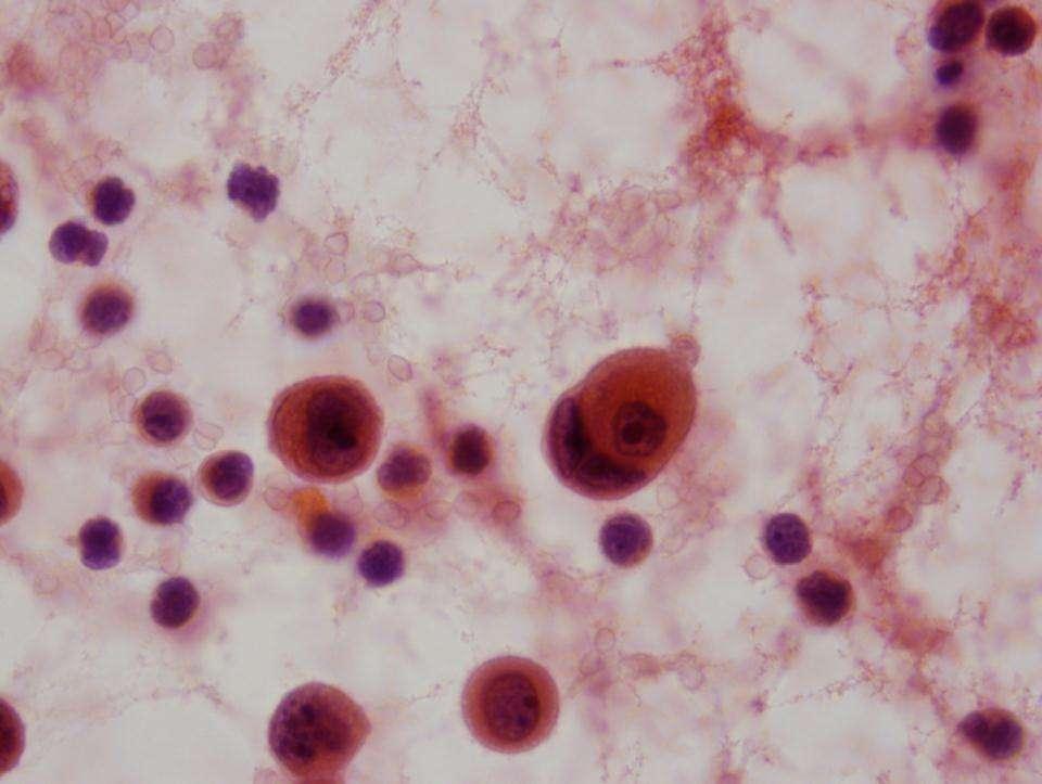
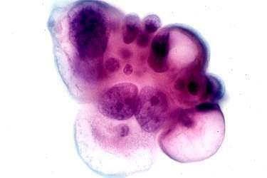
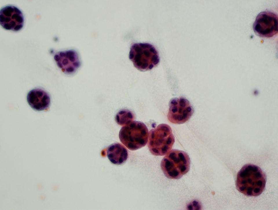
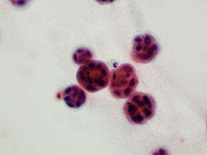
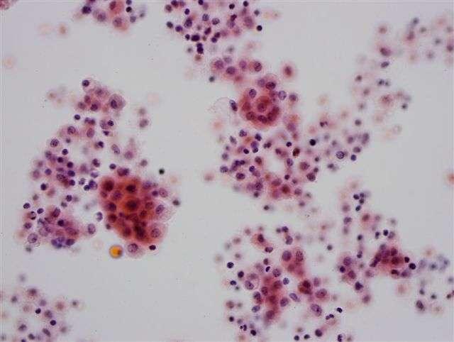
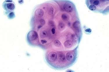
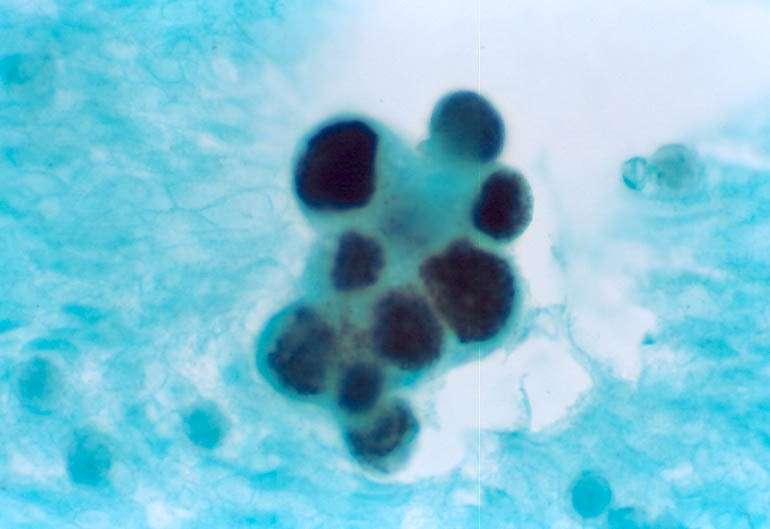
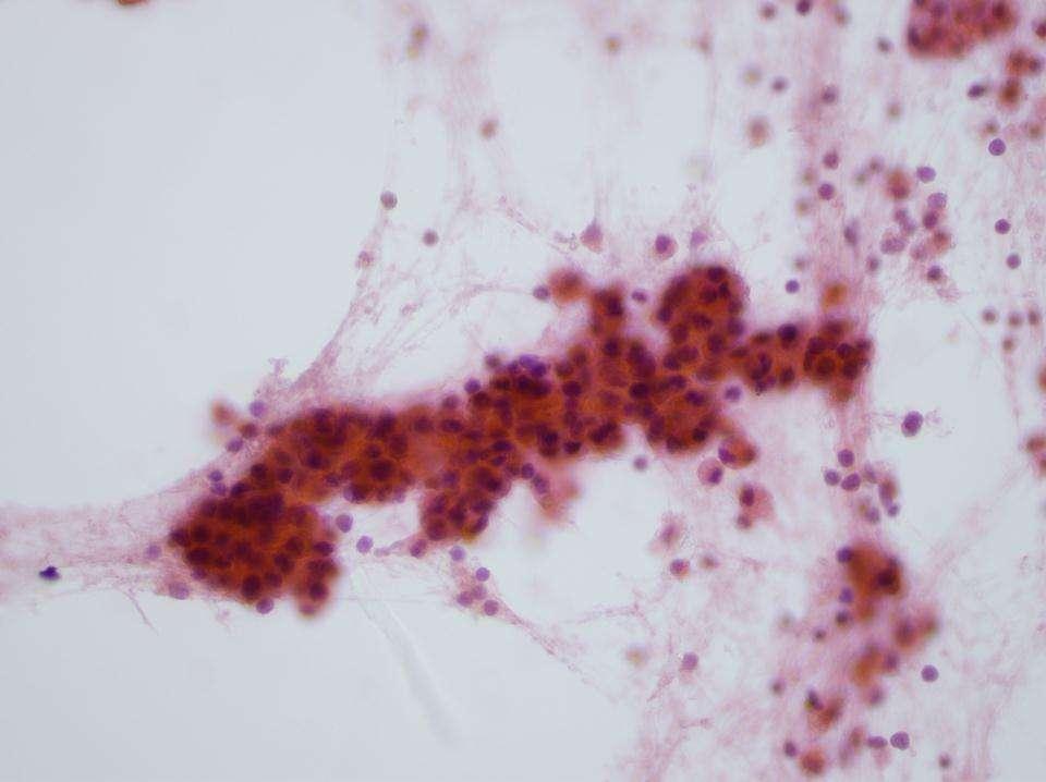
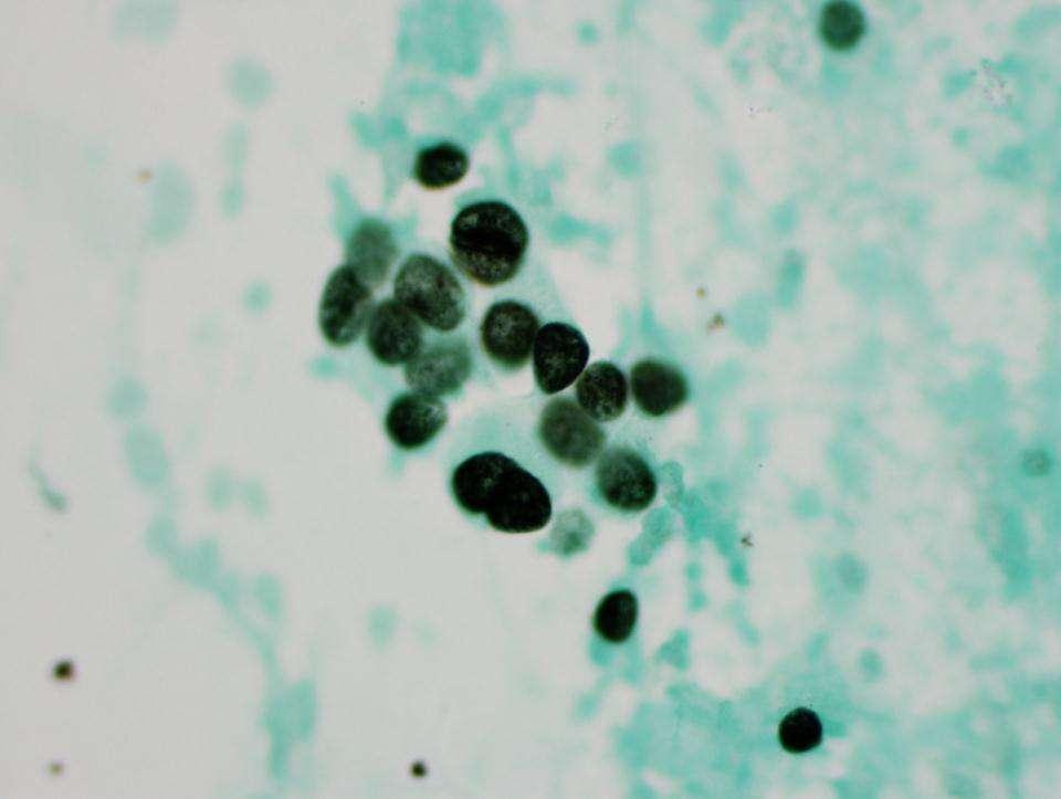
9 Reactive Mesothelium
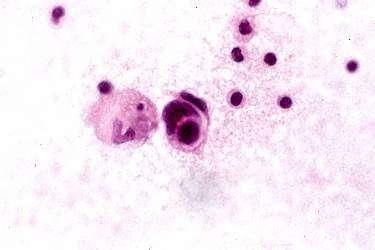
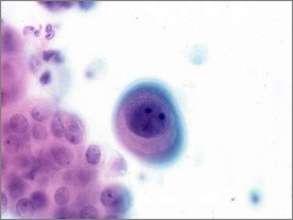
10 Signet Ring Cells Look for them in Groups!
11
12 Malignant
13 Benign
14 By cytomorphology Cellular Pattern Cellular Morphology
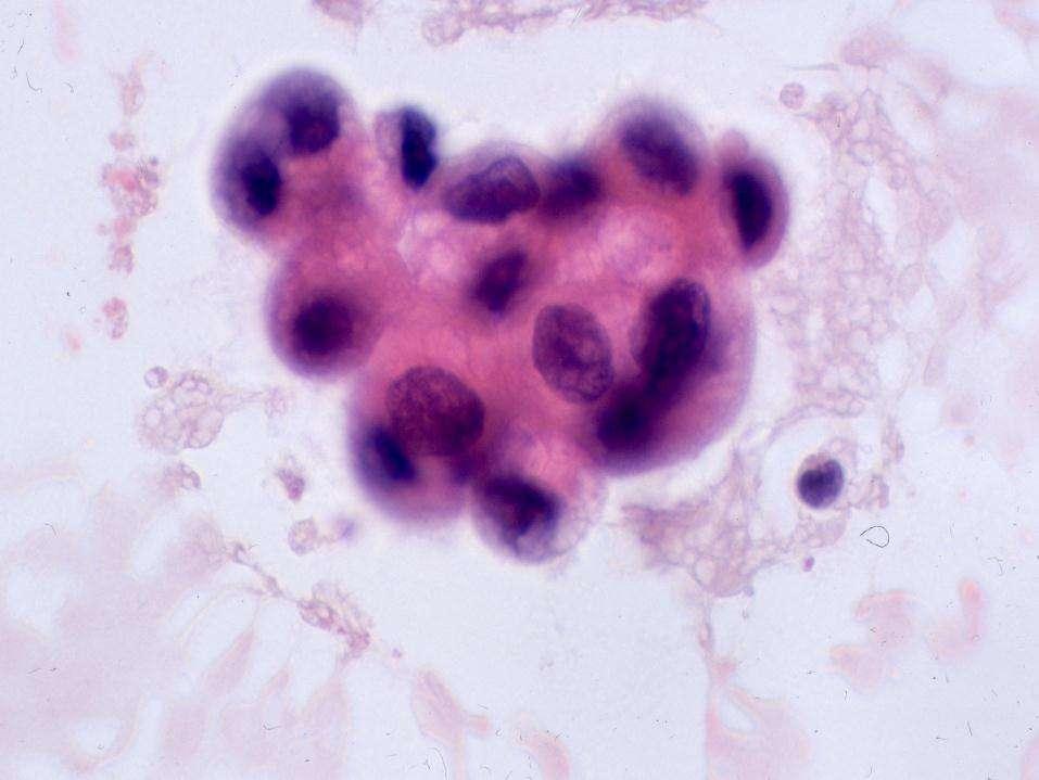
15 Malignant Ascitic Fluid Cellular Pattern Cells in Clusters Isolated Cells
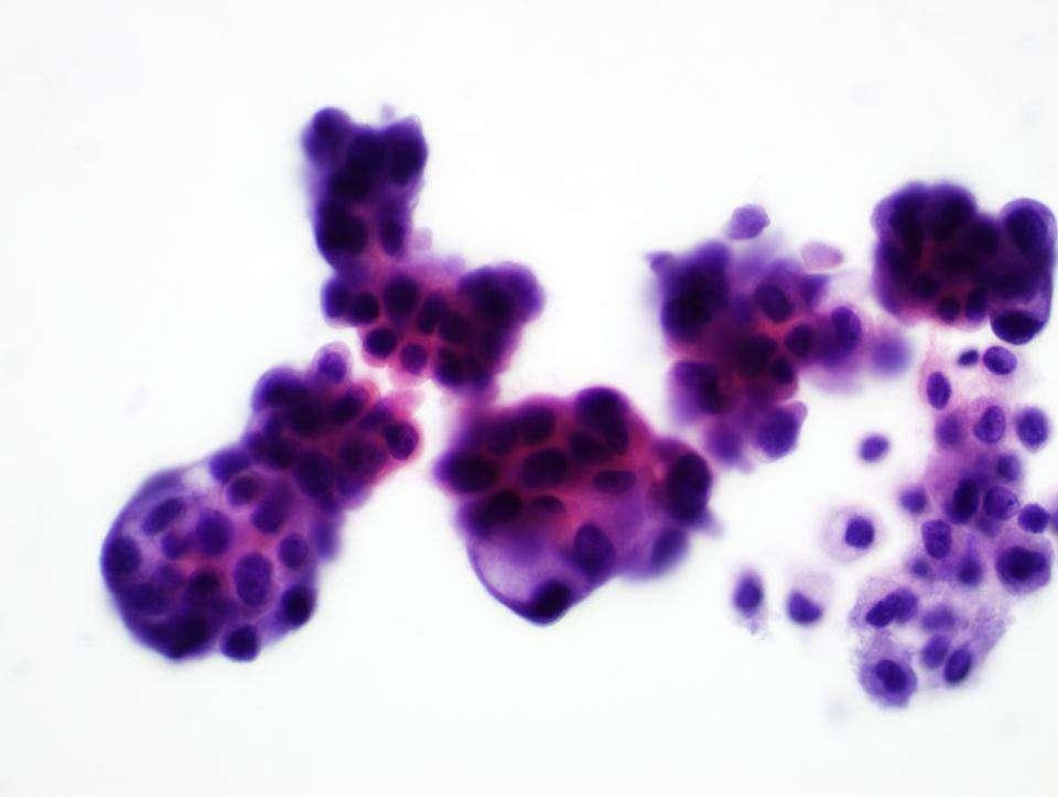
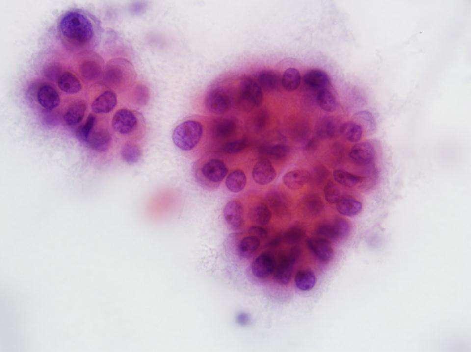
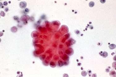
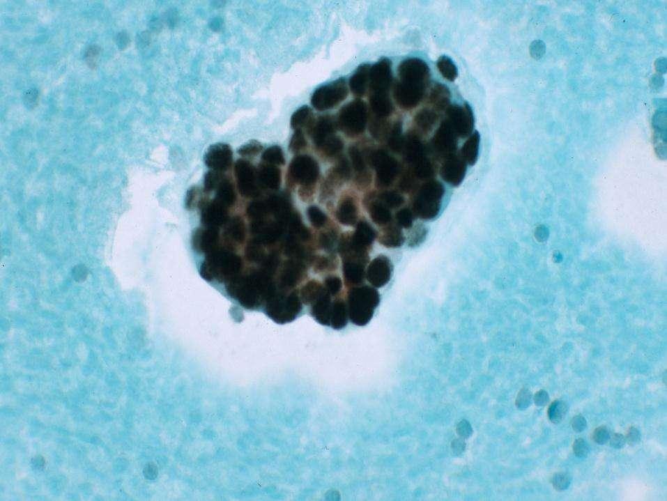
16 Malignant Ascitic Fluid: Cells in Clusters Cells Tight and compact Smooth borders Background No cells Reactive mesothelial cells Diagnosis : Cytomorphology
17
18
19
20 Malignant Effusions: Cells in clusters Differential Diagnosis Carcinoma Malignant Mesothelioma Diagnosis : Cytomorphology & Immunocytochemistry
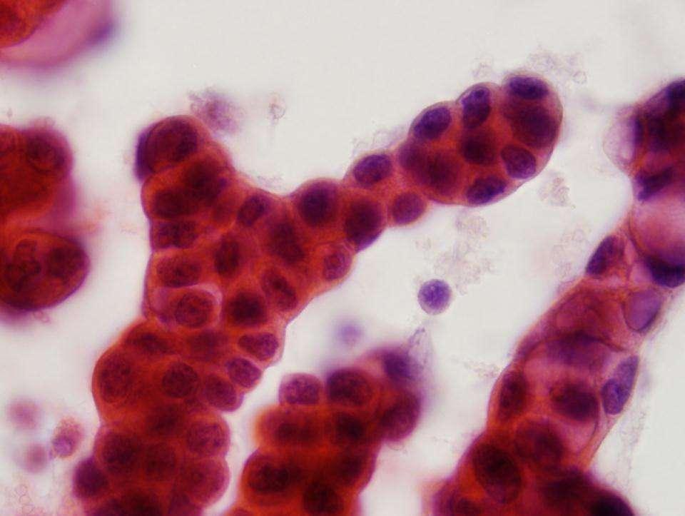
21 Malignant Effusions: Isolated Cells Abnormal single cells May be overlooked in low power Look for small clusters
22 Malignant Effusions: Isolated Cells Abnormal Cell Morphology Pleomorphism High N/C ratio Hyperchromasia Abnormal nucleoli Clumped, irregular chromatin Intraluminal mucin Diagnosis : Cytomorphology & Immunocytochemistry 3
23
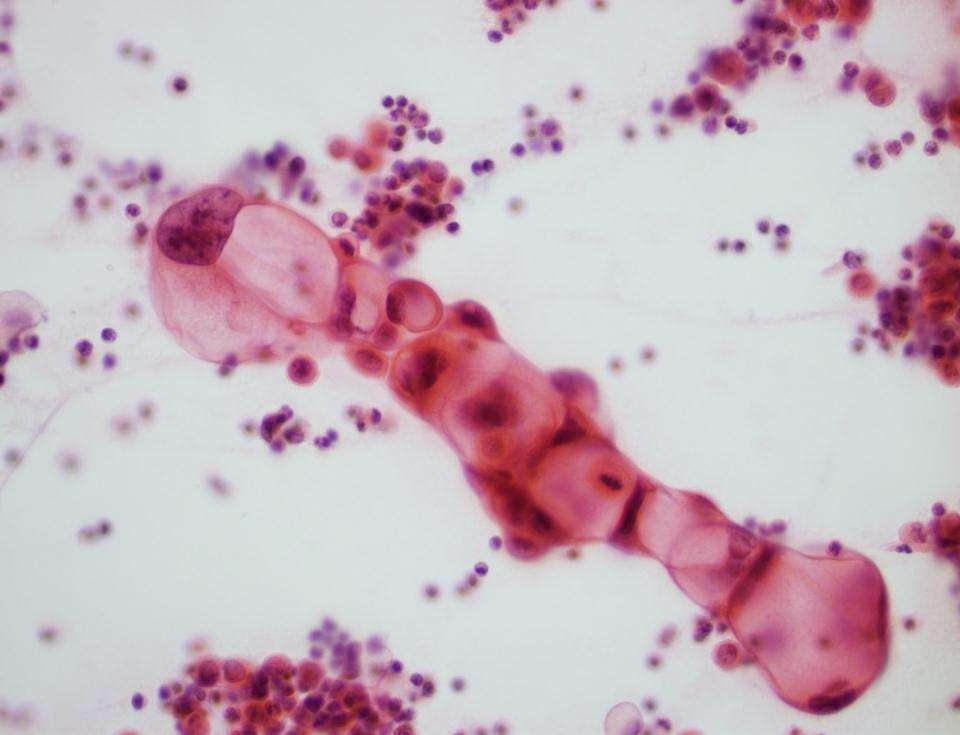
24 Malignant Ascitic Fluid Site of origin by Cytomorphology Tight cell balls in breast Ca. Psammoma bodies in serous Papillary Ca. Indian filling in breast, gastric and pancreatic Ca. Signet ring cells in breast, gastric and ovarian Ca. Keratin pearls in squamous cell Ca. Melanin in malignant melanoma. Intranuclear inclusions in Adenocarcinoma of lung lipidic, papillary thyroid carcinoma and melanomas Knobby clusters in mesotheliomas

25 Cell balls in Breast Ca.

26 Psammoma Bodies

27 Cellular Chain

28 Keratin Pearl

29 Knobby cluster in Mesothelioma
30 Malignant Effusions Specific Type and Site of Origin Diagnosis : Cytomorphology & Immunocytochemistry
31 Is It Malignant? Yes? No Mesothelioma Others Adenocarcinoma Site of Origin Small Cell Ca Lymphoma Squamous cell Ca Others
32 When to Use Immunocytochemistry in Ascitic fluid Cytology

33 Alamo 1917
34 Immunocytochemistry in Cytology University of Miami Experience Acta Cytol 1980; 24:
35 IHC Applications University of Miami Diagnosis/Classification 65% Prognosticators/Predictors 18% Target therapy, Others 17%
36 How Often? University of Miami Percent of our Total Cases Surgical Pathology 5.9% Cytopathology 4.9% Autopsy Pathology 18%
37 Type of Specimen ICC in Cytology FNA 55% Effusion 41% Others 4%
38 Diagnostic IHC Facts IHC is an important diagnostic tool in tumor pathology Traditionally used on histologic material and cytologic cell blocks The technique is not widely used in diagnostic cytology
39 Why IHC is Not Widely Used in Cytology? Limited cytologic material Problems in interpretation Lack of specific markers to differentiate benign from malignant cells
40 Technical Considerations Use cell block if possible (Cellular) Use alcohol fixation (95% isopropyl) Alcohol- fixed, Pap- stained archival slides can be used No de-staining is necessary Most cytology samples can be used
41 Immunocytochemistry Not good in: Air-dried slides Diff-Quick-stained slides De-stained slides (cellular antigens maybe removed) Slides with plastic coverslip
42 Immunocytochemistry Filter preparation Not good in: Serous fluid specimens with excess blood and proteins Wash specimen or use Saccomanno solution
43 Immunocytochemistry Fixation 95% isopropyl alcohol Buffered formalin Formol-acetone Mixture of ethanol & formalin
44 Immunocytochemistry Fixation Prolonged fixation (wks/months) in formalin may result in antigenic loss Prolonged fixation in alcoholbased fixatives is not a major problem
45 Easy 3-Step Procedure 1. Use a diamond pen to mark the cells on the back of the slide 2. Remove the coverslip 3. Start your routine IHC/ICC procedure
46 Immunocytochemistry Using Archival Slides Removal of coverslip may be difficult When diagnostic slides are limited, ICC can be performed on a previously negative slide
47 IHC=ICC Technique No technical alterations needed for cytologic specimens

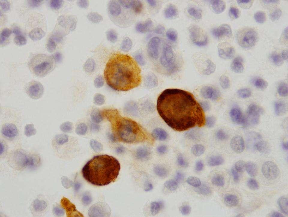
48 True Positive EMA Calretinin
49 You Should Know your Antibody
50 ICC in Diagnostic Cytology Applications Tumor Diagnosis/Classification Prognostic/Predictor Markers Target Therapy
51 ICC in Diagnostic Cytology Applications Tumor Diagnosis/Classification Prognostic/Predictor Markers Target Therapy
52 Selection of Markers Cytomorphology Clinical Information Working Diagnosis Differential Diagnosis Selection of ICC Markers Final Interpretation
53 ICC in Diagnostic Cytology Selection of Markers tailor-made Approach When the differential diagnosis is narrowed down, usually not more than 2-3 markers are needed ( tailor-made ) In many occasions only one marker is used to confirm the working diagnosis
54 Diagnosis/Classification Our 3-Step Approach 1. Define a specific differential Dx 2. Select a small panel of ICC markers 3. Combine cytomorphology and ICC
55 Is It Malignant? ICC Yes? No Mesothelioma ICC Adenocarcinoma ICC Site of Origin Others ICC Small Cell Ca Lymphoma Squamous cell Ca Others
56 Is It Malignant? ICC Yes? No Mesothelioma Others Adenocarcinoma Site of Origin Small Cell Ca Lymphoma Squamous cell Ca Others

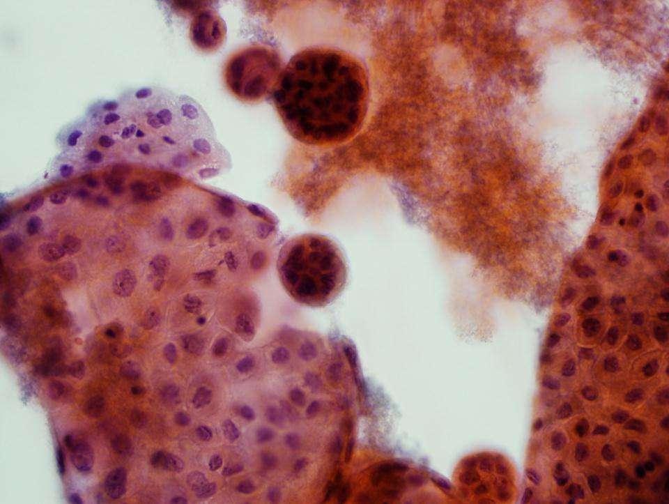
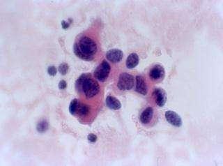
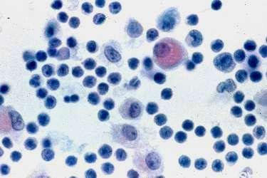
57 First Step.. Reactive Mesothelial cells versus Malignant Process
58 The reactive mesothelial cells may group. If so, the grouping usually presents as loose clusters, without nuclear overlapping.
59
60 Differential Diagnosis of Atypical cells in Ascitic Fluid Reactive Mesothelial Cells Malignant Mesothelioma Malignant Morphology NO YES Resemble Mesothelial Cells YES YES Adenocarcinoma YES NO
61 Commonly Used Markers In Effusions EMA: Malignant: adenocarcinoma, malignant mesothelioma CEA: Malignant: adenocarcinoma Ber-EP4: Malignant: adenocarcinoma LeuM1: Malignant: adenocarcinoma Desmin: Benign: reactive mesothelium
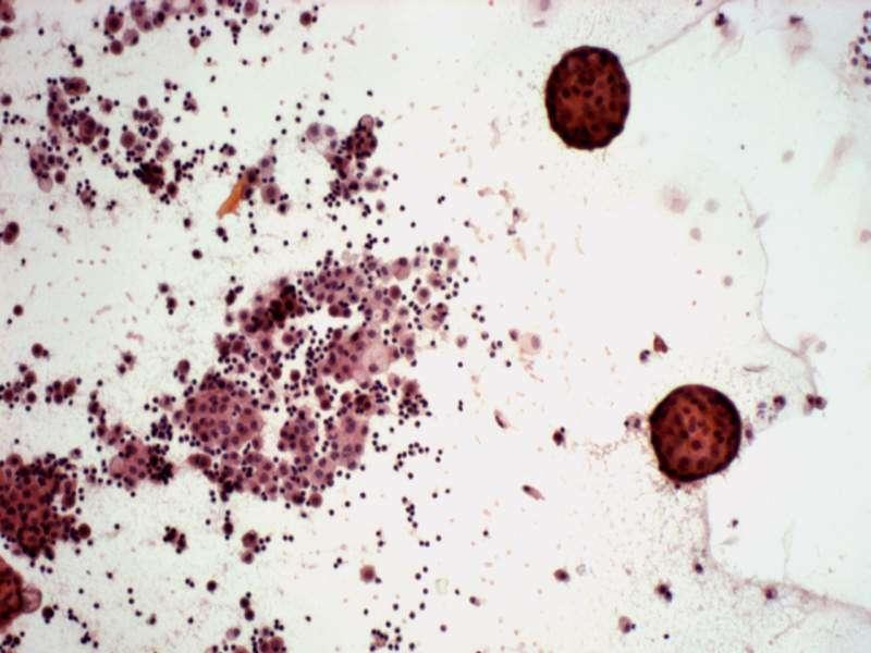
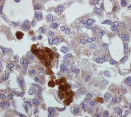
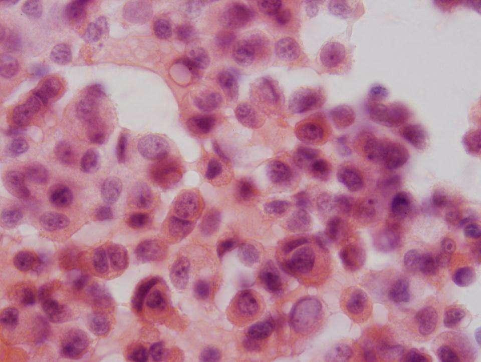
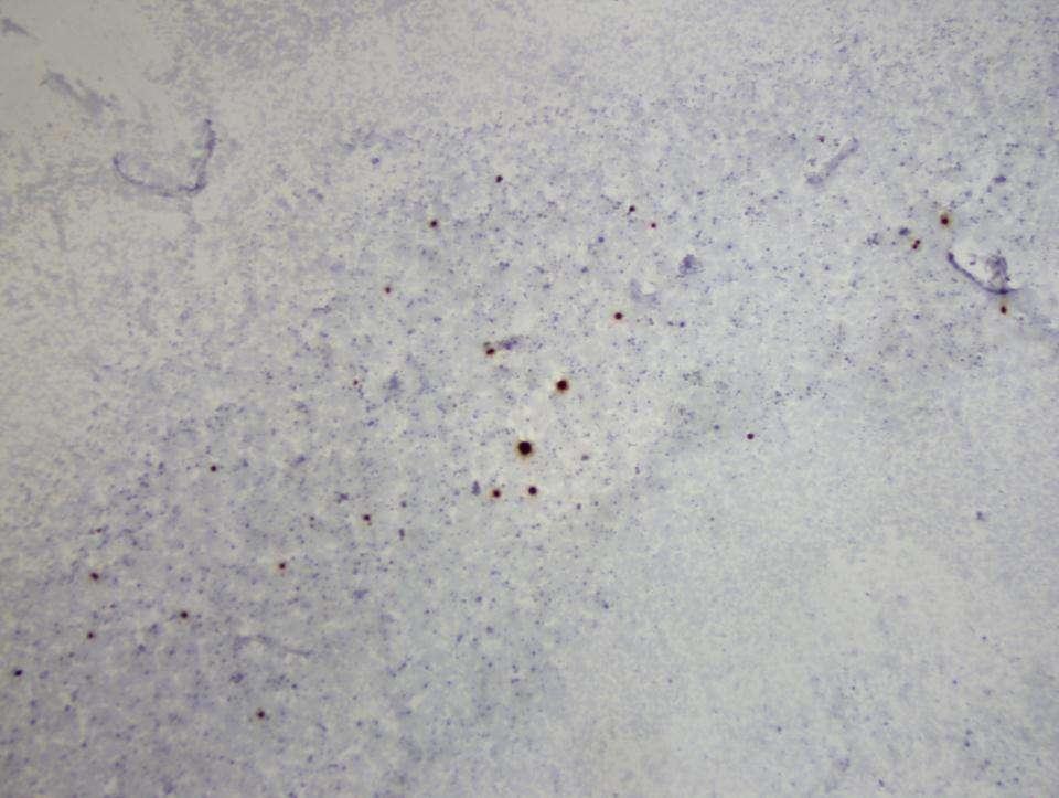
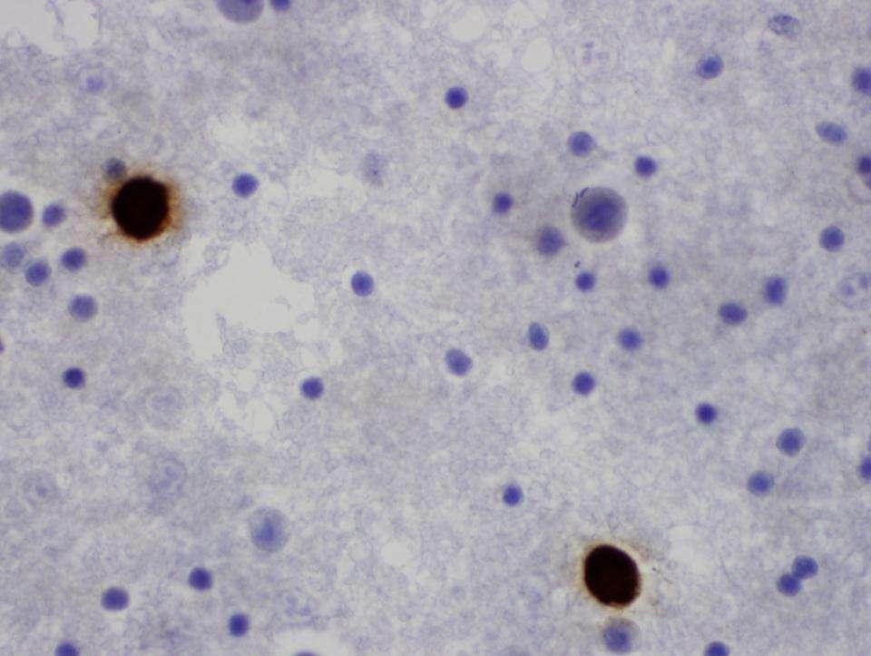
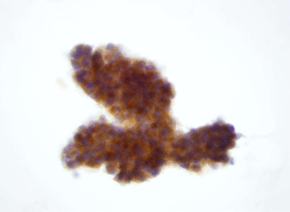
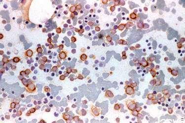
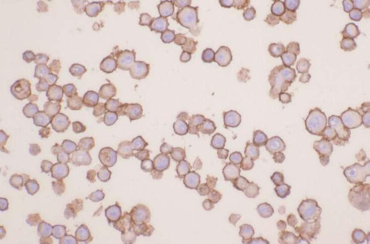
62 In our laboratory, EMA (clone E29) is the most frequently used antibody in defining atypical cells in effusions.
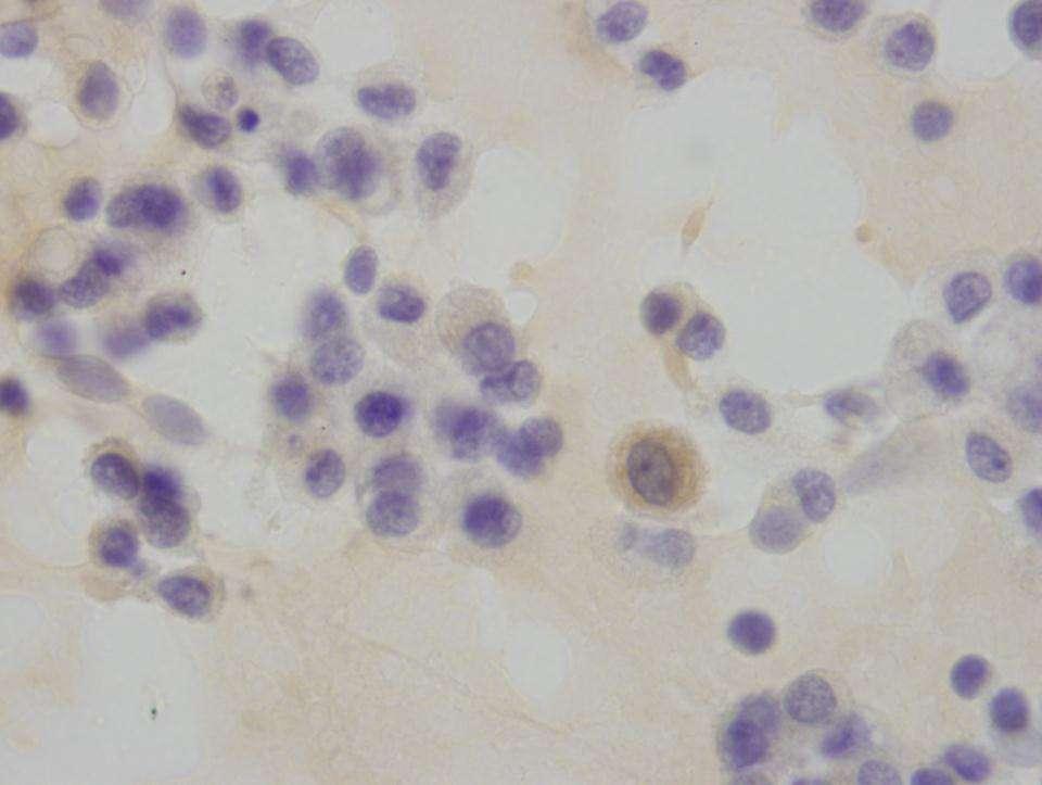
63 Reactive Mesothelium vs. Adenocarcinoma and Mesothelioma EMA Reactive Adenocarcinoma Mesothelioma Negative Positive (Cytoplasm) Positive (membrane)
64 In our experience, the most useful limited panel of ICC includes: EMA Calretinin Nuclear and intracytoplasmic positivity for calretinin and negativity for EMA confirms a reactive mesothelial proliferation. Acta Cytol 2000; 44 : 854 Diag Cytopathol 2008, 34:
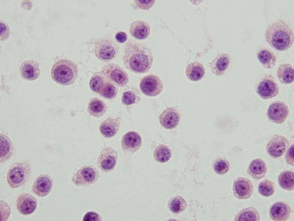
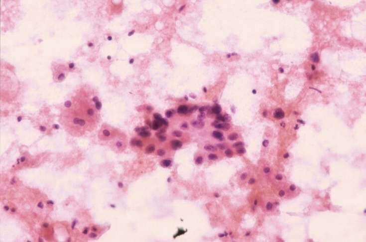
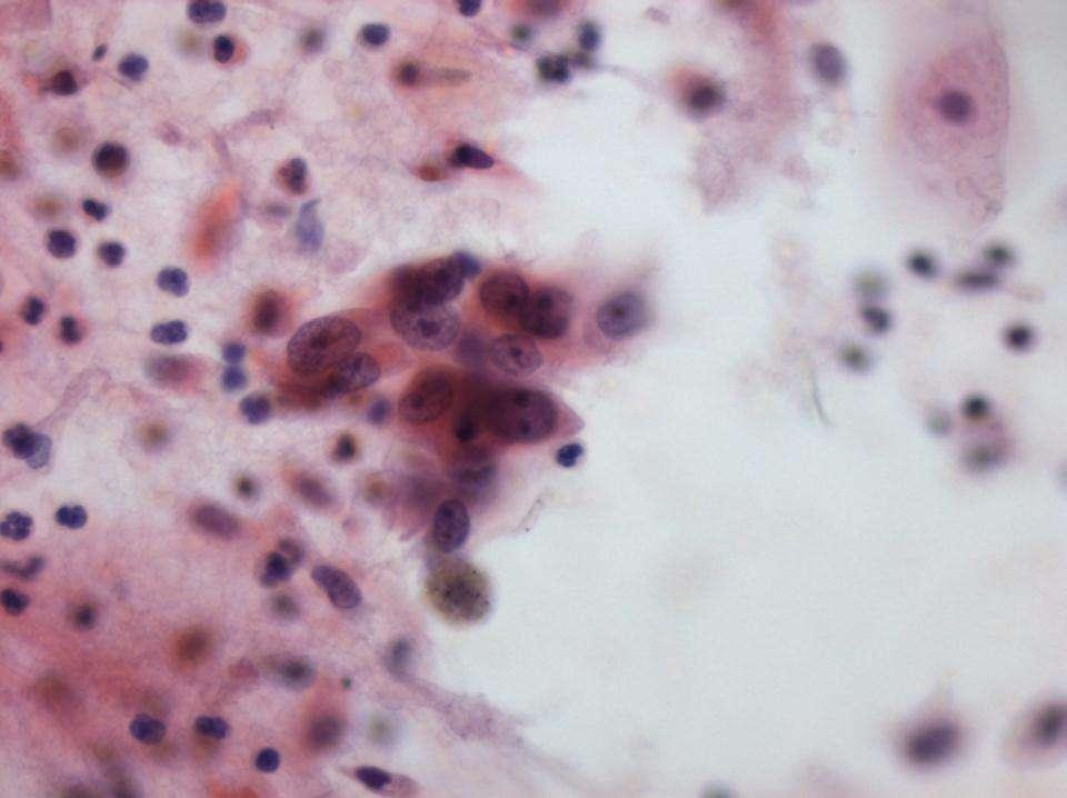
65 Reactive Mesothelium
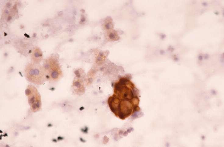
66 EMA
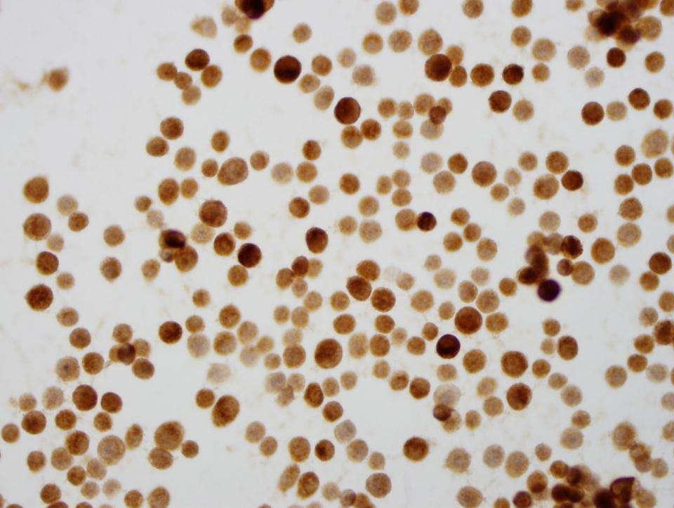
67 Calretinin
68

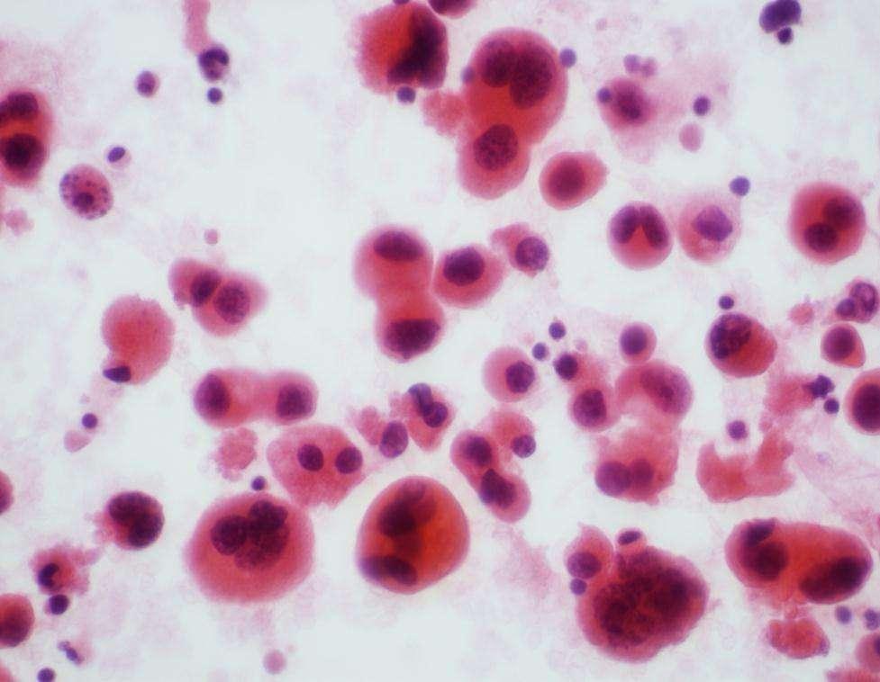
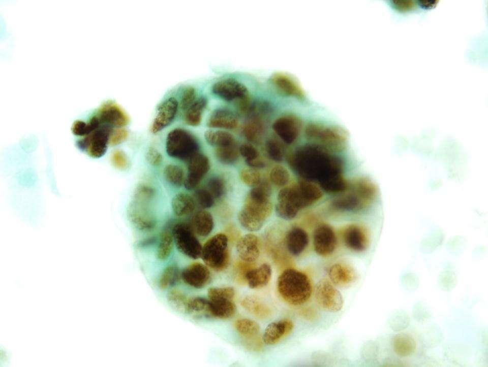
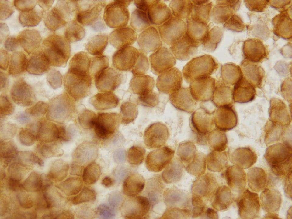
69 EMA Positivity Strong, Intracytoplasmic & Easily seen on Low Power EMA
70 PAP
71 EMA
72
73 EMA

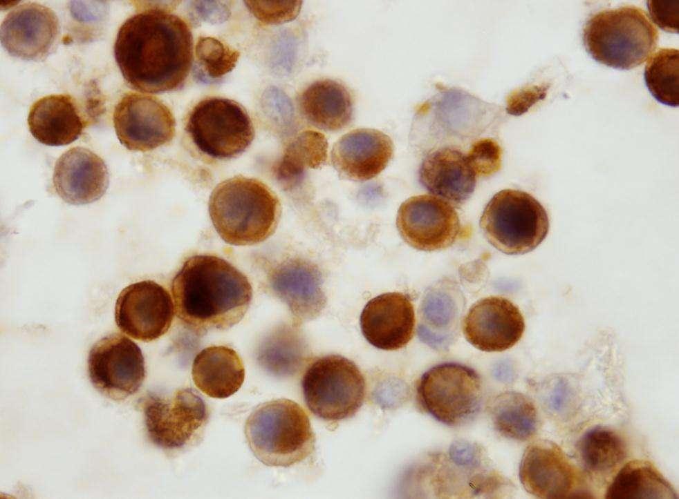
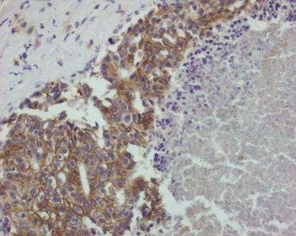
74 EMA Positive in MM: Strong Membrane/Cytoplasmic

75 EMA Positive in MM Strong Cytoplasmic Strong Membrane/Cytoplasmic
76 Positive EMA in Serous Effusions Represents adenocarcinoma, if: Easily seen on low power Is strong and intracytoplasmic
77 EMA
78 EMA
79 Is It Malignant? Yes? No Mesothelioma ICC Adenocarcinoma Site of Origin Others Small Cell Ca Lymphoma Squamous cell Ca Others
80 Malignant Mesotheliomas in Effusions Low Power Small or large 3D groups Knobby clusters
81
82

83
84 Resemblance to Mesothelial Cells
85 Malignant Mesothelioma in Effusions Differential Diagnosis of Mesothelioma Cytomorphology Electron microscopy Cytochemistry Immunocytochemistry (ICC)
86 When Malignant Mesothelioma Mimics Adenocarcinomas Use ICC
87 In our experience, the most useful limited panel of ICC includes: EMA Calretinin Nuclear and intracytoplasmic positivity for calretinin and Positivity for EMA confirms a Malignant Mesothelioma Acta Cytol 2000; 44 : 854 Diag Cytopathol 2008, 34:

88 Calretinin Malignant Mesothelioma
89 Ascitic Fluid Malignant Mesothelioma Vs Lung Adenocarcinoma Calret TTF-1 CEA D2-40 MM Pos Neg Neg Pos LA Neg Pos Pos Neg
90 Is It Malignant? Yes? No Mesothelioma Others Adenocarcinoma ICC Site of Origin Small Cell Ca Lymphoma Squamous cell Ca Others
91 Adenocarcinoma in Ascitic Fluid Primary Sites in Adult Male Adenocarcinoma GI tract- Pancreas GU Lung
92 Adenocarcinoma in Ascitic Fluid Primary Sites in Adult Female Adenocarcinoma Ovary Breast GI Tract-Pancreas Lung 3
93 Breast/GYN adenocarcinoma
94 ER
95 ER-1D5 In Fluids
96 Remember! Be careful with the use of ER in peritoneal effusions of female patients Benign epithelial inclusions may cause false positive results First establish the malignant nature of the cells by cytomorphology
97 Adenocarcinoma of Lung Vs. Colonic Carcinoma TTF-1 CK20 CK7 Adenoca of Lung Colonic Carcinoma - + -
98 ICC Markers for Colon Cancer CK 7 CK 20 CDX-2 CEA Negative Positive Positive Positive
99 CK20
100 Hepatocellular Carcinoma vs Metastatic Adenocarcinoma CK7 HCA Hepatocellular Ca - + Adenocarcinoma + -
101
102 HCA
103 Hepatocellular Carcinoma Vs. Renal Cell Carcinoma HCA RCA EMA Hepatocellular Ca Renal Cell Ca
104
105 RCA
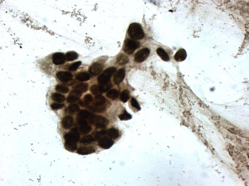
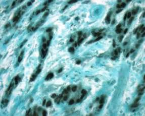
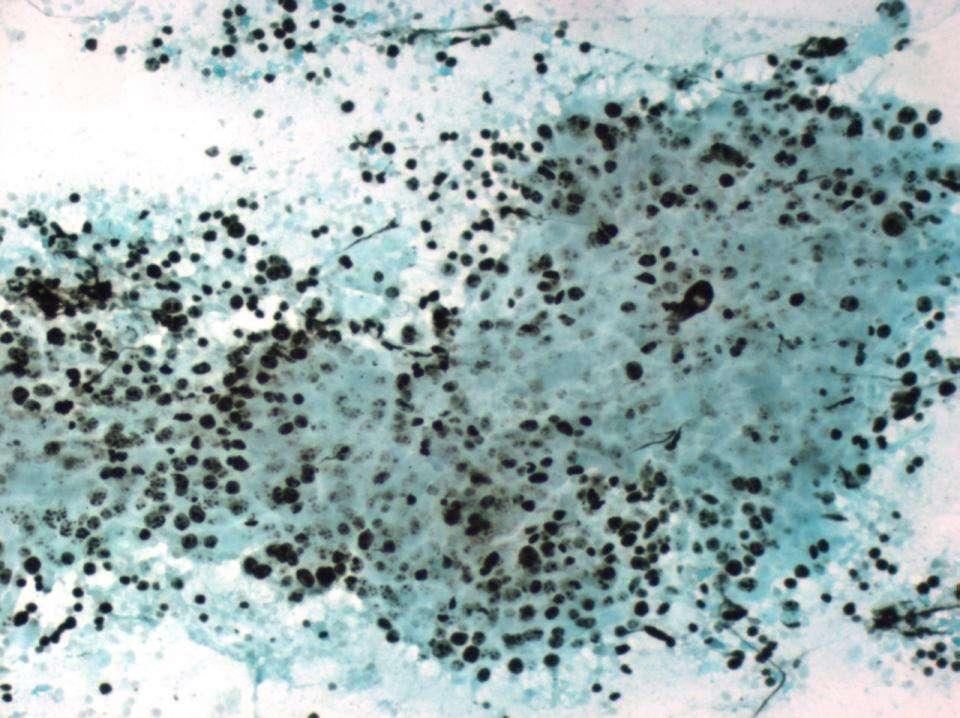
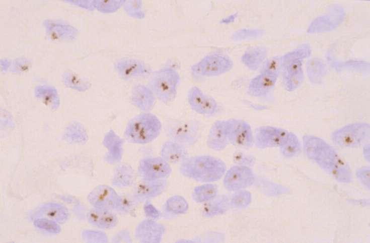
106 TTF-1 in Lung Adenoca. TTF-1 is useful for diagnosis of lung adenocarcinomas in effusions Only nuclear staining must be considered positive Cancer Cytopathol 96: , 2002
107
108 TTF-1

109 TTF-1; Neg.
110 Is It Malignant? Yes? No Mesothelioma Adenocarcinoma Site of Origin Others ICC Small Cell Ca Lymphoma Squamous cell Ca Others
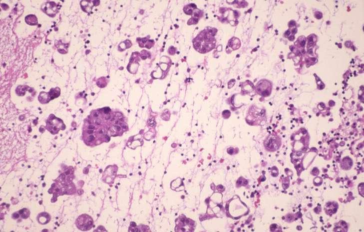
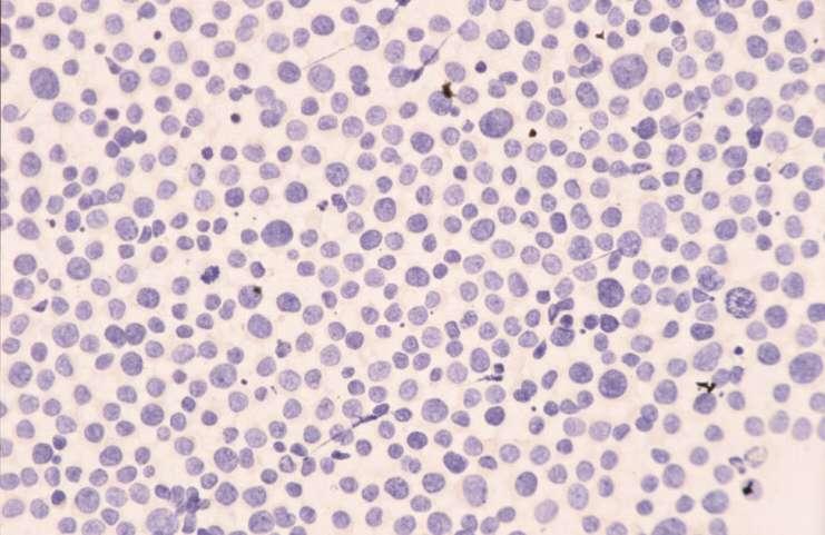
111 Small Cell Carcinoma in Ascitic Fluid Low Power Tight cell balls Indian file/chain Isolated cells may be overlooked High Power Nuclear molding Coarse chromatin Wrinkled nuclear membrane Occasional cells with nucleoli
112 Lung Carcinoma Non- Small vs Small Cell CK SYN CHR TTF-1 Non-Small + -/+ - +/- Small Cell +(dot) + +/- +
113
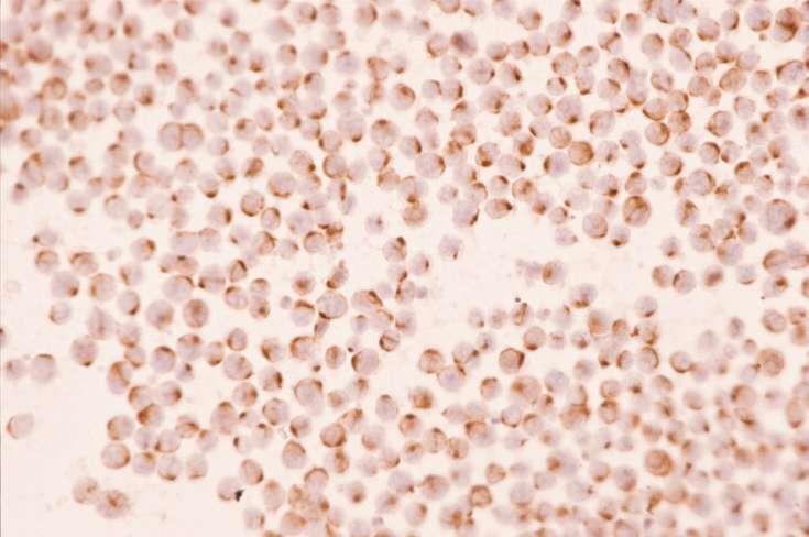
114 CK
115 Small Cell Carcinoma in Ascitic Fluid Differential Diagnosis Malignant lymphoma Small blue cell tumors
116 ICC in Differential Diagnosis of Small Cell Malignancies LCA KER CHR DES NB Small Cell Ca - + +/- - - Lymphoma Rhabdomyosarcoma Neuroblastoma
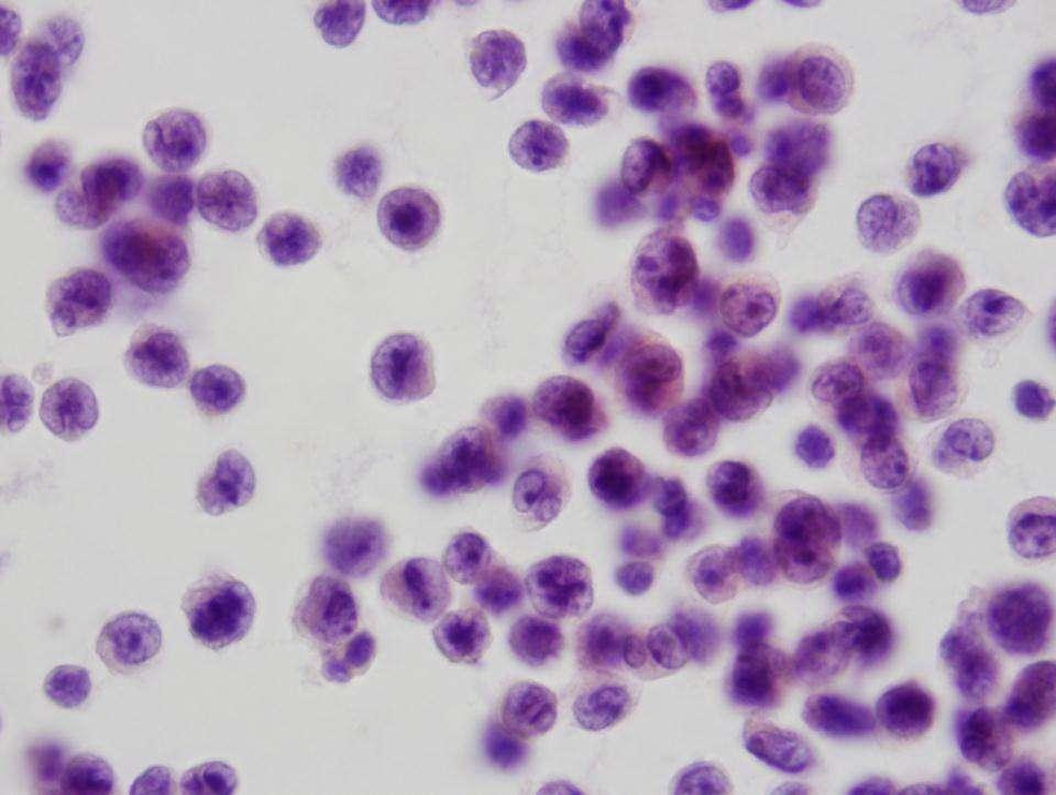
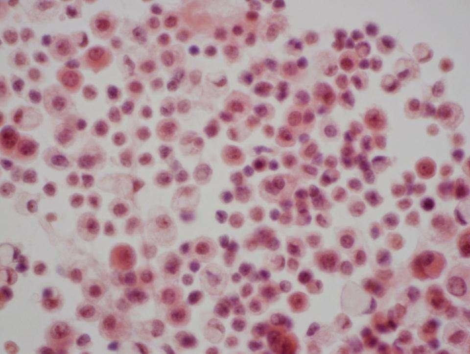
117 Malignant Lymphoma in Ascitic Fluid Low Power Isolated Cells
118
119 Malignant Lymphoma in Ascitic Fluid High Power Nuclear variation in size and shape Nuclear indentation/convolution Vesicular nuclei with prominent nucleoli Individual cell necrosis (apoptosis) Scant, basophilic cytoplasm, rarely well preserved
120
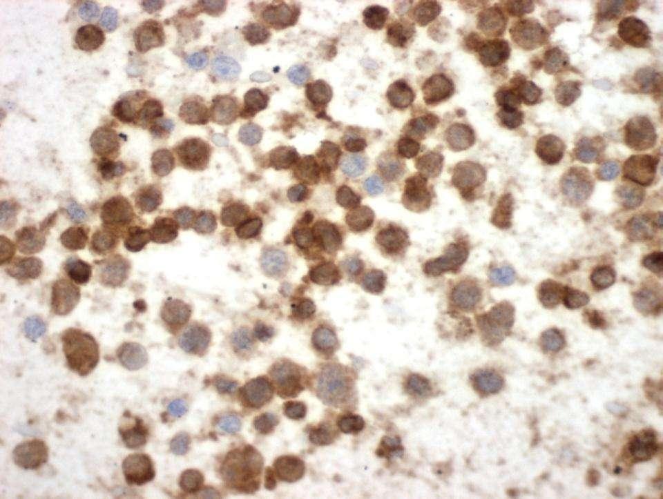
121 CD20

122 CD 79


123 Malignant Lymphoma in Ascitic Fluid HHV8 associated lymphoma
124 Lymphoma vs. Carcinoma vs. Germinoma vs. Melanoma Favor Lymphoma Only isolated cells Nuclear clefts Apoptotic cells Immunocytochemistry LCA ( + ) Keratin ( - ) PLAP ( - ) S100 ( - )
125 Small Mature-Looking Lymphocytes in Effusions Differential Diagnosis Chronic pleuritis (TB) Small cell lymphomas Chronic lymphocytic leukemia Waldenstrom s macroglobulinemia
126
127 Lymphocytes in Effusions Effusion CD45 CD20 CD3 Type (LCA) (B-cell) (T-cell) Benign Malignant + + -
128 CD3

129 CD 20 It is an Artifact
130 Ancillary Techniques to Rule Out Malignant Lymphoma Flow cytometry Gene rearrangement

131
132 Squamous Cell Carcinomas are Rare in Effusions Site of Origin Lung Cervix Skin Esophagus Diagnostic Difficulties Tumor cells do not shed May be mistaken for poorly differentiated adenocarcinomas or mesotheliomas

133 Squamous cell ca

134 p63 Squamous cell ca
135 Remember! Squamous carcinoma cells are usually overlooked in body cavity fluid cytology - Only few cells shed They might be confused with necrotic /degenerative mesothelial cells p63 and p40 are very helpful to detect squamous cells Cancer Cytopathol 2009; 117: 46-50
136 Carcinoma vs Melanoma CK S100 HMB45 Carcinoma + -/+ - Melanoma - + +
137
138 HMB 45
139 Melanoma Markers S100 Protein +++ HMB Melan-A ++ Tyrosinase ++
140 ICC in Diagnostic Cytology Applications Tumor Diagnosis/Classification Prognostic/Predictor Markers Target Therapy
141 ER-1D5

142 Ki-67
143 Detection of HER2 in cytology ICC, FISH, CISH Predictive Value NOT standard of Care for Breast CA Diagn Cytopathol 1994; 11:
144 HER2-ICC

145 Her2 gene Chr 17
146 HER2-CISH

147 CD20 Rituximab (Rituxan )
148 ICC in Diagnostic Cytology Applications Tumor Diagnosis/Classification Prognostic/Predictor Markers Target Therapy
149 NSCLC: Target Therapy tyrosine kinase inhibitors (TKI) first-line therapy in patients with advanced lung adenocarcinoma with EGFR mutations adenocarcinomas with ALK rearrangements are responsive to crizotinib (AlK inhibitor). Patients with KRAS or BRAF mutation do not respond to TKI, ALKI Arch Pathol Lab Med 2013, 137:
150 NSCLC: Target Therapy patients with adenocarcinoma or NSCLC, not otherwise specified (NSCLC- NOS),are more responsive to pemetrexed than those squamous cell carcinoma squamous cell carcinoma is associated with life-threatening hemorrhage in patients treated with bevacizumab; therefore, it is contraindicated in lung cancer patients with this histology. Arch Pathol Lab Med 2013, 137:
151
152 TTF-1

153 p63 Squamous Cell Carcinoma
154 ICC Limitations Large 3D cellular clusters in cytospin samples Histiocytes, macrophages, cells in mitosis, tumor giant cells Look for single cells or smaller 2D groups AM J Clin Pathol 1990; 94:
155 ICC Limitations Lack of internal control Negative results in ICC are not as meaningful as positive reactions Diag Cytopathol 1986; 81-2, 1986
156 Final Words. Use our 3-step approach: Define a specific differential Dx Select a small panel of ICC markers Combine Cytomorphology and ICC
157 Final Words. ICC can be used on previously alcohol-fixed Pap-stained slides without de-staining The technique does not require any modification of the routine ICC staining protocol


158 Springer, 2007 Demos, 2011
159 ASCP Workshops Diagnostic problems in body cavity fluid cytology; a practical approach. Immunocytochemistry in Diagnostic Cytology: Values and Limitations Parvin Ganjei-Azar MD, FASCP Mercè Jordà MD, PhD, FASCP

160 University of Miami Health System Sylvester Comprehensive Cancer Center
Cytological Sub-classification of Lung Cancer: Morphologic and Molecular Characteristics. Mercè Jordà, University of Miami
 Cytological Sub-classification of Lung Cancer: Morphologic and Molecular Characteristics Mercè Jordà, University of Miami Mortality Lung cancer is the most frequent cause of cancer incidence and mortality
Effusion Cytology: Diagnostic Challenges
 Effusion Cytology: Diagnostic Challenges Tarik M. Elsheikh, MD Professor and Medical Director, Anatomic Pathology Cleveland Clinic Outside Consult Case 45 year old woman, presented with nausea, dyspnea,
Serous Effusions. Spasenija Savic Prince, MD Pathology, University Hospital Basel, Switzerland
 Serous Effusions Spasenija Savic Prince, MD Pathology, University Hospital Basel, Switzerland Serous membrane Body cavities: Pleural Pericardial Peritoneal Effusion = Excess of fluid 80% Benign 20% Malignant
Applications of Flow Cytometry in Diagnostic Cytology of Body Cavity Fluids
 Applications of Flow Cytometry in Diagnostic Cytology of Body Cavity Fluids Awtar Krishan, PhD. Professor, Department of Pathology University of Miami School of Medicine akrishan@med.miami.edu Beckman
Case 1. Slide 1 History: 65 year old male presents with bilateral pleural effusions, a 40 pack year smoking history and peripheral and hilar lung
 Case 1. Slide 1 History: 65 year old male presents with bilateral pleural effusions, a 40 pack year smoking history and peripheral and hilar lung masses. Specimen shown is from a tap of the pleural effusion.
SELECTED DILEMMAS IN RESPIRATORY CYTOPATHOLOGY (2 CASES)
") SELECTED DILEMMAS IN RESPIRATORY CYTOPATHOLOGY (2 CASES) Dr. Mariamma Joseph Professor of Pathology Division Head Cytopathology Department of Pathology and Laboratory Medicine LHSC and Western University
SELECTED DILEMMAS IN RESPIRATORY CYTOPATHOLOGY (2 CASES) Dr. Mariamma Joseph Professor of Pathology Division Head Cytopathology Department of Pathology and Laboratory Medicine LHSC and Western University
Presentation material is for education purposes only. All rights reserved URMC Radiology Page 1 of 98
 Presentation material is for education purposes only. All rights reserved. 2011 URMC Radiology Page 1 of 98 Radiology / Pathology Conference February 2011 Brooke Koltz, Cytopathology Resident Presentation
Introduction. 23 rd Annual Seminar in Pathology. FLUIDS, Part 1. Pittsburgh, PA Gladwyn Leiman UVMMC, VT
 23 rd Annual Seminar in Pathology Pittsburgh, PA Gladwyn Leiman UVMMC, VT FLUIDS, Part 1 "Blue walls", Claudia Hansen, 2009 Introduction o Challenging to everyone o Almost any benign or malignant process
ACCURACY OF IMMUNOHISTOCHEMISTRY IN EVALUATION
 POL J PATHOL 2011; 2: 95-100 ACCURACY OF IMMUNOHISTOCHEMISTRY IN EVALUATION OF MALIGNANT PLEURAL AND PERITONEAL EFFUSIONS FERESHTEH ENSANI, FARNAZ NEMATIZADEH, GITI IRVANLOU Department of Cytology, Cancer
Serous effusion Objectives. Cytology of Serous Effusions From basics to challenges
 Cytology of Serous Effusions From basics to challenges Cytology of Serous Effusions From basics to challenges Pınar Fırat, MD, MIAC Department of Pathology, İstanbul University, İstanbul Faculty of Medicine,
Applications of IHC. Determination of the primary site in metastatic tumors of unknown origin
 Applications of IHC Determination of the primary site in metastatic tumors of unknown origin Classification of tumors that appear 'undifferentiated' by standard light microscopy Precise classification
BOSNIAN-TURKISH CYTOPATHOLOGY SCHOOL June 18-19, 2016 Sarajevo. Case Discussions. 60 year old woman Routine gynecologic control LBC
 BOSNIAN-TURKISH CYTOPATHOLOGY SCHOOL June 18-19, 2016 Sarajevo Case Discussions Prof Dr Sıtkı Tuzlalı Tuzlalı Pathology Laboratory 60 year old woman Routine gynecologic control LBC 1 2 Endometrial thickening
Cytyc Corporation - Case Presentation Archive - October 2001
 ThinPrep Pap Test History: 82 Year Old Female Specimen Type: Peritoneal Washings Case provided by Dr. Berle Stratton, Southwest Washington Medical Center, Vancouver, Washington. *The images, analysis and
How to Recognize Gynecologic Cancer Cells from Pelvic Washing and Ascetic Specimens
 How to Recognize Gynecologic Cancer Cells from Pelvic Washing and Ascetic Specimens Wenxin Zheng, M.D. Professor of Pathology and Gynecology University of Arizona zhengw@email.arizona.edu http://www.zheng.gynpath.medicine.arizona.edu/index.html
Immunohistochemistry on Fluid Specimens: Technical Considerations
 Immunohistochemistry on Fluid Specimens: Technical Considerations Blake Gilks Dept of Pathology University of British Columbia, Vancouver, BC, Canada Disclosures None Learning Objectives At the end of
Follow up of the Guidelines for Cytopathologic Diagnosis of Malignant Mesothelioma
 Follow up of the Guidelines for Cytopathologic Diagnosis of Malignant Mesothelioma Assoc. Prof. Katalin Dobra, Senior Lecturer in Molecular Pathology Karolinska University Hospital Stockholm, Sweden Disclosure
ACCME/Disclosures. Diagnosing Mesothelioma in Limited Tissue Samples. Papanicolaou Society of Cytopathology Companion Meeting March 12 th, 2016
 Diagnosing Mesothelioma in Limited Tissue Samples Papanicolaou Society of Cytopathology Companion Meeting March 12 th, 2016 Sanja Dacic, MD, PhD University of Pittsburgh ACCME/Disclosures GENERAL RULES
Problem 1: Differential of Neuroendocrine Carcinoma 3/23/2017. Disclosure of Relevant Financial Relationships
 Differential of Neuroendocrine Carcinoma Alain C. Borczuk,MD Weill Cornell Medicine Disclosure of Relevant Financial Relationships USCAP requires that all faculty in a position to influence or control
INTRODUCTION TO PATHOLOGICAL TECHNIQUES. 1. Types of routine biopsy procedures 2. Special exams (IHC, FISH)
") INTRODUCTION TO PATHOLOGICAL TECHNIQUES 1. Types of routine biopsy procedures 2. Special exams (IHC, FISH) Biopsy-Indications Diffuse/multifocal lesions (neoplastic, inflammatory, etc) Etiology of the
INTRODUCTION TO PATHOLOGICAL TECHNIQUES 1. Types of routine biopsy procedures 2. Special exams (IHC, FISH) Biopsy-Indications Diffuse/multifocal lesions (neoplastic, inflammatory, etc) Etiology of the
Value of antimesothelioma HBME 1 in the diagnosis of inflammatory and malignant pleural effusions
 Romanian Journal of Morphology and Embryology 2006, 47(4):351 355 ORIGINAL PAPER Value of antimesothelioma HBME 1 in the diagnosis of inflammatory and malignant pleural effusions LILIANA MOCANU 1), ANCA
Hyperchromatic Crowded Groups: What is Your Diagnosis? Session 3000
 Hyperchromatic Crowded Groups: What is Your Diagnosis? Session 3000 Thomas A. Bonfiglio, M.D. Professor Emeritus, Pathology and Laboratory Medicine University of Rochester Disclosures In the past 12 months,
EBUS-TBNA Diagnosis and Staging of Lung Cancer
 EBUS-TBNA Diagnosis and Staging of Lung Cancer Nirag Jhala MD, MIAC Professor of Pathology and Lab Med. Director of Anatomic Pathology and Cytopathology Lewis Katz School of Medicine@ Temple University
Diagnostic Cytology of Cancer Cases
 Diagnostic Cytology of Cancer Cases Somporn Techangamsuwan Companion Animal Cancer Research Unit (CAC-RU) Department of Pathology, Faculty of Veterinary Science, Chulalongkorn University 1 Tumor or Non-tumor
ACCME/Disclosures. Case 4 USCAP Pulmonary Panel Case 4 History
 Case 4 USCAP Pulmonary Panel 2016 Andrew Churg, MD Department of Pathology Vancouver General Hospital & University of British Columbia Vancouver, BC achurg@mail.ubc.ca. ACCME/Disclosures The USCAP requires
Outline 11/2/2017. Pancreatic EUS-FNA general aspects. Cytomorphologic features of solid neoplasms/lesions of the pancreas
 ENDOSCOPIC ULTRASOUND GUIDED-FINE NEEDLE ASPIRATION CYTOLOGY OF PANCREAS Khalid Amin M.D. Assistant Professor Department of Laboratory Medicine and Pathology University of Minnesota Outline Pancreatic
Mesothelioma: diagnostic challenges from a pathological perspective. Naseema Vorajee August 2016
 Mesothelioma: diagnostic challenges from a pathological perspective Naseema Vorajee August 2016 Naseema.vorajee@nhls.ac.za Pleural diseases (whether neoplastic, reactive or infective) may have similar
Objectives. Atypical Glandular Cells. Atypical Endocervical Cells. Reactive Endocervical Cells
 2013 California Society of Pathologists 66 th Annual Meeting San Francisco, CA Atypical Glandular Cells to Early Invasive Adenocarcinoma: Cervical Cytology and Histology Christina S. Kong, MD Associate
Gynecologic Cytopathology: Glandular lesions
 Gynecologic Cytopathology: Glandular lesions Lin Wai Fung (MSc, MPH, CMIAC) 17/4/2014 Glandular lesions of the uterus Endocervix Endometrium Normal endocervical cells Sheets, strips well-preserved architecture:
Key Words: effusion; carcinoma; immunocytochemistry; direct smear; cytology
 The Application of Immunocytochemistry to Direct Smears in the Diagnosis of Effusions Stewart M. Knoepp, M.D., Ph.D., { Jeremiah Placido, M.D., { Kristina L. Fields, B.S., Dafydd Thomas, M.D., Ph.D., and
CYTOPATHOLOGIC AND MOLECULAR DIAGNOSTIC ISSUES IN LUNG CYTOPATHOLOGY
 COMPANION MEETING Four Ps of Pulmonary Cytopathology: Procedural, Predictive, Personalized and Participatory CYTOPATHOLOGIC AND MOLECULAR DIAGNOSTIC ISSUES IN LUNG CYTOPATHOLOGY Prof. Fernando Schmitt
Impact of immunostaining of pulmonary and mediastinal cytology
 Impact of immunostaining of pulmonary and mediastinal cytology Harman Sekhon MD, PhD Director of Cytopathology Head of Ottawa-site Ontario Tumour Bank June 20, 2014 Disclaimer Pfizer: Honorarium-Advisory
Immunohistochemical classification of lung carcinomas and mesotheliomas. Prof. Mogens Vyberg NordiQC Institute of Pathology Aalborg, Denmark
 Immunohistochemical classification of lung carcinomas and mesotheliomas Prof. Mogens Vyberg NordiQC Institute of Pathology Aalborg, Denmark Endobronchial ultrasound guided transbronchial needle biopsy
Case 3 - GYN. History: 66 year old, routine Pap test. Dr. Stelow
 Case 3 - GYN History: 66 year old, routine Pap test Dr. Stelow Case 3 66 year year old woman Routine Pap Test Cytologic Features 3 dimensional clusters of cells with small to moderate amount of
A 53 year-old woman with a lung mass, right hilar mass and mediastinal adenopathy.
 November 2015 Case of the Month A 53 year-old woman with a lung mass, right hilar mass and mediastinal adenopathy. Contributed by: Rasha Salama, M.D., IU Department of Pathology and Laboratory Medicine
Thyroid master class. Thyroid Fine needle aspiration cytology and liquid-based techniques: Hologic and Becton Dickinson
 Thyroid master class Thyroid Fine needle aspiration cytology and liquid-based techniques: Hologic and Becton Dickinson Principle of LBC Collection of cells in liquid medium Immediate fixation Processor-prepared
Histopathological diagnosis of CUP
 Histopathological diagnosis of CUP Dr Karin Oien karin.oien@glasgow.ac.uk Disclosure slide Dr Karin Oien has no financial interests in any company mentioned in this presentation. Dr Karin Oien is conducting
Oncocytic-Appearing Salivary Gland Tumors. Oncocytic, Cystic, Mucinous, and High Grade Salivary Gland Tumors SALIVARY GLAND FNA: PART II
 William C. Faquin, MD, PhD Professor of Pathology Harvard Medical School Director of Head and Neck Pathology Massachusetts Eye and Ear Massachusetts General Hospital SALIVARY GLAND FNA: PART II Oncocytic,
Cytology for the Endocrinologist. Nicole Massoll M.D
 Cytology for the Endocrinologist Nicole Massoll M.D Objectives Discuss slide preperation Definitions of adequacy ROSE (Rapid On-Site Evaluation) Thyroid Cytology Adequacy Nicole Massoll M.D. University
From Morphology to Molecular Pathology: A Practical Approach for Cytopathologists Part 1-Cytomorphology. Songlin Zhang, MD, PhD LSUHSC-Shreveport
 From Morphology to Molecular Pathology: A Practical Approach for Cytopathologists Part 1-Cytomorphology Songlin Zhang, MD, PhD LSUHSC-Shreveport I have no Conflict of Interest. FNA on Lymphoproliferative
4/12/2018. MUSC Pathology Symposium Kiawah Island April 18, Jesse K. McKenney, MD
 MUSC Pathology Symposium Kiawah Island April 18, 2018 Jesse K. McKenney, MD 1 Urothelial Carcinoma with Alternative Differentiation 2 Urothelial Carcinoma with Alternative Differentiation Recognition as
LUNG CANCER. pathology & molecular biology. Izidor Kern University Clinic Golnik, Slovenia
 LUNG CANCER pathology & molecular biology Izidor Kern University Clinic Golnik, Slovenia 1 Pathology and epidemiology Small biopsy & cytology SCLC 14% NSCC NOS 4% 70% 60% 50% 63% 62% 61% 62% 59% 54% 51%
Cutaneous metastases. Thaddeus Mully. University of California, San Francisco Professor, Departments of Pathology and Dermatology
 Cutaneous metastases Thaddeus Mully University of California, San Francisco Professor, Departments of Pathology and Dermatology DISCLOSURE OF RELATIONSHIPS WITH INDUSTRY Thaddeus Mully Course C005 Essential
Salivary Gland Cytology
 Salivary Gland Cytology Diagnostic challenges and potential pitfalls Tarik M. Elsheikh, MD Professor and Medical Director Anatomic Pathology Cleveland Clinic FNA Salivary Gland Lesions Indications Distinguish
LGM International, Inc.
 Liqui-PREP TM Cytology Atlas Preface The following pictures are examples with descriptions of cytology slides processed with the Liqui-PREP TM System.. The descriptions are reviewed by Pathologists. It
Case year female. Routine Pap smear
 Case 1 57 year female Routine Pap smear Diagnosis? 1. Atypical glandular cells of unknown significance (AGUS) 2. Endocervical AIS 3. Endocervical adenocarcinoma 4. Endometrial adenocarcinoma 5. Adenocarcinoma
DIAGNOSTIC DILEMMA. Case Reports Clinical history. Materials and Methods
 DIAGNOSTIC DILEMMA A Metastatic Renal Carcinoid Tumor Presenting as Breast Mass: A Diagnostic Dilemma Farnaz Hasteh, M.D., 1 Robert Pu, M.D., Ph.D., 2 and Claire W. Michael, M.D. 2 * We present clinicopathological
Almost any suspected tumor can be aspirated easily and safely. Some masses are more risky to aspirate including:
 DOES THIS PATIENT HAVE CANCER? USING IN-HOUSE CYTOLOGY TO HELP YOU MAKE THIS DIAGNOSIS. Joyce Obradovich, DVM, Diplomate, ACVIM (Oncology) Animal Cancer & Imaging Center, Canton, Michigan Almost every
and management of lung cancer Maureen F. Zakowski, M.D. Memorial Sloan-Kettering Cancer Center
 The new role of cytology in the diagnosis and management of lung cancer Maureen F. Zakowski, M.D. Memorial Sloan-Kettering Cancer Center Outline Role of cytology in the diagnosis of lung cancer Non-small
Pancreatitis: A Potential Pitfall in Endoscopic Ultrasound Guided Pancreatic FNA
 Pancreatitis: A Potential Pitfall in Endoscopic Ultrasound Guided Pancreatic FNA Jack Yang, MD Department of Pathology, Medical University of South Carolina Objectives Understand the indication of EUS
Differential diagnosis of HCC
 Hepatocellular Carcinoma Quest for an Ideal Immunohistochemical Panel Sanjay Kakar, MD UCSF Differential diagnosis of HCC Hepatocellular lesions Adenoma, FNH, HG dysplasia Adenocarcinoma CholangioCA, metastasis
Urinary Cytology. Spasenija Savic Prince, MD Pathology, University Hospital Basel, Switzerland
 Urinary Cytology Spasenija Savic Prince, MD Pathology, University Hospital Basel, Switzerland Outline Pre-analytics The Paris System (TPS): Background Diagnostic categories Morphologic criteria for each
SQUAMOUS CELLS: Atypical squamous cells (ASC) - of undetermined significance (ASC-US) - cannot exclude HSIL (ASC-H)
 - of undetermined significance (ASC-US) - cannot exclude HSIL (ASC-H)") SQUAMOUS CELLS: Atypical squamous cells (ASC) - of undetermined significance (ASC-US) - cannot exclude HSIL (ASC-H) ASC refers to cytologic changes suggestive of SIL, which are qualitativley or quantitatively
SQUAMOUS CELLS: Atypical squamous cells (ASC) - of undetermined significance (ASC-US) - cannot exclude HSIL (ASC-H) ASC refers to cytologic changes suggestive of SIL, which are qualitativley or quantitatively
The 24 th Congress Of The IAP-Arab Division KHARTOUM - SUDAN Arab School of Pathology Cytology workshop December 5-6, 2012
 International Academy of Pathology - Arab Division The 24 th Congress Of The IAP-Arab Division KHARTOUM - SUDAN Arab School of Pathology Cytology workshop December 5-6, 2012 Body Fluid Cytology Mousa Al-Abbadi,
The role of immunohistochemistry in surgical pathology of the uterine corpus and cervix
 The role of immunohistochemistry in surgical pathology of the uterine corpus and cervix Prof. Ben Davidson, MD PhD Department of Pathology, Norwegian Radium Hospital, Oslo University Hospital, Oslo, Norway
FNA of Thyroid. Toward a Uniform Terminology With Management Guidelines. NCI NCI Thyroid FNA State of the Science Conference
 FNA of Thyroid NCI NCI Thyroid FNA State of the Science Conference Toward a Uniform Terminology With Management Guidelines Thyroid Thyroid FNA Cytomorphology NCI Thyroid FNA State of the Science Conference
Lung Tumor Cases: Common Problems and Helpful Hints
 Lung Tumor Cases: Common Problems and Helpful Hints Brandon T. Larsen, MD, PhD Senior Associate Consultant Department of Laboratory Medicine and Pathology Mayo Clinic Arizona Arizona Society of Pathologists
Anaplastic Large Cell Lymphoma (of T cell lineage)
") Anaplastic Large Cell Lymphoma (of T cell lineage) Definition T-cell lymphoma comprised of large cells with abundant cytoplasm and pleomorphic, often horseshoe-shaped nuclei CD30+ Most express cytotoxic
Anaplastic Large Cell Lymphoma (of T cell lineage) Definition T-cell lymphoma comprised of large cells with abundant cytoplasm and pleomorphic, often horseshoe-shaped nuclei CD30+ Most express cytotoxic
LUNG CANCER PATHOLOGY: UPDATE ON NEUROENDOCRINE LUNG TUMORS
 LUNG CANCER PATHOLOGY: UPDATE ON NEUROENDOCRINE LUNG TUMORS William D. Travis, M.D. Attending Thoracic Pathologist Memorial Sloan Kettering Cancer Center New York, NY PULMONARY NE TUMORS CLASSIFICATION
Urinary Bladder: WHO Classification and AJCC Staging Update 2017
 Urinary Bladder: WHO Classification and AJCC Staging Update 2017 Houston Society of Clinical Pathologists 58 th Annual Spring Symposium Houston, TX April 8, 2017 Jesse K. McKenney, MD Classification
Pancreatic malignant tumors are the fifth leading cause of cancerrelated
 44 CANCER CYTOPATHOLOGY Cytologic Criteria for Well Differentiated Adenocarcinoma of the Pancreas in Fine-Needle Aspiration Biopsy Specimens Fan Lin, M.D., Ph.D. 1 Gregg Staerkel, M.D. 2 1 Department of
EBUS-FNAB: HOW TO OPTIMIZE YOUR CYTOLOGY SAMPLES, LHSC EXPERIENCE. Dr. Mariamma Joseph Division Head of Cytopathology LHSC and Western University
 EBUS-FNAB: HOW TO OPTIMIZE YOUR CYTOLOGY SAMPLES, LHSC EXPERIENCE Dr. Mariamma Joseph Division Head of Cytopathology LHSC and Western University Objectives Brief overview of EBUS-FNA Strategies to optimize
DOI: /jemds/2014/1921 ORIGINAL ARTICLE
 CYTODIAGNOSIS OF GASTROINTESTINAL STROMAL TUMOURS (GISTs) ON ROMANOWSKY STAINED SMEARS Priyanka Agrawal 1, Subhash Agrawal 2, Atul Gupta 3, Anurag Gupta 4 HOW TO CITE THIS ARTICLE: Priyanka Agrawal, Subhash
The role of Electron Microscopy in the study of cytologic specimens. Elba A. Turbat-Herrera, MD
 The role of Electron Microscopy in the study of cytologic specimens. Elba A. Turbat-Herrera, MD Louisiana State University Health Sciences Center Shreveport, LA, USA Introduction The field of Cytology
Diagnosis of lung cancer. Diagnosis Subtyping. Morphologic features Approach to small samples
 Lung Cytology Lung Cytology Pınar Fırat,MD,MIAC Diagnosis of lung cancer Detection of infections Evaluation of interstitial diseases Morphologic features Approach to small samples Lung Cancer Second most
NEW IHC A n t i b o d i e s
 NEW IHC Antibodies TABLE OF CONTENTS NEW IHC ANTIBODIES from Cell Marque CITED1 (5H6).... 1 Claudin 7 (5D10F3).... 1 GATA1 (4F5).... 1 Transgelin (2A10C2).... 1 NEW IHC ANTIBODIES using RabMAb Technology
Biliary Tract Neoplasia: A Cyto-histologic Review. Michelle Reid, MD, MSc Professor of Pathology Director of Cytopathology Emory University Hospital
 Biliary Tract Neoplasia: A Cyto-histologic Review Michelle Reid, MD, MSc Professor of Pathology Director of Cytopathology Emory University Hospital Bile Duct Brushings (BDB) BDBs are the initial diagnostic
Case #1 FNA of nodule in left lobe of thyroid in 67 y.o. woman
 Challenging Cases Manon Auger M.D., F.R.C.P. (C) Professor, Department of Pathology McGill University Director, Cytopathology Laboratory McGill University it Health Center Case #1 FNA of nodule in left
Mody. AIS vs. Invasive Adenocarcinoma of the Cervix
 Common Problems in Gynecologic Pathology Michael T. Deavers, M.D. Houston Methodist Hospital, Houston, Texas Common Problems in Gynecologic Pathology Adenocarcinoma in-situ (AIS) of the Cervix vs. Invasive
Cytology and the Investigation of Carcinoma of Unknown Primary (CUP) Dr Anna Green ST5, St Thomas Hospital London, UK
 Dr Anna Green ST5, St Thomas Hospital London, UK") Cytology and the Investigation of Carcinoma of Unknown Primary (CUP) Dr Anna Green ST5, St Thomas Hospital London, UK Objectives Introduction to CUP Our experience of cytology and CUP Role of Cytology
Cytology and the Investigation of Carcinoma of Unknown Primary (CUP) Dr Anna Green ST5, St Thomas Hospital London, UK Objectives Introduction to CUP Our experience of cytology and CUP Role of Cytology
, , 2011 HODGKIN LYMPHOMA
 European Federation of Cytology Societies 4tu Annual Tutorial in Cytopathology Trieste, June 6-10, 2011 HODGKIN LYMPHOMA Classification The World Health Organization Classification of Lymphomas (2001)
Diagnosis of a granular cell tumour at the abdominal wall using fine needle aspiration cytology and histology: Case report
 Case Report Diagnosis of a granular cell tumour at the abdominal wall using fine needle aspiration cytology and histology: Case report Journal of International Medical Research 2015, Vol. 43(4) 592 596!
Evolution of Pathology
 1 Traditional pathology Molecular pathology 2 Evolution of Pathology Gross Pathology Cellular Pathology Morphologic Pathology Molecular/Predictive Pathology Antonio Benivieni (1443-1502): First autopsy
Dr. Issraa Ali Hussein
 CLINICAL 09888888;rCYTOLOGY Dr. Issraa Ali Hussein objectives Define diagnostic cytology (clinical cytology). Explain the differences between histopathology and cytopathology. Recognize the methods for
MECHANISMS OF HUMAN DISEASE: LABORATORY SESSION CYTOPATHOLOGY Monday, April 26, 2013 FACULTY COPY
 GOAL: MECHANISMS OF HUMAN DISEASE: LABORATORY SESSION CYTOPATHOLOGY Monday, April 26, 2013 FACULTY COPY 1. Understated the role of cytopathology in the clinical management of the patient and recognize
What is New in the 2015 WHO Lung Cancer Classification? Zhaolin Xu, MD, FRCPC, FCAP
 What is New in the 2015 WHO Lung Cancer Classification? Zhaolin Xu, MD, FRCPC, FCAP Professor, Dept of Pathology, Dalhousie University, Canada Pulmonary Pathologist and Cytopathologist, QEII HSC Senior
Diagnostic IHC in lung and pleura pathology
 Diagnostic IHC in lung and pleura pathology Mogens Vyberg Professor of Clinical Pathology Director of NordiQC Aalborg University Hospital, Aalborg, Denmark WHO 2004 and Web Malignant mesothelioma Epithelioid
HOW TO GET THE MOST INFORMATION FROM A TUMOR BIOPSY
 HOW TO GET THE MOST INFORMATION FROM A TUMOR BIOPSY 7 TH Annual New York Lung Cancer Symposium Saturday, November 10, 2012 William D. Travis, M.D. Attending Thoracic Pathologist Memorial Sloan Kettering
Essentials of Fluid Cytology
 Essentials of Fluid Cytology Gia-Khanh Nguyen 2018 Essentials of Fluid Cytology Gia-Khanh Nguyen, MD, FRCPC Professor Emeritus Department of Laboratory Medicine and Pathology Faculty of Medicine and Dentistry
Neoplasia 2018 Lecture 2. Dr Heyam Awad MD, FRCPath
 Neoplasia 2018 Lecture 2 Dr Heyam Awad MD, FRCPath ILOS 1. List the differences between benign and malignant tumors. 2. Recognize the histological features of malignancy. 3. Define dysplasia and understand
Journal of Cytology & Histology
 ISSN: 2157-7099 ojournal of Cytology & Hist logy Journal of Cytology & Histology Sen, et al., 2015, 6:2 DOI: 10.4172/2157-7099.1000314 Research Article Article Open Open Access Morphometric Analysis and
Prepared By Jocelyn Palao and Layla Faqih
 Prepared By Jocelyn Palao and Layla Faqih The structure of the suspected atypical cell should always be compared to the structure of other similar, benign, cells which are present in the smears. The diagnosis
Update on Thyroid FNA The Bethesda System. Shikha Bose M.D. Associate Professor Cedars Sinai Medical Center
 Update on Thyroid FNA The Bethesda System Shikha Bose M.D. Associate Professor Cedars Sinai Medical Center Thyroid Nodules Frequent occurrence Palpable: 4-7% of adults Ultrasound: 10-31% Majority benign
40th European Congress of Cytology Liverpool, UK, 2-5 th October 2016
 40th European Congress of Cytology Liverpool, UK, 2-5 th October 2016 EUS FNA of abdominal organs: An approach to reporting and triage for ancillary testing Date and time: Sunday 2 nd October 2016 15.00-16.30
Disclosure of Relevant Financial Relationships NON-SMALL CELL LUNG CANCER: 70% PRESENT IN ADVANCED STAGE
 MORPHOLOGY AND MOLECULAR TESTING IN NON-SMALL CELL OF LUNG NEW FRONTIEIRS IN CYTOPATHOLOGY PRACTICE American Society for Cytopathology San Antonio, Texas Sunday March 5, 2017 Disclosure of Relevant Financial
QUALITY ASSURANCE PROGRAM CYTOLOGY CYCLE 01/2018 (TRIAL)
") [Pick the Date] FINAL REPORT QUALITY ASSURANCE PROGRAM CYTOLOGY CYCLE 01/2018 (TRIAL) NOTES FROM THE COORDINATOR 1. For this cycle 01/2018, a total of 32 pen drives had been circulated. Twenty-eight institutions
[Pick the Date] FINAL REPORT QUALITY ASSURANCE PROGRAM CYTOLOGY CYCLE 01/2018 (TRIAL) NOTES FROM THE COORDINATOR 1. For this cycle 01/2018, a total of 32 pen drives had been circulated. Twenty-eight institutions
Immunohistochemistry in Bone and Soft Tissue Tumors. Sahar Rassi Zankoul, MD
 Immunohistochemistry in Bone and Soft Tissue Tumors Sahar Rassi Zankoul, MD Introduction Bone tumors represent a wide variety of tumors of various origins and malignant potentials. These different tumor
Thyroid Nodules: Understanding FNA Cytology (The Bethesda System for Reporting of Thyroid Cytopathology) Shamlal Mangray, MB, BS
 Shamlal Mangray, MB, BS") Thyroid Nodules: Understanding FNA Cytology (The Bethesda System for Reporting of Thyroid Cytopathology) Shamlal Mangray, MB, BS Attending Pathologist Rhode Island Hospital, Providence, RI DISCLOSURE:
Thyroid Nodules: Understanding FNA Cytology (The Bethesda System for Reporting of Thyroid Cytopathology) Shamlal Mangray, MB, BS Attending Pathologist Rhode Island Hospital, Providence, RI DISCLOSURE:
Predictors of Malignancy in Thyroid Fine-Needle Aspirates Cyst Fluid Only Cases
 Predictors of Malignancy in Thyroid Fine-Needle Aspirates Cyst Fluid Only Cases Can Potential Clues of Malignancy Be Identified? Mohammad Jaragh, MD 1 ; V. Bessie Carydis, MMedSci (Cytol) 1 ; Christina
Respiratory Cytology and Ancillary diagnostic techniques. Dr Alex Rice Royal Brompton Hospital
 Respiratory Cytology and Ancillary diagnostic techniques Dr Alex Rice Royal Brompton Hospital Overview Specialist Cardiothoracic centre BAL specimens and cell differential counts EBUS Diagnostic pitfalls
Cytologic Evaluation of the Enlarged Neck Node: FNAC Utility in Metastatic Neck Disease
 ISPUB.COM The Internet Journal of Pathology Volume 6 Number 2 Cytologic Evaluation of the Enlarged Neck Node: FNAC Utility in Metastatic Neck Disease I Bagwan, S Kane, R Chinoy Citation I Bagwan, S Kane,
ACCME/Disclosures. Case History 4/13/2016. USCAP GU Specialty Conference Case 3. Ann Arbor, MI
 USCAP GU Specialty Conference Case 3 March 2016 L. Priya Kunju, M.D. University of Michigan Health System Ann Arbor, MI University of Michigan Health System ACCME/Disclosures The USCAP requires that anyone
Respiratory Tract Cytology
 Respiratory Tract Cytology 40 th European Congress of Cytology Liverpool, UK Momin T. Siddiqui M.D. Professor of Pathology and Laboratory Medicine Director of Cytopathology Emory University Hospital, Atlanta,
FNA Cytology of Metastatic Malignancies of Unknown Primary Site
 FNA Cytology of Metastatic Malignancies of Unknown Primary Site Tarik M. Elsheikh Jan F. Silverman Pathologic Diagnosis of Metastasis Smaller specimens, less invasive techniques FNA cytology is highly
Cerebral Parenchymal Lesions: I. Metastatic Neoplasms
 Chapter 4 Cerebral Parenchymal Lesions: I. Metastatic Neoplasms After one has reasonably ruled out the possibility of a nonneoplastic diagnosis (see Chap. 3), one is left with considering a diagnosis of
Award Top Quizzes For Residents
 Award Top Quizzes For Residents Giovanni Negri, MD, Bolzano (Italy) Eva M. Wojcik MD, Department of Pathology, Loyola University, Chicago (USA) Esther D. Rossi MD, PhD, MIAC, Division of Anatomic Pathology
Morphologic and Immunocytochemical Performances of Effusion Cell Blocks Prepared Using 3 Different Methods
 Anatomic Pathology / Performance of Different Cell Block Preparation Techniques Morphologic and Immunocytochemical Performances of Effusion Cell Blocks Prepared Using 3 Different Methods Xin Jing, MD,
Potential Pitfalls for False Suspicion of Papillary Thyroid Carcinoma:
 SUPPLEMENT 1 SPECIAL ISSUE: CYTOPATHOLOGY OF THE THYROID GLAND Guest Editor: Zubair Baloch Potential Pitfalls for False Suspicion of Papillary Thyroid Carcinoma: A Cytohistologic Review of 22 Cases Xin
International Journal of Health Sciences and Research ISSN:
 International Journal of Health Sciences and Research www.ijhsr.org ISSN: 2249-9571 Original Research Article Utility of Modified Cell Block Technique in Cases of Pleural Effusion Suspected of Malignancy
Case # year old man with a 2 cm right kidney mass
 Case # 4. 52 year old man with a 2 cm right kidney mass Figure 1 Figure 2 Figure 3 Figure 4 Diagnosis: Negative/Non-diagnostic Normal kidney tissue Fine needle aspiration (FNA) of the kidney is performed
Objectives. Salivary Gland FNA: The Milan System. Role of Salivary Gland FNA 04/26/2018
 Salivary Gland FNA: The Milan System Dr. Jennifer Brainard Section Head Cytopathology Cleveland Clinic Objectives Introduce the Milan System for reporting salivary gland cytopathology Define cytologic