Ultrasound in Peripheral Nerve Interventions
|
|
|
- Nancy Bruce
- 5 years ago
- Views:
Transcription
1 Ultrasound in Peripheral Nerve Interventions John L. Lin, M.D. Shepherd Center Assistant Clinical Professor Emory University, School of Medicine
2 Outline Ultrasound basics Nerve blocks in physiatric setting Ultrasound with nerve blocks
3 Ultrasound Basics Sound wave energy is transformed when it interacts upon different medium Reflected Absorbed Transformed
4 Sound Wave Transformation Frequency Energy heat, electricity, mechanical energy / vibration etc. Density of median -> impedance High impedance difference -> high echogenicity Mineral, fluid, gas

5 Ultrasound Machines Transforms electricity to ultrasound Receives reflected ultrasound and transforms to electricity Display of digital images


6 Ultrasound transducers Transforms electricity to vibrations of the piezoelectric crystals Higher frequency -> higher resolution, lower penetration MHz

7 J Ultrasound Med 2002; 21:315
8 Sonographic Appearance Fluid anechoic Muscle relatively hypoechoic Tendon hyperechoic Nerve hyperechoic Bone, cortex hyperechoic with acoustic shadow
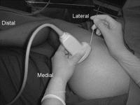
9 Lateral Medial Adductor Longus Adductor Brevis Femur Adductor Magnus Sciatic Nerve Shibata, Yasuyuki, and Misako Harato. Ultrasound-Guided Anterior Sciatic Nerve Block.
10 Doppler Effect Sound wave travelling away decreases in frequency Sound wave travelling towards increases in frequency

11 Doppler Imaging 3%2DVASC%2DBICPA&systemcode=e&div=ultra
12 Anisotropy
13 Anisotropy
14 Nerve Blocks Management of spasticity, contractures Pre-serial casting transient blocks Pre-manipulation blocks Diagnostic / therapeutic
15 Ultrasound Imaging for Nerve Blocks Brachial Plexus Suprascapular Median elbow, carpal tunnel Ulnar Radial Femoral Obturator Sciatic Tibial

16 Brachial Plexus Preparation for regional shoulder manipulations Anesthetic block


17

18 AnaesthesiaUK: Home>>Resources>>Anatomy>>Anatomy for anaesthetists>>applied anatomy for upper limb blocks (August 30, 2011)

19 Brachial Plexus Anatomical Appraoch
")

20 The New York School of Regional Anesthesia>> Home>> Nerve Blocks >>Ultrasound-Guided Techniques >>Ultrasound-Assisted Nerve Blocks (14/03/2009) (Aug 30, 2011) Brachial Plexus Interscalene Approach

21 Brachial Plexus - Interscalene

22 Brachial Plexus - Interscalene
23
24

25 `

26

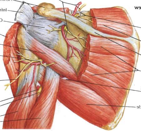
27 Brachial Plexus Suprascapular Artery

28 Yasuyuki Shibata. The Suprascapular Artery and the Brachial Plexus.

29 Yasuyuki Shibata. The Suprascapular Artery and the Brachial Plexus.


30 Brachial Plexus - Supraclavicular The New York School of Regional Anesthesia>> Home>> Nerve Blocks >>Ultrasound-Guided Techniques >>Ultrasound-Assisted Nerve Blocks (14/03/2009) (Aug 30, 2011)

31 Supraclavicular Suprascapular Nerve

32 Brachial Plexus Infraclavicular

33 Brachial Plexus Infraclavicular Yasuyuki Shibata. Ultrasound-Guided Infraclavicular Brachial Plexus Block.

34 Brachial Plexus Infraclavicular Yasuyuki Shibata. Ultrasound-Guided Infraclavicular Brachial Plexus Block.

35 Musculocutaneous Nerve

 http://www.nysora.")

36 The New York School of Regional Anesthesia>> Home>> Nerve Blocks >>Ultrasound-Guided Techniques >>Ultrasound-Assisted Nerve Blocks (14/03/2009) (Aug 30, 2011)
37 Musculocutaneous and Axillary AnaesthesiaUK: Home>>Resources>>Anatomy>>Anatomy for anaesthetists>>applied anatomy for upper limb blocks (August 30, 2011)

38 Musculocutaneous and Axillary AnaesthesiaUK: Home>>Resources>>Anatomy>>Anatomy for anaesthetists>>applied anatomy for upper limb blocks (August 30, 2011)


39 The New York School of Regional Anesthesia>> Home>> Nerve Blocks >>Ultrasound-Guided Techniques >>Ultrasound- Assisted Nerve Blocks (14/03/2009)
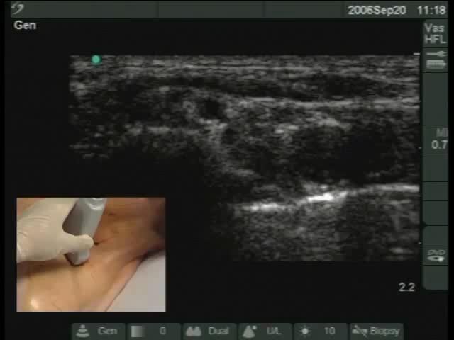
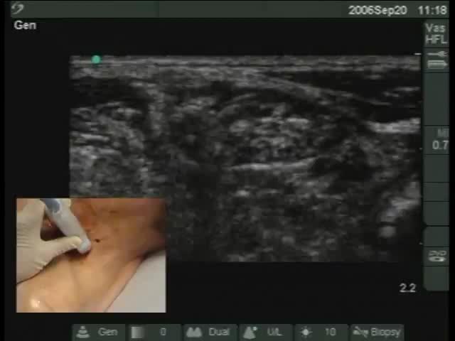
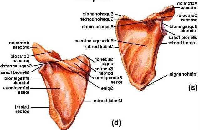
40 Suprascapular Nerve Preparation for regional shoulder manipulations In conjunction with other blocks Axillary nerve Intra-articular Acromio-clavicular Sub-acromial bursa
41 Suprascapular Nerve

42 Suprascapular Nerve Pain Physician 2007; 10:

43 Pain Physician 2007; 10:
44
45 Median Nerve Diagnostic / Therapeutic blocks Carpal tunnel injections
46 Median Nerve
47
48 Carpal Tunnel Anatomy Daniel Shelton, Carpal Tunnel: Bifid Median Nerve -SonoSite Case Study, SonoSite
49 Carpal Tunnel Anatomy Daniel Shelton, Carpal Tunnel: Bifid Median Nerve -SonoSite Case Study, SonoSite
50 Carpal Tunnel Anatomy Daniel Shelton, Carpal Tunnel: Bifid Median Nerve -SonoSite Case Study, SonoSite
51 Carpal Tunnel Anatomy Daniel Shelton, Carpal Tunnel: Bifid Median Nerve -SonoSite Case Study, SonoSite
52 Carpal Tunnel Anatomy Daniel Shelton, Carpal Tunnel: Bifid Median Nerve -SonoSite Case Study, SonoSite

53

54 Proximal Carpal Tunnel Probe Placement Daniel Shelton, Carpal Tunnel: Bifid Median Nerve -SonoSite Case Study, SonoSite

55 Proximal Carpal Tunnel Probe Placement - Transverse Ultrasound Evaluation for Carpal Tunnel Syndrome, Viewsono.com

56 Daniel Shelton, Carpal Tunnel: Bifid Median Nerve -SonoSite Case Study, SonoSite

57 Daniel Shelton, Carpal Tunnel: Bifid Median Nerve -SonoSite Case Study, SonoSite

58 Daniel Shelton, Carpal Tunnel: Bifid Median Nerve -SonoSite Case Study, SonoSite

59 Ultrasound Evaluation for Carpal Tunnel Syndrome, Viewsono.com

60 Ultrasound Evaluation for Carpal Tunnel Syndrome, Viewsono.com

61 Proximal Carpal Tunnel Probe Placement - Longitudinal Ultrasound Evaluation for Carpal Tunnel Syndrome, Viewsono.com

62 Proximal Carpal Tunnel Probe Placement - Longitudinal Ultrasound Evaluation for Carpal Tunnel Syndrome, Viewsono.com

63 Daniel Shelton, Carpal Tunnel: Bifid Median Nerve -SonoSite Case Study, SonoSite

64 Daniel Shelton, Carpal Tunnel: Bifid Median Nerve -SonoSite Case Study, SonoSite

65 Median Nerve Edema Daniel Shelton, Carpal Tunnel: Bifid Median Nerve -SonoSite Case Study, SonoSite
66 Carpal Retinaculum Release

67
68 Ulnar Nerve Finger / wrist flexor dennervations Pre splinting / casting Epicondylar groove

69 Radial Nerve
70 Femoral Nerve Extensor contracture of quadriceps Diagnosis of the extent of tone versus soft tissue contracture prior to placing a temporary intrathecal baclofen infusion pump
71 Femoral Nerve Lateral Medial

72 Lateral Medial Femoral Nerve Femoral Artery Iliopsoas Muscle

73

74
75 Obturator Nerve Adductor spasticity / contractures Primarily for neurolysis

76 Obturator Nerve

77 Obturator Nerve Rabah, Danny M. and Ahmed K. Thallaj. Phase 1 Study, Safety and Feasibility of Ultrasound Guided Obturator Nerve Block in Transurethral Surgery

78 Rabah, Danny M. and Ahmed K. Thallaj. Phase 1 Study, Safety and Feasibility of Ultrasound Guided Obturator Nerve Block in Transurethral Surgery

79 Rabah, Danny M. and Ahmed K. Thallaj. Phase 1 Study, Safety and Feasibility of Ultrasound Guided Obturator Nerve Block in Transurethral Surgery

80 Rabah, Danny M. and Ahmed K. Thallaj. Phase 1 Study, Safety and Feasibility of Ultrasound Guided Obturator Nerve Block in Transurethral Surgery

81 Rabah, Danny M. and Ahmed K. Thallaj. Phase 1 Study, Safety and Feasibility of Ultrasound Guided Obturator Nerve Block in Transurethral Surgery

82 Rabah, Danny M. and Ahmed K. Thallaj. Phase 1 Study, Safety and Feasibility of Ultrasound Guided Obturator Nerve Block in Transurethral Surgery

83 Rabah, Danny M. and Ahmed K. Thallaj. Phase 1 Study, Safety and Feasibility of Ultrasound Guided Obturator Nerve Block in Transurethral Surgery
84 US Guided Obturator Nerve Block Rabah, Danny M. and Ahmed K. Thallaj. Phase 1 Study, Safety and Feasibility of Ultrasound Guided Obturator Nerve Block in Transurethral Surgery 18 consecutive men, ages 54-95, mean 69 Mean time for procedure 4.2 min Successful block 97.2%

85 Yasuyuki Shibata. Ultrasound-Guided Obturator Nerve Block.

86 Yasuyuki Shibata. Ultrasound-Guided Obturator Nerve Block.
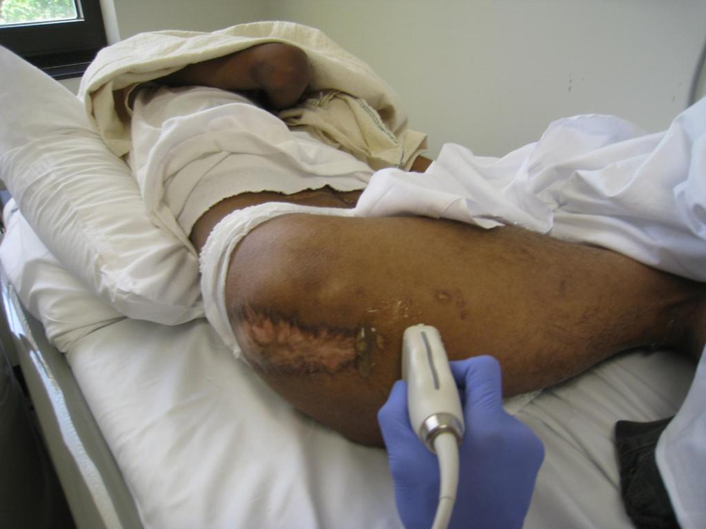
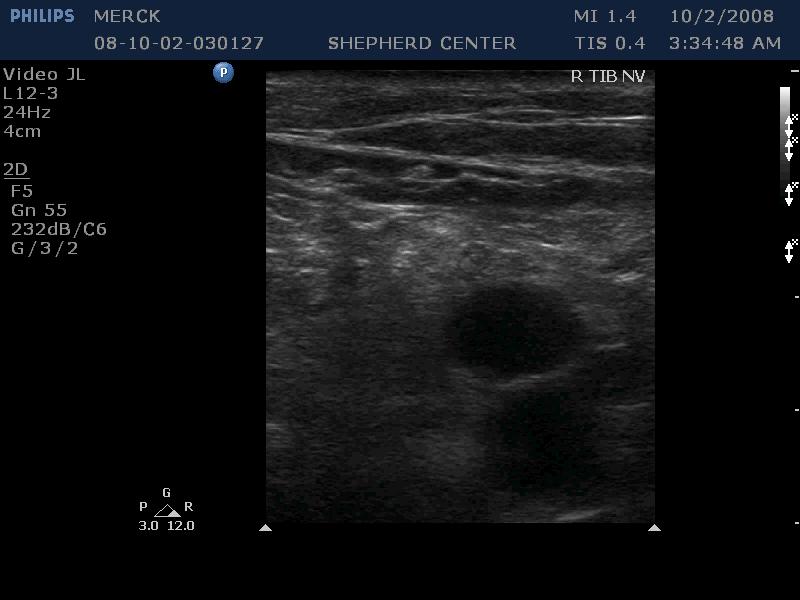
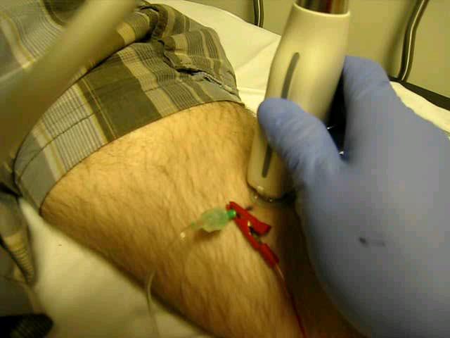
87 Sciatic / Tibial Nerves Pre-serial short leg casting of ankle contractures Diagnostic block Deformity, e.g. hammer toe refractory to intramuscular motor point blocks Versus diagnostic motor point blocks

88 Sciatic Nerve

89 Sciatic Nerve

90
91

92
93 Sciatic Nerve
94 Shibata, Yasuyuki, and Misako Harato. Ultrasound-Guided Anterior Sciatic Nerve Block.
95 Lateral Medial Adductor Longus Adductor Brevis Femur Adductor Magnus Sciatic Nerve Shibata, Yasuyuki, and Misako Harato. Ultrasound-Guided Anterior Sciatic Nerve Block.

96 Shibata, Yasuyuki, and Misako Harato. Ultrasound-Guided Anterior Sciatic Nerve Block.
97 Shibata, Yasuyuki, and Misako Harato. Ultrasound-Guided Anterior Sciatic Nerve Block.

98 Shibata, Yasuyuki, and Misako Harato. Ultrasound-Guided Anterior Sciatic Nerve Block.

99 Shibata, Yasuyuki, and Misako Harato. Ultrasound-Guided Anterior Sciatic Nerve Block.

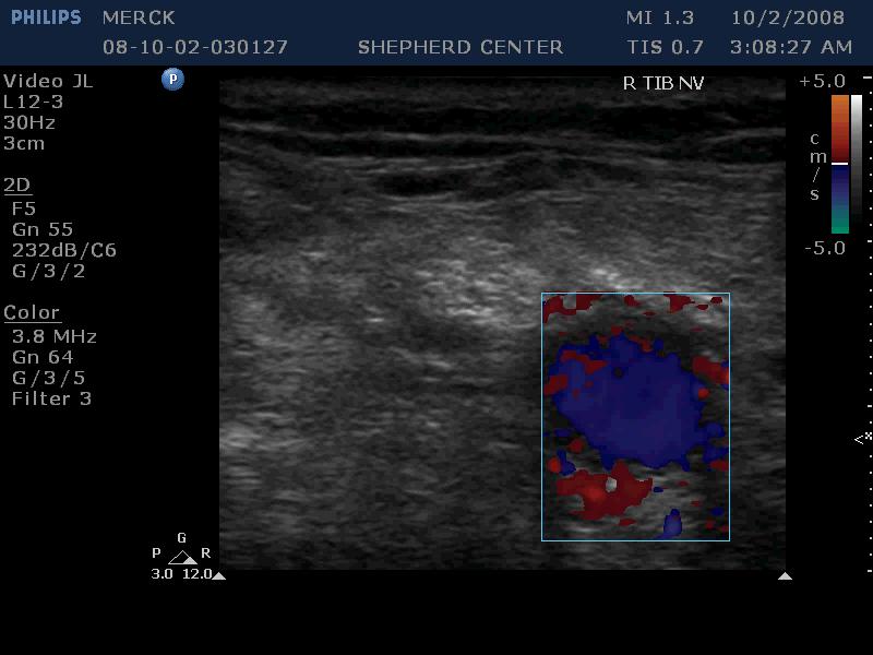
100 Tibial Nerve
101
102

103
104
105
106

107 Resources Atlas of Ultrasound- and Nerve Stimulation-Guided Regional Anesthesia Anatomical overlay of key muscles Real time videos on DVD

108 Resources Atlas of Ultrasound- and Nerve Stimulation-Guided Regional Anesthesia Extensive list of detailed peripheral nerve blocks

109 Resources Atlas of Ultrasound- Guided Regional Anesthesia: Expert Consult Online videos

110 Resources Ultrasound Guided Regional Anesthesia and Pain Medicine Numerous pain / sensory nerve blocks
111

112 Resources Ultrasound in Anesthetic Practice (with DVD)
 Alexander Levitov, Paul Mayo, Anthony")
113 Resources Critical Care Ultrasonography (with DVD) Alexander Levitov, Paul Mayo, Anthony Slonim
 Philip M. Hopkins, Andrew R. Bodenham, Scott T.")
114 Resources Practical Ultrasound in Anesthesia for Critical Care and Pain Management (with CD) Philip M. Hopkins, Andrew R. Bodenham, Scott T. Reeves
115
Ultrasound Guided Regional Nerve Blocks
 Ultrasound Guided Regional Nerve Blocks In the country of the blind the one eyed man is King -Deciderius Erasmus (1466-1536) Objectives Benefits of Regional Anesthesia Benefits of US guidance Role of ultrasound
inerve Guide to Nerves 2009
 inerve Guide to Nerves 2009 A guide to self learning and self assessment Context: The following guide is intended to help interpret the sono-anatomy and follow a systematic stepwise approach to the practice
Year 2004 Paper one: Questions supplied by Megan
 QUESTION 47 A 58yo man is noted to have a right foot drop three days following a right total hip replacement. On examination there is weakness of right ankle dorsiflexion and toe extension (grade 4/5).
Terminology Tissue Appearance
 By Marc Nielsen, MD Advantages/Disadvantages Generation of Image Ultrasound Machine/Transducer selection Modes of Ultrasound Terminology Tissue Appearance Scanning Technique Real-time Portable No ionizing
Ultrasound Physics and Knobology Alan Macfarlane. Consultant Anaesthetist Glasgow Royal Infirmary
 Ultrasound Physics and Knobology Alan Macfarlane Consultant Anaesthetist Glasgow Royal Infirmary RAPM 2009; 34: 40-46 Ultrasound Proficiency Understanding US image generation and device operation Image
Nerves of the upper limb Prof. Abdulameer Al-Nuaimi. E. mail:
 Nerves of the upper limb Prof. Abdulameer Al-Nuaimi E-mail: a.al-nuaimi@sheffield.ac.uk E. mail: abdulameerh@yahoo.com Brachial plexus Median nerve After originating from the brachial plexus in the axilla,
Human Anatomy and Physiology I Laboratory Spinal and Peripheral Nerves and Reflexes
 Human Anatomy and Physiology I Laboratory Spinal and Peripheral Nerves and Reflexes 1 This lab involves the second section of the exercise Spinal Cord, Spinal Nerves, and the Autonomic Nervous System,
Principles of Ultrasound. Cara C. Prideaux, M.D. University of Utah PM&R Sports Medicine Fellow March 14, 2012
 Principles of Ultrasound Cara C. Prideaux, M.D. University of Utah PM&R Sports Medicine Fellow March 14, 2012 None Disclosures Outline Introduction Benefits and Limitations of US Ultrasound (US) Physics
Table of contents. Foreword. Preface. 1 Introduction Historical Perspective 00
 Table of contents Foreword Preface 1 Introduction 00 1.1 Historical Perspective 00 2 Fundamentals of musculoskeletal ultrasound 00 2.1 Frequency and wavelength 00 2.2 Generating ultrasound waves 00 2.3
Ultrasound Use in Anaesthesia
 Trainee Name: 1 Ultrasound Use in Anaesthesia Assessments to accompany Workbook for anaesthetic trainees in North Queensland 2010 Authors: Mark Fairley, Emile Kurukchi, Andrew Potter 2 Trainee Name: Ultrasound
Basic Physics of Ultrasound and Knobology
 WELCOME TO UTMB Basic Physics of Ultrasound and Knobology By Daneshvari Solanki, FRCA Laura B. McDaniel Distinguished Professor Anesthesiology and Pain Medicine University of Texas Medical Branch Galveston,
Brachial plexus blockade within the interscalene groove involves local anesthetic
 Interscalene Brachial Plexus Block- How I do it. Part 1 of a 2 part discussion on technique. Stuart Grant Professor of Anesthesiology Duke University Medical Center Durham NC Brachial plexus blockade within
Ultrasonography of Peripheral Nerve -upper extremity
 Ultrasonography of Peripheral Nerve -upper extremity Department of Physical Medicine and Rehabilitation Korea University Guro Hospital Korea University College of Medicine Yoon Joon Shik Normal median
Index. Note: Page numbers of article titles are in boldface type.
 Note: Page numbers of article titles are in boldface type. A ACJ. See Acromioclavicular joint (ACJ) Acromioclavicular joint (ACJ) procedures of, 557 559 Ankle and foot procedures of, 649 671 (See also
Anatomy Workshop Upper Extremity David Ebaugh, PT, PhD Workshop Leader. Lab Leaders: STATION I BRACHIAL PLEXUS
 Anatomy Workshop Upper Extremity David Ebaugh, PT, PhD Workshop Leader Lab Leaders: STATION I BRACHIAL PLEXUS A. Posterior cervical triangle and axilla B. Formation of plexus 1. Ventral rami C5-T1 2. Trunks
Ultrasonography of the wrist - a step-by-step approach to study protocols and normal findings
 Ultrasonography of the wrist - a step-by-step approach to study protocols and normal findings Poster No.: C-1779 Congress: ECR 2016 Type: Educational Exhibit Authors: R. R. Domingues Madaleno, A. P. Pissarra,
Surgery Under Regional Anesthesia
 Surgery Under Regional Anesthesia Jean Daniel Eloy, MD Assistant Professor Residency Program Director Rutgers-New Jersey Medical School Rutgers The State University of New Jersey Peripheral Nerve Block
BRACHIAL PLEXUS. DORSAL SCAPULAR NERVE (C5) supraclavicular branch innervates rhomboids (major and minor) and levator scapulae
 supraclavicular branch innervates rhomboids (major and minor) and levator scapulae") THE BRACHIAL PLEXUS DORSAL SCAPULAR NERVE (C5) supraclavicular branch innervates rhomboids (major and minor) and levator scapulae SCHEMA OF THE BRACHIAL PLEXUS THE BRACHIAL PLEXUS PHRENIC NERVE supraclavicular
THE BRACHIAL PLEXUS DORSAL SCAPULAR NERVE (C5) supraclavicular branch innervates rhomboids (major and minor) and levator scapulae SCHEMA OF THE BRACHIAL PLEXUS THE BRACHIAL PLEXUS PHRENIC NERVE supraclavicular
Introduction to Ultrasound Examination of the Hand and upper
 Introduction to Ultrasound Examination of the Hand and upper Emil Dionysian, M.D. Ultrasound of upper ext. Upside Convenient Opens another exam dimension Can be like a stethoscope Helps 3-D D visualization
213: HUMAN FUNCTIONAL ANATOMY: PRACTICAL CLASS 1: Proximal bones, plexuses and patterns
 213: HUMAN FUNCTIONAL ANATOMY: PRACTICAL CLASS 1: Proximal bones, plexuses and patterns CLAVICLE Examine an isolated clavicle and compare it with a clavicle on an articulated skeleton. Viewed from above,
Nerves of Upper limb. Dr. Brijendra Singh Professor & Head Department of Anatomy AIIMS Rishikesh
 Nerves of Upper limb Dr. Brijendra Singh Professor & Head Department of Anatomy AIIMS Rishikesh 1 Objectives Origin, course & relation of median & ulnar nerves. Motor & sensory distribution Carpal tunnel
Sonoanatomy Of The Brachial Plexus With Single Broad Band-High Frequency (L17-5 Mhz) Linear Transducer
 Linear Transducer") ISPUB.COM The Internet Journal of Anesthesiology Volume 11 Number 2 Sonoanatomy Of The Brachial Plexus With Single Broad Band-High Frequency (L17-5 Mhz) Linear A Thallaj Citation A Thallaj.. The Internet
ISPUB.COM The Internet Journal of Anesthesiology Volume 11 Number 2 Sonoanatomy Of The Brachial Plexus With Single Broad Band-High Frequency (L17-5 Mhz) Linear A Thallaj Citation A Thallaj.. The Internet
Biceps Brachii. Muscles of the Arm and Hand 4/4/2017 MR. S. KELLY
 Muscles of the Arm and Hand PSK 4U MR. S. KELLY NORTH GRENVILLE DHS Biceps Brachii Origin: scapula Insertion: radius, fascia of forearm (bicipital aponeurosis) Action: supination and elbow flexion Innervation:
3/20/2017. Disclosures. Ultrasound Fundamentals. Ultrasound Fundamentals. Bone Anatomy. Tissue Characteristics
 Disclosures Images of ultrasound equipment in this presentation are not an endorsement Fundamentals of Musculoskeletal Ultrasound Physics and Knobology Shane A. Shapiro, M.D. Assistant Professor Orthopedic
Peripheral Nerve Ultrasound
 Peripheral Nerve Ultrasound Jon A. Jacobson, M.D. Professor of Radiology Director, Division of Musculoskeletal Radiology University of Michigan Normal Peripheral Nerve Ultrasound appearance: Hypoechoic
Anatomy of the Musculoskeletal System
 Anatomy of the Musculoskeletal System Kyle E. Rarey, Ph.D. Department of Anatomy & Cell Biology and Otolaryngology University of Florida College of Medicine Outline of Presentation Vertebral Column Upper
Functional anatomy and variability of the blood vessels of the upper and lower limbs. Anastasia Bendelic Human Anatomy Departament
 Functional anatomy and variability of the blood vessels of the upper and lower limbs Anastasia Bendelic Human Anatomy Departament Plan: 1. Variations of the branching pattern of the aortic arch 2. Arterial
Netter's Anatomy Flash Cards Section 6 List 4 th Edition
 Netter's Anatomy Flash Cards Section 6 List 4 th Edition https://www.memrise.com/course/1577581/ Section 6 Upper Limb (66 cards) Plate 6-1 Humerus and Scapula: Anterior View 1.1 Acromion 1.2 Greater tubercle
Ultrasound Evaluation of Masses
 Ultrasound Evaluation of Masses Jon A. Jacobson, M.D. Professor of Radiology Director, Division of Musculoskeletal Radiology University of Michigan Disclosures: Consultant: Bioclinica Advisory Panel: GE,
Ultrasound-guided nerve blocks in the emergency department
 Full Text Online @ www.onlinejets.org Case Series DOI: 10.4103/0974-2700.58655 Ultrasound-guided nerve blocks in the emergency department Sanjeev Bhoi, Amit Chandra 1, Sagar Galwankar 2 Department of Emergency
Ultrasound Guided Lower Extremity Blocks
 Ultrasound Guided Lower Extremity Blocks CONTENTS: 1. Femoral Nerve Block 2. Popliteal Nerve Block Updated December 2017 1 1. Femoral Nerve Block Indications Surgery involving the knee, anterior thigh,
Point-of-Care Ultrasound: An Introduction
 Point-of-Care Ultrasound: An Introduction Delegation Teaching Package for Registered Respiratory Therapists and Anesthesia Assistants Developed by: Rob Bryan RRT, AA Edited by: Kelly Hassall RRT, FCSRT,
Dr Kelly Jones Anesthesiologist at Northwest Orthopedics
 Dr Kelly Jones Anesthesiologist at Northwest Orthopedics Decrease narcotic use in the immediate post operative period. Better Pain Control Less side effects then General Anesthesia Sedation Post operative
Anatomy of Peripheral Nerve 가톨릭대학교 재활의학과 김재민
 Anatomy of Peripheral Nerve 가톨릭대학교 재활의학과 김재민 Contents US appearance of nerves Scanning technique Peripheral nerve pathology Nerves of arm Nerves of leg US Appearance of Nerve Multiple longitudinal hypoechoic
ULTRASOUND GUIDED NERVE BLOCKS
 2 ULTRASOUND GUIDED NERVE BLOCKS Elizabeth Kwan, MD UCSF High Risk Emergency Medicine 2014 1 Instructors Kristin Berona Reza Danesh Sally Graglia Daniel Kievlan Starr Knight Allison Mulcahy Carmen Partida
Foreword by Vincent W. S. Chan... Preface... Acknowledgments... Contributors...
 Contents Foreword by Vincent W. S. Chan... Preface... Acknowledgments... Contributors... vii ix xi xxv I Imaging in Interventional Pain Management and Basics of Ultrasonography 1 Imaging in Interventional
Al-Balqa Applied University
 Al-Balqa Applied University Faculty Of Medicine *You can use this checklist as a guide to you for the lab. the items on this checklist represent the main features of the models that you have to know for
High-resolution ultrasound of the elbow - didactic approach.
 High-resolution ultrasound of the elbow - didactic approach. Poster No.: C-2358 Congress: ECR 2014 Type: Educational Exhibit Authors: C. M. Olchowy, M. Lasecki, U. Zaleska-Dorobisz; Wroclaw/PL Keywords:
INTRODUCTION. Getting the best scan. Choosing a probe. Choosing the frequency
 Getting the best scan Choosing a probe Select the most appropriate probe for the particular scan required. s vary in their: operating frequency range higher ultrasound frequencies provide better discrimination
The Upper Limb III. The Brachial Plexus. Anatomy RHS 241 Lecture 12 Dr. Einas Al-Eisa
 The Upper Limb III The Brachial Plexus Anatomy RHS 241 Lecture 12 Dr. Einas Al-Eisa Brachial plexus Network of nerves supplying the upper limb Compression of the plexus results in motor & sensory changes
perivascular, sensory and motor effects, 62, side effects, 64 and subfascial hematoma, 221 axillary plexus, 6 7 axonotmesis, 221, 222
 249 Index A abscess, and continuous peripheral nerve block, 244 Achilles tendon rupture, 173 and saphenous nerve block, 208 and sural nerve block, 209 and tibial nerve block, 203, 204, 210, 211 acromion,
Surgical Care at the District Hospital. EMERGENCY & ESSENTIAL SURGICAL CARE
 Surgical Care at the District Hospital 1 18 Orthopedic Trauma Key Points 2 18.1 Upper Extremity Injuries Clavicle Fractures Diagnose fractures from the history and by physical examination Treat with a
Management of Brachial Plexus & Peripheral Nerves Blast Injuries. First Global Conflict Medicine Congress
 Management of Brachial Plexus & Peripheral Nerves Blast Injuries Joseph BAKHACH First Global Conflict Medicine Congress Hand & Microsurgery Department American University of Beirut Medical Centre Brachial
I-A-1) Non-specific thickening of synovial membrane
 Non-specific thickening of synovial membrane") I-A-1) Non-specific thickening of synovial membrane Grayscale Metatarsal Power Doppler Dorsal aspect of metatarsophalangeal joint in right 1 st toe, longitudinal view Asterisks indicate non-specific thickening
I-A-1) Non-specific thickening of synovial membrane Grayscale Metatarsal Power Doppler Dorsal aspect of metatarsophalangeal joint in right 1 st toe, longitudinal view Asterisks indicate non-specific thickening
Manual therapy approach to the Patient with Carpal Tunnel Syndrome.
 Manual therapy approach to the Patient with Carpal Tunnel Syndrome www.fisiokinesiterapia.biz Symptoms and Signs Thumb, index, middle, and radial aspect of ring finger Hand Pain Paresthesia Numbness Pins
The Elbow 3/5/2015. The Elbow Scanning Sequence. * Anterior Joint (The anterior Pyramid ) * Lateral Epicondyle * Medial Epicondyle * Posterior Joint
 * Lateral Epicondyle * Medial Epicondyle * Posterior Joint") Scanning Sequence * Anterior Joint (The anterior Pyramid ) * Lateral Epicondyle * Medial Epicondyle * Posterior Joint Anterior Elbow Pyramid Courtesy of Jay Smith, MD. Vice chair PMR Mayo Clinic Rochester,
Scanning Sequence * Anterior Joint (The anterior Pyramid ) * Lateral Epicondyle * Medial Epicondyle * Posterior Joint Anterior Elbow Pyramid Courtesy of Jay Smith, MD. Vice chair PMR Mayo Clinic Rochester,
STRUCTURAL BASIS OF MEDICAL PRACTICE EXAMINATION 5. September 30, 2011
 STRUCTURAL BASIS OF MEDICAL PRACTICE EXAMINATION 5 September 30, 2011 PART l. Answer in the space provided. (12 pts) 1. Identify the structures. (2 pts) EXAM NUMBER A. Suprascapular nerve B. Axillary nerve
Background & Indications Probe Selection
 Teresa S. Wu, MD, FACEP Director, EM Ultrasound Program & Fellowship Co-Director, Simulation Based Training Program & Fellowship Associate Program Director, EM Residency Program Maricopa Medical Center
Nerve Injury. 1) Upper Lesions of the Brachial Plexus called Erb- Duchene Palsy or syndrome.
 Upper Lesions of the Brachial Plexus called Erb- Duchene Palsy or syndrome.") Nerve Injury - Every nerve goes to muscle or skin so if the nerve is injured this will cause paralysis in the muscle supplied from that nerve (paralysis means loss of function) then other muscles and other
Nerve Injury - Every nerve goes to muscle or skin so if the nerve is injured this will cause paralysis in the muscle supplied from that nerve (paralysis means loss of function) then other muscles and other
Musculoskeletal Ultrasound: Basics, Utility, and Clinical Applications
 Musculoskeletal Ultrasound: Basics, Utility, and Clinical Applications Andrew Lavigne, MD, FRCPC Physical Medicine and Rehabilitation CSCN Diplomat (EMG) Dip Sport Medicine Eugene Maida, MD, PGY-4 Resident
Introduction to Ultrasound Guided Region Anesthesia
 Introduction to Ultrasound Guided Region Anesthesia Brian D. Sites, MD Dept of Anesthesiology Dartmouth-Hitchcock Medical Center INTRODUCTION Welcome to Introduction to Ultrasound Guided Regional Anesthesia.
The arm: *For images refer back to the slides
 The arm: *For images refer back to the slides Muscles of the arm: deltoid, triceps (which is located at the back of the arm), biceps and brachialis (it lies under the biceps), brachioradialis (it lies
Lab # 2: Spinal Cord & Nerves, Reflexes and General Senses. A & P II Spring, 2014
 Lab # 2: Spinal Cord & Nerves, Reflexes and General Senses A & P II Spring, 2014 Objectives Be able to identify specified spinal cord structures and spinal nerves on models Be familiar with spinal nerve
Where should you palpate the pulse of different arteries in the lower limb?
 Where should you palpate the pulse of different arteries in the lower limb? The femoral artery In the femoral triangle, its pulse is easily felt just inferior to the inguinal ligament midway between the
musculoskeletal system anatomy nerves of the lower limb 2 done by: Dina sawadha & mohammad abukabeer
 musculoskeletal system anatomy nerves of the lower limb 2 done by: Dina sawadha & mohammad abukabeer #Sacral plexus : emerges from the ventral rami of the spinal segments L4 - S4 and provides motor and
The Lower Limb II. Anatomy RHS 241 Lecture 3 Dr. Einas Al-Eisa
 The Lower Limb II Anatomy RHS 241 Lecture 3 Dr. Einas Al-Eisa Tibia The larger & medial bone of the leg Functions: Attachment of muscles Transfer of weight from femur to skeleton of the foot Articulations
Pragmatic ultrasound in the diagnosis of soft tissue rheumatic pain. Plamen Todorov
 Pragmatic ultrasound in the diagnosis of soft tissue rheumatic pain Plamen Todorov INTRODUCTION Soft tissue rheumatism: nonsystemic, focal pathological syndromes involving the periarticular structures.
MSK Imaging Conference. 07/22/2016 Eman Alqahtani, MD, MPH R3/PGY4 UCSD Radiology
 MSK Imaging Conference 07/22/2016 Eman Alqahtani, MD, MPH R3/PGY4 UCSD Radiology A 51 years old female with chronic thumb pain, and inability to actively flex the thumb interphalyngeal joint Possible trigger
Muscles of the Hip 1. Tensor Fasciae Latae O: iliac crest I: lateral femoral condyle Action: abducts the thigh Nerve: gluteal nerve
 Muscles of the Hip 1. Tensor Fasciae Latae O: iliac crest I: lateral femoral condyle Action: abducts the thigh Nerve: gluteal nerve 2. Gluteus Maximus O: ilium I: femur Action: abduct the thigh Nerve:
Program. Thematic Session: Dynamic Observation by US. 1) Dynamic ultrasonographic examination for the diagnosis of Achilles tendon ruptures
 Dynamic ultrasonographic examination for the diagnosis of Achilles tendon ruptures") Program Thematic Session: Dynamic Observation by US 1) Dynamic ultrasonographic examination for the diagnosis of Achilles tendon ruptures 2) Dynamic ultrasonographic examination for baseball elbow 3) The
Program Thematic Session: Dynamic Observation by US 1) Dynamic ultrasonographic examination for the diagnosis of Achilles tendon ruptures 2) Dynamic ultrasonographic examination for baseball elbow 3) The
Introduction to Musculoskeletal Ultrasound. Disclosures. Evidence Based Medicine Key References 8/30/2017
 Introduction to Musculoskeletal Ultrasound Johannes Roth MD, PhD, FRCPC, RhMSUS Professor of Pediatrics University of Ottawa Gurjit S Kaeley MBBS, MRCP, RhMSUS Professor of Medicine Division Chief Director
musculoskeletal system anatomy muscles of foot sheet done by: dina sawadha & mohammad abukabeer
 musculoskeletal system anatomy muscles of foot sheet done by: dina sawadha & mohammad abukabeer Extensor retinaculum : A- superior extensor retinaculum (SER) : originates from the distal ends of the tibia
Demonstrate the bony features of Cl and C2 vertebrae evident on this Xray
 SUBJECT: ANATOMY 7 September 2007 am. TOPIC: X-ray: Lateral C spine NUMBER: JL Demonstrate the bony features of Cl and C2 vertebrae evident on this Xray 1 Odontoid peg (dens) 2 Bodies of Cl andc2 3 anterior
Misc Anatomy. Upper Limb! 2. Lower Limb! 5. Venous Drainage! Head & neck! 8
 Misc Anatomy Upper Limb! 2 Arteries!... 2 Veins!... 2 Spaces!... 4 Lower Limb! 5 Arteries!... 5 Venous Drainage!... 6 Spaces!... 7 Head & neck! 8 Artery!... 8 Ultrasound View for IJ CVL!... 8 Arteries
Lab Activity 13. Spinal Cord. Portland Community College BI 232
 Lab Activity 13 Spinal Cord Portland Community College BI 232 Definitions Tracts: collections of axons in CNS Nerves:collections of axons in PNS Ganglia: collections of neuron cell bodies in PNS Nucleus
How to Triage Orthopaedic Care. David W. Gray, M.D.
 How to Triage Orthopaedic Care David W. Gray, M.D. OBJECTIVES: Define basic assessments skills needed to identify orthopedic injuries Differentiate when an orthopedic injury is a medical emergency Determine
Location Terms. Anterior and posterior. Proximal and Distal The term proximal (Latin proximus; nearest) describes where the appendage joins the body.
 describes where the appendage joins the body.") HUMAN ANAT OMY Location Terms Anterior and posterior In human anatomical usage, anterior refers to the front of the individual. Similarly, posterior refers to the back of the subject. In standard anatomical
HUMAN ANAT OMY Location Terms Anterior and posterior In human anatomical usage, anterior refers to the front of the individual. Similarly, posterior refers to the back of the subject. In standard anatomical
Basic of Ultrasound Physics E FAST & Renal Examination. Dr Muhammad Umer Ihsan MBBS,MD, DCH CCPU,DDU1,FACEM
 Basic of Ultrasound Physics E FAST & Renal Examination Dr Muhammad Umer Ihsan MBBS,MD, DCH CCPU,DDU1,FACEM What is Sound? Sound is Mechanical pressure waves What is Ultrasound? Ultrasounds are sound waves
WHAT CAN ULTRASOUND SEE IN THE CARPAL TUNNEL REGION?
 WHAT CAN ULTRASOUND SEE IN THE CARPAL TUNNEL REGION? Jay Smith, M.D. CMO, Sonex Health LLC June 2017 Modern day ultrasound (US) machines provide a powerful combination of submillimeter resolution and dynamic
Chapter 2 Pitfalls in Musculoskeletal Ultrasound
 Chapter 2 Pitfalls in Musculoskeletal Ultrasound Violeta Maria Vlad MD, PhD Introduction Taking a good ultrasound (US) picture is an art. Interpreting it is a science. This is in fact everything US is
ULTRASOUND GUIDED TECHNIQUES FOR PERIOPERATIVE PAIN MANAGEMENT IN TOTAL KNEE ARTHOPLASTY
 No. 11 28 July 2017 ULTRASOUND GUIDED TECHNIQUES FOR PERIOPERATIVE PAIN MANAGEMENT IN TOTAL KNEE ARTHOPLASTY S Bobaker Moderator: Dr Y Hookamchand School of Clinical Medicine Discipline of Anaesthesiology
BRACHIAL PLEXUS 11/12/2014 كيف تتكون الضفيرة FORMATION ENLARGEMENT (INTUMESCENCE) OF THE SPINAL CORD. Grey matter. Cervical intumescence - C 6 - T 2
 OF THE SPINAL CORD. Grey matter. Cervical intumescence - C 6 - T 2") BRACHIAL PLEXUS Prof. Fawzy Elnady ENLARGEMENT (INTUMESCENCE) OF THE SPINAL CORD Grey matter Cervical intumescence - C 6 - T 2 Lumbar intumescence - L 4 S 2 كيف تتكون الضفيرة FORMATION The ventral rami
BRACHIAL PLEXUS Prof. Fawzy Elnady ENLARGEMENT (INTUMESCENCE) OF THE SPINAL CORD Grey matter Cervical intumescence - C 6 - T 2 Lumbar intumescence - L 4 S 2 كيف تتكون الضفيرة FORMATION The ventral rami
Fascial Compartments of the Upper Arm
 Fascial Compartments of the Upper Arm The upper arm is enclosed in a sheath of deep fascia and has two fascial septa: 1- Medial fascial septum (medial intermuscular septum): attached to the medial supracondylar
rotation of the hip Flexion of the knee Iliac fossa of iliac Lesser trochanter Femoral nerve Flexion of the thigh at the hip shaft of tibia
 Anatomy of the lower limb Anterior & medial compartments of the thigh Dr. Hayder The fascia lata encloses the entire thigh like a sleeve/stocking. Three intramuscular fascial septa (lateral, medial, and
Diagnostic Ultrasound. Sutiporn Khampunnip, M.D.
 Diagnostic Ultrasound Sutiporn Khampunnip, M.D. Definition of Ultrasound Ultrasound is simply sound waves, like audible sound. High-frequency sound and refers to mechanical vibrations above 20 khz. Human
*the Arm* -the arm extends from the shoulder joint (proximal), to the elbow joint (distal) - it has one bone ; the humerus which is a long bone
, to the elbow joint (distal) - it has one bone ; the humerus which is a long bone") *the Arm* -the arm extends from the shoulder joint (proximal), to the elbow joint (distal) - it has one bone ; the humerus which is a long bone - muscles in the arm : *brachialis muscle *Biceps brachii
*the Arm* -the arm extends from the shoulder joint (proximal), to the elbow joint (distal) - it has one bone ; the humerus which is a long bone - muscles in the arm : *brachialis muscle *Biceps brachii
Regional Anaesthesia
 Regional Anaesthesia Basic Sciences Anaesthetic Course Dr Vassilis ATHANASSOGLOU FRCA Consultant Anaesthetist and Lecturer in Medicine Oxford University Hospitals NHS Trust, UK Geneva, 5th June 2017 Faculty
Axilla and Brachial Region
 L 4 A B O R A T O R Y Axilla and Brachial Region BRACHIAL PLEXUS 5 Roots/Rami (ventral rami C5 T1) 3 Trunks Superior (C5, C6) Middle (C7) Inferior (C8, T1) 3 Cords Lateral Cord (Anterior Superior and Anterior
The Essentials Tissue Characterization and Knobology
 The Essentials Tissue Characterization and Knobology Randy E. Moore, DC, RDMS RMSK No relevant financial relationships Ultrasound The New Standard of Care Musculoskeletal sonography has become the standard
region of the upper limb between the shoulder and the elbow Superiorly communicates with the axilla.
 1 region of the upper limb between the shoulder and the elbow Superiorly communicates with the axilla. Inferiorly, a number of important structures pass between arm & forearm through cubital fossa. 2 medial
ARM Brachium Musculature
 ARM Brachium Musculature Coracobrachialis coracoid process of the scapula medial shaft of the humerus at about its middle 1. flexes the humerus 2. assists to adduct the humerus Blood: muscular branches
The Elbow Scanning Protocol
 The Elbow Scanning Protocol Diagnostic Imaging of the Elbow: Introduction The elbow maybe considered as consisting of four quadrants, anterior, medial, lateral and posterior. Ultrasound would normally
WELCOME! Introduction to Bedside Ultrasound
 WELCOME! Introduction to Bedside Ultrasound TEACHERS University of California-Irvine School of Medicine Nathan Molina nathan.d.molina@gmail.com Trevor Plescia taplescia90@gmail.com Jack Silva jpsilva42@gmail.com
High-resolution Ultrasound of the Thenar Motor Branch of the Median Nerve
 High-resolution Ultrasound of the Thenar Motor Branch of the Median Nerve Poster No.: C-1727 Congress: ECR 2016 Type: Scientific Exhibit Authors: F. Zaottini, J. Smith, S. Airaldi, C. Martinoli ; Genova/IT,
Key Relationships in the Upper Limb
 Key Relationships in the Upper Limb This list contains some of the key relationships that will help you identify structures in the lab. They are organized by dissection assignment as defined in the syllabus.
USRA OF THE UPPER EXTREMITY
 USRA OF THE UPPER EXTREMITY Christian R. Falyar, DNAP, CRNA Department of Nurse Anesthesia Virginia Commonwealth University Disclosure Statement of Financial Interest I, Christian Falyar, DO NOT have a
STRUCTURAL BASIS OF MEDICAL PRACTICE EXAMINATION 5 October 6, 2006
 STRUCTURAL BASIS OF MEDICAL PRACTICE EXAMINATION 5 October 6, 2006 PART l. Answer in the space provided. (8 pts) 1. Identify the structures. (2 pts) B C A. _pisiform B. _ulnar artery A C. _flexor carpi
Ultraheli kasutamine anestesioloogias
 Ultraheli kasutamine anestesioloogias Andres Sell SA TÜK Anestesioloogia ja intensiivravi kliiniku üldanestesioloogia osakond - Tsentraalsete veenide kanüleerimine - Perifeersete närvipõimikute / närvide
Leg. Dr. Heba Kalbouneh Associate Professor of Anatomy and Histology
 Leg Dr. Heba Kalbouneh Associate Professor of Anatomy and Histology Skin of the Leg Cutaneous Nerves Medially: The saphenous nerve, a branch of the femoral nerve supplies the skin on the medial surface
HUMAN BODY COURSE LOWER LIMB NERVES AND VESSELS
 HUMAN BODY COURSE LOWER LIMB NERVES AND VESSELS October 22, 2010 D. LOWER LIMB MUSCLES 2. Lower limb compartments ANTERIOR THIGH COMPARTMENT General lfunction: Hip flexion, knee extension, other motions
Ultrasound Guidance in Anesthesia Practice
 Objectives Ultrasound Guidance in Anesthesia Practice Maria Hirsch, CRNA, DNAP Describe the basic principles of ultrasound Explore the various applications of ultrasound in anesthesia practice Discuss
Index. Note: Page numbers of article titles are in boldface type.
 Neurol Clin N Am 20 (2002) 605 617 Index Note: Page numbers of article titles are in boldface type. A ALS. See Amyotrophic lateral sclerosis (ALS) Amyotrophic lateral sclerosis (ALS) active denervation
Human Anatomy Biology 351
 Human Anatomy Biology 351 Lower Limb Please place your name on the back of the last page of this exam. You must answer all questions on this exam. Because statistics demonstrate that, on average, between
Peripheral Nervous Sytem: Upper Body
 Peripheral Nervous Sytem: Upper Body MSTN121 - Neurophysiology Session 10 Department of Myotherapy Cervical Plexus Accessory nerve (CN11 + C1-5) Motor: trapezius and sternocleidomastoid Greater auricular
Duputytren's Contracture
 Disclaimer This movie is an educational resource only and should not be used to manage Orthopaedic Health. All decisions about must be made in conjunction with your Physician or a licensed healthcare provider.
Organization of the Lower Limb
 Organization of the Lower Limb Limb Development Lower limb develops in an aterolateral position at the level of the L2 to S3 trunk segments Great toe positioned cephalic direction with the soles of the
Part 1: Communication between CNS & PNS
 Ch. 6: Peripheral Nervous System Objectives: 1. Communication between CNS & PNS: afferent (sensory) pathway versus efferent (motor) pathway of information. 2. Regulation of somatic (voluntary) motor system
Current Developments in the Prevention and Treatment of Repetitive Motion Injuries of the Upper Extremity
 Current Developments in the Prevention and Treatment of Repetitive Motion Injuries of the Upper Extremity D. Mowry 1 Mowry, D. 1995. Current Development in the Prevention and Treatment of Repetitive Motion
TRAINING TOOLS THE MOST ADVANCED. ULTRASOUND
 THE MOST ADVANCED ULTRASOUND TRAINING TOOLS Introducing NYSORA ULTRASOUND SIMULATORS Developed by opinion leaders and educators, NYSORA ULTRASOUND SIMULATORS are the most anatomically accurate, tissue-like