Diagnostic Ultrasound. Sutiporn Khampunnip, M.D.
|
|
|
- Helen Cassandra Fowler
- 5 years ago
- Views:
Transcription

1 Diagnostic Ultrasound Sutiporn Khampunnip, M.D.

2 Definition of Ultrasound Ultrasound is simply sound waves, like audible sound. High-frequency sound and refers to mechanical vibrations above 20 khz. Human ears can hear sounds with frequencies between 20 Hz and 20 khz.
3 Ultrasound frequencies commonly used for medical diagnosis are between 2 MHz and 15 MHz.
4 Generation of ultrasound pulse Ultrasound transducers (or probes) contain multiple piezoelectric crystals which are interconnected electronically and vibrate in response to an applied electric current.
5 Imaging by ultrasound Ultrasound imaging is performed by emitting a pulse, which is partly reflected from a boundary between two tissue structures, and partly transmitted The reflection depends on the difference in impedance of the two tissues. Different structures will reflect different amount of the emitted energy, and thus the reflected signal from different depths will have different amplitudes as shown below. The time before a new pulse is sent out, is dependent of the maximum desired depth that is desired to image.
6
7 Ultrasound wave length and frequency Proper selection of transducer frequency is an important concept for providing optimal image resolution in diagnostic and procedural US. High-frequency ultrasound waves (short wavelength) generate images of high axial resolution. Low-frequency waves (long wavelength) offer images of lower resolution but can penetrate to deeper structures due to a lower degree of attenuation
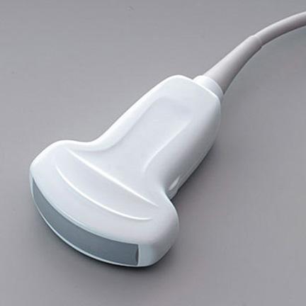
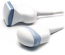
8 Ultrasound Instruments Ultrasound machines convert the echoes received by the transducer into visible dots, which form the anatomic image on ultrasound screen A linear transducer can produce parallel scan lines and rectangular display, called a linear scan Curved transducer yields a curvilinear scan and arcshaped image
9 Bioeffect and Safety Ultrasound application could produce a biologic effect can be characterized into two aspects: heating and mechanical. The generation of heat increases as ultrasound intensity or frequency is increased.

10

11 Curved transducer
12 linear transducer

13 Transvaginal transducer

14 4 4 D transducer

15 Ultrasound mode A mode is the oldest ultrasound modality Simple one-dimensional ultrasound image is generated

16 B mode Two-dimensional image of the area B mode can provide a cross sectional image through the area of interest

17 Doppler mode Produces a color-coded map of Doppler shifts superimposed onto a B-mode ultrasound image Blood flow direction depends on whether the motion is toward or away from the transducer. Selected by convention, red and blue colors provide information about the direction and velocity of the blood flow.

18 Power Doppler mode Up to five times more sensitive in detecting blood flow than color Doppler and it is less dependent on the scanning angle. Not provide any information on the direction and speed of blood flow

19 M mode A single beam in an ultrasound scan can be used to produce a picture with a motion signal Movement of a structure such as a heart valve can be depicted in a wave-like manner.
 to hyperechoic. Gas bubbles may also be seen. Doppler will demonstrate absence of central perfusion.")
20 Liver abscess Liver abscesses are typically poorly demarcated with a variable appearance, ranging from predominantly hypoechoic (still with some internal echoes however) to hyperechoic. Gas bubbles may also be seen. Doppler will demonstrate absence of central perfusion.


21 Cirrhosis


22 Acute cholecystitis


23 Pancreatitis

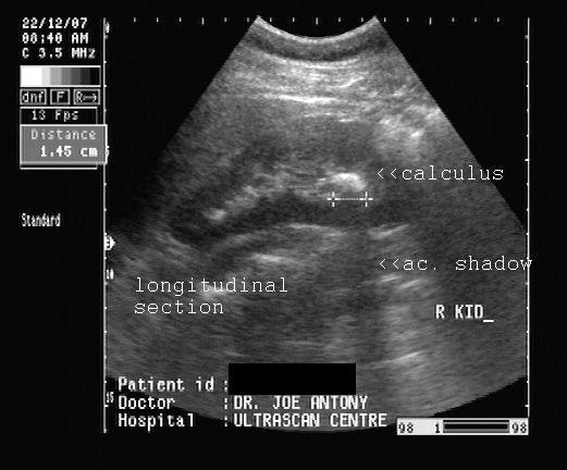


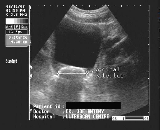
24 Renal calculi and hydronephrosis


25 Appendicitis


26 Torsion testis VS orchitis


27 Urinary bladder lesion


28 Other bowel lesion
Basic Physics of Ultrasound and Knobology
 WELCOME TO UTMB Basic Physics of Ultrasound and Knobology By Daneshvari Solanki, FRCA Laura B. McDaniel Distinguished Professor Anesthesiology and Pain Medicine University of Texas Medical Branch Galveston,
Principles of Ultrasound. Cara C. Prideaux, M.D. University of Utah PM&R Sports Medicine Fellow March 14, 2012
 Principles of Ultrasound Cara C. Prideaux, M.D. University of Utah PM&R Sports Medicine Fellow March 14, 2012 None Disclosures Outline Introduction Benefits and Limitations of US Ultrasound (US) Physics
1 Fundamentals. Basic Definitions and Physics Principles. Fundamentals
 1 To become versed in the language of ultrasonography, it is necessary to review some of the basic principles of physics. The wave physics principles of ordinary (i.e., audible) sound apply to ultrasound
Basic of Ultrasound Physics E FAST & Renal Examination. Dr Muhammad Umer Ihsan MBBS,MD, DCH CCPU,DDU1,FACEM
 Basic of Ultrasound Physics E FAST & Renal Examination Dr Muhammad Umer Ihsan MBBS,MD, DCH CCPU,DDU1,FACEM What is Sound? Sound is Mechanical pressure waves What is Ultrasound? Ultrasounds are sound waves
Physical Principles of Ultrasound
 Physical Principles of Ultrasound Grateful appreciation to Richard A. Lopchinsky, MD, FACS and Nancy H. Van Name, RDMS, RTR, and MarleneKattaron, RDMS 2000 UIC All Rights Reserved. Course Objectives Identify
Terminology Tissue Appearance
 By Marc Nielsen, MD Advantages/Disadvantages Generation of Image Ultrasound Machine/Transducer selection Modes of Ultrasound Terminology Tissue Appearance Scanning Technique Real-time Portable No ionizing
ULTRASOUND. OB/Gyn (Core) Ultrasound PIEZOELECTRIC EFFECT. Principles of Ultrasound Physics and Instrumentation. Nathan Pinkney, BS, CDOS
 Ultrasound PIEZOELECTRIC EFFECT. Principles of Ultrasound Physics and Instrumentation. Nathan Pinkney, BS, CDOS") 1 OB/Gyn (Core) Ultrasound Principles of Ultrasound Physics and Instrumentation Nathan Pinkney, BS, CDOS Philadelphia College of Osteopathic Medicine 2016 ULTRASOUND CATEGORIES OF SOUND INFRASOUND = below
1 OB/Gyn (Core) Ultrasound Principles of Ultrasound Physics and Instrumentation Nathan Pinkney, BS, CDOS Philadelphia College of Osteopathic Medicine 2016 ULTRASOUND CATEGORIES OF SOUND INFRASOUND = below
Introduction to Biomedical Imaging
 Alejandro Frangi, PhD Computational Imaging Lab Department of Information & Communication Technology Pompeu Fabra University www.cilab.upf.edu Basic principles. Comparison to X-rays Ultrasound > 20kHz
Ultrasound 10/1/2014. Basic Echocardiography for the Internist. Mechanical (sector) transducer Piezoelectric crystal moved through a sector sweep
 transducer Piezoelectric crystal moved through a sector sweep") Ultrasound Basic Echocardiography for the Internist Carol Gruver, MD, FACC UT Erlanger Cardiology Mechanical wave of compression and rarefaction Requires a medium for transmission Ultrasound frequency
Ultrasound Basic Echocardiography for the Internist Carol Gruver, MD, FACC UT Erlanger Cardiology Mechanical wave of compression and rarefaction Requires a medium for transmission Ultrasound frequency
ULTRASOUND IMAGING EE 472 F2018. Prof. Yasser Mostafa Kadah
 ULTRASOUND IMAGING EE 472 F2018 Prof. Yasser Mostafa Kadah www.k-space.org Recommended Textbook Diagnostic Ultrasound: Physics and Equipment, 2nd ed., by Peter R. Hoskins (Editor), Kevin Martin (Editor),
Point-of-Care Ultrasound: An Introduction
 Point-of-Care Ultrasound: An Introduction Delegation Teaching Package for Registered Respiratory Therapists and Anesthesia Assistants Developed by: Rob Bryan RRT, AA Edited by: Kelly Hassall RRT, FCSRT,
Ultrasound Physics & Terminology
 Ultrasound Physics & Terminology This module includes the following: Basic physics terms Basic principles of ultrasound Ultrasound terminology and terms Common artifacts seen Doppler principles Terms for
Preamble (disclaimer)
") Preamble (disclaimer) PHYSICS AND PRINCIPLES OF HEAD/NECK ULTRASOUND Joseph C. Sniezek, MD FACS LTC, MC, USA Otolaryngology/H&N Surgery Tripler Army Medical Center 1. I am not a physicist 2. ACS has recommended
Preamble (disclaimer) PHYSICS AND PRINCIPLES OF HEAD/NECK ULTRASOUND Joseph C. Sniezek, MD FACS LTC, MC, USA Otolaryngology/H&N Surgery Tripler Army Medical Center 1. I am not a physicist 2. ACS has recommended
The Physics of Ultrasound. The Physics of Ultrasound. Claus G. Roehrborn. Professor and Chairman. Ultrasound Physics
 The Physics of Ultrasound Pipe Organ 10-8000 Emission Dog 452-1080 Man 85-1100 Spectrum Bat 10,000-120,000 Porpoise 7000-120,000 Claus G. Roehrborn Professor and Chairman 10 20 Cycles per second Reception
Sound in medicine. CH.12. Dr.Rajaa أ.م.د. رجاء سهيل جنم جامعة تكريت كلية طب االسنان. General Properties of Sound
 CH.12. Dr.Rajaa Sound in medicine أ.م.د. رجاء سهيل جنم جامعة تكريت كلية Sound : It is the audible waves of frequency between 20 Hz and 20 khz. Infrasound : refers to the sound of frequency below the normal
Diploma of Medical Ultrasonography (DMU) Physical Principles of Ultrasound and Instrumentation Syllabus
 Physical Principles of Ultrasound and Instrumentation Syllabus") Diploma of Medical Ultrasonography (DMU) Physical Principles of Ultrasound and Instrumentation Syllabus Page 1 of 7 11/18 Candidates are expected to cover all of the content of this syllabus when preparing
Diploma of Medical Ultrasonography (DMU) Physical Principles of Ultrasound and Instrumentation Syllabus Page 1 of 7 11/18 Candidates are expected to cover all of the content of this syllabus when preparing
Ultrasound Principles cycle Frequency Wavelength Period Velocity
 ! Teresa S. Wu, MD, FACEP Director, EM Ultrasound Program & Fellowship Co-Director, Simulation Based Training Program & Fellowship Associate Program Director, EM Residency Program Maricopa Medical Center
Basic Ultrasound Physics Board Review Questions
 Basic Ultrasound Physics Board Review Questions Sidney K. Edelman, PhD ESP Ultrasound The Woodlands, TX Question 1 What is the wavelength of 2 MHz sound in soft tissue? 1. 1.54 mm 2. 0.75 mm 3. 0.75 cm
Ultrasound. Principles of Medical Imaging. Contents. Prof. Dr. Philippe Cattin. MIAC, University of Basel. Oct 17th, 2016
 Ultrasound Principles of Medical Imaging Prof. Dr. Philippe Cattin MIAC, University of Basel Contents Abstract 1 Image Generation Echography A-Mode B-Mode M-Mode 2.5D Ultrasound 3D Ultrasound 4D Ultrasound
Ultrasound Physics and Knobology Alan Macfarlane. Consultant Anaesthetist Glasgow Royal Infirmary
 Ultrasound Physics and Knobology Alan Macfarlane Consultant Anaesthetist Glasgow Royal Infirmary RAPM 2009; 34: 40-46 Ultrasound Proficiency Understanding US image generation and device operation Image
Introduction & Physics of ED Ultrasound. Objectives. What? - Limited Studies. Who? - ED Docs
 Introduction & Physics of ED Ultrasound Martine Sargent, MD Ultrasound Director, Assistant Professor UCSF Department of Emergency Medicine San Francisco General Hospital & Trauma Center Objectives Who?
Ultrasound Physics & Doppler
 Ultrasound Physics & Doppler Endocrine University 2018 Mark Lupo, MD, FACE, ECNU Objectives Review the essential components of ultrasound physics in neck sonography Demonstrate the importance of ultrasound
Diagnostic approach to heart disease
 Diagnostic approach to heart disease Initial work up History Physical exam Chest radiographs ECG Special studies Echocardiography Cardiac catheterization Echocardiography principles Technique of producing
Ultrasound Knobology
 Ultrasound Knobology Raj Dasgupta MD, FACP, FCCP, FASSM Assistant Professor of Clinical Medicine Pulmonary / Critical Care / Sleep Medicine University of Southern California (USC) Objectives Physics of
Basic Physics of Ultrasound in Transesophageal Echocardiography
 SPECIAL ARTICLE IJUTPC Basic Physics of Ultrasound in Transesophageal Echocardiography Basic Physics of Ultrasound in Transesophageal Echocardiography 1 Mary Korula, 2 Ravi Hebballi 1 Senior Consultant,
DIGITAL IMAGE PROCESSING IN ULTRASOUND IMAGES
 DIGITAL IMAGE PROCESSING IN ULTRASOUND IMAGES Kamaljeet Kaur Computer Science & Engineering Department Guru Nanak Dev Engg. College, Ludhiana. Punjab-India meetk.89@gmail.com ABSTRACT-- Image processing
Introduction to Ultrasound Guided Region Anesthesia
 Introduction to Ultrasound Guided Region Anesthesia Brian D. Sites, MD Dept of Anesthesiology Dartmouth-Hitchcock Medical Center INTRODUCTION Welcome to Introduction to Ultrasound Guided Regional Anesthesia.
Medical Imaging. By: Engr. Joseph Ronald Canedo
 Medical Imaging By: Engr. Joseph Ronald Canedo Medical Sonography (Ultrasound) is an ultrasound-based diagnostic imaging technique used to visualize muscles and internal organs, their size, structures
Supplement (videos)
") Supplement (videos) Ruben s tube (sound): http://www.youtube.com/watch?v=gpcquuwqayw Doppler US (diagnostic use): http://www.youtube.com/watch?v=fgxzg-j_hfw http://www.youtube.com/watch?v=upsmenyoju8 High
Supplement (videos) Ruben s tube (sound): http://www.youtube.com/watch?v=gpcquuwqayw Doppler US (diagnostic use): http://www.youtube.com/watch?v=fgxzg-j_hfw http://www.youtube.com/watch?v=upsmenyoju8 High
Ultrasound in Anesthesia: Applying Scientific Principles to Clinical Practice
 AANA Journal Course Update for Nurse Anesthetists 3 6 CE Credits* Ultrasound in Anesthesia: Applying Scientific Principles to Clinical Practice Christian R. Falyar, CRNA, DNAP The use of ultrasound as
Dr Emma Chung. Safety first - Physical principles for excellent imaging
 Safety first - Physical principles for excellent imaging Dr Emma Chung Lecturer in Medical Physics, University of Leicester Clinical Scientist, University Hospitals of Leicester NHS Trust Thanks to Caroline
Ultrasound guidance in regional anesthesia has
 Ultrasound and Regional Anesthesia Artifacts and Pitfall Errors Associated With Ultrasound-Guided Regional Anesthesia. Part I: Understanding the Basic Principles of Ultrasound Physics and Machine Operations
The 2 nd Cambridge Advanced Emergency Ultrasound Course
 The 2 nd Cambridge Advanced Emergency Ultrasound Course Addenbrooke s Hospital Cambridge Sept 2008 1 2 Faculty! UK! USA! Australia! Toshiba! Emergency Medicine! Radiology 3 Programme! Day 1 Introduction
Concepts of Imaging and Knobology
 Concepts of Imaging and Knobology Pravin Patil, MD FACC FASE Associate Professor of Medicine Director, Cardiovascular Disease Training Program Lewis Katz School of Medicine at Temple University Disclosures
High resolution ultrasound scanner for skin imaging
 High resolution ultrasound scanner for skin imaging Christine Turlat Sales Director Atys medical 17 Parc d Arbora 69510 SOUCIEU EN JARREST Atys company Principle of ultrasound imaging DERMCUP Normal image
ULTRASOUND NOMENCLATURE
 Chapter 1: Ultrasound Nomenclature, Image Orientation, and Basic Instrumentation CYNTHIA SIKOWSKI Ultrasound waves are sound waves that have a frequency exceeding 20,000 Hz. When sound waves are transmitted
Descriptions of NDT Projects Fall 2004 October 31, 2004
 Descriptions of NDT Projects Fall 2004 October 31, 2004 Introduction There are two separate NDT labs in Magister: ULTRA for ultrasound and EDDY for eddy current. Both labs are equipped with mechanical
WELCOME! Introduction to Bedside Ultrasound
 WELCOME! Introduction to Bedside Ultrasound TEACHERS University of California-Irvine School of Medicine Nathan Molina nathan.d.molina@gmail.com Trevor Plescia taplescia90@gmail.com Jack Silva jpsilva42@gmail.com
Table of contents. Foreword. Preface. 1 Introduction Historical Perspective 00
 Table of contents Foreword Preface 1 Introduction 00 1.1 Historical Perspective 00 2 Fundamentals of musculoskeletal ultrasound 00 2.1 Frequency and wavelength 00 2.2 Generating ultrasound waves 00 2.3
1. Fig. 1 shows data for the intensity of a parallel beam of X-rays after penetration through varying thicknesses of a material
 1. Fig. 1 shows data for the intensity of a parallel beam of X-rays after penetration through varying thicknesses of a material. intensity / MW m 2 thickness / mm 0.91 0.40 0.69 0.80 0.52 1.20 0.40 1.60
Application of Phased Array Radar Theory to Ultrasonic Linear Array Medical Imaging System
 Application of Phased Array Radar Theory to Ultrasonic Linear Array Medical Imaging System R. K. Saha, S. Karmakar, S. Saha, M. Roy, S. Sarkar and S.K. Sen Microelectronics Division, Saha Institute of
Lesson 07: Ultrasound Transducers. This lesson contains 73 slides plus 16 multiple-choice questions.
 Lesson 07: Ultrasound Transducers This lesson contains 73 slides plus 16 multiple-choice questions. This lesson was derived from pages 33 through 42 in the textbook: Ultrasound Transducers Ultrasound Transducers
Ultrasound: Past and Present. Lecturer: Dr. John M Hudson, PhD
 Ultrasound: Past and Present Lecturer: Dr. John M Hudson, PhD Disclosures 2 No conflicts of interest to declare Course Outline 3 1. Survey of ultrasound physics & applications 2. (Sep 21) 3. (Sep 28) 4.
Exam Practice Guide. Units 1 & 2 Physics: Detailed Study 5 - Investigations: Medical physics Examination Questions
 Exam Practice Guide Units 1 & 2 Physics: Detailed Study 5 - Investigations: Medical physics Examination Questions Key Features: 22 original examination style questions on all examinable topics. Full solutions
What is Ultrasound? What is Ultrasound? B A. Basic Principles of Ultrasound. Basic Principles of Ultrasound. Basic Principles of Ultrasound
 Introduction to Ultrasound Principles Mani Montazemi, RDMS Baylor College of Medicine Division of Maternal-Fetal Medicine Department of Obstetrics and Gynecology Manager, Maternal Fetal Center Imaging
Ultrasonic Testing Level I:
 Ultrasonic Testing Level I: 1- Sound Wave - Introduction - ASNT Level I - Sound Wave Propagation - Velocity / Frequency / Wave Length - Acoustic Impedance - Energy / Intensity 2- Ultrasound Wave Modes
What is Ultrasound? Resolution Image production Attenuation Imaging modes Ultrasound artifacts... 7
 What is Ultrasound?... 1 Resolution... 3 Image production... 3 Attenuation... 4 Imaging modes... 5 Ultrasound artifacts... 7 0 What is Ultrasound? High frequency sound of frequencies 2-50 MHz is used in
Emergency Medicine Interest Group (EMIG) 2016
 2016") Emergency Medicine Interest Group (EMIG) 2016 Welcome to the flipped classroom (learning objectives summary) for the 2016 Emergency Medicine Interest Group (EMIG) Procedures Workshop. Overview - Tuesday
Emergency Medicine Interest Group (EMIG) 2016 Welcome to the flipped classroom (learning objectives summary) for the 2016 Emergency Medicine Interest Group (EMIG) Procedures Workshop. Overview - Tuesday
Tissue Strain Analytics Virtual Touch Tissue Imaging and Quantification
 Whitepaper Tissue Strain Analytics Virtual Touch Tissue Imaging and Quantification ACUSON S2000 Ultrasound System Answers for life. Page 1 Tissue Strain Analytics: Virtual Touch Tissue Imaging and Quantification
Development of Ultrasound Based Techniques for Measuring Skeletal Muscle Motion
 Development of Ultrasound Based Techniques for Measuring Skeletal Muscle Motion Jason Silver August 26, 2009 Presentation Outline Introduction Thesis Objectives Mathematical Model and Principles Methods
Pulse-Echo Ultrasound Imaging. Resolution in Ultrasound Imaging. Doppler Ultrasound. Resolution vs Penetration. Medical Imaging (EL582/BE620/GA4426)
") Medical Imaging (EL582/BE620/GA4426) Pulse-Echo Ultrasound Imaging Ultrasound Imaging Lecture 2 Daniel (Dan) Turnbull, Ph.D. Skirball Institute and Dept of Radiology NYU School of Medicine (daniel.turnbull@med.nyu.edu)
Medical Imaging (EL582/BE620/GA4426) Pulse-Echo Ultrasound Imaging Ultrasound Imaging Lecture 2 Daniel (Dan) Turnbull, Ph.D. Skirball Institute and Dept of Radiology NYU School of Medicine (daniel.turnbull@med.nyu.edu)
Children's (Pediatric) Ultrasound - Abdomen
 Ultrasound - Abdomen") Scan for mobile link. Children's (Pediatric) Ultrasound - Abdomen Children s (pediatric) ultrasound imaging of the abdomen is a safe, noninvasive test that uses sound waves to produce a clear picture of
Scan for mobile link. Children's (Pediatric) Ultrasound - Abdomen Children s (pediatric) ultrasound imaging of the abdomen is a safe, noninvasive test that uses sound waves to produce a clear picture of
3/20/2017. Disclosures. Ultrasound Fundamentals. Ultrasound Fundamentals. Bone Anatomy. Tissue Characteristics
 Disclosures Images of ultrasound equipment in this presentation are not an endorsement Fundamentals of Musculoskeletal Ultrasound Physics and Knobology Shane A. Shapiro, M.D. Assistant Professor Orthopedic
An Overview of Ultrasound Testing For Lesion Detection in Human Kidney
 Journal of Tomography System & Sensors Application Vol.1, Issue 1, June 2018 An Overview of Ultrasound Testing For Lesion Detection in Human Kidney Aina Fadhilah Abd Rahim 1, Zawin Najah Abd Halim 1, Jaysuman
Confident and Conclusive Diagnosis with Flexible Solutions
 Internal Medicine Confident and Conclusive Diagnosis with Flexible Solutions ALPINION MEDICAL SYSTEMS We are Ultrasound Professionals Ultrasound is widely used as the gold-standard for internal medicine
FOR IMMEDIATE RELEASE
 FOR IMMEDIATE RELEASE Hitachi to launch ALOKA ARIETTA 850, the flagship model of the ARIETTA series diagnostic of ultrasound platforms This premium model features superior image quality, seamless workflow,
DOW-RAD, DOW DIAGNOSTIC COMPLEX, DUHS TRAINING PROGRAM HANDBOOK 2013
 DOW-RAD, DOW DIAGNOSTIC COMPLEX, DUHS TRAINING PROGRAM HANDBOOK 2013 CERTIFICATE COURSE INVASCULAR/DOPPLER ULTRASOUND: Introduction: Ultrasound is an evolving technology with wide spectrum application
Underwater Acoustic Measurements in Megahertz Frequency Range.
 Underwater Acoustic Measurements in Megahertz Frequency Range. Current State and Prospects of Development in Russia Alexander M. Enyakov,, Many medical applications of underwater acoustic measurements
Image optimization for critical care US
 Image optimization for critical care US 1 Although we assume you are already familiar with focused US in the ED, it might not hurt to revise the basics: Machines & transducers US appearance of normal tissues
Human Systems. Technology - Ultrasounds
 Human Systems Technology - Ultrasounds What is General Ultrasound Imaging? Ultrasound imaging, also called ultrasound scanning or sonography, involves exposing part of the body to high-frequency sound
Linear Ultrasonic Wave Propagation in Biological Tissues
 Indian Journal of Biomechanics: Special Issue (NCBM 7-8 March 29) Linear Ultrasonic Wave Propagation in Biological Tissues Narendra D Londhe R. S. Anand 2, 2 Electrical Engineering Department, IIT Roorkee,
Lesson 03: Sound Wave Propagation and Reflection. This lesson contains 15 slides plus 14 multiple-choice questions.
 Lesson 03: Sound Wave Propagation and Reflection This lesson contains 15 slides plus 14 multiple-choice questions. Accompanying text for the slides in this lesson can be found on pages 8 through 14 in
The table below shows the density and velocity of waves in two different substances. Density / kg m 3 Velocity / m s 1
 Q1.(a) When ultrasound is incident at an interface between two different media some energy is transmitted and some is reflected. The ratio of the reflected energy intensity I r to the incident energy intensity
Chapter 1. Principles of medical ultrasound. Overview. Background history: first steps to the piezo-electric effect.
 Chapter 1 Principles of medical ultrasound GRAHAM ARTHURS, PATRICK HILL AND TREVOR FRANKEL Overview This chapter provides an introduction to the ultrasound process for trainees in anesthesia and other
Endobronchial Ultrasound
 Endobronchial Ultrasound l Armin Ernst Editors Felix J.F. Herth Endobronchial Ultrasound An Atlas and Practical Guide 13 Editors Armin Ernst, MD Chief, Section of Interventional Pulmonology; Director,
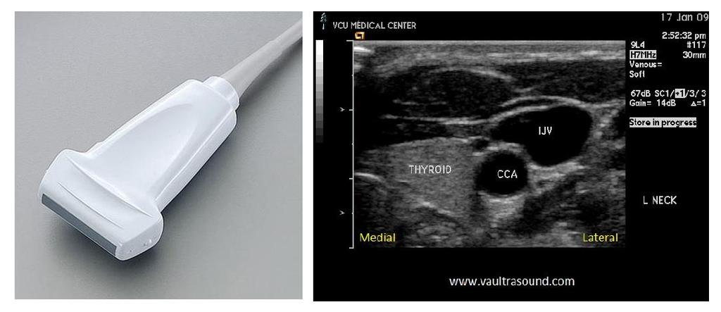
Ultrasonography of the Neck as an Adjunct to FNA. Nicole Massoll M.D.
 Ultrasonography of the Neck as an Adjunct to FNA Nicole Massoll M.D. Basic Features of Head and Neck Ultrasound and Anatomy Nicole Massoll M.D. University of Arkansas for Medical Sciences, Little Rock
CONTENTS. Test Number cpd Tanya Reynolds (Nat. Dip. Diag. Rad., B. Tech. Diag. Rad., B. Tech. Ultrasound)
") CONTENTS page 1-15 page 16 BASIC 2-DIMENSIONAL ULTRASOUND PRINCIPLES Multiple Choice Test Test Number cpd 41640 Tanya Reynolds (Nat. Dip. Diag. Rad., B. Tech. Diag. Rad., B. Tech. Ultrasound) Tanya is
CONTENTS page 1-15 page 16 BASIC 2-DIMENSIONAL ULTRASOUND PRINCIPLES Multiple Choice Test Test Number cpd 41640 Tanya Reynolds (Nat. Dip. Diag. Rad., B. Tech. Diag. Rad., B. Tech. Ultrasound) Tanya is
General Ultrasound. What is General Ultrasound Imaging?
 Scan for mobile link. General Ultrasound Ultrasound imaging uses sound waves to produce pictures of the inside of the body. It is used to help diagnose the causes of pain, swelling and infection in the
Basic Training Programme. 16 Februrary 2018, ROTTERDAM. Pre and Post-Course Test Answers
 Basic Training Programme 16 Februrary 2018, ROTTERDAM Pre and Post-Course Test Answers Your details: Name: Conference registration number/ BT delegate number: Email address: Are you already performing
Chapter 3 Physical Principles of Ultrasound of the Male Genitalia
 Chapter 3 Physical Principles of Ultrasound of the Male Genitalia Bruce R. Gilbert and Pat Fox Fulgham Introduction The value of ultrasound evaluation of the male genitalia depends, in large part, on the
S1Stephanie J. Doniger
 Section 1 Ultrasound fundamentals Introduction S1Stephanie J. Doniger Pediatric Emergency Medicine is a relatively new field of medicine developed in the 1980s. Since its inception, several advancements
Principles of echocardiography for the anesthesiologist.
 Principles of echocardiography for the anesthesiologist. Nikolaos Skubas, MD PhD Abstract Ultrasound-based diagnostic techniques are now part of the cardiological patients chart, while echocardiography
4.17. RESEARCHING MODELS WITH AN ULTRASONIC ECHOSCOPE
 4.17. RESEARCHING MODELS WITH AN ULTRASONIC ECHOSCOPE Purpose of experiment Determine the main characteristics of ultrasound waves, and the distances and positions of models using an ultrasonic echoscope.
Chapter 14. Imaging Artifacts
 Chapter 14 Image Artifacts The complex physical interactions that occur between an ultrasound beam and human anatomy and the intricate and sophisticated technological components of a sonographic imaging
Introduction. Cardiac Imaging Modalities MRI. Overview. MRI (Continued) MRI (Continued) Arnaud Bistoquet 12/19/03
 MRI (Continued) Arnaud Bistoquet 12/19/03") Introduction Cardiac Imaging Modalities Arnaud Bistoquet 12/19/03 Coronary heart disease: the vessels that supply oxygen-carrying blood to the heart, become narrowed and unable to carry a normal amount
Introduction Cardiac Imaging Modalities Arnaud Bistoquet 12/19/03 Coronary heart disease: the vessels that supply oxygen-carrying blood to the heart, become narrowed and unable to carry a normal amount
Optimising your Doppler settings for an accurate PI. Alison McGuinness Mid Yorks Hospitals
 Optimising your Doppler settings for an accurate PI Alison McGuinness Mid Yorks Hospitals Applications Both maternal uterine and fetal circulations can be studied with doppler sonography Uterine arteries
4.17. RESEARCHING MODELS WITH AN ULTRASONIC ECHOSCOPE
 4.17. RESEARCHING MODELS WITH AN ULTRASONIC ECHOSCOPE Purpose of experiment Determine the main characteristics of ultrasound waves, and the distances and positions of models using an ultrasonic echoscope.
Do you think the ultrasound experiments on mice were justified?
 Q1.(a) Explain what ultrasound is. (b) Ultrasound is used for pre-natal scanning. This is much safer than using X-rays. However, doctors were only sure ultrasound was safe after experiments on mice. Do
Doppler Basic & Hemodynamic Calculations
 Doppler Basic & Hemodynamic Calculations August 19, 2017 Smonporn Boonyaratavej MD Division of Cardiology, Department of Medicine Chulalongkorn University Cardiac Center, King Chulalongkorn Memorial Hospital
17.4 Sound and Hearing
 You can identify sounds without seeing them because sound waves carry information to your ears. People who work in places where sound is very loud need to protect their hearing. Properties of Sound Waves
Ultrasound in Medicine
 Ultrasound in Medicine Experimental Equipment for Medical Education Universities Colleges Medical Schools Medical and Med-Technical Training Education can befun! WELCOME TO GAMPT Devices and accessories
Point- of- care Ultrasound Workshop
 Point- of- care Ultrasound Workshop January 13, 2011 Shailaja J. Hayden, MD Fellow, Pulmonary & CriHcal Care Medicine Disclosures None. The VA Puget Sound Health Care System is accredited by the Washington
Roaa M.Hussein Professor, Department of Physics, College of Science, Ramadi, Iraq. Abstract:
 Abstract: Ultrasound Waves Employment in the Medical Diagnostic for Lıver and Gallbladder Faik H. Antar Professor, Department of Physics, College of Science, AL- Anbar University, Ramadi, Iraq. Ultrasound
2015 ARDMS Sonography Principles & Instrumentation Job Task Analysis Summary Report
 P a g e 1 2015 ARDMS Sonography Principles & Instrumentation Job Task Analysis Summary Report American Registry for Diagnostic Medical Sonography (ARDMS) P a g e 2 Table of Contents ABOUT THE REPORT...
Flaw Assessment Using Shear wave Phased array Ultrasonic Transducer
 18th World Conference on Nondestructive Testing, 16-20 April 2012, Durban, South Africa Flaw Assessment Using Shear wave Phased array Ultrasonic Transducer Byungsik YOON AUTHOR 1, Hee-Jong LEE CO-AUTHOR
Evaluation of the Quality of Thick Fibre Composites Using Immersion and Air- Coupled Ultrasonic Techniques
 ECNDT 2006 - We.1.6.4 Evaluation of the Quality of Thick Fibre Composites Using Immersion and Air- Coupled Ultrasonic Techniques Kaj K. BORUM, Risø National Laboratory, Materials Research Department, Roskilde,
Vikram Dogra, M.D. Professor of Radiology, Urology & BME Department of Imaging Sciences University Of Rochester Medical Center
 Ultrasound of the Scrotum Vikram Dogra, M.D. Professor of Radiology, Urology & BME Department of Imaging Sciences University Of Rochester Medical Center Etiologies of Acute Scrotal Pain Epididymitis/Orchitis
Guide to Small Animal Vascular Imaging using the Vevo 770 Micro-Ultrasound System
 Guide to Small Animal Vascular Imaging using the Vevo 770 Micro-Ultrasound System January 2007 Objectives: After completion of this module, the participant will be able to accomplish the following: Understand
Job Task Analysis for ARDMS Abdomen Data Collected: June 30, 2011
 Job Task Analysis for ARDMS Abdomen Data Collected: June 30, 2011 Reported: Analysis Summary for: Abdomen Examination Survey Dates 06/13/2011-06/26/2011 Invited Respondents 6,000 Surveys with Demographics
Physical Principles of Ultrasound
 Physical Principles of Ultrasound Pat F. Fulgham 2 Introduction The use of ultrasound is fundamental to the practice of urology. In order for urologists to best use this technology on behalf of their patients,
General Ultrasound. What is General Ultrasound Imaging?
 Scan for mobile link. General Ultrasound What is General Ultrasound Imaging? Ultrasound is safe and painless, and produces pictures of the inside of the body using sound waves. Ultrasound imaging, also
Abdominal Ultrasound
 Abdominal Ultrasound What is Ultrasound Imaging of the Abdomen? What are some common uses of the procedure? How should I prepare? What does the equipment look like? How does the procedure work? How is
Breast Imaging Essentials
 Breast Imaging Essentials Module 9 Transcript 2016 ASRT. All rights reserved. Breast Imaging Essentials Module 9 Breast Ultrasound 1. ASRT Animation 2. Welcome Welcome to Module 9 of Breast Imaging Essentials
iu22 Liver Shear Wave ElastPQ
 iu22 Liver Shear Wave ElastPQ Clinical Case Study Lucy Wang Clinical Application Specialist ASEAN Case Study: History: 58-year-old male patient, hepatitis B virus (HBV) carrier, with non clinical symptoms
Abdomen Sonography Examination Content Outline
 Abdomen Sonography Examination Content Outline (Outline Summary) # Domain Subdomain Percentage 1 2 3 Anatomy, Perfusion, and Function Pathology, Vascular Abnormalities, Trauma, and Postoperative Anatomy
An abdominal ultrasound produces a picture of the organs and other structures in the upper abdomen.
 Scan for mobile link. Ultrasound - Abdomen Ultrasound imaging of the abdomen uses sound waves to produce pictures of the structures within the upper abdomen. It is used to help diagnose pain or distention
Numerical Modelling of Ultrasonic Phased Array Transducers and Their Application
 ECNDT 2006 - Mo.2.7.5 Numerical Modelling of Ultrasonic Phased Array Transducers and Their Application Prashanth Kumar CHINTA, René MARKLEIN, University of Kassel, Department of Electrical Engineering
Ultrasound Applied Physics
 Ultrasound Applied Physics University of Toronto Department of Medical Imaging Applied Physics Mini-Course #3 2016 Ultrasound Laboratory Manual and Examination Booklet 1/21/2016 Ultrasound Applied Physics
The Value. Ultrasound System. Professional Concept for. Women s. Health
 The Value Innovative Ultrasound System Professional Concept for Women s Health 9 Beyond Women s Healthcare E-CUBE 9 - Powerful Imaging Performance for All Women s Health Diagnostic Applications Diagnostic
(sound with frequency) above hertz / 20 khz. frequencies above (human) audible range. (sound) cannot be heard by humans 2
 above hertz / 20 khz. frequencies above (human) audible range. (sound) cannot be heard by humans 2") M. (a) any two from: (sound with frequency) above 20 000 hertz / 20 khz frequencies above (human) audible range (sound) cannot be heard by humans 2 either two appropriate points gain mark each either both
M. (a) any two from: (sound with frequency) above 20 000 hertz / 20 khz frequencies above (human) audible range (sound) cannot be heard by humans 2 either two appropriate points gain mark each either both