Introduction to Musculoskeletal Ultrasound. Disclosures. Evidence Based Medicine Key References 8/30/2017
|
|
|
- Tamsin Nicholson
- 6 years ago
- Views:
Transcription
1 Introduction to Musculoskeletal Ultrasound Johannes Roth MD, PhD, FRCPC, RhMSUS Professor of Pediatrics University of Ottawa Gurjit S Kaeley MBBS, MRCP, RhMSUS Professor of Medicine Division Chief Director of Musculoskeletal Ultrasound University of Florida College of Medicine, Jacksonville. Disclosures Johannes Roth MD, PhD, FRCPC, RhMSUS Nothing to disclose Gurjit S Kaeley MBBS, MRCP Unrestricted equipment loan from Esaote Evidence Based Medicine Key References Stefano Bianchi, Carlo Martinoli.Ultrasound of the Musculoskeletal System. Springer M Backhaus et al. Guidelines for musculoskeletal ultrasound in Rheumatology. Ann Rheum Dis 2001;60: Wakefield RJ et al. Proceedings from the OMERACT Special Interest Group for Musculoskeletal Ultrasound including definitions for ultrasonographic pathology. J Rheumatol 2005; 32:
2 Objectives After attending this session you should be able to Understand the basics of ultrasound scanning Recognize sonographic appearances of skin, tendons, muscles, nerves, bone, fluid collections and inflammatory lesions. Able to perform a basic knee sonographic exam Understand the principles of ultrasound guided joint injections Understand the application of musculoskeletal ultrasound to rheumatological practice. Agenda Introduction Basic Physics Concepts Terminology Basic principles of ultrasound scanning Identify normal tissues Sonography of the Knee Musculoskeletal Ultrasound in Rheumatological Practice Guided interventions Billing and Coding Ultrasound Equipment Ultrasound machine Transducers: High frequency (High resolution, less penetration) Low frequency (Low resolution, deep penetration) Gel Transducer cover Computer/printer 2
 Image information is provided by energy of waves reflected from")
 Medical ultrasound commonly is in the 5-22 MHz range.")
3 What is ultrasound? Ultrasound imaging uses transmission and reflection of high frequency longitudinal mechanical waves (ultra sonic waves) Image information is provided by energy of waves reflected from surfaces between different tissues Wave propagation speed allows calculation of distance Frequency Human hearing is in the 20-20,000 Hz range. Frequency of an ultrasound wave is above 20,000 Hz (20 KHz) Medical ultrasound commonly is in the 5-22 MHz range. Generation of ultrasound beam 1) conversion of electrical to mechanical (sound) = converse piezoelectric effect (Gabriel Lippman 1881). 2) Conversion sound to electrical energy = piezoelectric effect (Pierre Curie 1880) one piezoelectric crystal = ultrasound wave, summation of waves=ultrasound beam 3

4 Reflection Strong specular reflections bright dots (hyperechoic), gallstone Weak diffuse reflections grey dots (hypoechoic), solid organs No reflection dark dots (anechoic), fluid Deep structures hypoechoic, attenuation limits beam transmission Reflection Reflection at the interface of two tissues depends on Difference in the acoustic impedance of the tissues Angle of incidence of the sound beam Reflection of sound increases with the increase in difference in acoustic impedance. Typical reflection at soft tissue interfaces 99.9% soft tissue air 40% muscle and bone < 1% soft tissue to soft tissue Terminology Echoic - white Hyperechoic white/grey Isoechoic - grey Hypoechoic grey/dark grey Anechoic - black 4

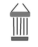
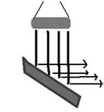
5 Direction of the beam Anisotropy Ultrasound Grayscale or B-Mode Doppler. 5
 The observed frequency of")


6 Doppler Effect Christian Doppler ( ) The observed frequency of a wave depends on the relative speed of the source and the observer. Born in Salzburg Professor of Mathematics and Physics in Prague, Banská Štiavnica/Slovakia and Vienna Doppler Effect Doppler detects the Frequency shift ft fr 6


7 Doppler equation 2 ft V cosine Frequency (fd) = ft - fr = C Velocity V and Angle dependent Cosine 90 degrees = 0 Detects only movement towards or away from the transducer Doppler modalities Color Doppler Angle dependent Determines mean velocity Image relates to velocity Power Doppler Angle independent can be displayed with colour Summarizes all signals Signal relates to number of erythrocytes Enough Physics Let s get started 7


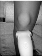
8 How to hold the probe The need for gel a lot of gel!!! 8


9 Pressure and Gel! Instrument settings Knobology after choosing your preset adjust: Frequency Depth Focus Gain PRF for Doppler Then say cheese and push the freeze button The Impact of Ultrasonic Frequency on Attenuation Frequency = Attenuation; Attenuation = Penetration 9

10 Frequency: Penetration vs Resolution 10 MHz 18 MHz DEPTH Should be adjusted so area of interest is centred, seen completely but is not too large or too small. Focus Should be at the level of the area of interest 10

11 GAIN Adjusts the amplification of echoes on the monitor Brightness Compensates for attenuation Will increase brightness of the entire image TGC TGC = Time Gain Compensation Adjusts the Gain at specific depths Agenda Introduction Identify normal tissues Skin and subcutaneous tissue Muscles Nerves Tendons and demonstrate anisotropy Bone and cartilage Blood vessels Joint structure Selected artifacts Sonography of the Knee Musculoskeletal Ultrasound in Rheumatological Practice Guided interventions Billing and Coding 11


12 Which tissues are important? Fluid anechoic, posterior enhancement, compressible 0.2 cm Fluid Compression (Doppler!) Can be more difficult in deeper structures 12


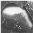
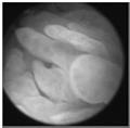
13 Cartilage Homogeneous anechoic layer lining the bony cortex with superficial and deep margins that characteristically appear thin, sharp, continuous and regularly hyperechoic Careful technique and positioning is everything! Blood Vessels 13
")

 with a thin")
 halo in transverse or thin")


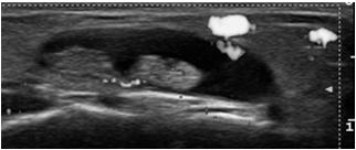
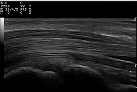
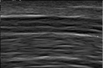
14 Tendon and tendon sheath Hyperechoic (relative to subdermal fat) fibrillar pattern (ie, hyperechoic parallel lines in longitudinal planes and hyperechoic dots in transverse planes) with a thin hypoechoic (relative to tendon fibres) halo in transverse or thin hypoechoic lines above and below the tendon in longitudinal views. Tendon fibrillar Long Trans Enthesis Achilles Tendon Fat Pad Bursa / Fibrocartilag Calcaneus Fibrocartilage 14
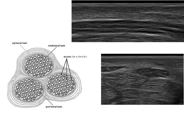
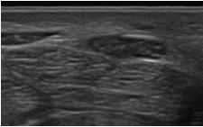
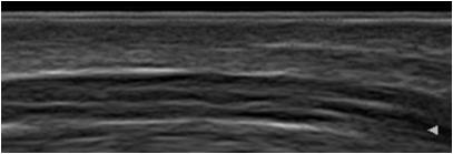
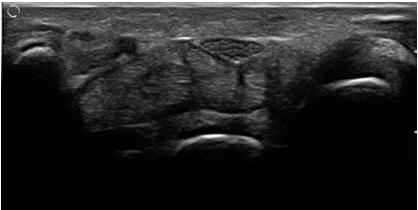
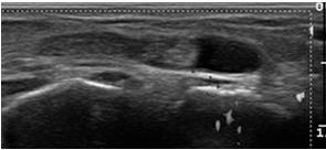
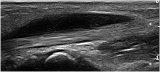
15 Nerve fascicular Nerve fascicular Tendon and Nerve Median nerve Median N Flexor tendon Flexor tendon Flexor T 15


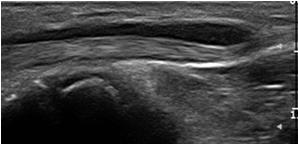
16 Bone and Muscle And now try it yourself!!! Practical Session Objectives Identify Bone Joint Structures Muscle Tendons Nerve Demonstrate Anisotropy Artifact 16
17 Musculoskeletal Ultrasound of The Knee Objectives Scan familiar anatomy This session is aimed to give you a taste of what you can do with familiar knowledge. We will be asking you to perform limited scanning. Full standard scans are not difficult to perform once basic training has been acquired Guidelines for Musculoskeletal Ultrasound in Rheumatology Knee Standard scans of the knee 1. Suprapatellar longitudinal scan in neutral position 2. Suprapatellar transverse scan in neutral position 3. Suprapatellar transverse scan in maximal flexion 4. Infrapatellar longitudinal scan 5. Infrapatellar transverse scan 6. Medial longitudinal scan 7. Lateral longitudinal scan 8. Posterior medial longitudinal scan 9. Posterior lateral longitudinal scan 10. Posterior transverse scan 17


 30")

18 Knee - Synovial Spaces All illustrations in this presentation adapted from C Martinoli Knee Ligaments and Tendons BF ITB QT SM LCL LCL MCL MCL ST Lateral Anterior Medial Suprapatellar longitudinal scan (30 Flexion) 30 Flexion Different positions might be necessary for Doppler!!! straight 18


")


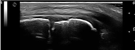
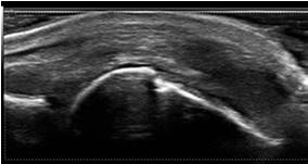
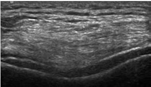
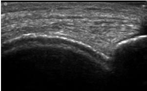
19 Suprapatellar longitudinal scan (30 Flexion) Fat pad Patella Fat pad Recess Femur Growth Plate Schmidt-WA ARD 2004: Sagittal midline 2.4 ± 1.2 mm Suprapatellar transverse scan (30 flexion) Pat Tendon Tendon Femur Bone Suprapatellar transverse scan in maximal flexion Schmidt-WA ARD 2004: Cartilage 3.1 ± 0.7 mm 19


20 The parapatellar recess Lateral Medial lateral retinaculum PATELLA PATELLA FEMUR FEMUR medial retinaculum Infrapatellar long prox Location of Bursa Tendon Patella Hoffa Schmidt-WA ARD 2004: Patella tendon 2 cm distal Patella 3.2 ± 0.7 mm Infrapatellar longitudinal distal Bursa infrapatellaris superficialis Hoffa Bursa infrapat profunda Tibia 20
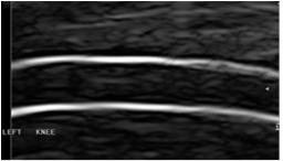
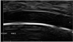
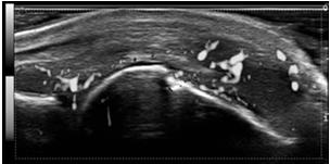
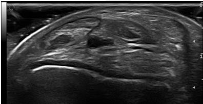
21 Infrapatellar transverse scan Patella Tendon Condyle Gout Double Contour Sign Knee: Maximum Flexion Transverse Long Double Contour Interface Sign Chondrocalcinosis 21

22 Practical Session Objectives Scan and identify Quadriceps tendon in orthogonal planes Patellar Ligament in Orthogonal planes Inspect the femoral condyle cartilage with the knee in maximal flexion. Agenda Introduction Identify normal tissues Sonography of the Knee Musculoskeletal Ultrasound in Rheumatological Practice Guided interventions Billing and Coding US Guided Injection Region: Suprapatellar Recess Diag copyright Primal and Servier 22


23 Accessing the Recess Confirming the Position Longitudenally Injecting Under Visualization 23

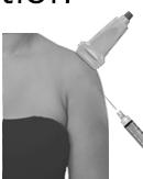
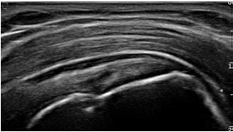
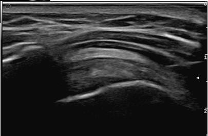
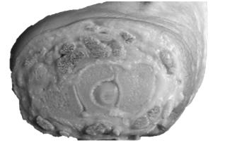
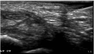

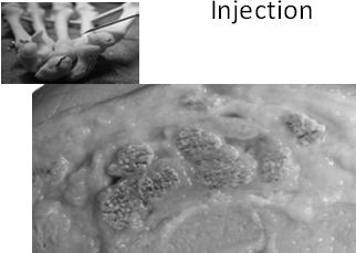
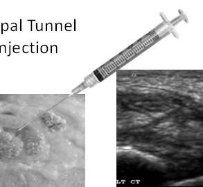
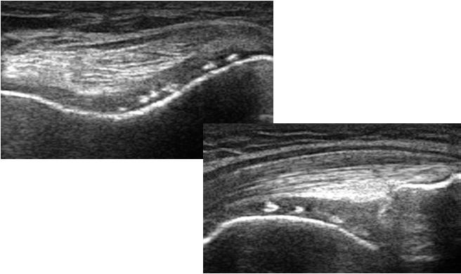
24 Acute Podagra: 1 st MTP Aspiration US Guided SASD Injection Deltoid Fat Stripe SASD SST SASD Subacromial subdeltoid bursa Carpal Tunnel Injection 24


25 2 nd MCP INJECTION Metacarpal Head Metacarpal Head Agenda Introduction Identify normal tissues Sonography of the Knee Musculoskeletal Ultrasound in Rheumatological Practice Detection of common pathology Guided interventions Billing and Coding Ultrasound Billing and documentation Verify your State s nuances in what is allowable for medicare Document diagnosis and indication for ultrasound Complete Scan: Directed Scan: Right sided scan Code 76881/2 modifier RT, use second modifier 59 for upper and lower extremity scan Left sided scan Code 76881/2 modifier LT, use second modifier 59 for upper and lower extremity scan Bilateral scan Bill twice using above codes 25
26 Procedure Billing and documentation Procedure code Use appropriate procedure code Ultrasound guided needle placement Code CHANGED IN 2015 SEE NEXT 2 SLIDES Use modifier 25 with office visit code Document Diagnosis Indication for US guidance Procedure documentation Aftercare Must have a permanent US picture of needle in target site New for Medicare Billing Code Changes Arthrocentesis, aspiration and/or injection, small joint or bursa (e.g., fingers, toes); with ultrasound guidance, with permanent recording and reporting Arthrocentesis, aspiration and/or injection, intermediate joint or bursa (e.g., temporomandibular, acromioclavicular, wrist, elbow or ankle, olecranon bursa); with ultrasound guidance, with permanent recording and reporting Arthrocentesis, aspiration and/or injection, large joint or bursa (e.g., shoulder, hip, knee, subacromial bursa); with ultrasound guidance, with permanent recording and reporting 2015 Code Changes Procedure CPT 2014 CPT 2015 Small Joint Injection + MSK US Intermediate Joint Injection + MSK US Large Joint Injection + MSK US Tendon Sheath Injection + MSK US No change Tendon Origin / Insert. Injection + MSK US No change Carpal Tunnel Injection + MSK US No change Injection, enzyme Dupuytren s + MSK US No change Ganglion Cyst Injection + MSK US No change ACR Update Nov 6 th 2014 ( ) 26
27 QUESTIONS? 27
Principles of Ultrasound. Cara C. Prideaux, M.D. University of Utah PM&R Sports Medicine Fellow March 14, 2012
 Principles of Ultrasound Cara C. Prideaux, M.D. University of Utah PM&R Sports Medicine Fellow March 14, 2012 None Disclosures Outline Introduction Benefits and Limitations of US Ultrasound (US) Physics
3/20/2017. Disclosures. Ultrasound Fundamentals. Ultrasound Fundamentals. Bone Anatomy. Tissue Characteristics
 Disclosures Images of ultrasound equipment in this presentation are not an endorsement Fundamentals of Musculoskeletal Ultrasound Physics and Knobology Shane A. Shapiro, M.D. Assistant Professor Orthopedic
The Essentials Tissue Characterization and Knobology
 The Essentials Tissue Characterization and Knobology Randy E. Moore, DC, RDMS RMSK No relevant financial relationships Ultrasound The New Standard of Care Musculoskeletal sonography has become the standard
Table of contents. Foreword. Preface. 1 Introduction Historical Perspective 00
 Table of contents Foreword Preface 1 Introduction 00 1.1 Historical Perspective 00 2 Fundamentals of musculoskeletal ultrasound 00 2.1 Frequency and wavelength 00 2.2 Generating ultrasound waves 00 2.3
Chapter 2 Pitfalls in Musculoskeletal Ultrasound
 Chapter 2 Pitfalls in Musculoskeletal Ultrasound Violeta Maria Vlad MD, PhD Introduction Taking a good ultrasound (US) picture is an art. Interpreting it is a science. This is in fact everything US is
Ultrasound Physics and Knobology Alan Macfarlane. Consultant Anaesthetist Glasgow Royal Infirmary
 Ultrasound Physics and Knobology Alan Macfarlane Consultant Anaesthetist Glasgow Royal Infirmary RAPM 2009; 34: 40-46 Ultrasound Proficiency Understanding US image generation and device operation Image
Terminology Tissue Appearance
 By Marc Nielsen, MD Advantages/Disadvantages Generation of Image Ultrasound Machine/Transducer selection Modes of Ultrasound Terminology Tissue Appearance Scanning Technique Real-time Portable No ionizing
ELENI ANDIPA General Hospital of Athens G. Gennimatas
 ELENI ANDIPA General Hospital of Athens G. Gennimatas Technological advances over the last years have caused a dramatic improvement in ultrasound quality and resolution An established imaging modality
Pragmatic ultrasound in the diagnosis of soft tissue rheumatic pain. Plamen Todorov
 Pragmatic ultrasound in the diagnosis of soft tissue rheumatic pain Plamen Todorov INTRODUCTION Soft tissue rheumatism: nonsystemic, focal pathological syndromes involving the periarticular structures.
Scoring and Grading B-Mode Synovitis and Doppler findings in pediatric MSKUS. Johannes Roth MD PhD FRCPC RhMSUS
 Scoring and Grading B-Mode Synovitis and Doppler findings in pediatric MSKUS Johannes Roth MD PhD FRCPC RhMSUS Pathology - Definition Synovitis Synovitis on ultrasonography in children B-mode and Doppler
Basic Physics of Ultrasound and Knobology
 WELCOME TO UTMB Basic Physics of Ultrasound and Knobology By Daneshvari Solanki, FRCA Laura B. McDaniel Distinguished Professor Anesthesiology and Pain Medicine University of Texas Medical Branch Galveston,
Ultrasound Physics & Terminology
 Ultrasound Physics & Terminology This module includes the following: Basic physics terms Basic principles of ultrasound Ultrasound terminology and terms Common artifacts seen Doppler principles Terms for
Basic of Ultrasound Physics E FAST & Renal Examination. Dr Muhammad Umer Ihsan MBBS,MD, DCH CCPU,DDU1,FACEM
 Basic of Ultrasound Physics E FAST & Renal Examination Dr Muhammad Umer Ihsan MBBS,MD, DCH CCPU,DDU1,FACEM What is Sound? Sound is Mechanical pressure waves What is Ultrasound? Ultrasounds are sound waves
I-A-1) Non-specific thickening of synovial membrane
 Non-specific thickening of synovial membrane") I-A-1) Non-specific thickening of synovial membrane Grayscale Metatarsal Power Doppler Dorsal aspect of metatarsophalangeal joint in right 1 st toe, longitudinal view Asterisks indicate non-specific thickening
I-A-1) Non-specific thickening of synovial membrane Grayscale Metatarsal Power Doppler Dorsal aspect of metatarsophalangeal joint in right 1 st toe, longitudinal view Asterisks indicate non-specific thickening
Ultrasound Guided Injections
 Ultrasound Guided Injection Technique More accurate injections Better Results! 1 Benefits: Increased Level of Certainty ie : really know how accurate PRP/Prolotherapy Avoid damage to articular cartilage
Standardised. knee. scanning of the. Basic pathology. Nemanja Damjanov. University of Belgrade Institute of Rheumatology
 Standardised scanning of the Nemanja Damjanov University of Belgrade Institute of Rheumatology knee Basic pathology Disclosure Lecturer: Pfizer, Abbvie, Roche, MSD, Boehringer-Ingelheim, Gedeon Richter,
Introduction to Ultrasound Guided Region Anesthesia
 Introduction to Ultrasound Guided Region Anesthesia Brian D. Sites, MD Dept of Anesthesiology Dartmouth-Hitchcock Medical Center INTRODUCTION Welcome to Introduction to Ultrasound Guided Regional Anesthesia.
Diagnostic Ultrasound. Sutiporn Khampunnip, M.D.
 Diagnostic Ultrasound Sutiporn Khampunnip, M.D. Definition of Ultrasound Ultrasound is simply sound waves, like audible sound. High-frequency sound and refers to mechanical vibrations above 20 khz. Human
Shane A. Shapiro, M.D. Assistant Professor, Orthopedic Surgery Mayo Clinic 2012 MFMER slide MFMER slide-3
 Ultrasound Foot and Ankle Pathology Disclosures None relevant Shane A. Shapiro, M.D. Assistant Professor, Orthopedic Surgery Mayo Clinic Florida @ShaneShapiroMD 2012 MFMER slide-2 Foot and Ankle Fundamentals
Ultrasound in Peripheral Nerve Interventions
 Ultrasound in Peripheral Nerve Interventions John L. Lin, M.D. Shepherd Center Assistant Clinical Professor Emory University, School of Medicine Outline Ultrasound basics Nerve blocks in physiatric setting
WELCOME! Introduction to Bedside Ultrasound
 WELCOME! Introduction to Bedside Ultrasound TEACHERS University of California-Irvine School of Medicine Nathan Molina nathan.d.molina@gmail.com Trevor Plescia taplescia90@gmail.com Jack Silva jpsilva42@gmail.com
Ultrasound Knobology
 Ultrasound Knobology Raj Dasgupta MD, FACP, FCCP, FASSM Assistant Professor of Clinical Medicine Pulmonary / Critical Care / Sleep Medicine University of Southern California (USC) Objectives Physics of
Ultrasound in Rheumatology
 Arthritis Research UK Primary Care Centre Winner of a Queen s Anniversary Prize For Higher and Further Education 2009 Ultrasound in Rheumatology Alison Hall Consultant MSK Sonographer/Research Fellow Primary
Ultrasound Evaluation of Masses
 Ultrasound Evaluation of Masses Jon A. Jacobson, M.D. Professor of Radiology Director, Division of Musculoskeletal Radiology University of Michigan Disclosures: Consultant: Bioclinica Advisory Panel: GE,
Knee, Ankle, and Foot: Normal and Abnormal Features with MRI and Ultrasound Correlation. Disclosures. Outline. Joint Effusion. Suprapatellar recess
 Knee, Ankle, and Foot: Normal and Abnormal Features with MRI and Ultrasound Correlation Jon A. Jacobson, M.D. Professor of Radiology Director, Division of Musculoskeletal Radiology University of Michigan
Index. Note: Page numbers of article titles are in boldface type.
 Note: Page numbers of article titles are in boldface type. A ACJ. See Acromioclavicular joint (ACJ) Acromioclavicular joint (ACJ) procedures of, 557 559 Ankle and foot procedures of, 649 671 (See also
Preamble (disclaimer)
") Preamble (disclaimer) PHYSICS AND PRINCIPLES OF HEAD/NECK ULTRASOUND Joseph C. Sniezek, MD FACS LTC, MC, USA Otolaryngology/H&N Surgery Tripler Army Medical Center 1. I am not a physicist 2. ACS has recommended
Preamble (disclaimer) PHYSICS AND PRINCIPLES OF HEAD/NECK ULTRASOUND Joseph C. Sniezek, MD FACS LTC, MC, USA Otolaryngology/H&N Surgery Tripler Army Medical Center 1. I am not a physicist 2. ACS has recommended
Ultrasonography of the Neck as an Adjunct to FNA. Nicole Massoll M.D.
 Ultrasonography of the Neck as an Adjunct to FNA Nicole Massoll M.D. Basic Features of Head and Neck Ultrasound and Anatomy Nicole Massoll M.D. University of Arkansas for Medical Sciences, Little Rock
limbsandthings.com Knee Aspiration & Injection Trainer with Ultrasound Capability User Guide For more skills training products visit
 Knee Aspiration & Injection Trainer with Ultrasound Capability Product No: 70103 User Guide For more skills training products visit limbsandthings.com Limbs & Things Ltd. Sussex Street, St Philips Bristol,
Ultrasound of the Knee
 Ultrasound of the Knee Jon A. Jacobson, M.D. Professor of Radiology Director, Division of Musculoskeletal Radiology University of Michigan Disclosures: Consultant: Bioclinica Book Royalties: Elsevier Advisory
Ultrasound Principles cycle Frequency Wavelength Period Velocity
 ! Teresa S. Wu, MD, FACEP Director, EM Ultrasound Program & Fellowship Co-Director, Simulation Based Training Program & Fellowship Associate Program Director, EM Residency Program Maricopa Medical Center
Disclosure. Pre-Procedural Considerations. Transducer Selection. Sterile Procedure. Sterile Procedure. Ultrasound Guided Foot and Ankle Injections
 Ultrasound Guided Foot and Ankle Injections Disclosure No relevant financial relationships exist Shane A. Shapiro, M.D. Assistant Professor, Orthopedic Surgery Mayo Clinic Florida @ShaneShapiroMD 2012
Ultrasound of the Knee Joint. Jun Sung Park,M.D. Bundang General Hospital Dept. of Rehabilitation Medicine
 Ultrasound of the Knee Joint Jun Sung Park,M.D. Bundang General Hospital Dept. of Rehabilitation Medicine Clinical History and P/E Chronic or Acute Symptoms Chronic Sx. : possible of systemic articular
Ultrasound Physics & Doppler
 Ultrasound Physics & Doppler Endocrine University 2018 Mark Lupo, MD, FACE, ECNU Objectives Review the essential components of ultrasound physics in neck sonography Demonstrate the importance of ultrasound
Reporting Ultrasound Findings and Diagnosis
 Reporting Ultrasound Findings and Diagnosis Rodina Nestorova MD Rheumatology Centre St. Irina, Sofia Bulgarian MSUS Society Basic MSU Course 14-16 Jan 2016 Plovdiv, Bulgaria ULTRASOUND REPORT COLLECTION
INTRODUCTION. Getting the best scan. Choosing a probe. Choosing the frequency
 Getting the best scan Choosing a probe Select the most appropriate probe for the particular scan required. s vary in their: operating frequency range higher ultrasound frequencies provide better discrimination
CONTENTS. Test Number cpd Tanya Reynolds (Nat. Dip. Diag. Rad., B. Tech. Diag. Rad., B. Tech. Ultrasound)
") CONTENTS page 1-15 page 16 BASIC 2-DIMENSIONAL ULTRASOUND PRINCIPLES Multiple Choice Test Test Number cpd 41640 Tanya Reynolds (Nat. Dip. Diag. Rad., B. Tech. Diag. Rad., B. Tech. Ultrasound) Tanya is
CONTENTS page 1-15 page 16 BASIC 2-DIMENSIONAL ULTRASOUND PRINCIPLES Multiple Choice Test Test Number cpd 41640 Tanya Reynolds (Nat. Dip. Diag. Rad., B. Tech. Diag. Rad., B. Tech. Ultrasound) Tanya is
Ultrasound in Rheumatology
 Ultrasound in Rheumatology Alison Hall Consultant MSK Sonographer Research Institute for Primary Care & Health Sciences, Keele University Department of Rheumatology, Cannock Hospital, Royal Wolverhampton
Musculoskeletal ultrasound education for sports medicine fellows: a suggested/potential curriculum by the American Medical Society for Sports Medicine
 The appendix to this paper is published online only. To view this fi le please visit the journal online (http://bjsm. bmj.com) 1 Mayo Clinic College of Medicine, Mayo Clinic Sports Medicine Center, Rochester,
Knee Ultrasonography step by step
 Knee Ultrasonography step by step Poster No.: C-2809 Congress: ECR 2018 Type: Educational Exhibit Authors: J. A. Torres de Abreu Macedo, N. Pereira da Silva, A. I. Aguiar, F. Alves, F. Caseiro Alves; Coimbra/PT
The Elbow 3/5/2015. The Elbow Scanning Sequence. * Anterior Joint (The anterior Pyramid ) * Lateral Epicondyle * Medial Epicondyle * Posterior Joint
 * Lateral Epicondyle * Medial Epicondyle * Posterior Joint") Scanning Sequence * Anterior Joint (The anterior Pyramid ) * Lateral Epicondyle * Medial Epicondyle * Posterior Joint Anterior Elbow Pyramid Courtesy of Jay Smith, MD. Vice chair PMR Mayo Clinic Rochester,
Scanning Sequence * Anterior Joint (The anterior Pyramid ) * Lateral Epicondyle * Medial Epicondyle * Posterior Joint Anterior Elbow Pyramid Courtesy of Jay Smith, MD. Vice chair PMR Mayo Clinic Rochester,
Introduction & Physics of ED Ultrasound. Objectives. What? - Limited Studies. Who? - ED Docs
 Introduction & Physics of ED Ultrasound Martine Sargent, MD Ultrasound Director, Assistant Professor UCSF Department of Emergency Medicine San Francisco General Hospital & Trauma Center Objectives Who?
Lesson 03: Sound Wave Propagation and Reflection. This lesson contains 15 slides plus 14 multiple-choice questions.
 Lesson 03: Sound Wave Propagation and Reflection This lesson contains 15 slides plus 14 multiple-choice questions. Accompanying text for the slides in this lesson can be found on pages 8 through 14 in
Background & Indications Probe Selection
 Teresa S. Wu, MD, FACEP Director, EM Ultrasound Program & Fellowship Co-Director, Simulation Based Training Program & Fellowship Associate Program Director, EM Residency Program Maricopa Medical Center
2015 ARDMS Musculoskeletal Sonographer Job Task Analysis Summary Report
 P a g e 1 2015 ARDMS Musculoskeletal Sonographer Job Task Analysis Summary Report American Registry for Diagnostic Medical Sonography (ARDMS) P a g e 2 Table of Contents ABOUT THE REPORT... 3 METHODOLOGY...
Focused Musculoskeletal Ultrasound
 Focused Musculoskeletal Ultrasound David Lewis Consultant Emergency Medicine Ipswich (Club Doctor, Ipswich Town FC) Advanced Emergency Ultrasound Objectives! General principles! Musculoskeletal anatomy!
Musculoskeletal Ultrasound. Technical Guidelines SHOULDER
 Musculoskeletal Ultrasound Technical Guidelines SHOULDER 1 Although patient s positioning for shoulder US varies widely across different Countries and Institutions reflecting multifaceted opinions and
Musculoskeletal Ultrasound: Basics, Utility, and Clinical Applications
 Musculoskeletal Ultrasound: Basics, Utility, and Clinical Applications Andrew Lavigne, MD, FRCPC Physical Medicine and Rehabilitation CSCN Diplomat (EMG) Dip Sport Medicine Eugene Maida, MD, PGY-4 Resident
The Physics of Ultrasound. The Physics of Ultrasound. Claus G. Roehrborn. Professor and Chairman. Ultrasound Physics
 The Physics of Ultrasound Pipe Organ 10-8000 Emission Dog 452-1080 Man 85-1100 Spectrum Bat 10,000-120,000 Porpoise 7000-120,000 Claus G. Roehrborn Professor and Chairman 10 20 Cycles per second Reception
Physical Principles of Ultrasound
 Physical Principles of Ultrasound Grateful appreciation to Richard A. Lopchinsky, MD, FACS and Nancy H. Van Name, RDMS, RTR, and MarleneKattaron, RDMS 2000 UIC All Rights Reserved. Course Objectives Identify
ULTRASOUND. OB/Gyn (Core) Ultrasound PIEZOELECTRIC EFFECT. Principles of Ultrasound Physics and Instrumentation. Nathan Pinkney, BS, CDOS
 Ultrasound PIEZOELECTRIC EFFECT. Principles of Ultrasound Physics and Instrumentation. Nathan Pinkney, BS, CDOS") 1 OB/Gyn (Core) Ultrasound Principles of Ultrasound Physics and Instrumentation Nathan Pinkney, BS, CDOS Philadelphia College of Osteopathic Medicine 2016 ULTRASOUND CATEGORIES OF SOUND INFRASOUND = below
1 OB/Gyn (Core) Ultrasound Principles of Ultrasound Physics and Instrumentation Nathan Pinkney, BS, CDOS Philadelphia College of Osteopathic Medicine 2016 ULTRASOUND CATEGORIES OF SOUND INFRASOUND = below
Musculoskeletal Ultrasound of the Knee, Foot and ankle
 Musculoskeletal Ultrasound of the Knee, Foot and ankle ADVANCED TEAM PHYSICIAN COURSE SAN DIEGO, CALIFORNIA DECEMBER 11TH 2016 Jonathan S. Halperin MD Learning objec-ves: Understand the basics of knee,
What is Ultrasound? Resolution Image production Attenuation Imaging modes Ultrasound artifacts... 7
 What is Ultrasound?... 1 Resolution... 3 Image production... 3 Attenuation... 4 Imaging modes... 5 Ultrasound artifacts... 7 0 What is Ultrasound? High frequency sound of frequencies 2-50 MHz is used in
Introduction to Ultrasound Examination of the Hand and upper
 Introduction to Ultrasound Examination of the Hand and upper Emil Dionysian, M.D. Ultrasound of upper ext. Upside Convenient Opens another exam dimension Can be like a stethoscope Helps 3-D D visualization
Ultrasound in Sports Medicine
 Ultrasound in Sports Medicine CASES AND USES T I F FA N Y T S AY, M D T O W S O N O R T H O PA E D I C A S S O C I AT E S T H E P R I M A R Y C A R E A P P R O A C H T O T R E AT I N G T H E I N J U R
The Shoulder. Systematically scanning the shoulder provides extremely useful diagnostic information. The Shoulder
 1 ! The most ACCESSIBLE to sonographic exam! The most MOBILE and VULNERABLE extremity AND Systematically scanning the shoulder provides extremely useful diagnostic information! The Goal for this section
Point-of-Care Ultrasound: An Introduction
 Point-of-Care Ultrasound: An Introduction Delegation Teaching Package for Registered Respiratory Therapists and Anesthesia Assistants Developed by: Rob Bryan RRT, AA Edited by: Kelly Hassall RRT, FCSRT,
1 Fundamentals. Basic Definitions and Physics Principles. Fundamentals
 1 To become versed in the language of ultrasonography, it is necessary to review some of the basic principles of physics. The wave physics principles of ordinary (i.e., audible) sound apply to ultrasound
ULTRASOUND IMAGING EE 472 F2018. Prof. Yasser Mostafa Kadah
 ULTRASOUND IMAGING EE 472 F2018 Prof. Yasser Mostafa Kadah www.k-space.org Recommended Textbook Diagnostic Ultrasound: Physics and Equipment, 2nd ed., by Peter R. Hoskins (Editor), Kevin Martin (Editor),
Diploma of Medical Ultrasonography (DMU) Physical Principles of Ultrasound and Instrumentation Syllabus
 Physical Principles of Ultrasound and Instrumentation Syllabus") Diploma of Medical Ultrasonography (DMU) Physical Principles of Ultrasound and Instrumentation Syllabus Page 1 of 7 11/18 Candidates are expected to cover all of the content of this syllabus when preparing
Diploma of Medical Ultrasonography (DMU) Physical Principles of Ultrasound and Instrumentation Syllabus Page 1 of 7 11/18 Candidates are expected to cover all of the content of this syllabus when preparing
Ultrasonography of Peripheral Nerve -upper extremity
 Ultrasonography of Peripheral Nerve -upper extremity Department of Physical Medicine and Rehabilitation Korea University Guro Hospital Korea University College of Medicine Yoon Joon Shik Normal median
US finding of the shoulder (with live demonstration) 인제의대상계백병원 안재기
 인제의대상계백병원 안재기") US finding of the shoulder (with live demonstration) 인제의대상계백병원 안재기 Shoulder US Biceps tendon & Rotator Cuff Long Head of Biceps Tendon Subscapularis tendon Supraspinatus tendon Infraspinatus tendon Teres
US finding of the shoulder (with live demonstration) 인제의대상계백병원 안재기 Shoulder US Biceps tendon & Rotator Cuff Long Head of Biceps Tendon Subscapularis tendon Supraspinatus tendon Infraspinatus tendon Teres
DIGITAL IMAGE PROCESSING IN ULTRASOUND IMAGES
 DIGITAL IMAGE PROCESSING IN ULTRASOUND IMAGES Kamaljeet Kaur Computer Science & Engineering Department Guru Nanak Dev Engg. College, Ludhiana. Punjab-India meetk.89@gmail.com ABSTRACT-- Image processing
Sound in medicine. CH.12. Dr.Rajaa أ.م.د. رجاء سهيل جنم جامعة تكريت كلية طب االسنان. General Properties of Sound
 CH.12. Dr.Rajaa Sound in medicine أ.م.د. رجاء سهيل جنم جامعة تكريت كلية Sound : It is the audible waves of frequency between 20 Hz and 20 khz. Infrasound : refers to the sound of frequency below the normal
What is Ultrasound? What is Ultrasound? B A. Basic Principles of Ultrasound. Basic Principles of Ultrasound. Basic Principles of Ultrasound
 Introduction to Ultrasound Principles Mani Montazemi, RDMS Baylor College of Medicine Division of Maternal-Fetal Medicine Department of Obstetrics and Gynecology Manager, Maternal Fetal Center Imaging
Cadaveric Musculoskeletal Ultrasound Guided Injection Course
 Cadaveric Musculoskeletal Ultrasound Guided Injection Course David Baker HCPC, MCSP, PG Cert Musculoskeletal Sonography Robert Mast BSc Physiotherapy HCPC, MCSP, PgCert Musculoskeletal Sonography Suresh
Emergency Medicine Interest Group (EMIG) 2016
 2016") Emergency Medicine Interest Group (EMIG) 2016 Welcome to the flipped classroom (learning objectives summary) for the 2016 Emergency Medicine Interest Group (EMIG) Procedures Workshop. Overview - Tuesday
Emergency Medicine Interest Group (EMIG) 2016 Welcome to the flipped classroom (learning objectives summary) for the 2016 Emergency Medicine Interest Group (EMIG) Procedures Workshop. Overview - Tuesday
RHEUMUS. Ciclo de Palestras Imagem Médica 2014 Sistemas Interactivos para a Saúde (Mest. Inf. Médica)
") RHEUMUS Ciclo de Palestras Imagem Médica 2014 Sistemas Interactivos para a Saúde (Mest. Inf. Médica) Who we are ENERMETER is a technological based company working in the development of innovative solutions
RHEUMUS Ciclo de Palestras Imagem Médica 2014 Sistemas Interactivos para a Saúde (Mest. Inf. Médica) Who we are ENERMETER is a technological based company working in the development of innovative solutions
Lateral Elbow Pathology
 Lateral Elbow Pathology Jon A. Jacobson, M.D. Professor of adiology Director, Division of Musculoskeletal adiology University of Michigan Disclosures: Consultant: Bioclinica Advisory Board: GE, Philips
Clinical Practice Guideline. Ultrasound in Rheumatological Settings. Version
 Clinical Practice Guideline Ultrasound in Rheumatological Settings Version 1.1.2017 November 2017 Table of Contents Abbreviations...3 Introduction...4 Diagnostic Musculoskeletal Ultrasound...6 Definition
Image optimization for critical care US
 Image optimization for critical care US 1 Although we assume you are already familiar with focused US in the ED, it might not hurt to revise the basics: Machines & transducers US appearance of normal tissues
CLASSIFICATION OF JOINTS STRUCTURAL VS FUNCTIONAL
 CHAPTER 8 JOINTS CLASSIFICATION OF JOINTS STRUCTURAL VS FUNCTIONAL The most moveable type of joint is a 1) Synarthrosis 2) Amphiarthrosis 3) Diarthrosis FIBROUS JOINTS Figure 8.1 Fibrous joints. (a) Suture
Introduction to Ultrasound Guided Shoulder Injections. Alison Hall Consultant Sonographer Keele University Cannock Chase Hospital
 Introduction to Ultrasound Guided Shoulder Injections Alison Hall Consultant Sonographer Keele University Cannock Chase Hospital Safe Robust Aim: to provide a service that is Cost effective To enable patients
Ultrasound in Rheumatological Conditions:
 Ultrasound in Rheumatological Conditions: Status and Perspectives Nancy A. Chauvin, MD Assistant Professor of Radiology Director of Musculoskeletal Imaging The Children s Hospital of Philadelphia University
2019 RHEUMATOLOGIC ULTRASOUND (RhUS) CURRICULUM SUPPLEMENT TO THE AMERICAN COLLEGE OF RHEUMATOLOGY 2015 CORE CURRICULUM OUTLINE
 CURRICULUM SUPPLEMENT TO THE AMERICAN COLLEGE OF RHEUMATOLOGY 2015 CORE CURRICULUM OUTLINE") 2019 RHEUMATOLOGIC ULTRASOUND (RhUS) CURRICULUM SUPPLEMENT TO THE AMERICAN COLLEGE OF RHEUMATOLOGY 2015 CORE CURRICULUM OUTLINE TABLE OF CONTENTS I. MEDICAL KNOWLEDGE II. PATIENT CARE III. PRACTICE-BASED
2019 RHEUMATOLOGIC ULTRASOUND (RhUS) CURRICULUM SUPPLEMENT TO THE AMERICAN COLLEGE OF RHEUMATOLOGY 2015 CORE CURRICULUM OUTLINE TABLE OF CONTENTS I. MEDICAL KNOWLEDGE II. PATIENT CARE III. PRACTICE-BASED
ULTRASOUND NOMENCLATURE
 Chapter 1: Ultrasound Nomenclature, Image Orientation, and Basic Instrumentation CYNTHIA SIKOWSKI Ultrasound waves are sound waves that have a frequency exceeding 20,000 Hz. When sound waves are transmitted
Case study #11 Rt. knee
 The patient is a 55 year old female who presents with bilateral knee pain. Patient is a collegiate softball coach and has a very active lifestyle and career that is hampered by her chronic knee pain. She
Urgent Cases and Foreign Bodies
 Urgent Cases and Foreign Bodies Catherine J. Brandon, MD, MS University of Michigan Ann Arbor, MI, USA Introduction: Patients added on to the schedule from the emergency department or as urgent add-on
9/18/18. Welcome- MSK Ultrasound Workshop. Introduction to Musculoskeletal Ultrasound. Acknowledgement of Country. The Workshop.
 Acknowledgement of Country Welcome- MSK Ultrasound Workshop I would like to acknowledge that this meeting is being held on the traditional lands of the Wurundjeri and Boonwurrung people and pay my respect
Proceedings of the 10th International Congress of World Equine Veterinary Association
 www.ivis.org Proceedings of the 10th International Congress of World Equine Veterinary Association Jan. 28 Feb. 1, 2008 - Moscow, Russia Next Congress: Reprinted in IVIS with the permission of the Conference
BATES VISUAL GUIDE TO PHYSICAL EXAMINATION. OSCE 4: Knee Pain
 BATES VISUAL GUIDE TO PHYSICAL EXAMINATION OSCE 4: Knee Pain This video format is designed to help you prepare for objective structured clinical examinations, or OSCEs. You are going to observe and participate
Basic Ultrasound Physics Board Review Questions
 Basic Ultrasound Physics Board Review Questions Sidney K. Edelman, PhD ESP Ultrasound The Woodlands, TX Question 1 What is the wavelength of 2 MHz sound in soft tissue? 1. 1.54 mm 2. 0.75 mm 3. 0.75 cm
Joints of the Lower Limb II
 Joints of the Lower Limb II Lecture Objectives Describe the components of the knee and ankle joint. List the ligaments associated with these joints and their attachments. List the muscles acting on these
Descriptions of NDT Projects Fall 2004 October 31, 2004
 Descriptions of NDT Projects Fall 2004 October 31, 2004 Introduction There are two separate NDT labs in Magister: ULTRA for ultrasound and EDDY for eddy current. Both labs are equipped with mechanical
Dr Emma Chung. Safety first - Physical principles for excellent imaging
 Safety first - Physical principles for excellent imaging Dr Emma Chung Lecturer in Medical Physics, University of Leicester Clinical Scientist, University Hospitals of Leicester NHS Trust Thanks to Caroline
Supplement (videos)
") Supplement (videos) Ruben s tube (sound): http://www.youtube.com/watch?v=gpcquuwqayw Doppler US (diagnostic use): http://www.youtube.com/watch?v=fgxzg-j_hfw http://www.youtube.com/watch?v=upsmenyoju8 High
Supplement (videos) Ruben s tube (sound): http://www.youtube.com/watch?v=gpcquuwqayw Doppler US (diagnostic use): http://www.youtube.com/watch?v=fgxzg-j_hfw http://www.youtube.com/watch?v=upsmenyoju8 High
Ultrasonic Testing Level I:
 Ultrasonic Testing Level I: 1- Sound Wave - Introduction - ASNT Level I - Sound Wave Propagation - Velocity / Frequency / Wave Length - Acoustic Impedance - Energy / Intensity 2- Ultrasound Wave Modes
High resolution ultrasound scanner for skin imaging
 High resolution ultrasound scanner for skin imaging Christine Turlat Sales Director Atys medical 17 Parc d Arbora 69510 SOUCIEU EN JARREST Atys company Principle of ultrasound imaging DERMCUP Normal image
Ultrasound of Shoulder Pathology and Intervention 서울대학교병원재활의학과 김기원
 Ultrasound of Shoulder Pathology and Intervention 서울대학교병원재활의학과 김기원 Ultrasound for Shoulder Disorder Advantage Dynamic evaluation Immediate clinical correlation + Intervention Weakness Diagnostic accuracy?
Breast Imaging Essentials
 Breast Imaging Essentials Module 9 Transcript 2016 ASRT. All rights reserved. Breast Imaging Essentials Module 9 Breast Ultrasound 1. ASRT Animation 2. Welcome Welcome to Module 9 of Breast Imaging Essentials
Basic Training Programme. 16 Februrary 2018, ROTTERDAM. Pre and Post-Course Test Answers
 Basic Training Programme 16 Februrary 2018, ROTTERDAM Pre and Post-Course Test Answers Your details: Name: Conference registration number/ BT delegate number: Email address: Are you already performing
Learning Objectives. Frequency: resolution and depth. The Evolution of Ultrasound Technology. Systems are smaller and portable
 9:45 10:45am Ultrasound for the PCP SPEAKER Richard Hoppmann, MD, FACP Presenter Disclosure Information The following relationships exist related to this presentation: Richard Hoppmann, MD, FACP, has no
Sonography of Knee and Calf Pain: the differential considerations
 Sonography of Knee and Calf Pain: the differential considerations Dr. Lisa L. S.Wong Consultant Radiologist St Paul s Hospital Outline Ultrasound techniques Common pathologies in calf and posterior knee
Abdominal Ultrasound
 Abdominal Ultrasound Imaging Control Buttons Depth The organ imaged should take up 3/4 of the screen Frequency = Penetration Use high frequencies (harmonics) for fluid filled and superficial structures
Development of Ultrasound Based Techniques for Measuring Skeletal Muscle Motion
 Development of Ultrasound Based Techniques for Measuring Skeletal Muscle Motion Jason Silver August 26, 2009 Presentation Outline Introduction Thesis Objectives Mathematical Model and Principles Methods
Needle visualization with ZONARE ultrasound systems
 Needle visualization with ZONARE ultrasound systems This material provides a general overview of ultrasound guided needle imaging and techniques and is not intended to replace formal training or education
Benefits of Aspiration and Injection JOINT INJECTIONS. Injection Indications. Mechanism of Action 1/11/2016
 Benefits of Aspiration and Injection JOINT INJECTIONS Mark Niedfeldt, M.D. Medical College of Wisconsin Decrease or resolution of pain Decrease or resolution of inflammation Decrease or resolution of effusion
Guidelines for musculoskeletal ultrasound in rheumatology
 Ann Rheum Dis 2001;60:641 649 641 REVIEW Rheumatology and Clinical Immunology, Charité University Hospital, Humboldt University, Berlin, Germany M Backhaus G-R Burmester Rheumatology and Physical Medicine,
Roaa M.Hussein Professor, Department of Physics, College of Science, Ramadi, Iraq. Abstract:
 Abstract: Ultrasound Waves Employment in the Medical Diagnostic for Lıver and Gallbladder Faik H. Antar Professor, Department of Physics, College of Science, AL- Anbar University, Ramadi, Iraq. Ultrasound
MUSCULOSKELETAL ULTRASOUND
 MUSCULOSKELETAL ULTRASOUND A brief overview of diagnostic and therapeutic applications in musculoskeletal medicine. By Elmer G. Pinzon, MD, MPH and Randy E. Moore, DC, RDMS The use of musculoskeletal ultrasound