3/20/2017. Disclosures. Ultrasound Fundamentals. Ultrasound Fundamentals. Bone Anatomy. Tissue Characteristics
|
|
|
- Crystal Hancock
- 5 years ago
- Views:
Transcription


 = hyperechoic appearance (bright) Medium density structures (organs) look grey slide-3 slide-4 Tissue Characteristics Bone Adipose Muscle Tendon Ligament Vasculature")
1 Disclosures Images of ultrasound equipment in this presentation are not an endorsement Fundamentals of Musculoskeletal Ultrasound Physics and Knobology Shane A. Shapiro, M.D. Assistant Professor Orthopedic Surgery Mayo Clinic College of Medicine Mayo Center for Regenerative Medicine slide-1 2 Ultrasound Fundamentals US transducer emits and receives sound waves Electrical current is applied to an array of piezoelectric crystals inside transducer Vibrational energy is created (ultrasound waves) Transducer is coupled to the body by ultrasound gel Waves move through body and are attenuated, reflected, and/or scattered Electrical signal from reflected waves are received back by transducer and processed by machine to generate an image on the screen. Ultrasound Fundamentals Depending on sound waves received, anatomy takes on different appearance (echoes) High water content appears dark (few reflections/echoes) = anechoic/hypoechoic Bone and tendon (heavy signal reflection) = hyperechoic appearance (bright) Medium density structures (organs) look grey slide-3 slide-4 Tissue Characteristics Bone Adipose Muscle Tendon Ligament Vasculature Nerves Cartilage Bone Anatomy Hyperechoic MSK Signature or Homebase slide-5 slide-6 1


2 Tissue Characteristics Muscle Sonography Adipose Tissue Cartilage Largely hypoechoic with Fibro-adipose septa slide- 7 slide-8 Tendon Sonographic Appearance Long Axis = Fascicular structure - multiple closely spaced lines in parallel Short Axis = multiple echogenic dots, starry night appearance Ligament Sonographic Appearance Striated Hyper/Hypoechoic anisotropy Uniform Compact slide-9 slide-10 Peripheral Nerve Anatomy Axons are cellular extensions Groups of axons surrounded by perineurium is a fasicle. Perineurium not visible but fat outside perineureum gives the nerve honeycomb appearance Epineurium surrounds perineurium Contains variable quantities of fat Peripheral Nerve Ultrasound Peripheral nerves 2-10mm Nerves may be Hypo- or Hyperechoic Numbers of fasicles Quantity of fat Surrounding tissues slide-11 2

 Specific joints: Shoulder, knee, elbow, wrist, hip, etc.")


3 Transducers Linear transducer Compact transducer Curved transducer Transducer Selection 8-13 MHz linear array probe Swiss Army Knife Good for most MSK 7-15 MHz hockey stick probe Smaller linear footprint Easier for gel standoff 4-9 MHz curved array probe Deeper structures Wider field of view slide-13 slide-14 System Presets Unique to individual machines, software General (MSK, Small parts, nerve) Specific joints: Shoulder, knee, elbow, wrist, hip, etc. Manufacture presets, or custom settings Standardize Vocabulary Probe Translation Probe Rotation Toggling Heel Toe Ultrasound Artifacts Anisotropy Variation in appearance of structure based on angle of ultrasound beam Reverberation smooth flat object Increased through Transmission (posterior acoustic enhancement) sound waves through fluid Acoustic shadowing Anechoic area extending deep to a structure caused by absorption or reflection of sound waves Anisotropy Angle of insonation Subtle changes alter the picture Diffraction and Scattering slide-17 slide-18 3


4 Anisotropy Heel-Toe Maneuver Peroneal Tubercle Peroneal Tubercle slide-19 slide-20 Wag Tail / Toggle Reverberation slide Acoustic Shadowing Increased through Transmission

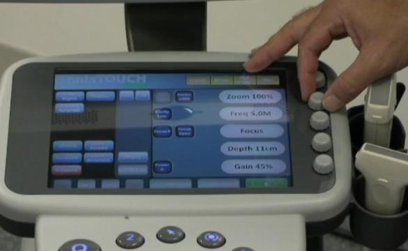

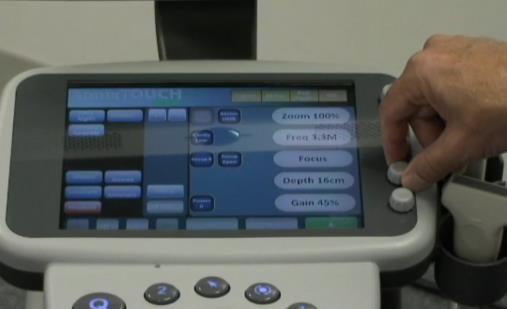
5 Image Optimization Knobology Frequency Brightness (Gain) Contrast (Dynamic Range) Depth Focal Zone Power Doppler slide-25 slide-26 Frequency Frequency 13 mhz 10 mhz Depth Image Depth 29 slide-30 5
 Contrast (Dynamic Range) Depth Focal Zone")


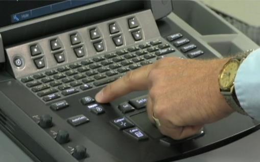
6 Focal Zones Selected by Target Depth Power Doppler Frequency Brightness (Gain) Contrast (Dynamic Range) Depth Focal Zone Power Doppler Depth = 2.5cm slide-31 slide-32 Miscellaneous Concepts Needle Movement Heel-toe Wag tail / Toggle Needle movement Tissue harmonics Power Doppler / color flow B-Steer Virtual convex Gel standoff Extended Field of View slide-33 slide-34 B-Steer Virtual Convex Simulates curved probe Gives wider field of view Eliminates keyhole effect May lose crossbeam/detail slide-35 slide-36 6


7 Virtual Convex Gel Standoff Allows for change in needle approach Obtain better visibility with needle parallel to probe Deactivates cross-beam technology Loses the improved detail of cross beam harmonics slide-38 Gel Standoff Extended Field of View slide-39 slide-40 Normal vs. Pathology Sonopalpation Lateral Facet slide-41 slide- 42 7


")


8 Dynamic Examination Measurements Digital Calipers Tissue Thickness Mass Size Circumferential Area Peripheral nerve slide Image Storage Diagnostic Billing CPT code Ultrasound, extremity, nonvascular, real-time with image documentation; complete CPT code Ultrasound, extremity, nonvascular, real-time with image documentation; limited All diagnostic ultrasound examinations require permanently recorded images in the patient record. Images do not need to be submitted with the claim. Images can be stored as printed images, on a tape or electronic medium. Documentation of the study must be available to the insurer upon request. A written report of all ultrasound studies should be maintained in the patient's record Diagnostic US Documentation Injection Billing - CHANGES 1/1/15 Standard Joint, Bursa, Tendon Injections do not accompany CPT 76942, US Guidance for Needle Placement when billing Medicare as of January 2015 (20600, and now have the language without ultrasound guidance) Ganglion, Tendon or Nerve procedure injection codes may still be used with needle guidance CPT US Guidance for Needle Placement in Small, Medium, Large Joints - CPT 20604, 20606, Requires documented indication Requires description of procedure Requires permanently stored image of target localization and needle May be used in combo with Diagnostic Codes if justified and appropriately documented 47 slide-48 8

.")
9 Injection Documentation Conclusion Procedure: Joint Aspiration / Injection Procedure Indication: Symptomatic relief, glenohumeral osteoarthritis. Informed Consent: Prior to starting the procedure, the patient s identity was verified, pertinent available records were reviewed, the nature of the procedure was explained along with risks, benefits and alternatives. Consent was signed. The appropriate sites of the procedure were confirmed directly with the patient, verified, and marked. A pre-procedure pause was performed for final verification of all the above. Location: Right shoulder. Preparation and Technique: Skin prep ChoraPrep, sterile preparation of site (in usual fashion). Local anesthesia with lidocaine (1 ml, 1% strength, without epinephrine). An initial pre-injection ultrasound evaluation was performed to survey the relevant anatomy. Approach (lateral, posterior, under continuous ultrasound guidance using a 4S curved array probe at a frequency of 5 mhz, in plane approach, per the protocol originally published by Zwar, et. al. in the American Journal of Roentgenology, July 2004 Vol. 183.). Sterile needle used (size #22 gauge, length 2.5 inch). Joint injected with Kenalog 40mg 4ml 1% Lidocaine without epinephrine. Procedure tolerated well. No Complications. Patient Instructions: Ice shoulder tonight, gradually resume use as pain permits. Practice, but don't practice on patients slide-49 slide-50 9
Disclosure. Pre-Procedural Considerations. Transducer Selection. Sterile Procedure. Sterile Procedure. Ultrasound Guided Foot and Ankle Injections
 Ultrasound Guided Foot and Ankle Injections Disclosure No relevant financial relationships exist Shane A. Shapiro, M.D. Assistant Professor, Orthopedic Surgery Mayo Clinic Florida @ShaneShapiroMD 2012
Principles of Ultrasound. Cara C. Prideaux, M.D. University of Utah PM&R Sports Medicine Fellow March 14, 2012
 Principles of Ultrasound Cara C. Prideaux, M.D. University of Utah PM&R Sports Medicine Fellow March 14, 2012 None Disclosures Outline Introduction Benefits and Limitations of US Ultrasound (US) Physics
The Essentials Tissue Characterization and Knobology
 The Essentials Tissue Characterization and Knobology Randy E. Moore, DC, RDMS RMSK No relevant financial relationships Ultrasound The New Standard of Care Musculoskeletal sonography has become the standard
Shane A. Shapiro, M.D. Assistant Professor, Orthopedic Surgery Mayo Clinic 2012 MFMER slide MFMER slide-3
 Ultrasound Foot and Ankle Pathology Disclosures None relevant Shane A. Shapiro, M.D. Assistant Professor, Orthopedic Surgery Mayo Clinic Florida @ShaneShapiroMD 2012 MFMER slide-2 Foot and Ankle Fundamentals
Terminology Tissue Appearance
 By Marc Nielsen, MD Advantages/Disadvantages Generation of Image Ultrasound Machine/Transducer selection Modes of Ultrasound Terminology Tissue Appearance Scanning Technique Real-time Portable No ionizing
WELCOME! Introduction to Bedside Ultrasound
 WELCOME! Introduction to Bedside Ultrasound TEACHERS University of California-Irvine School of Medicine Nathan Molina nathan.d.molina@gmail.com Trevor Plescia taplescia90@gmail.com Jack Silva jpsilva42@gmail.com
Basic Physics of Ultrasound and Knobology
 WELCOME TO UTMB Basic Physics of Ultrasound and Knobology By Daneshvari Solanki, FRCA Laura B. McDaniel Distinguished Professor Anesthesiology and Pain Medicine University of Texas Medical Branch Galveston,
Ultrasound Guided Injections
 Ultrasound Guided Injection Technique More accurate injections Better Results! 1 Benefits: Increased Level of Certainty ie : really know how accurate PRP/Prolotherapy Avoid damage to articular cartilage
Ultrasound Physics and Knobology Alan Macfarlane. Consultant Anaesthetist Glasgow Royal Infirmary
 Ultrasound Physics and Knobology Alan Macfarlane Consultant Anaesthetist Glasgow Royal Infirmary RAPM 2009; 34: 40-46 Ultrasound Proficiency Understanding US image generation and device operation Image
Preamble (disclaimer)
") Preamble (disclaimer) PHYSICS AND PRINCIPLES OF HEAD/NECK ULTRASOUND Joseph C. Sniezek, MD FACS LTC, MC, USA Otolaryngology/H&N Surgery Tripler Army Medical Center 1. I am not a physicist 2. ACS has recommended
Preamble (disclaimer) PHYSICS AND PRINCIPLES OF HEAD/NECK ULTRASOUND Joseph C. Sniezek, MD FACS LTC, MC, USA Otolaryngology/H&N Surgery Tripler Army Medical Center 1. I am not a physicist 2. ACS has recommended
Ultrasound Knobology
 Ultrasound Knobology Raj Dasgupta MD, FACP, FCCP, FASSM Assistant Professor of Clinical Medicine Pulmonary / Critical Care / Sleep Medicine University of Southern California (USC) Objectives Physics of
Ultrasound Principles cycle Frequency Wavelength Period Velocity
 ! Teresa S. Wu, MD, FACEP Director, EM Ultrasound Program & Fellowship Co-Director, Simulation Based Training Program & Fellowship Associate Program Director, EM Residency Program Maricopa Medical Center
Basic of Ultrasound Physics E FAST & Renal Examination. Dr Muhammad Umer Ihsan MBBS,MD, DCH CCPU,DDU1,FACEM
 Basic of Ultrasound Physics E FAST & Renal Examination Dr Muhammad Umer Ihsan MBBS,MD, DCH CCPU,DDU1,FACEM What is Sound? Sound is Mechanical pressure waves What is Ultrasound? Ultrasounds are sound waves
Introduction to Musculoskeletal Ultrasound. Disclosures. Evidence Based Medicine Key References 8/30/2017
 Introduction to Musculoskeletal Ultrasound Johannes Roth MD, PhD, FRCPC, RhMSUS Professor of Pediatrics University of Ottawa Gurjit S Kaeley MBBS, MRCP, RhMSUS Professor of Medicine Division Chief Director
Musculoskeletal Ultrasound Fundamentals
 Fundamentals Benjamin D. Levine, M.D. Associate Professor of Radiology Musculoskeletal Imaging Dept. of Radiological Sciences UCLA Health System I. Image Optimization II. Image Interpretation Artifacts
1 Fundamentals. Basic Definitions and Physics Principles. Fundamentals
 1 To become versed in the language of ultrasonography, it is necessary to review some of the basic principles of physics. The wave physics principles of ordinary (i.e., audible) sound apply to ultrasound
Ultrasonography of the Neck as an Adjunct to FNA. Nicole Massoll M.D.
 Ultrasonography of the Neck as an Adjunct to FNA Nicole Massoll M.D. Basic Features of Head and Neck Ultrasound and Anatomy Nicole Massoll M.D. University of Arkansas for Medical Sciences, Little Rock
Table of contents. Foreword. Preface. 1 Introduction Historical Perspective 00
 Table of contents Foreword Preface 1 Introduction 00 1.1 Historical Perspective 00 2 Fundamentals of musculoskeletal ultrasound 00 2.1 Frequency and wavelength 00 2.2 Generating ultrasound waves 00 2.3
Needle visualization with ZONARE ultrasound systems
 Needle visualization with ZONARE ultrasound systems This material provides a general overview of ultrasound guided needle imaging and techniques and is not intended to replace formal training or education
Diagnostic Ultrasound. Sutiporn Khampunnip, M.D.
 Diagnostic Ultrasound Sutiporn Khampunnip, M.D. Definition of Ultrasound Ultrasound is simply sound waves, like audible sound. High-frequency sound and refers to mechanical vibrations above 20 khz. Human
Ultrasound Guidance Needle Techniques
 Ultrasound Guidance Needle Techniques Dr TANG Ho-ming AED/UCH USG Guidance Needle Techniques Commonly used in EM 1. Vessel cannulation-peripheral & central 2. Foreign body removal 3. Peripheral nerve/plexus
INTRODUCTION. Getting the best scan. Choosing a probe. Choosing the frequency
 Getting the best scan Choosing a probe Select the most appropriate probe for the particular scan required. s vary in their: operating frequency range higher ultrasound frequencies provide better discrimination
Introduction to Ultrasound Guided Region Anesthesia
 Introduction to Ultrasound Guided Region Anesthesia Brian D. Sites, MD Dept of Anesthesiology Dartmouth-Hitchcock Medical Center INTRODUCTION Welcome to Introduction to Ultrasound Guided Regional Anesthesia.
Background & Indications Probe Selection
 Teresa S. Wu, MD, FACEP Director, EM Ultrasound Program & Fellowship Co-Director, Simulation Based Training Program & Fellowship Associate Program Director, EM Residency Program Maricopa Medical Center
Chapter 2 Pitfalls in Musculoskeletal Ultrasound
 Chapter 2 Pitfalls in Musculoskeletal Ultrasound Violeta Maria Vlad MD, PhD Introduction Taking a good ultrasound (US) picture is an art. Interpreting it is a science. This is in fact everything US is
Lesson 07: Ultrasound Transducers. This lesson contains 73 slides plus 16 multiple-choice questions.
 Lesson 07: Ultrasound Transducers This lesson contains 73 slides plus 16 multiple-choice questions. This lesson was derived from pages 33 through 42 in the textbook: Ultrasound Transducers Ultrasound Transducers
The Physics of Ultrasound. The Physics of Ultrasound. Claus G. Roehrborn. Professor and Chairman. Ultrasound Physics
 The Physics of Ultrasound Pipe Organ 10-8000 Emission Dog 452-1080 Man 85-1100 Spectrum Bat 10,000-120,000 Porpoise 7000-120,000 Claus G. Roehrborn Professor and Chairman 10 20 Cycles per second Reception
Introduction & Physics of ED Ultrasound. Objectives. What? - Limited Studies. Who? - ED Docs
 Introduction & Physics of ED Ultrasound Martine Sargent, MD Ultrasound Director, Assistant Professor UCSF Department of Emergency Medicine San Francisco General Hospital & Trauma Center Objectives Who?
Musculoskeletal ultrasound education for sports medicine fellows: a suggested/potential curriculum by the American Medical Society for Sports Medicine
 The appendix to this paper is published online only. To view this fi le please visit the journal online (http://bjsm. bmj.com) 1 Mayo Clinic College of Medicine, Mayo Clinic Sports Medicine Center, Rochester,
Ultrasound in Peripheral Nerve Interventions
 Ultrasound in Peripheral Nerve Interventions John L. Lin, M.D. Shepherd Center Assistant Clinical Professor Emory University, School of Medicine Outline Ultrasound basics Nerve blocks in physiatric setting
Musculoskeletal Ultrasound: Basics, Utility, and Clinical Applications
 Musculoskeletal Ultrasound: Basics, Utility, and Clinical Applications Andrew Lavigne, MD, FRCPC Physical Medicine and Rehabilitation CSCN Diplomat (EMG) Dip Sport Medicine Eugene Maida, MD, PGY-4 Resident
Physical Principles of Ultrasound
 Physical Principles of Ultrasound Grateful appreciation to Richard A. Lopchinsky, MD, FACS and Nancy H. Van Name, RDMS, RTR, and MarleneKattaron, RDMS 2000 UIC All Rights Reserved. Course Objectives Identify
Ultrasound Physics & Terminology
 Ultrasound Physics & Terminology This module includes the following: Basic physics terms Basic principles of ultrasound Ultrasound terminology and terms Common artifacts seen Doppler principles Terms for
Clinical Protocols of the Anesthesiology Department at the Dartmouth-Hitchcock Medical Center: Techniques for lower extremity nerve blocks.
 Clinical Protocols of the Anesthesiology Department at the Dartmouth-Hitchcock Medical Center: Techniques for lower extremity nerve blocks. Authors from DHMC: Brian D. Sites, MD. Assistant Professor of
The Elbow 3/5/2015. The Elbow Scanning Sequence. * Anterior Joint (The anterior Pyramid ) * Lateral Epicondyle * Medial Epicondyle * Posterior Joint
 * Lateral Epicondyle * Medial Epicondyle * Posterior Joint") Scanning Sequence * Anterior Joint (The anterior Pyramid ) * Lateral Epicondyle * Medial Epicondyle * Posterior Joint Anterior Elbow Pyramid Courtesy of Jay Smith, MD. Vice chair PMR Mayo Clinic Rochester,
Scanning Sequence * Anterior Joint (The anterior Pyramid ) * Lateral Epicondyle * Medial Epicondyle * Posterior Joint Anterior Elbow Pyramid Courtesy of Jay Smith, MD. Vice chair PMR Mayo Clinic Rochester,
Ultrasound Physics & Doppler
 Ultrasound Physics & Doppler Endocrine University 2018 Mark Lupo, MD, FACE, ECNU Objectives Review the essential components of ultrasound physics in neck sonography Demonstrate the importance of ultrasound
What is Ultrasound? What is Ultrasound? B A. Basic Principles of Ultrasound. Basic Principles of Ultrasound. Basic Principles of Ultrasound
 Introduction to Ultrasound Principles Mani Montazemi, RDMS Baylor College of Medicine Division of Maternal-Fetal Medicine Department of Obstetrics and Gynecology Manager, Maternal Fetal Center Imaging
Common Applications for Sonography and Guided Intervention: Shoulder
 Common Applications for Sonography and Guided Intervention: Shoulder Jon A. Jacobson, M.D. Professor of Radiology Director, Division of Musculoskeletal Radiology University of Michigan Disclosures: Consultant:
Point-of-Care Ultrasound: An Introduction
 Point-of-Care Ultrasound: An Introduction Delegation Teaching Package for Registered Respiratory Therapists and Anesthesia Assistants Developed by: Rob Bryan RRT, AA Edited by: Kelly Hassall RRT, FCSRT,
Ultrasound guidance in regional anesthesia has
 Ultrasound and Regional Anesthesia Artifacts and Pitfall Errors Associated With Ultrasound-Guided Regional Anesthesia. Part I: Understanding the Basic Principles of Ultrasound Physics and Machine Operations
Ultrasound basics Part 1
 Ultrasound basics Part 1 'Ultrasound enhanced critical care medicine' Rohit Patel, MD University of Florida Health Director, Critical Care Ultrasound Surgical ICU Center for Intensive Care Gainesville,
Ultrasound. Principles of Medical Imaging. Contents. Prof. Dr. Philippe Cattin. MIAC, University of Basel. Oct 17th, 2016
 Ultrasound Principles of Medical Imaging Prof. Dr. Philippe Cattin MIAC, University of Basel Contents Abstract 1 Image Generation Echography A-Mode B-Mode M-Mode 2.5D Ultrasound 3D Ultrasound 4D Ultrasound
MUSCULOSKELETAL ULTRASOUND
 MUSCULOSKELETAL ULTRASOUND A brief overview of diagnostic and therapeutic applications in musculoskeletal medicine. By Elmer G. Pinzon, MD, MPH and Randy E. Moore, DC, RDMS The use of musculoskeletal ultrasound
Brachial plexus blockade within the interscalene groove involves local anesthetic
 Interscalene Brachial Plexus Block- How I do it. Part 1 of a 2 part discussion on technique. Stuart Grant Professor of Anesthesiology Duke University Medical Center Durham NC Brachial plexus blockade within
Focused Musculoskeletal Ultrasound
 Focused Musculoskeletal Ultrasound David Lewis Consultant Emergency Medicine Ipswich (Club Doctor, Ipswich Town FC) Advanced Emergency Ultrasound Objectives! General principles! Musculoskeletal anatomy!
What is Ultrasound? Resolution Image production Attenuation Imaging modes Ultrasound artifacts... 7
 What is Ultrasound?... 1 Resolution... 3 Image production... 3 Attenuation... 4 Imaging modes... 5 Ultrasound artifacts... 7 0 What is Ultrasound? High frequency sound of frequencies 2-50 MHz is used in
Dr Emma Chung. Safety first - Physical principles for excellent imaging
 Safety first - Physical principles for excellent imaging Dr Emma Chung Lecturer in Medical Physics, University of Leicester Clinical Scientist, University Hospitals of Leicester NHS Trust Thanks to Caroline
ULTRASOUND NOMENCLATURE
 Chapter 1: Ultrasound Nomenclature, Image Orientation, and Basic Instrumentation CYNTHIA SIKOWSKI Ultrasound waves are sound waves that have a frequency exceeding 20,000 Hz. When sound waves are transmitted
Diploma of Medical Ultrasonography (DMU) Physical Principles of Ultrasound and Instrumentation Syllabus
 Physical Principles of Ultrasound and Instrumentation Syllabus") Diploma of Medical Ultrasonography (DMU) Physical Principles of Ultrasound and Instrumentation Syllabus Page 1 of 7 11/18 Candidates are expected to cover all of the content of this syllabus when preparing
Diploma of Medical Ultrasonography (DMU) Physical Principles of Ultrasound and Instrumentation Syllabus Page 1 of 7 11/18 Candidates are expected to cover all of the content of this syllabus when preparing
Probe Selection A high frequency (7-12 MHz) linear array transducer should be used to visualize superficial structures (Image 1).
 linear array transducer should be used to visualize superficial structures (Image 1).") ! Teresa S. Wu, MD, FACEP Director, Emergency Ultrasound Program & Fellowships Co-Director, Women s Imaging Fellowship Maricopa Medical Center Associate Professor, Emergency Medicine Director, Simulation
! Teresa S. Wu, MD, FACEP Director, Emergency Ultrasound Program & Fellowships Co-Director, Women s Imaging Fellowship Maricopa Medical Center Associate Professor, Emergency Medicine Director, Simulation
ULTRASOUND. OB/Gyn (Core) Ultrasound PIEZOELECTRIC EFFECT. Principles of Ultrasound Physics and Instrumentation. Nathan Pinkney, BS, CDOS
 Ultrasound PIEZOELECTRIC EFFECT. Principles of Ultrasound Physics and Instrumentation. Nathan Pinkney, BS, CDOS") 1 OB/Gyn (Core) Ultrasound Principles of Ultrasound Physics and Instrumentation Nathan Pinkney, BS, CDOS Philadelphia College of Osteopathic Medicine 2016 ULTRASOUND CATEGORIES OF SOUND INFRASOUND = below
1 OB/Gyn (Core) Ultrasound Principles of Ultrasound Physics and Instrumentation Nathan Pinkney, BS, CDOS Philadelphia College of Osteopathic Medicine 2016 ULTRASOUND CATEGORIES OF SOUND INFRASOUND = below
Pragmatic ultrasound in the diagnosis of soft tissue rheumatic pain. Plamen Todorov
 Pragmatic ultrasound in the diagnosis of soft tissue rheumatic pain Plamen Todorov INTRODUCTION Soft tissue rheumatism: nonsystemic, focal pathological syndromes involving the periarticular structures.
Basics of US Regional Anaesthesia. November 2008
 Basics of US Regional Anaesthesia November 2008 Essential Physics HIGH frequency = great resolution but poor penetration LOW frequency = poor resolution but great penetration Potential Advantages of US
Image optimization for critical care US
 Image optimization for critical care US 1 Although we assume you are already familiar with focused US in the ED, it might not hurt to revise the basics: Machines & transducers US appearance of normal tissues
Ultrasound of Shoulder Pathology and Intervention 서울대학교병원재활의학과 김기원
 Ultrasound of Shoulder Pathology and Intervention 서울대학교병원재활의학과 김기원 Ultrasound for Shoulder Disorder Advantage Dynamic evaluation Immediate clinical correlation + Intervention Weakness Diagnostic accuracy?
Abdominal Ultrasound
 Abdominal Ultrasound Imaging Control Buttons Depth The organ imaged should take up 3/4 of the screen Frequency = Penetration Use high frequencies (harmonics) for fluid filled and superficial structures
Optimizing Breast Sonography
 Optimizing Breast Sonography Cindy Rapp BS, RDMS, FSDMS, FAIUM Denver, Colorado Breast Sonography general goal to make a more specific diagnosis than can be made with clinical and mammographic findings
Rotator Cuff and Biceps Pathology
 Rotator Cuff and Biceps Pathology Jon A. Jacobson, M.D. Professor of Radiology Director, Division of Musculoskeletal Radiology University of Michigan Disclosures: Consultant: Bioclinica Advisory Board:
ELENI ANDIPA General Hospital of Athens G. Gennimatas
 ELENI ANDIPA General Hospital of Athens G. Gennimatas Technological advances over the last years have caused a dramatic improvement in ultrasound quality and resolution An established imaging modality
2015 ARDMS Musculoskeletal Sonographer Job Task Analysis Summary Report
 P a g e 1 2015 ARDMS Musculoskeletal Sonographer Job Task Analysis Summary Report American Registry for Diagnostic Medical Sonography (ARDMS) P a g e 2 Table of Contents ABOUT THE REPORT... 3 METHODOLOGY...
Descriptions of NDT Projects Fall 2004 October 31, 2004
 Descriptions of NDT Projects Fall 2004 October 31, 2004 Introduction There are two separate NDT labs in Magister: ULTRA for ultrasound and EDDY for eddy current. Both labs are equipped with mechanical
Routine Quality Assurance Cookbook
 This Cookbook is a companion guide to the AIUM Routine Quality Assurance (QA) for Diagnostic Ultrasound Equipment document, which outlines the basic QA requirements for AIUM-accredited practices. The Guide
How do you use the AiM Vascular Mapping Kit?
 How do you use the AiM Vascular Mapping Kit? Protocol for Successful Skin Mapping Evaluate vessel anatomy, perform measurements, etc. according to your laboratory s protocol prior to applying the AiM device
The 2 nd Cambridge Advanced Emergency Ultrasound Course
 The 2 nd Cambridge Advanced Emergency Ultrasound Course Addenbrooke s Hospital Cambridge Sept 2008 1 2 Faculty! UK! USA! Australia! Toshiba! Emergency Medicine! Radiology 3 Programme! Day 1 Introduction
Do more at the point of care.
 SONIMAGE HS1 Compact Ultrasound System HEALTHCARE at the point of care. for wrists. shoulders. for hips. for knees. for feet. SONIMAGE HS1 Compact Ultrasound System with ultrasound. Supporting increased
Knobology for Dummies
 Knobology for Dummies Power On/Off Preset button Patient Information Entry Choose preset Transducer probes Connect and disconnect transducer Approach to the patient (machine placement, comfort, draping,
Ultrasound Guided Peripheral Intravenous Access
 Ultrasound Guided Peripheral Intravenous Access J. Christian Fox, MD, RDMS, FACEP, FAAEM, FAIUM Professor and Interim Chair of Emergency Medicine Director of Instructional Ultrasound University of California,
Innovation Meets Portability. Indeed. ALPINION MEDICAL SYSTEMS We are Ultrasound Professionals
 Innovation Meets Portability. Indeed. ALPINION MEDICAL SYSTEMS We are Ultrasound Professionals E-CUBE i7, the next step in confident diagnosis with excellent image performance. The innovative portable
Do more with ultrasound. Increased Diagnostic Confidence Earlier in the Care Path CASE STUDY KNEE
 Do more with ultrasound. Increased Diagnostic Confidence Earlier in the Care Path CASE STUDY KNEE Better clinical outcomes. Improved efficiencies. Increased patient satisfaction. Increased diagnostic confidence
Concepts of Imaging and Knobology
 Concepts of Imaging and Knobology Pravin Patil, MD FACC FASE Associate Professor of Medicine Director, Cardiovascular Disease Training Program Lewis Katz School of Medicine at Temple University Disclosures
CONTENTS. Test Number cpd Tanya Reynolds (Nat. Dip. Diag. Rad., B. Tech. Diag. Rad., B. Tech. Ultrasound)
") CONTENTS page 1-15 page 16 BASIC 2-DIMENSIONAL ULTRASOUND PRINCIPLES Multiple Choice Test Test Number cpd 41640 Tanya Reynolds (Nat. Dip. Diag. Rad., B. Tech. Diag. Rad., B. Tech. Ultrasound) Tanya is
CONTENTS page 1-15 page 16 BASIC 2-DIMENSIONAL ULTRASOUND PRINCIPLES Multiple Choice Test Test Number cpd 41640 Tanya Reynolds (Nat. Dip. Diag. Rad., B. Tech. Diag. Rad., B. Tech. Ultrasound) Tanya is
ULTRASOUND IMAGING EE 472 F2018. Prof. Yasser Mostafa Kadah
 ULTRASOUND IMAGING EE 472 F2018 Prof. Yasser Mostafa Kadah www.k-space.org Recommended Textbook Diagnostic Ultrasound: Physics and Equipment, 2nd ed., by Peter R. Hoskins (Editor), Kevin Martin (Editor),
ULTRASOUND QA SOLUTIONS. Ensure Accurate Screening, Diagnosis & Monitoring DOPPLER FLOW PHANTOMS MULTI-PURPOSE PHANTOMS TRANSDUCER TEST PHANTOMS
 ULTRASOUND QA SOLUTIONS Ensure Accurate Screening, Diagnosis & Monitoring DOPPLER FLOW PHANTOMS MULTI-PURPOSE PHANTOMS TRANSDUCER TEST PHANTOMS INNOVATORS IN ADVANCED ULTRASOUND TECHNIQUES Gammex is the
Index. Note: Page numbers of article titles are in boldface type.
 Note: Page numbers of article titles are in boldface type. A ACJ. See Acromioclavicular joint (ACJ) Acromioclavicular joint (ACJ) procedures of, 557 559 Ankle and foot procedures of, 649 671 (See also
S1Stephanie J. Doniger
 Section 1 Ultrasound fundamentals Introduction S1Stephanie J. Doniger Pediatric Emergency Medicine is a relatively new field of medicine developed in the 1980s. Since its inception, several advancements
Confident and Conclusive Diagnosis with Flexible Solutions
 Internal Medicine Confident and Conclusive Diagnosis with Flexible Solutions ALPINION MEDICAL SYSTEMS We are Ultrasound Professionals Ultrasound is widely used as the gold-standard for internal medicine
Transducer. ACUSON Juniper. Ultrasound System. siemens.com/healthineers
 Transducer ACUSON Juniper Ultrasound System siemens.com/healthineers Table of Contents 5C1 Transducer 3 7C2 Transducer 3 11M3 Transducer 4 12L3 Transducer 4 11L4 Transducer 5 16L4 Transducer 5 18H5 Transducer
Innovation Meets Portability. Indeed. ALPINION MEDICAL SYSTEMS We are Ultrasound Professionals
 Innovation Meets Portability. Indeed. ALPINION MEDICAL SYSTEMS We are Ultrasound Professionals E-CUBE i7, the next step in confident diagnosis with excellent image performance. The innovative portable
Ultrasound-Guided Shoulder Injections 인제대학교일산백병원 재활의학과 임길병
 Ultrasound-Guided Shoulder Injections 인제대학교일산백병원 재활의학과 임길병 How to improve needle visibility Advantages of Ultrasound in Procedures Real-time imaging Avoids radiation exposure But, interventions without
9/18/18. Welcome- MSK Ultrasound Workshop. Introduction to Musculoskeletal Ultrasound. Acknowledgement of Country. The Workshop.
 Acknowledgement of Country Welcome- MSK Ultrasound Workshop I would like to acknowledge that this meeting is being held on the traditional lands of the Wurundjeri and Boonwurrung people and pay my respect
Ultrasound - Musculoskeletal
 Ultrasound - Musculoskeletal What is Ultrasound Imaging of the Musculoskeletal System? Ultrasound imaging, also called ultrasound scanning or sonography, involves exposing part of the body to high-frequency
FOR IMMEDIATE RELEASE
 FOR IMMEDIATE RELEASE Hitachi to launch ALOKA ARIETTA 850, the flagship model of the ARIETTA series diagnostic of ultrasound platforms This premium model features superior image quality, seamless workflow,
OPHTHALMOLOGY AND ULTRASOUND
 Vet Times The website for the veterinary profession https://www.vettimes.co.uk OPHTHALMOLOGY AND ULTRASOUND Author : JAMES OLIVER Categories : Vets Date : April 28, 2008 JAMES OLIVER discusses why ultrasound
Disclosures. Objectives 5/4/2018. PT Management: What the Rheumatology Trainee Needs to Know. Physical Therapy in Spondyloarthritis
 Physical Therapy in Spondyloarthritis PT Management: What the Rheumatology Trainee Needs to Know Angelo Papachristos BSc, BScPT,CAFCI, MBA Advanced Practice Physiotherapist St. Michael s Hospital Toronto,
Focus on your needs. Ultrasound system HS60 SAMSUNG MEDISON CO., LTD. CT-HS60 V1.0-GI-FT EN
 Samsung Medison, an affiliate of Samsung Electronics, is a global medical company founded in 1985. With a mission to bring health and well-being to people's lives, the company manufactures diagnostic ultrasound
Chapter 3. Sonographic Image Interpretation
 Chapter 3 Sonographic Image Interpretation Sonograms are two-dimensional gray-scale images that allow assessment and diagnosis of many anatomic and pathologic changes that can occur in the human body.
Debbie Childs RDMS, RVT Sonographer Murphy Medical Center Murphy, NC
 Debbie Childs RDMS, RVT Sonographer Murphy Medical Center Murphy, NC Worked at Murphy Medical Center as a sonographer for 18 years Registered in Abdomen, OB/GYN, Breast, & Vascular Ultrasound ACR Accredited
Point of Care Ultrasound on the Field of Play K AT I E N ANOS, MD
 Point of Care Ultrasound on the Field of Play K AT I E N ANOS, MD H I GH P ERFORMANCE S PORTS MEDICINE P HYSI ATRIST, P R ACTICING S PORTS MEDI CINE No disclosures No disclosures Who am I? Objectives Over
Ultrasound in Medicine
 Ultrasound in Medicine Experimental Equipment for Medical Education Universities Colleges Medical Schools Medical and Med-Technical Training Education can befun! WELCOME TO GAMPT Devices and accessories
Quality in your daily practice
 ALPINION MEDICAL SYSTEMS Co., Ltd. Verdi Tower, 72, Digital-ro 26-gil, Guro-gu, Seoul, 08393, Korea Homepage www.alpinion.com E-mail international@alpinion.com TEL +82-2-3282-0900 FAX +82-2-851-5591 Standalone
Underwater Acoustic Measurements in Megahertz Frequency Range.
 Underwater Acoustic Measurements in Megahertz Frequency Range. Current State and Prospects of Development in Russia Alexander M. Enyakov,, Many medical applications of underwater acoustic measurements
DIGITAL IMAGE PROCESSING IN ULTRASOUND IMAGES
 DIGITAL IMAGE PROCESSING IN ULTRASOUND IMAGES Kamaljeet Kaur Computer Science & Engineering Department Guru Nanak Dev Engg. College, Ludhiana. Punjab-India meetk.89@gmail.com ABSTRACT-- Image processing
ICCUME All Domain recommendations Montreal, October 14,
 Domain 1 (Scope) ICCUME All Domain recommendations Montreal, October 14, 2017 10 1.1 Goal: The ICC will produce consensus recommendations on An integrated ultrasound curriculum for undergraduate medical
The complete, portable patient care solution
 The complete, portable patient care solution ACUSON P300 Ultrasound System www.siemens.com/ultrasound Answers for life. Mobile. Powerful. And extraordinarily versatile. Sleek design Portable Complete 2
Breast Ultrasound: Improving Your Skills & Patient Care
 Breast Ultrasound: Improving Your Skills & Patient Care Objectives Discuss US techniques available for image optimization. Review & compare the US appearances of benign & malignant masses. Cherie M. Kuzmiak,
Ultrasonic Testing Level I:
 Ultrasonic Testing Level I: 1- Sound Wave - Introduction - ASNT Level I - Sound Wave Propagation - Velocity / Frequency / Wave Length - Acoustic Impedance - Energy / Intensity 2- Ultrasound Wave Modes
Category Term Definition Comments 1 Major Categories 1a
 Working Lexicon Categories, Terms & Definitions Category Term Definition Comments 1 Major Categories 1a Physiologic Category (consistent with normal ovarian physiology) Follicle Simple 3 cm in premenopausal
Background & Indications Probe Selection
 Teresa S. Wu, MD, FACEP Director, EM Ultrasound Program & Fellowship Co-Director, Simulation Based Training Program & Fellowship Associate Program Director, EM Residency Program Maricopa Medical Center
Chapter 14. Imaging Artifacts
 Chapter 14 Image Artifacts The complex physical interactions that occur between an ultrasound beam and human anatomy and the intricate and sophisticated technological components of a sonographic imaging