Patterns and mechanisms of inflammatory skin conditions: the pathologist s survival kit SALVADOR J. DIAZ-CANO BAHRAIN, APRIL 2017
|
|
|
- David Barber
- 5 years ago
- Views:
Transcription

1 Patterns and mechanisms of inflammatory skin conditions: the pathologist s survival kit SALVADOR J. DIAZ-CANO BAHRAIN, APRIL 2017


2 Basic Elements of Lesions
3 Repair Injury Time & Intensity Cell damage oncosis Hyperplastic reaction Cell damage cytopathic Combined Inflammatory cellular Inflammatory Extracellular/vascular
4 Biological Basis of Elementary Lesions Cellular response Cell perpetuation pathway Cell differentiation pathway Activation of default pathway cell death Tissue effects Parenchyma and stroma interactions Combination of findings depending on the primary damage target
5 Biological Processes and Pathology Primary Biological Process Increased keratinocyte turnover Decreased keratinocyte turnover Inflammation Cell damage General Pathology Hyperplastic changes Lack of full cell differentiation Relative increase of cell loss Relative atrophy Full cell differentiation Decreased cell loss Vascular changes and permeability Cellular infiltrate Cell damage (stroma, vessels, epithelia) Reversible Irreversible Regeneration and reparation

6 Psoriasiform Acanthosis Dermatophytosis Psoriasis Seborrheic dermatitis Allergic contact / nummular dermatitis PRP Secondary syphilis Scabies, Norwegian type MF

7 Psoriasiform Dermatitis Neutrophils within parakeratosis Superficial perivascular dermatitis Psoriasis

8 Irregular Epidermal Hyperplasia Pseudoepitheliomatous hyperplasia Suppurative granulomatous dermatitis Deep mycosis, atypical mycobacterias

9 Markedly Thinned Epidermis Dermatomyositis/DLE DLE GVHD LP, atrophic LP-like keratosis Porokeratosis Lichen sclerosus Degos disease
10 Interface Dermatitis Leukocyte infiltration of the dermis Vacuolar change of basilar epidermis Papillary dermal melanophages Necrosis of keratinocytes AKA: lichenoid tissue reaction pattern, vacuolar interface dermatitis

11

12 Lichenoid Interface Dermatitis Wedge-shaped hypergranulosis Lichenoid infiltrate Lichen planus

13 Vacuolar Interface Dermatitis Necrotic keratinocytes Vacuolar alteration Lichenoid inflammation Superficial perivascular lymphohistiocytic Erythema Multiforme


14 Spongiotic Dermatitis Spongiosis around acrosyringia Miliaria rubra

15 Eosinophilic Spongiosis BP/HG, urticarial Allergic dermatitis Pemphigus vulgaris, urticarial Arthropod assault Dermatophytosis Incontinentia pigmenti, vesicular Toxic erythema of newborn

16 Lymphocytic Infiltrate DLE, tumid Pernio PLE Insect bite Erythema figuratum

17 Perivascular Lymphoplasmacytic Infiltrate Acrodermatitis chronica atrophicans Erythema chronicum migrans Secondary syphilis Necrobiosis lipoidica Morphea
18 Exocytosis vs. Epidermotropism EXOCYTOSIS EPIDERMOTROPISM Random through epidermis to surface Spongiotic tissue reaction Inflammatory processes Lower third/half epidermis Tendency to aggregate No/little spongiosis Feature of MF
19 Superficial and Deep Inflammation Light reaction Lymphoma Leprosy Lues Lichen striatus Lupus erythematosus Lipoidica (necrobiosis) Lepidoptera (+ other arthropods) Dermatophyte Reticular erythematous mucinosis Urticarial stages (BP) Gyrate erythemas Scleroderma (localized) Drug reactions 8Ls + DRUGS

20 Nodular Dermatitis Germinal centers in a dense nodular or diffuse infiltrate Delayed hypsersensitivity reaction

21 Abnormal Lymphocytes in Mixed Cell Infiltrate Tick bite reaction Herpesvirus Dermatophytosis Ruptured molluscum Kikuchi s disease Gianotti-Crosti Lymphomatoid papulosis MF

22 Dense Mixed Inflammation Granuloma faciale / Erythema elevatum diutinum Nodular scabies Tick bite reaction
23 Subcorneal Pustule Dermatophytosis Candidiasis Impetigo Suppurative infundibulitis Pemphigus foliaceus Pustular psoriasis Prurigo pigmentosa

24 Papillary Micro-abscesses DH / linear IgA dermatitis, drug eruptions Acquired EB DLE Bullous LE LCC vasculitis

25 Interstitial Neutrophils in Reticular Dermis Urticaria Cutis laxa DH / linear IgA dermatitis Bullous LE Fixed drug eruption Cellulitis Flea bite Pustular infundibular dermatitis LCC vasculitis Sweet s / PG Lymphomatoid papulosis

26 Cytopathic Dermatitis Acantholytic separation Multinucleated epithelial giant cells Herpesvirus infections

27 Ballooning EM Fixed drug eruption Mucha-Haberman Prurigo pigmentosa Hand-foot-mouth disease Herpesvirus Milker s nodule/orf Irritant contact dermatitis Burn

28 Fibrosing Dermatitis Thinned epidermis Thickened edematous / sclerotic papillary dermis Underlying mononuclear infiltrate Lichen sclerosus

29 Fibrosing Dermatitis Decreased number of adnexal structures Fibrosing inflammatory of neoplastic conditions
30 Elements of Pattern Recognition With/without epidermal changes Spongiotic, interface, hyperplastic, atrophic Distribution of inflammatory infiltrate Topography Superficial vs. superficial & deep Microanatomy Perivascular, interstitial, nodular, diffuse Type of inflammatory infiltrate Mononuclear Lymphocytes ± histiocytes Mixed Mono- and polymorphonuclear Polymorphonuclear ± eosinophils
31 Approach to Microscopic Diagnosis


32 Cytopathic dermatitis Psoriasiform dermatitis Intraepidermal bullous dermatitis Lichenoid dermatitis Vacuolar dermatitis Spongiotic dermatitis Subepidermal bullous dermatitis

33 Approach to Microscopic Diagnosis (2)


34 Vasculitides - dermatitis Nodular dermatitis Fibrosing dermatitis Perivascular dermatitis Diffuse dermatitis
35 Lesions by Topography Adnexal structures Folliculitis and perifolliculitis Follicular lesions with alopecia Subcutaneous soft tissue Panniculitis
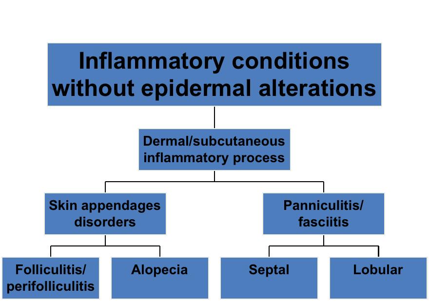


36 Folliculitis Septal panniculitis Perifolliculitis Lobular panniculitis

37 Perifolliculitis Peri-infundibular and perifollicular fibroplasia Long-standing perifolliculitis (traction alopecia, LPP)

38 Non-scarring Alopecia Catagen or telogen follicles NO inflammatory infiltrate Other findings: Plucked hair All telogen hairs Common baldness, telogen effluvium, trichotillomania

39 Mostly Septal Panniculitis Miescher s radial granuloma Linear spaces containing lipids Tiny collections of neutrophils Palisading histiocytes Erythema nodosum

40 Mostly Lobular Panniculitis Arteritis Fat necrosis Suppuration Granulomas Fibrosis Nodular vasculitis

41 Cell-Poor Subepidermal Blister Suction blister Gas gangrene Porphyria cutanea tarda Bullous dermatosis of hemodialysis Bullous amyloid Blister above scar Hypoxemia blister Electric current Second-degree burn Bullous pemphigoid EB Darier s disease, systematized epidermal nevus Grover s disease Porokeratosis Solar keratosis


42 Antigen Presentation, Inflammation & Hypersensitivity
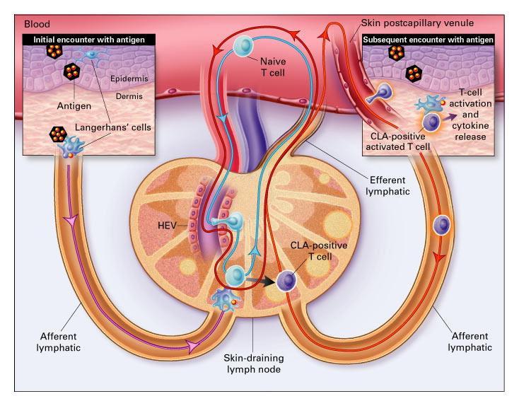
43 Antigen Processing


44 Inflammatory and Cellular Response after Injury


45 Mediator release Necrosis Mediator release Adhesion Soluble complexes Ag-Ab complex transfer CYTOTOXIC INACTIVATION ANAPHYLACTIC CELL-MEDIATED CYTOTOXICITY DELAYED HYPERSENSITIVITY GRANULOMATOUS REACTION

46 Interstitial Granulomatous Infiltrate Granuloma annulare Dermatofibroma Interstitial granulomatous dermatitis Necrobiosis lipoidica MF, interstitial type MF (granulomatous slack skin)

47 Palisaded Granuloma Granuloma annulare Gout Rheumatoid nodule Interstitial granulomatous dermatitis Necrobiosis lipoidica Necrobiotic xanthogranuloma

48 Sarcoidal granulomas Sarcoidal granulomatous pattern Cohesive epithelioid histiocytes Few or no lymphocytes Sarcoidosis, hallogenoderm

49 Tuberculoid granulomas Tuberculoid granulomatous pattern Collections of epithelioid histiocytes Lymphocytes and plasma cells Mycobacterial infections, leishmania, subcutaneous sarcoid


50 Suppurative granulomas Suppurative granulomatous inflammation Neutrophils and necrotic debris Histiocytes Few or no lymphocytes Rupture infundibular cyst, sporotrichosis, dermatophytes

51 Vasculitis Neutrophils, nuclear dust and fibrin in small blood vessel walls Leukocytoclastic vasculitis

52 Vasculitis Neutrophils, nuclear dust and fibrin in small blood vessel walls Thrombi within their lumina Septic vasculitis
53 Psoriasiform Dermatitis

54 Vacuolar Interface Dermatitis

55 Lichenoid Interface Dermatitis
56 Spongiotic Dermatitis
57 Superficial PV Dermatitis
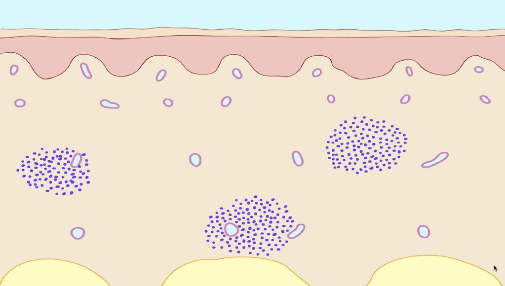
58 Nodular Dermatitis

59 Granulomatous Dermatitis
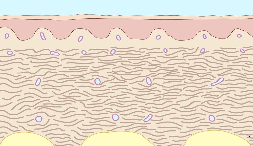
60 Fibrosing Dermatitis

61 Mostly Septal Panniculitis

62 Mostly Lobular Panniculitis


63 Pathology for ADR Evaluation Confirmation of Drug Etiology Patterns and Mechanisms Prognosis/Activity Eosinophils Necrotic keratinocytes Vascular changes Endothelial prominence Hyperpermeability Exanthematous Extrinsic vascular Spongiotic Vesiculo-spongiotic Intrinsic vascular ± granulomas Vesiculopustular ± lichenoid or psoriasiform component Extension of necrosis: Confluent or not Neutrophilic exocytosis: Isolated / Pustular Vascular damage: IVR, vasculitis Extension of inflammation: PV, interstitial, nodular, diffuse


64 a b c d


65 Patterns: Common Basic and Secondary Exanthematous Psoriasiform Lichenoid Commonest pattern Basic profile Chronicity Relative low risk Common as secondary pattern Certain drugs if pseudoepitheliomatous Chronicity Relative low risk Common as secondary pattern


66 Epithelial Damage and Necrosis Basal cells Multifocal Confluent Isolated, cytotoxic Barrier effect maintained Clustering, cytotoxic Barrier effect maintained Multifactorial Barrier effect lost


67 Vascular Changes and Damage Perivascular IVR Vasculitis Basic profile Permeability Minor vessel wall damage Permeability Major vessel wall damage


68 a b c d


69 Neutrophilic Activity Perivascular Single cell exocytosis Pustular Basic profile Spongiosis No keratinocyte damage/necrosis Spongiosis With keratinocyte damage/necrosis


70 Infiltrate Extension Basic - Perivascular PV + Interstitial Nodular Diffuse Low grade progressive Severe Severe & Tissue Damaging

71 Type I Hypersensitivity


72 IgG4-mediated Reactions
73 Inflammatory Skin Lesions Patterns are the result of the interaction of injury agents and tissue response, modulated in intensity and time Elementary lesions will depend on: basic epidermal reaction (based on cellular turnover and maturation), vascular and cellular inflammatory components, types of cells, and distribution of cells (topography and microanatomy) Activity should be evaluated a from target damage (epithelial necrosis, vasculitis), exocytosis (neutrophilic), and density of infiltrate Morphological and clinico-pathologic correlation should be included in the conclusion
My Method for Approaching Skin Biopsies
 My Method for Approaching Skin Biopsies P A U L H A U N, MD, MS, F A A D A S S I S T A N T P R O F E S S O R D E R M A T O L O G Y A N D D E R M A T O P A T H O L O G Y D E P A R T M E N T O F D E R M
Mucinoses Diverse group of disorders which have in common deposition of basophilic, finely granular and stringy material in the connective tissues of
 Cutaneous Mucinoses Nathan C. Walk, M.D. Mucinoses Diverse group of disorders which have in common deposition of basophilic, finely granular and stringy material in the connective tissues of the dermis.
Spongiotic Dermatitis
 Prepared by Kurt Schaberg Introduction to Inflammatory Dermpath Spongiotic Dermatitis intraepidermal intercellular edema (spongiosis) - presence of widened intercellular spaces between keratinocytes, with
Table of Contents: Part 1 Medical Dermatology. Chapter 1 Acneiform Disorders. Acne. Acne Vulgaris. Pomade Acne. Steroid Acne
 Table of Contents: Part 1 Medical Dermatology Chapter 1 Acneiform Disorders Acne Acne Vulgaris Pomade Acne Steroid Acne Infantile Acne Pediatric Perspectives Neonatal Acne (Acne Neonatorum) Pediatric Perspectives
Inflammatory Dermatopathology
 Inflammatory Dermatopathology Steven D. Billings Jenny Cotton Inflammatory Dermatopathology A Pathologist s Survival Guide Second Edition Steven D. Billings, MD Professor of Pathology and Co-Director
Inflammatory skin disease I Jade Wititsuwannakul, MD Chulalongkorn University, Thailand
 Inflammatory skin disease I Jade Wititsuwannakul, MD Chulalongkorn University, Thailand Superficial Perivascular Dermatitis Interface Dermatitis Vacuolar Dermatitis Lichenoid Dermatitis Barnhill Textbook
Inflammatory Skins. Dr W. Merchant St. James Hospital Leeds
 Inflammatory Skins Dr W. Merchant St. James Hospital Leeds Case1 51 M long standing plaque on back Main Features Low power; Not obvious Rather square edged biopsy. Increased thickness to dermal collagen
Principi ed Aggiornamenti in Dermatologia Roma, 6-7 Aprile Grand rounds. Lorenzo Cerroni, Graz
 Principi ed Aggiornamenti in Dermatologia Roma, 6-7 Aprile 2018 Grand rounds Lorenzo Cerroni, Graz "Computer palms" Described in patient using computer keyboards for long periods; similar features described
W. Kempf ] M. Hantschke ] H. Kutzner ] W. H.C. Burgdorf. Dermatopathology
![W. Kempf ] M. Hantschke ] H. Kutzner ] W. H.C. Burgdorf. Dermatopathology](/thumbs/95/126003533.jpg "W. Kempf ] M. Hantschke ] H. Kutzner ] W. H.C. Burgdorf. Dermatopathology") W. Kempf ] M. Hantschke ] H. Kutzner ] W. H.C. Burgdorf Dermatopathology W. Kempf M. Hantschke H. Kutzner W. H. C. Burgdorf Dermatopathology With 242 Color Figures 12 Werner Kempf MD Kempf und Pfaltz Histologische
W. Kempf ] M. Hantschke ] H. Kutzner ] W. H.C. Burgdorf Dermatopathology W. Kempf M. Hantschke H. Kutzner W. H. C. Burgdorf Dermatopathology With 242 Color Figures 12 Werner Kempf MD Kempf und Pfaltz Histologische
Supplementary Online Content
 Supplementary Online Content Ross NA, Chung H-J, Li Q, Andrews JP, Keller MS, Uitto J. Pityriasis rubra pilaris: a case series of patients. Published online March 9, 26. JAMA Dermatol. doi:./jamadermatol.26.9.
Introduction. A Short Review of Cutaneous Vasculitis. Introduction. Introduction. Introduction. Introduction
 A Short Review of Cutaneous Vasculitis Uma Sundram, MD, PhD Professor of Pathology, William Beaumont Oakland University School of Medicine Staff Dermatopathologist Beaumont Hospital-Royal Oak, MI September
Cutaneous Lymphoid Proliferations: A Comprehensive Textbook of Lymphocytic Infiltrates of the Skin
 Cutaneous Lymphoid Proliferations: A Comprehensive Textbook of Lymphocytic Infiltrates of the Skin Magro, Cynthia M., MD ISBN-13: 9780471695981 Table of Contents Chapter One: Introduction to the Classification
Basal cell carcinoma 5/28/2011
 Goal of this Presentation A practical approach to the diagnosis of cutaneous carcinomas and their mimics Thaddeus Mully, MD University of California San Francisco To review common non-melanoma skin cancers
CONDITIONS OF THE SKIN
 CONDITIONS OF THE SKIN UCSF/SFGH Family & Community Medicine Residency Program Educational Objectives I. Knowledge The resident will be able to discuss the definition, diagnosis, and initial management
Rash Decisions Approach to the patient with a skin condition
 National Conference for Nurse Practitioners April 25, 2014 Rash Decisions Approach to the patient with a skin condition Margaret A. Bobonich, DNP, FNP C, DCNP, FAANP Assistant Professor, Case Western Reserve
Dermatopathology Workshop Summary, Berlin 2004
 Dermatopathology Workshop Summary, Berlin 2004 David A. Whiting and Rolf Hoffmannw Baylor Hair Research and Treatment Center, Dallas, Texas, USA; wdermatology Practice, Freiburg, Germany Figure 1 Case
T he histological diagnosis of cutaneous
 1233 REVIEW My approach to superficial inflammatory dermatoses K O Alsaad, D Ghazarian... Superficial inflammatory dermatoses are very common and comprise a wide, complex variety of clinical conditions.
Pathology of the skin. Dr Fónyad László, 1sz. Patológiai és Kísérleti Rákkutató Intézet, SE
 Pathology of the skin Dr Fónyad László, 1sz. Patológiai és Kísérleti Rákkutató Intézet, SE The skin Biggest organ Kb. 1.8 nm Kb. 10 kg Most frequent site for tumor development (BCC) Pathology of the skin
Diagnose dermatologic conditions based on physical examination (visual recognition). The majority of the items will come from Group 1.
. The majority of the items will come from Group 1.") This document was developed by a committee of the American Board of Dermatology (ABD) for the purpose of preparing the BASIC Examination. The BASIC Exam is assessment of fundamental knowledge and skills.
This document was developed by a committee of the American Board of Dermatology (ABD) for the purpose of preparing the BASIC Examination. The BASIC Exam is assessment of fundamental knowledge and skills.
=ﻰﻤاﻤﺤﻠا ﺔﻴﻘﻠﺤﻠا ﺔذﺒاﻨﻠا
 1 / 15 Erythema Annulare Centrifugum and Other Figurate Erythemas The figurate erythemas include a variety of eruptions characterized by annular and polycyclic lesions. Classification of this group has
1/14/2018. Objectives
 2018 Pathology CME Cutaneous Hematopathology Maui, HI Jan 18 th 26 th Pseudolymphomas Alejandro A. Gru, M.D. Assistant Professor of Pathology & Dermatology Dermatopathology Division and Fellowship Director
Evaluation of Epidermal Reaction Pattern and Assessment of Histopathological Findings of Various Skin Disorders
 ORIGINAL RESEARCH www.ijcmr.com Evaluation of Epidermal Reaction Pattern and Assessment of Histopathological Findings of Various Skin Disorders Shweta Sharma 1, Dhara P Trivedi 2, Ronak Vyas 3 ABSTRACT
Index. derm.theclinics.com. Note: Page numbers of article titles are in boldface type.
 Note: Page numbers of article titles are in boldface type. A Abatacept for DLE, 493 for SLE, 497 Ablative therapies, localized, for cutaneous T-cell lymphoma, 502 506. See also Cutaneous T-cell lymphoma,
Important Decisions in Dermatopathology: The Clinico- Pathologic Correlation. Dermatopathology Specialists Needed. Changing Trends
 Important Decisions in Dermatopathology: The Clinico- Pathologic Correlation Uma Sundram, MD, PhD Departments of Pathology and Dermatology Stanford University May 29, 2008 Dermatopathology Specialists
A 40-year old male with follicular papule and pustule at central face area for 3 months
 A 40-year old male with follicular papule and pustule at central face area for 3 months GMS- Neg AFB-Neg Fite stain - neg HISTOPATHOLOGICAL DIFFERENTIAL DIAGNOSIS CASEOUS GRANULOMA INFECTION -MYCOBACTERIUM
Granuloma annulare is a benign self-limited disease, first described by Colcott-Fox 1 in 1895 and Radcliffe-Crocker in 1902.
 Granuloma Annulare Granuloma annulare is a benign self-limited disease, first described by Colcott-Fox 1 in 1895 and Radcliffe-Crocker in 1902. EPIDEMIOLOGY Granuloma annulare is a relatively common disorder.
BSD Self Assessment Workshop 7 th July 2013 CASE 27 RAC6123
 BSD Self Assessment Workshop 7 th July 2013 CASE 27 RAC6123 M55. 4/7 tender lesions on knee, legs and arms. Also iritis/ weight loss/headache, synovitis.?vasculitis. Sarcoidosis. Biopsy from left elbow
COURSE DESCRIPTION AND STUDY REGULATIONS
 COURSE DESCRIPTION AND STUDY REGULATIONS Course: SKIN AND VENEREAL DISEASES Course type: COMPULSORY ELECTIVE ECTS credits: 6 Nominated teacher(s): prof. dr. Tomaž Lunder, izr. prof. dr. Mateja Dolenc-Voljč
Course Regime. Course: SKIN AND VENEREAL DISEASES. Study Programme: Medicine. Year of the Course: 4 th study year.
 Komisija za študijske zadeve UL Medicinske fakultete Vrazov trg 2 SI-1000 Ljubljana E: ksz@mf.uni-lj.si T: +386 1 543 7700 Course Regime Course: SKIN AND VENEREAL DISEASES Study Programme: Medicine Year
page: 582 alphabetical Index by Causes picture cause basic lesion search contents print last screen viewed back next
 page: 582 Index by Causes basic lesion cause picture alphabetical Index by Causes page: 583 Mechanical factors Acquired digital fibrokeratoma,393 Angioma,418 Atopic dermatitis in the adult: xerosis, lichenification
Color Atlas of Differential Diagnosis in Dermatopathology
 Color Atlas of Differential Diagnosis in Dermatopathology Color Atlas of Differential Diagnosis in Dermatopathology Loren E Clarke md Vice President of Medical Affairs Dermatology Unit Myriad Genetics,
Proceedings of the Southern European Veterinary Conference - SEVC -
 Close this window to return to IVIS www.ivis.org Proceedings of the Southern European Veterinary Conference - SEVC - Sep. 30-Oct. 3, 2010, Barcelona, Spain Next SEVC Conference: Sep. 30-Oct. 2, 2011 -
22 year old QH mare with regionally extensive alopecia and scaling on one front limb and ventral chest (Figure 1 and 2).
.") 22 year old QH mare with regionally extensive alopecia and scaling on one front limb and ventral chest (Figure 1 and 2). Which of the following is the most likely disease? a. Sterile granuloma complex
22 year old QH mare with regionally extensive alopecia and scaling on one front limb and ventral chest (Figure 1 and 2). Which of the following is the most likely disease? a. Sterile granuloma complex
CUTANEOUS DRUG REACTIONS OR I WOULDN T HAVE SEEN IT, IF I HADN T BELIEVED IT Edmund J. Rosser Jr., DVM, DACVD
 CUTANEOUS DRUG REACTIONS OR I WOULDN T HAVE SEEN IT, IF I HADN T BELIEVED IT Edmund J. Rosser Jr., DVM, DACVD DERMATOLOGY Pathogenesis Immunologic: can involve Type I, II, III, IV hypersensitivity reactions.
DESCRIPTIONS FOR MED 3 ROTATIONS Dermatology A3S
 Regardless of your future field of practice, you will be exposed to a considerable amount of dermatology and this rotation provides you the chance to see a range of skin diseases. You will have the opportunity
CPC. Chutika Srisuttiyakorn, M.D. Kobkul Aunhachoke, M.D. Phramongkutklao Hospital Bangkok, Thailand
 CPC Chutika Srisuttiyakorn, M.D. Kobkul Aunhachoke, M.D. Phramongkutklao Hospital Bangkok, Thailand A 53 year-old woman with fever, facial swelling and rashes on face, trunk and upper extremities for 3
Contents. QAaptm-2. CAaptei-3. CAaptm-4. Cftapte%-5. Qfiaptvt-6. QhapteK-7. Qkaptefc-8 Clinical Immunology and Allergy 71
 Contents Ckaptm-1 Aaatomy, Physiology, Embryology, Bacteriology and Pathology ~ 1 Anatomy 1 Physiology 10 Embryology 14 Pathology 19 Bacteriology 22 Laboratory and other aids in dermatological pratice
Index. Angiosarcoma diagnosis, 47 lymphedema-related vs. non-lymphedemarelated, 48
 A Acneiform rash biopsy, 134 cetuximab, EGFR, 132 133 diagnosis, 131 patient history, 131 134 treatment, 134 135 Acne vulgaris, 109 AGA. See Androgenetic alopecia Alopecia areata, 148 American Joint Committee
New Haven, Connecticut
 New Haven, Connecticut Yale University Main Campus Yale mascot: Handsome Dan Cutaneous Lymphomas Tony Subtil, MD, MBA Associate Professor Yale University Cutaneous Lymphomas: 1. Intro 2. CTCL/NK 3. CBCL
Diagnosis? 2/17/2018. Case year old female. F045 Dermpath Case Challenge
 Case 1 75 year old female F045 Dermpath Case Challenge Tammie Ferringer, MD Depts of Dermatology and Pathology Danville, PA tferringer@geisinger.edu 1 year history of undiagnosed joint pain and 6 month
4. Pityriasis lichenoides
 Go Back to the Top To Order, Visit the Purchasing Page for Details usually more than 5 cm in diameter and accompanied by poikiloderma. Some but not all patients may develop mycosis fungoides (Fig. 22.35).
Some skin conditions
 Some skin conditions Some skin conditions Acute Inflammatory Dermatoses Chronic Inflammatory Dermatoses Blistering (Bullous) Diseases Panniculitis Disorders of Epidermal Appendages -Urticaria -Acute eczematous
Immunobullous Diseases: Review and Update. May P. Chan, MD Associate Professor of Pathology and Dermatology University of Michigan
 Immunobullous Diseases: Review and Update May P. Chan, MD Associate Professor of Pathology and Dermatology University of Michigan Diagnosis of Immunobullous Diseases Clinical H&E DIF DIAGNOSIS IIF ELISA
COPYRIGHTED MATERIAL. Introduction CHAPTER 1. Introduction
 CHAPTER 1 Introduction OVERVIEW The clinical features of skin lesions are related to the underlying pathological processes. Broadly skin conditions fall into three clinical groups: (a) those with a well-defined
Case No. 5; Slide No. B13/8956/2
 Interface diseases Case No. 5; Slide No. B13/8956/2 Histological findings Severe hydropic vacuolation of epidermal and follicular basal cells/ interface dermatitis Multifocally apoptotic keratinocytes
Rashes Not To Be Missed In Children
 May 2016 Rashes Not To Be Missed In Children Dr Chan Yuin Chew Dermatologist Dermatology Associates Gleneagles Medical Centre Scope of presentation Focus on rashes May lead to significant morbidity if
DERMATOLOGY SKIN DISEASE: APPROACH TO DIAGNOSIS
 DERMATOLOGY SKIN DISEASE: APPROACH TO DIAGNOSIS History Clinical Examination List and Prioritise Differentials Diagnostic Testing/Trials (eg Treatment Trial) Correlate All Findings History Signalment age,
CHRONIC INFLAMMATION
 CHRONIC INFLAMMATION Chronic inflammation is an inflammatory response of prolonged duration often for months, years or even indefinitely. Its prolonged course is proved by persistence of the causative
Actinic keratosis (AK): Dr Sarma s simple guide
: Dr Sarma s simple guide") Actinic keratosis (AK): Dr Sarma s simple guide Actinic keratosis is a very common lesion that you will see in your day-to-day practice. First, let me explain the name Actinic keratosis. It means keratosis
Actinic keratosis (AK): Dr Sarma s simple guide Actinic keratosis is a very common lesion that you will see in your day-to-day practice. First, let me explain the name Actinic keratosis. It means keratosis
Primer of Immunohistochemistry (Leukocytic)
") Primer of Immunohistochemistry (Leukocytic) Paul K. Shitabata, M.D. Dermatopathology Institute Torrance, CA BENIGN LYMPHOID SKIN LESIONS CAPABLE OF SIMULATING LYMPHOMA -Jessner s lymphoid infiltrate -Dermal-subcutaneous
Primer of Immunohistochemistry (Leukocytic) Paul K. Shitabata, M.D. Dermatopathology Institute Torrance, CA BENIGN LYMPHOID SKIN LESIONS CAPABLE OF SIMULATING LYMPHOMA -Jessner s lymphoid infiltrate -Dermal-subcutaneous
Original Contribution
 Direct Immunofluorescence Test of Skin Biopsy Samples Results of 204 Cases Kabir AN, 1 Das RK, 2 Kamal M 3 Direct immunofluorescence (DIF) test of skin and renal biopsy specimens is being done on regular
ISPUB.COM. A Case of Actinic Lichen Planus. K Choi, H Kim, H Kim, Y Park INTRODUCTION CASE REPORT
 ISPUB.COM The Internet Journal of Dermatology Volume 8 Number K Choi, H Kim, H Kim, Y Park Citation K Choi, H Kim, H Kim, Y Park.. The Internet Journal of Dermatology. 2009 Volume 8 Number. Abstract The
An Approach to Common and not so Common Rashes in the Office FMF 2014 Christie Freeman MD, CCFP, DipPDerm, MSc
 An Approach to Common and not so Common Rashes in the Office FMF 2014 Christie Freeman MD, CCFP, DipPDerm, MSc 1 Common Rashes Tinea Corporis: Annular- this is not the only criteria Advancing erythematous
Classification: 1. Infective: 2. Traumatic: 3. Idiopathic: Recurrent Aphthous Stomatitis (RAS) 4. Associated with systemic disease:
 4. Associated with systemic disease:") Classification: 1. Infective: 2. Traumatic: 3. Idiopathic: Recurrent Aphthous Stomatitis (RAS) 4. Associated with systemic disease: Hematological GIT Behcet s HIV 5. Associated with dermatological diseases:
Classification: 1. Infective: 2. Traumatic: 3. Idiopathic: Recurrent Aphthous Stomatitis (RAS) 4. Associated with systemic disease: Hematological GIT Behcet s HIV 5. Associated with dermatological diseases:
EXPERIMENTAL THERMAL BURNS I. A study of the immediate and delayed histopathological changes of the skin.
 EXPERIMENTAL THERMAL BURNS I A study of the immediate and delayed histopathological changes of the skin. RJ Brennan, M.D. and B. Rovatti M.D. The purpose of this study was to determine the progressive
Subspecialty Rotation: Dermatology
 Subspecialty Rotation: Dermatology Faculty: Wesley Galen, M.D. GOAL: Prevention, Counseling and Screening (Dermatology). Understand the pediatrician's role in preventing illness and dysfunction related
MOSTLY SEPTAL PANNICULITIS
 Margot S. Peters, M.D. Professor of Dermatology and Laboratory Medicine & Pathology, Mayo Clinic PANNICULITIS C017 Essential Dermatopathology, What You Need to Know for Clinical Practice: Inflammatory.
neuropeptides have been found to be involved in emotionally induced urticaria. Dominantly inherited cutaneous and neurocutaneous porphyrias
 Urticaria From Wikipedia, the free encyclopedia Jump to: navigation, search "Hives" redirects here. For other uses, see Hive. Urticaria Classification and external resourcesicd-10l50.icd- 9708DiseasesDB13606MedlinePlus000845eMedicineemerg/628
BSD SELF-ASSESSMENT CASES 21-24
 BSD SELF-ASSESSMENT CASES 21-24 EDINBURGH, 2 JULY 2018 LASZLO IGALI CASE 21 CLINICAL HISTORY Female, 43 years, nodule on left side of face/jaw angle area. Slow growth over years. MACRO (NOT GIVEN) Skin
B. Autoimmune blistering diseases
 Go Back to the Top To Order, Visit the Purchasing Page for Details formation immediately above the basal layer. The dermal papillae, which are covered by basal cells in the single layer that is left in
A. Erythema multiforme and related diseases
 Go Back to the Top To Order, Visit the Purchasing Page for Details Chapter Erythema, Erythroderma (Exfoliative Dermatitis) Erythema is caused by telangiectasia or hyperemia in the papillary and reticular
Benign and malignant epithelial lesions: Seborrheic keratosis: A common benign pigmented epidermal tumor occur in middle-aged or older persons more
 Benign and malignant epithelial lesions: Seborrheic keratosis: A common benign pigmented epidermal tumor occur in middle-aged or older persons more common on the trunk; but extremities, head and neck are
Inflammatory Dermatoses of the Vulva for the General/Gyn Pathologist with emphasis in the lichenoid pattern
 Inflammatory Dermatoses of the Vulva for the General/Gyn Pathologist with emphasis in the lichenoid pattern By Konstantinos Linos MD, FCAP, FASDP Bone, Soft Tissue and Dermatopathology Assistant Professor
Self assessment test handout Poznan, December 1 3, 2016
 1 Self assessment test handout Poznan, December 1 3, 2016 Wojciech Biernat MD PhD, Cases 1 6 Case 1 An 85 year old man presented with a tumor of the left buttock. The correct diagnosis is: C. Carcinoma
Citation The Journal of Dermatology, 37(8), available at
, available at") NAOSITE: Nagasaki University's Ac Title Two cases of blaschkitis with promi Author(s) Utani, Atsushi Citation The Journal of Dermatology, 37(8), Issue Date 2010-08 URL Right http://hdl.handle.net/10069/25634
NAOSITE: Nagasaki University's Ac Title Two cases of blaschkitis with promi Author(s) Utani, Atsushi Citation The Journal of Dermatology, 37(8), Issue Date 2010-08 URL Right http://hdl.handle.net/10069/25634
Index. Note: Page numbers of article titles are in boldface type.
 Note: Page numbers of article titles are in boldface type. A Adhesive tape impression 1 Diff Quik in EPD diagnosis, 583 Allergy(ies). See also Food allergy; specific types, e.g., Culicoides hypersensitivity
Dermatopathology. Dr. Rafael Botella Estrada. Hospital La Fe de Valencia
 Dermatopathology Dr. Rafael Botella Estrada. Hospital La Fe de Valencia DERMATOPATHOLOGY CASE CHALLENGE: RECOGNIZING MIMIS AND MASQUERADERS Rosalie Elenitsas. University of Pennsylvania Spectrum Lupus
Histopathology: granulomatous inflammation, including tuberculosis
 Histopathology: granulomatous inflammation, including tuberculosis These presentations are to help you identify basic histopathological features. They do not contain the additional factual information
More Non-infectious Granulomatous Diseases! Karolyn Wanat, MD Assistant Professor, Dermatology & Pathology University of Iowa
 More Non-infectious Granulomatous Diseases! Karolyn Wanat, MD Assistant Professor, Dermatology & Pathology University of Iowa Conflicts of Interest/Disclosure None Classification/Overview 1) Necrobiotic/Palisading
Antonella Tosti Fredric Brandt Endowed Professor of Dermatology & Cutaneous Surgery
 Dermoscopy in the evaluation and treatment of hair loss Antonella Tosti Fredric Brandt Endowed Professor of Dermatology & Cutaneous Surgery DISCLOSURE OF RELATIONSHIPS WITH INDUSTRY Antonella Tosti, MD
Pimples and Boils!! Dr Nathan Harvey Anatomical Pathology, PathWest
 Pimples and Boils!! Dr Nathan Harvey Anatomical Pathology, PathWest Overview & Learning Objectives Review the cardinal signs/symptoms of acute inflammation Review the histological features of acute inflammation
Psoriatic Scarring Alopecia
 Psoriatic Scarring Alopecia Ming-Hsiung Yeh 1 Tsen-Fang Tsai 2 Cheng-Hsiang Hsiao 3 Psoriasis affects approximately 0.123% of the population in China. Scalp is the first site of involvement in nearly 50%
CLINCOPATHOLOGICAL CASE
 CLINCOPATHOLOGICAL CASE Generalized vesiculo-bullous and pustular eruption in an adult man Hassab El-Naby H, MD, El-Khalawany M, MD Department of Dermatology, Al-Azhar University, Cairo, Egypt CLINICAL
CD30 + cells in benign inflammatory infiltrate of some dermatological diseases. Abstract. Latef M. El Balshy. Benha University-Benha, Egypt.
 CD30 + cells in benign inflammatory infiltrate of some dermatological diseases 1 Asmaa M. El Refaeie, 1 Osama H. Abdel Salam, 1 Sherine H.Abd EL-Rahman and 2 Abdel Latef M. El Balshy. 1 Dermatology & Andrology
Cutanous Manifestation of Lupus Erythematosus. Presented By: Dr. Naif S. Al Shahrani Salman Bin Abdaziz university
 Cutanous Manifestation of Lupus Erythematosus Presented By: Dr. Naif S. Al Shahrani Salman Bin Abdaziz university A 50-year old lady, who is otherwise healthy, presented to the dermatology clinic with
Granulomatous dermatitis. Philip E. LeBoit, M.D. Depts. of Pathology and Dermatology University of California, San Francisco
 Granulomatous dermatitis Philip E. LeBoit, M.D. Depts. of Pathology and Dermatology University of California, San Francisco Granulomatous dermatitis refers to inflammatory skin diseases in which histiocytes
Clinical Differential Diagnosis 4/16/2018 DERMATOPATHOLOGY OF THE GENITALIA AND BREAST NO CONFLICTS TO DISCLOSE
 DERMATOPATHOLOGY OF THE GENITALIA AND BREAST JOHN S. METCALF, MD Professor of Pathology and Dermatology MUSC NO CONFLICTS TO DISCLOSE Clinical Differential Diagnosis Plaques: Erythematous Inflammatory
Histopathology: skin pathology
 Histopathology: skin pathology These presentations are to help you identify, and to test yourself on identifying, basic histopathological features. They do not contain the additional factual information
What's New in Oncodermatopathology: Immunotherapy Reactions
 What's New in Oncodermatopathology: Immunotherapy Reactions Emily Y. Chu, M.D., Ph.D. Assistant Professor of Dermatology & Pathology and Laboratory Medicine Hospital of the University of Pennsylvania March
Inpatient dermatopathology. Inpatient dermatopathology. Inpatient dermatopathology: Histologic patterns that matter most
 Inpatient dermatopathology: Histologic patterns that matter most Beth S. Ruben, M.D. Associate Clinical Professor University of California, San Francisco Departments of Dermatology and Pathology Dermatopathology
Tips on getting the most from your alopecia pathology reports. D irector, H a ir C linic, Boston Medical C e n ter
 Tips on getting the most from your alopecia pathology reports Lynne J. Goldberg, MD J a g Bhawan Professor o f Dermatology a n d Pathology & Laboratory Medicine Boston U n iversity School of Medicine D
DISCLOSURE OF RELEVANT RELATIONSHIPS WITH INDUSTRY. Daniel A. West, MD I HAVE NO RELEVENT RELATIONSHIPS WITH ANY COMPANIES
 DISCLOSURE OF RELEVANT RELATIONSHIPS WITH INDUSTRY Daniel A. West, MD I HAVE NO RELEVENT RELATIONSHIPS WITH ANY COMPANIES The most likely diagnosis is: A. Allergic contact dermatitis B. Pemphigus vulgaris
Tips on Evaluation and Diagnosis of Scarring Alopecias. Melissa Peck Piliang, MD Dermatology and Anatomic Pathology Cleveland Clinic
 Tips on Evaluation and Diagnosis of Scarring Alopecias Melissa Peck Piliang, MD Dermatology and Anatomic Pathology Cleveland Clinic Disclosures I do not have any relevant relationships with industry Investigator:
Tufted folliculitis of the scalp: a distinctive clinicohistological variant of folliculitis decalvans
 British Journal of Dermatology 1998; 138: 799 805. Tufted folliculitis of the scalp: a distinctive clinicohistological variant of folliculitis decalvans G.ANNESSI Department of Dermatology and Dermatopathology,
Mycosis Fungoides and Variants
 Mycosis Fungoides and Variants Jennifer Madison McNiff, M.D. Associate Professor, Dermatology and Pathology Yale University School of Medicine Classic mycosis fungoides The most common cutaneous lymphoma
第 32 回日本皮膚病理組織学会学術大会診断投票結果
 第 32 回日本皮膚病理組織学会学術大会診断投票結果 口演 1 Drug eruption 13, うち erythema multiforme 1, Interface dermatitis 1, GVHD type 1 Cutaneous reaction due to CCR4 3, うち Dysplastic epidermal hyperplasia 2, Adverse reaction
Skin Pathology. SCBM342-Systemic Pathology. Somphong Narkpinit, M.D. Department of Pathobiology Faculty of Science, Mahidol university
 Skin Pathology SCBM342-Systemic Pathology Somphong Narkpinit, M.D. Department of Pathobiology Faculty of Science, Mahidol university Email: somphong.nar@mahidol.ac.th Dermatologic history: details of onset
Lymphoma and Pseudolymphoma
 Lymphoma and Pseudolymphoma Laura B. Pincus, MD Co-Director, Cutaneous Lymphoma Clinic Associate Professor Dermatology and Pathology University of California, San Francisco I HAVE NO RELEVANT RELATIONSHIPS
Pathology of the skin. 2nd Department of Pathology, Semmelweis University
 Pathology of the skin 2nd Department of Pathology, Semmelweis University Histology of the skin Epidermis: Stratum corneum Stratum granulosum Stratum spinosum Stratum basale Dermis: papillary and reticular
S003 CPC Self-Assessment
 S003 CPC Self-Assessment Alina G. Bridges, D.O. Associate Professor Program Director, Dermatopathology Fellowship Department of Dermatology, Division of Dermatopathology and Cutaneous Immunopathology Mayo
THERE IS A GROUP OF PAtients. Defining Urticarial Dermatitis. A Subset of Dermal Hypersensitivity Reaction Pattern
 STUDY Defining Urticarial Dermatitis A Subset of Dermal Hypersensitivity Reaction Pattern Steven Kossard, FACD; Ian Hamann, FACD; Barbara Wilkinson, BSc Background: Urticarial dermatitis may represent
المركب النموذج--- سبيتز وحمة = Type Spitz's Nevus, Compound SPITZ NEVUS 1 / 7
 SPITZ NEVUS 1 / 7 Epidemiology An annual incidence rate of 1.4 cases of Spitz nevus per 100,000 individuals has been estimated in Australia, compared with 25.4 per 100,000 individuals for cutaneous melanoma
Guttate psoriasis =ﻒدﺼﻠا ﻲﻄﻘﻨﻠا
 1 / 69 Psoriasis Psoriasis may be divided into psoriasis vulgaris, generalized pustular psoriasis, and localized pustular ps Psoriasis Vulgaris Clinical Features 2 / 69 Psoriasis vulgaris is a common chronic
Psoraisis = ﻒدﺼﻠا 1 / 84
 1 / 84 2 / 84 3 / 84 4 / 84 5 / 84 6 / 84 Psoriasis Psoriasis may be divided into psoriasis vulgaris, generalized pustular psoriasis, and localized pustular ps Psoriasis Vulgaris Clinical Features 7 /
Rameshwar Gutte and Uday Khopkar
 Extragenital unilateral lichen sclerosus et atrophicus in a child: a case report Rameshwar Gutte and Uday Khopkar Department of Dermatolgy, Seth GSMC and KEM Hospital, Parel, Mumbai-400012, India Egyptian
Autoimmune Diseases with Oral Manifestations
 Autoimmune Diseases with Oral Manifestations Martin S. Greenberg DDS, FDS RCSEd Professor Emeritus Department of Oral Medicine University of Pennsylvania Disclosure Statement I have no actual or potential
2/18/19. Case 1. Question
 Case 1 Which of the following can present with granulomatous inflammation? A. Sarcoidosis B. Necrobiotic xanthogranulma C. Atypical mycobacterial infection D. Foreign Body Reaction E. All of the above
Skin Disorders of the Nose in Dogs
 Customer Name, Street Address, City, State, Zip code Phone number, Alt. phone number, Fax number, e-mail address, web site Skin Disorders of the Nose in Dogs (Canine Nasal Dermatoses) Basics OVERVIEW Conditions
Paul K. Shitabata, M.D. Dermatopathology Institute
 Paul K. Shitabata, M.D. Dermatopathology Institute Technical Considerations Storage of slides at room temperature