22/04/2015. Dermoscopy of Melanoma. Ilsphi Browne. Overview
|
|
|
- Cecily Robertson
- 5 years ago
- Views:
Transcription
 Colour Patterns Global features Local features")


1 Dermoscopy of Melanoma Ilsphi Browne Overview The device Dermoscopic criteria (terminology) Colour Patterns Global features Local features Approach to diagnosing pigmented lesions Other uses in general practice The device Also called: Dermatoscopy Epiluminoscopy Epiluminescent microscopy DermNet NZ. Dermoscopy Unless otherwise stated, images in this presentation are published with permission from the New Zealand Dermatological Society, Inc. Published online at: 4 LSK


2 Instruments Non polarized light (always contact) Contact liquid mineral oil, immersion oil, alcohol, water Instruments Polarized light contact non contact 2


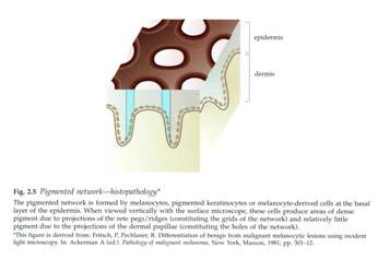
3 Dermoscopic criteria Terminology used in dermoscopy Colour Black Epidermis Light to dark brown Dermal epidermal junction Ink spot naevus Blue grey Papillary dermis Benign mole Steel blue Reticular dermis Superficial spreading melanoma Subcutaneous tissue Blue naevus 6 LSK Colour White Red Red black/blue black Regressive, scar like regions Cherry angioma Blood clot White Yellow Purple Keratin Keratin Haemangioma 7 LSK


4 Pattern: Global features Reticular pattern Globular pattern Pattern: Global features Pseudonetwork 9 LSK Pattern: Global features Homogeneous pattern Starburst pattern Cobblestone pattern Parallel pattern Lacunar pattern Unspecific pattern 4


5 Pattern: Local features Dots Globules Streaks Radial streaming Pseudopods Moth eaten borders Pattern: Local features Hypopigmentation Regression structures Blue white veil Blue grey ovoid nests Spoke wheel like structures Ulceration Pattern: Local features Comedo like openings Milia like cysts Fissures and ridges Central white patch Leaf like areas Fingerprint structures 14 LSK

 Dots/globules distributed")
 Five or six")


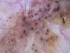
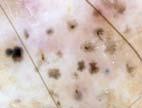
6 Pattern: Local features Red lacuna Wreath like vessels Arborising vessels Comma vessels Dotted vessels Hairpin vessels 15 LSK Melanoma features Global features Multicomponent pattern (3 or more patterns) Parallel pattern (along ridges; palms & soles only) Local features Atypical pigment network (branched, broken up, thickened, asymmetrical) Dots/globules distributed irregularly and of different sizes and shapes Asymmetrical blotches (featureless colours) Focal irregular streaking or peripheral linear projections (radial streaming and pseudopods) Five or six colours (black, brown, tan, grey, blue, red, white) Blue white veil over part of the lesion White scar like depigmentation Blue pepper like granules Irregular linear or dotted vessels, or polymorphous vascular pattern especially with milky red areas On face: grey dots, pseudonetwork, rhomboidal structures, asymmetrical pigmented follicles, annular granular structures On palms/soles: parallel ridge, irregular 6
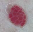
7 Approach to diagnosing pigmented lesions Two step procedure for differential diagnosis of pigmented skin lesions: Step 1: Differentiate between melanocytic and non melanocytic lesions Step 2: Differentiate between benign melanocytic lesions and melanoma Stolz W, et al. In: Argenziano G, et al., eds. First Congress of the International Dermoscopy Society (IDS) Apr 27 29; Naples. Dermatology. 2006;212: Step 1 Melanocytic vs. non melanocytic lesions Criteria Pigment network Pseudonetwork Aggregated globules Branched streaks Parallel pattern NO Criteria Homogeneous blue area NO YES YES Melanocytic lesion Blue naevus Criteria Milia-like cysts Comedo-like openings Fissure ridgeshairpin bessels Moth-eaten border Fingerprint structures Network-like structures NO YES Seborrhoeic keratosis 17 Soyer HP, et al. Color Atlas of Melanocytic Lesions of the Skin. Germany: Springer-Verlag; LSK Step 1 Melanocytic vs. non melanocytic lesions NO Criteria Red, blue-black lacunae Red-bluish to red-black homogeneous areas NO Criteria Arborising vessels Leaf-like areas Blue-grey ovoid nests Large blue-grey globules Spoke-wheel areas Ulceration Criteria None of the above NO YES YES YES Haemangioma Angiokeratoma Basal cell carcinoma Melanocytic lesion Soyer HP, et al. Color Atlas of Melanocytic Lesions of the Skin. Germany: Springer Verlag; LSK
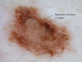
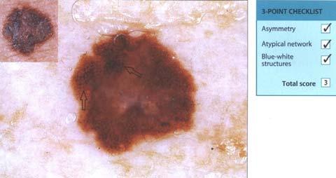
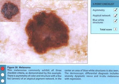
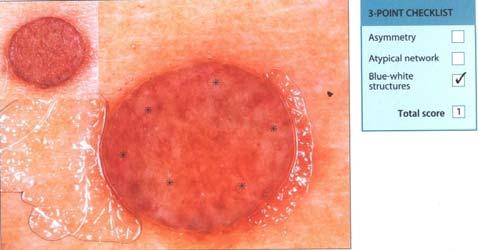
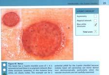
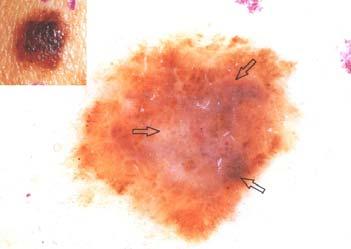
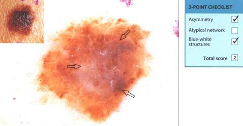
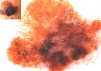
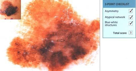
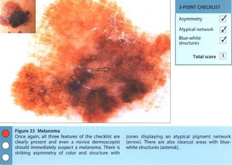
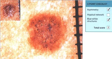
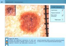
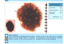
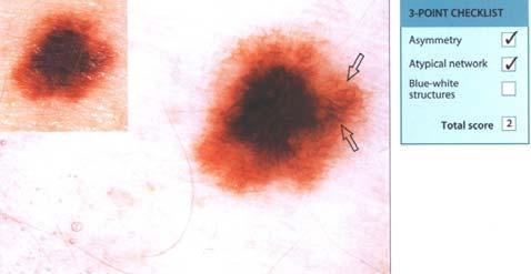
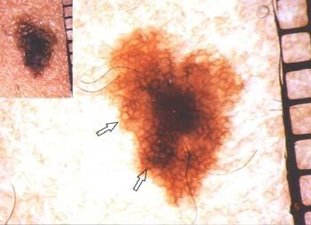
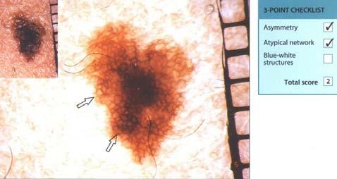
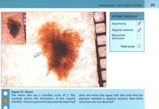
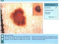
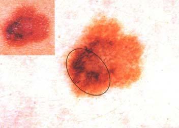
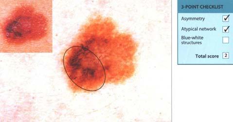
8 Diagnostic algorithms Second step Pattern analysis Pehamberger et al. J Inv Dermatol 1993 ABCD rule Stolz et al. Eur J Dermatol 1994 Menzies method Menzies et al. Arch Dermatol point check list Argenziano et al. Arch Dermatol 1998 Modified ABC rule JAAD point checklist Argenziano 2003 CASH JAAD 2007 Step 2 Benign vs. malignant melanocytic lesions 3 point checklist 1,2 Asymmetry Atypical network Blue white structures If lesion fulfils 2 criteria = suspicious lesion; biopsy 7 point checklist 3 Major criteria Points Atypical pigment network 2 Blue-white veil 2 Atypical vascular pattern 2 Minor criteria Irregular streaks 1 Irregular pigmentation 1 Irregular dots/globules 1 Regression structures 1 Score < 3 = Benign Score 3 = Malignant melanoma 1. Campos do Carmo G, Ramos e Silva M. Int J Dermatol. 2008;47: DermNet NZ. Three point checklist course/3 point checklist.html. 3. Argenziano G, et al. Arch Dermatol. 1998;134: LSK point checklist 8
9 Sensitivity 96.3 % 3 point checklist Sensitivity 96.3 % Specificity LOW 3 point checklist 1. Asymmetry 3 point checklist 9
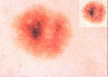
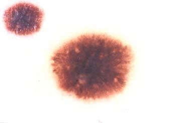
10 3 point checklist 1. Asymmetry (Asymmetry of colour and structure in one or two perpendicular axes. NOT shape) 10
11 11
12 12
 2.")
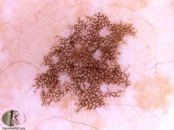
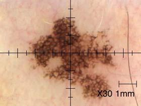
13 3 point checklist 1. Asymmetry (Asymmetry of colour and structure in one or two perpendicular axes. NOT shape) 2. Atypical network 3 point checklist 1. Asymmetry (Asymmetry of colour and structure in one or two perpendicular axes) 2. Atypical network (Pigment network with irregular holes and thick lines) 13
14 14
 3. Blue white structures 3 point checklist 1. Asymmetry (Asymmetry of colour and structure in one or two perpendicular axes) 2.")
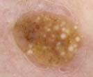
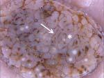
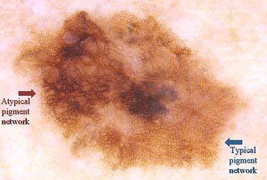
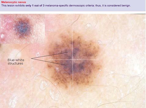
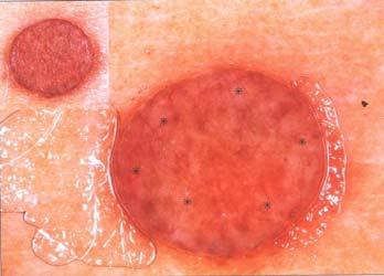
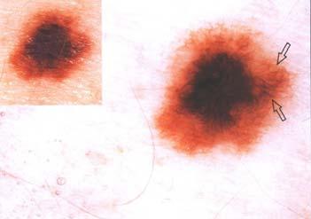
15 Typical pigment network Atypical pigment network 3 point checklist 1. Asymmetry (Asymmetry of colour and structure in one or two perpendicular axes) 2. Atypical network (Pigment network with irregular holes and thick lines) 3. Blue white structures 3 point checklist 1. Asymmetry (Asymmetry of colour and structure in one or two perpendicular axes) 2. Atypical network (Pigment network with irregular holes and thick lines) 3. Blue white structures (Any type of blue and/or white colour. Unless it occupies entire lesion) 15


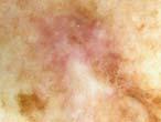
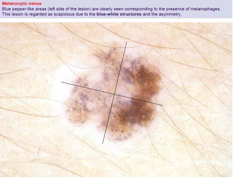
16 Blue white structures Pigmented melanophages or melanocytes of the dermis blue Thickened stratum corneum white Single most significant dermoscopic finding of invasive melanoma. Sensitivity of 51% and specificity of 97% If occupies entire lesion NOT blue white structures as in blue naevus 16

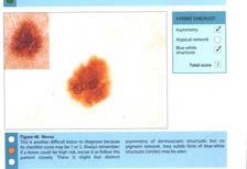
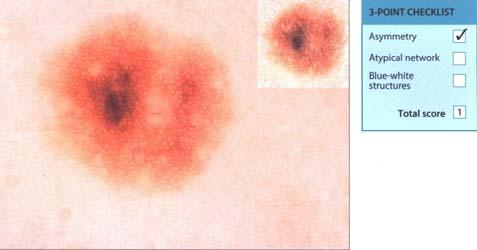
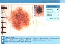
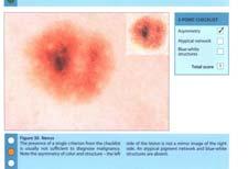
17 2 out of 3 = bx 3 point checklist 2 out of 3 = bx because... 3 point checklist 3 point checklist Using the 3 point checklist, determine if the following lesions require biopsy 17


18 Asymmetry Atypical network Blue white structures Asymmetry Atypical broad pigment network Blue white structures 7 point checklist Using the 7 point checklist, determine if the following lesions require biopsy 18


19 Case 1 7 point checklist Major criteria Points Atypical pigment network 2 Blue-white veil 2 Atypical vascular pattern 2 Minor criteria Irregular streaks 1 Irregular pigmentation 1 Irregular dots/globules 1 Regression structures 1 Case 7 point checklist Major criteria Points Atypical pigment network 2 Blue-white veil 2 Atypical vascular pattern 2 Minor criteria Irregular streaks 1 Irregular pigmentation 1 Irregular dots/globules 1 Regression structures 1 Case 3 7 point checklist Major criteria Points Atypical pigment network 2 Blue-white veil 2 Atypical vascular pattern 2 Minor criteria Irregular streaks 1 Irregular pigmentation 1 Irregular dots/globules 1 Regression structures 1 19
20 20
21 21

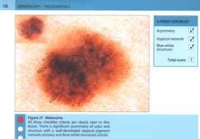
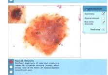
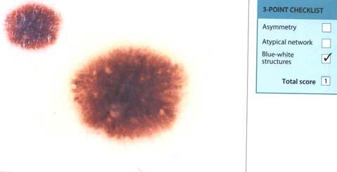
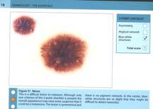
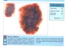
22 Cases Dermoscopy: The essentials 1 22
23 2 23
24 24
25 3 25
26 4 26
27 5 27
28 28
29 6 29
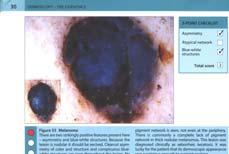
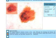
30 7 30
31 8 31
32 32

33 9 33
34 10 34
35 35


36 Other uses of dermoscopy in general practice Burrows Common wart Cutaneous lupus Lichen planus Porokeratosis Sebaceous hyperplasia 35 LSK Summary Use dermatoscope in correct manner Step 1?Pigmented lesion Step 2 Use algorythm Correlate clinically! Thank you 36
Fundamentals of dermoscopy
 Fundamentals of dermoscopy Learning objectives Upon completion of this session, participants should be able to: describe the basic principles of dermoscopy identify features associated with pigmented and
Appendix : Dermoscopy
 Go Back to the Top To Order, Visit the Purchasing Page for Details APP Appendix : Dermoscopy Dermoscopy, also known as dermatoscopy, epiluminoscopy and epiluminescent microscopy, is an effective non-invasive
Disclosure. Objectives. PAFP CME Conference Lou Mancano MD, FAAFP Reading Health System November 18, 2016
 PAFP CME Conference Lou Mancano MD, FAAFP Reading Health System November 18, 2016 1 Disclosure The speaker has no conflict of interest, financial agreement, or working affiliation with any group or organization.
Introduction to Dermoscopy. Nicholas Compton, MD June 16, 2010
 Introduction to Dermoscopy Nicholas Compton, MD June 16, 2010 Overview What is dermoscopy Brief history Types of dermoscopy General approach to lesion of interest 2 step algorithm 3-point checklist Practice
Dermoscopy: Recognizing Top Five Common In- Office Diagnoses
 Dermoscopy: Recognizing Top Five Common In- Office Diagnoses Vu A. Ngo, DO Department of Family Medicine and Dermatology Choctaw Nation Health Services Authority Learning Objectives Introduction to dermoscopy
Dermoscopy Quiz 3-Point Checklist Algorithm
 Dermoscopy Quiz 3-Point Checklist Algorithm GLOBAL PATTERN Globular LOCAL CRITERIA Aggregated globules Milia-like cysts 3 POINT CHECK LIST Symmetrical No abnormal net Slight Blue-white veil BENIGN MELANOCYTIC
MODULE 1. LOCAL AND GENERAL CRITERIA IN PIGMENTED MELANOCYTIC LESIONS.
 DERMOSCOPY TEACHING PROGRAMME Dermoscopy Teaching Programme Module 1 MODULE 1. LOCAL AND GENERAL CRITERIA IN PIGMENTED MELANOCYTIC LESIONS. Dermoscopy is a non-invasive in vivo technique that provides
It can be helpful in some cases of actinic keratosis, Bowen s disease and squamous cell carcinoma
 Dermoscopy Introduction, Terminology and Structures (to be read in conjunction with the Diagnostic Dermoscopic Algorithm) Copyright to Cunliffe TP (Jan. 2017) All rights reserved Introduction Dermoscopy
INTRODUCTION HOUSEKEEPING June 11 th Dr John Adams Dermatologist/Dermoscopist MOLEMAP NZ/Australia MOLESAFE USA
 INTRODUCTION HOUSEKEEPING June 11 th 2015 Dr John Adams Dermatologist/Dermoscopist MOLEMAP NZ/Australia MOLESAFE USA Program Skin cancer statistics. Dermoscopy description and usefulness. Patient /lesion
Clinical and Dermoscopic Features of Thin Nodular Melanoma
 Clinical and Dermoscopic Features of Thin Nodular Melanoma A study of the International Dermoscopy Society Coordinator: Dr. Alexander J. Stratigos and colleagues, alstrat2@gmail.com ** Extended to May
Dermoscopy in everyday practice. What and Why? When in doubt cut it out? Trilokraj Tejasvi MD
 Dermoscopy in everyday practice Trilokraj Tejasvi MD Assistant Professor, Department of Dermatology, Director Teledermatology services, University of Michigan, Faculty Associate, GLOBAL REACH, Michigan
Key factors in successfully integrating dermoscopy into your clinical practice
 Key factors in successfully integrating dermoscopy into your clinical practice S051 Dilemmas and challenges in skin cancer therapies and management Monday, March 4 th 2019 (9AM-12PM) Room 209A 10:56-11:09AM
Dermoscopy. Enhanced Diagnostic Ability: Pigmented Lesions. Ted Rosen, MD Baylor College of Medicine Houston, Texas
 Dermoscopy Enhanced Diagnostic Ability: Pigmented Lesions Ted Rosen, MD Baylor College of Medicine Houston, Texas Faculty Disclosure Statement No conflicts relevant to this workshop! Sir William Osler
comedo-like openings (clods, brown or orange & circles) milia-like cysts (dots or clods, white) 1/29/18 Dotted vessels are also commonly seen in SCC
 milia-like cysts (dots or clods, white) 1/29/18 Dotted vessels are also commonly seen in SCC") Brown circles Dotted vessels are also commonly seen in SCC Step1 1. Nevus (unequivocal) 2. DF/IDN 3. BCC 4. SCC Network Patchy network Peripheral network & central hypopigmentation DF: network with central
Brown circles Dotted vessels are also commonly seen in SCC Step1 1. Nevus (unequivocal) 2. DF/IDN 3. BCC 4. SCC Network Patchy network Peripheral network & central hypopigmentation DF: network with central
Dermoscopy. Synonyms. Dermoscopy. Definition. Dermoscopy opens up a world of colour and structure that can t be seen with the naked eye
 Synonyms Dermoscopy Australasian College of Dermatologists G.P Training Module Dermoscopy Dermatoscopy Epiluminescence microscopy Skin surface microscopy Incident light microscopy Oil immersion microscopy
Dermoscopy. Sir William Osler. Dermoscopy. Dermoscopy. Melanoma USA Primary Care Update Faculty Disclosure Statement
 Diagnostic Ability: Pigmented Lesions Ted Rosen, MD Baylor College of Medicine Houston, Texas Enhanced 2010 Primary Care Update Faculty Disclosure Statement Ted Rosen, MD Speakers Bureau: Abbott, Amgen,
Basics in Dermoscopy
 Basics in Dermoscopy Manal Bosseila Professor of Dermatology, Cairo University Member of European Academy Dermatology & Venereology EADV Member of International Dermoscopy Society IDS Member of Aesthetic
6/17/2018. Breaking Bad (Part 1) Dermoscopy of Brown(ish) Things. Bad?
 Dermoscopy of Brown(ish) Things. Bad?") Breaking Bad (Part 1) Dermoscopy of Brown(ish) Things Jennie T. Clarke, MD ssociate Professor of Dermatology University of Utah School of Medicine Bad? 1 Brown(ish) Things Bad Melanoma Pigmented basal
Breaking Bad (Part 1) Dermoscopy of Brown(ish) Things Jennie T. Clarke, MD ssociate Professor of Dermatology University of Utah School of Medicine Bad? 1 Brown(ish) Things Bad Melanoma Pigmented basal
Skin Cancer A Personal Approach. Dr Matthew Strack Dunedin New Zealand
 Skin Cancer A Personal Approach Dr Matthew Strack Dunedin New Zealand Outline Dermoscopy Instruments and setup Photochemosurgery Clinical Aim: Leave with 2-3 ideas JLE Benign Junctional Nevus Management
Dermoscopy STFM Richard Usatine, MD 5/2/16. Disclosure Statement: Some Dermatoscopes. Dermoscopy Video. Thanks to Dr.
 Disclosure Statement: Dermoscopy STFM 2016 Richard P. Usatine, MD, FAAFP Professor, Family and Community Medicine Professor, Dermatology and Cutaneous Surgery Medical Director, Clinic University of Texas
Dermoscopy-a BRIEF introduction
 Dermoscopy-a BRIEF introduction Aim of presentation -to tell you what dermoscopy is -to show some of what it can do -point the interested learner to further resources Overview of dermoscopy Dermoscopy
The impact of GP sub-specialisation and dermatoscopy use on diagnostic accuracy for melanomas in Australia
 The impact of GP sub-specialisation and dermatoscopy use on diagnostic accuracy for melanomas in Australia Cliff Rosendahl, Gail Williams, Diann Eley, Tobias Wilson, Greg Canning, Jeffrey Keir, Ian McColl,
Non-melanocytic Patterns
 Non-melanocytic Lesions Non-melanocytic Patterns Michelle Tarbox, MD Assistant Professor of Dermatology and Dermatopathology Texas Tech University Health Sciences Center 2018 Seborrheic keratoses Acanthotic
10/3/2018. Dermoscopy: Looking beneath the surface of the skin. Dermoscopy for Family Medicine 10/11/2018
 Dermoscopy for Family Medicine 10/11/2018 Jane M. Grant-Kels, MD, FAAD Founding Chair Emeritus, Dept of Dermatology Professor of Dermatology, Pathology & Pediatrics Director of the Cut Oncology Ctr & Melanoma
Dermoscopy, the use of a handheld
 ONLINE EXCLUSIVE Dermoscopy in family medicine: A primer Dermoscopy allows you to see deeper into the skin than with the naked eye. Here s how you can make use of it to spot malignant conditions sooner.
Non-Melanocytic Pattern Dermoscopy
 Non-Melanocytic Pattern Dermoscopy I have no conflicts of interest to disclose Except that I LOVE dermoscopy Michelle Tarbox, MD Assistant Professor of Dermatology and Dermatopathology Texas Tech University
Malignant non-melanocytic lesions
 Malignant non-melanocytic lesions Course C023: Fundamentals of Dermoscopy March 4, 2019, 11:20 AM - 11:50 PM Room: 146B Jason B. Lee, MD Professor & Vice Chair Director of Dermatopathology & Pigmented
50 interactive dermoscopic case discussions Dr Stephen Hayes
 50 interactive dermoscopic case discussions Dr Stephen Hayes Annotations will be found on your memory drive, as will 100 case discussions and other learning material Melanoma 2mm thick Ugly duckling-one
Skin lesions The Good and the Bad. Dr Virginia Hubbard Ipswich Hospital NHS Trust Barts and the London School of Medicine and Dentistry
 Skin lesions The Good and the Bad Dr Virginia Hubbard Ipswich Hospital NHS Trust Barts and the London School of Medicine and Dentistry Case 1 32 year old woman Australian Lesion on back New hair growing
STUDY. Epiluminescence Microscopy for the Diagnosis of Doubtful Melanocytic Skin Lesions
 STUDY Epiluminescence Microscopy for the Diagnosis of Doubtful Melanocytic Skin Lesions Comparison of the ABCD Rule of Dermatoscopy and a New 7-Point Checklist Based on Pattern Analysis Giuseppe Argenziano,
Regression 2/3/18. Histologically regression is characterized: melanosis fibrosis combination of both. Distribution: partial or focal!
 Regression Margaret Oliviero MSN, ARNP Harold S. Rabinovitz MD Histologically regression is characterized: melanosis fibrosis combination of both Distribution: partial or focal! Dermatoscopic terminology
Common Benign Lesions and Skin Cancers. 22nd May 2015 Dr Mark Foley
 Common Benign Lesions and Skin Cancers 22nd May 2015 Dr Mark Foley Thank you for downloading this file. This intended to supplement the presentation given at the NZ Wound Care Conference, it is not intended
What is Dermoscopy? Early Dermoscopes. Deciphering Dermoscopy: Terminology, Features & Algorithms 6/17/2018
 Deciphering Dermoscopy: Terminology, Features & Algorithms Where did it come from and why do we use it? Jennie T. Clarke, MD Associate Professor of Dermatology University of Utah School of Medicine What
Aspects on in vivo imaging techniques for diagnostics of pigmented skin lesions
 Thesis for the Degree of Doctor of Philosophy Aspects on in vivo imaging techniques for diagnostics of pigmented skin lesions Karin Terstappen (Westerhoff) Department of Dermatology and Venereology Institure
Acral and Mucosal Dermoscopy
 Acral and Mucosal Dermoscopy Caroline C. Kim, MD Assistant Professor, Department of Dermatology Harvard Medical School Director, Pigmented Lesion Clinic Associate Director, Cutaneous Oncology Program Beth
Revised Pattern Analysis: a method for the accurate diagnosis of pigmented skin lesions
 Dermatoscopy for Students A concise outline of: Revised Pattern Analysis: a method for the accurate diagnosis of pigmented skin lesions And Chaos and Clues: a decision algorithm for routine practice to
BJD British Journal of Dermatology. Summary. What s already known about this topic? CLINICAL AND LABORATORY INVESTIGATIONS
 CLINICAL AND LABORATORY INVESTIGATIONS BJD British Journal of Dermatology Pigmented nodular melanoma: the predictive value of dermoscopic features using multivariate analysis M.A. Pizzichetta, 1 H. Kittler,
Dermoscopic Features of Non-Pigmented Eccrine Poromas in. Department of Dermatology, Shinshu University School of Medicine,
 Original article Dermoscopic Features of Non-Pigmented Eccrine Poromas in Association with their Histopathological Features Akane Minagawa, Hiroshi Koga,* Masaomi Takahashi, + Kenji Sano, + Ryuhei Okuyama,
Diagnosis of Lentigo Maligna Melanoma. Steven Q. Wang, M.D. Memorial Sloan-Kettering Cancer Center Basking Ridge, NJ
 Diagnosis of Lentigo Maligna Melanoma Steven Q. Wang, M.D. Memorial Sloan-Kettering Cancer Center Basking Ridge, NJ Conflict of Interest: None Topics Epidemiology and Natural History Clinical and Histologic
Abrupt Intralesional Color Change on Dermoscopy as a New Indicator of Early Superficial Spreading Melanoma in a Japanese Woman
 Published online: June 24, 2015 1662 6567/15/0072 0123$39.50/0 This is an Open Access article licensed under the terms of the Creative Commons Attribution-NonCommercial 3.0 Unported license (CC BY-NC)
Graph-based Pigment Network Detection in Skin Images
 Graph-based Pigment Network Detection in Skin Images M. Sadeghi a,b, M. Razmara a, M. Ester a, T. K. Lee a,b,c, M. S. Atkins a a Simon Fraser University, 8888 University Drive, Burnaby, BC, Canada, V5A1S6;
Pathology of the skin. 2nd Department of Pathology, Semmelweis University
 Pathology of the skin 2nd Department of Pathology, Semmelweis University Histology of the skin Epidermis: Stratum corneum Stratum granulosum Stratum spinosum Stratum basale Dermis: papillary and reticular
Describe the functions of the vertebrate integumentary system. Discuss the structure of the skin and how it relates to function.
 Chapter 5 Describe the functions of the vertebrate integumentary system. Discuss the structure of the skin and how it relates to function. Explain the basis for different skin colors. Describe the structure
STUDY. Scott W. Menzies, MB,BS, PhD; Karin Westerhoff, MD; Harold Rabinovitz, MD; Alfred W. Kopf, MD; William H. McCarthy, MBBS, MEd; Brian Katz
 STUDY Surface Microscopy of Pigmented Basal Cell Carcinoma Scott W. Menzies, MB,BS, PhD; Karin Westerhoff, MD; Harold Rabinovitz, MD; Alfred W. Kopf, MD; William H. McCarthy, MBBS, MEd; Brian Katz Objectives:
Dermoscopy of non-pigmented skin lesions: a literature review
 Hong Kong J. Dermatol. Venereol. (2017) 25, 13-21 Review Article Dermoscopy of non-pigmented skin lesions: a literature review S Thomas, X Li, HP Soyer In this article, we will review benchmark dermoscopic
Benign versus Cancerous Lesions How to tell the difference FMF 2014 Christie Freeman MD, CCFP, DipPDerm, MSc
 1 Benign versus Cancerous Lesions How to tell the difference FMF 2014 Christie Freeman MD, CCFP, DipPDerm, MSc Benign lesions Seborrheic Keratoses: Warty, stuck-on Genetics and birthdays Can start in late
Multiple Primary Melanoma in a Thai Male: A Case Report
 Case Report Multiple Primary Melanoma in a Thai Male: A Case Report J Med Assoc Thai 2014; 97 (Suppl. 2): S234-S238 Full text. e-journal: http://www.jmatonline.com Kittisak Payapvipapong MD*, Pinyapat
STUDY. Identification of Clinically Featureless Incipient Melanoma Using Sequential Dermoscopy Imaging
 STUDY Identification of Clinically Featureless Incipient Melanoma Using Sequential Dermoscopy Imaging Harald Kittler, MD; Pascale Guitera, MD; Elisabeth Riedl, MD; Michelle Avramidis, MD; Ligia Teban,
Reports on Scientific Meetings
 Hong Kong J. Dermatol. Venereol. (2016) 24, 146-153 The Hong Kong Society of Dermatology and Venereology Annual Scientific Meeting 2016 Reported by BTH Chan, CT Chau, CW Chow, CC Koh, WYK Lam, BS Tong,
Introduction to Dermoscopy. Disclosure. Introduction
 Introduction to Dermoscopy 1 Disclosure Dr. Deborah Bren has no conflict of interest, financial agreement, or working affiliation with any group or organization. 2 Introduction Deborah A. Bren, DO Family
Yes. Breaking Bad II: Dermoscopy of Pink-ish Things. Does it Fit? Yes 6/17/2018. Yes. Joslyn Kirby, MD, MS, MEd
 Breaking Bad II: Dermoscopy of Pink-ish Things Joslyn Kirby, MD, MS, MEd Yes Observe Yes Step 2. Fit a Benign Nevus Pattern? Does it Fit? Step 1: Melanocytic? pigment network, globules, homogeneous? No
Prediction without Pigment: a decision algorithm for non-pigmented skin malignancy
 DERMATOLOGY PRACTICAL & CONCEPTUAL www.derm101.com Prediction without Pigment: a decision algorithm for non-pigmented skin malignancy Cliff Rosendahl 1, Alan Cameron 1, Philipp Tschandl 2, Agata Bulinska
Mole mapping and monitoring. Dr Stephen Hayes. Associate Specialist in Dermatology, University Hospital Southampton
 Mole mapping and monitoring Dr Stephen Hayes Associate Specialist in Dermatology, University Hospital Southampton Outline of presentation The melanoma epidemic Benefits of early detection Risks of the
Melanocytic Global Patterns Reticular Globular Cobblestone Homogeneous Starburst Multicomponent Nonspecific
 Step 1 Step 2 DERMOSCOPIC ANALYSIS CHECKLIST Melanocytic vs Nonmelanocytic Pigment network Brown dots / globules Homogeneous blue global pattern Acral patterns By default Melanocytic Global Patterns Reticular
Case Report Micromelanomas: A Review of Melanomas 2mmand a Case Report
 Case Reports in Oncological Medicine, Article ID 206260, 4 pages http://dx.doi.org/10.1155/2014/206260 Case Report Micromelanomas: A Review of Melanomas 2mmand a Case Report Sharad P. Paul 1,2,3 1 Skin
Histopathological and SIAscopic Correlation of Pigmented Skin Lesions
 Histopathological and SIAscopic Correlation of Pigmented Skin Lesions Professor Sujatha Fernando MBBS(Hon), MSc(London, Distinction), FRSTM&H, FRCPA, FIAC, FACTM Senior Consultant in Anatomical Pathology,
DERMATOLOGY PRACTICAL & CONCEPTUAL. Gabriel Salerni 1,2, Teresita Terán 3, Carlos Alonso 1,2, Ramón Fernández-Bussy 1 ABSTRACT
 DERMATOLOGY PRACTICAL & CONCEPTUAL www.derm101.com The role of dermoscopy and digital dermoscopy follow-up in the clinical diagnosis of melanoma: clinical and dermoscopic features of 99 consecutive primary
Dermatopathology: The tumor is composed of keratinocytes which show atypia, increase mitoses and abnormal mitoses.
 Squamous cell carcinoma (SCC): A common malignant tumor of keratinocytes arising in the epidermis, usually from a precancerous condition: 1- UV induced actinic keratosis, usually of low grade malignancy.
Principles of Dermatoscopy of Pigmented Skin Lesions
 Principles of Dermatoscopy of Pigmented Skin Lesions Wilhelm Stolz, U. Semmelmayer, K. Johow, and Walter H. C. Burgdorf There has been a dramatic increase in the incidence of malignant melanoma in most
Histopathology: skin pathology
 Histopathology: skin pathology These presentations are to help you identify, and to test yourself on identifying, basic histopathological features. They do not contain the additional factual information
STUDY. Characteristic Epiluminescent Microscopic Features of Early Malignant Melanoma on Glabrous Skin
 Characteristic Epiluminescent Microscopic Features of Early Malignant Melanoma on Glabrous Skin A Videomicroscopic Analysis STUDY Shinji Oguchi, MD; Toshiaki Saida, MD, PhD; Yoko Koganehira, MD; Sachiko
Review of vasculature visualized on dermoscopy
 doi: 10.1111/1346-8138.13686 Journal of Dermatology 2017; 44: 525 532 REVIEW ARTICLE Review of vasculature visualized on dermoscopy Yaei TOGAWA Department of Dermatology, Chiba University Graduate School
IV.4. Early Evolution of Melanoma (Small-Diameter Melanoma)
") Chapter Early Evolution of Melanoma (Small-Diameter Melanoma) Robert J. Friedman, Melanie Warycha, Michele Farber, Dina Gutkowicz-Krusin, Harold Rabinovitz, David Polsky, Margaret Oliviero, Darrell S.
Chapter Early Evolution of Melanoma (Small-Diameter Melanoma) Robert J. Friedman, Melanie Warycha, Michele Farber, Dina Gutkowicz-Krusin, Harold Rabinovitz, David Polsky, Margaret Oliviero, Darrell S.
Principles of Anatomy and Physiology
 Principles of Anatomy and Physiology 14 th Edition CHAPTER 5 The Integumentary System Introduction The organs of the integumentary system include the skin and its accessory structures including hair, nails,
STUDY. Dermoscopic Characteristics of Congenital Melanocytic Nevi Affecting Acral Volar Skin
 STUDY Dermoscopic Characteristics of Congenital Melanocytic Nevi Affecting Acral Volar Skin Akane Minagawa, MD; Hiroshi Koga, MD; Toshiaki Saida, MD, PhD Objective: To characterize the dermoscopic features
Benign and malignant epithelial lesions: Seborrheic keratosis: A common benign pigmented epidermal tumor occur in middle-aged or older persons more
 Benign and malignant epithelial lesions: Seborrheic keratosis: A common benign pigmented epidermal tumor occur in middle-aged or older persons more common on the trunk; but extremities, head and neck are
Chronology of lichen planus-like keratosis features by dermoscopy: a summary of 17 cases
 DERMATOLOGY PRACTICAL & CONCEPTUAL www.derm101.com Chronology of lichen planus-like keratosis features by dermoscopy: a summary of 17 cases Soko Watanabe 1, Mizuki Sawada 1, Itaru Dekio 1, Sumiko Ishizaki
VACAVILLE DERMATOLOGY
 Connecting the Dots on those Spots NANDAN V. KAMATH, M.D. VACAVILLE DERMATOLOGY Sources All of the photos were taken with permission from the Dermnet NZ website - Dermnet New Zealand after communicating
Finding Melanoma. Is not easy!
 Finding Melanoma Is not easy! Finding Melanoma Victoria mean depth at diagnosis is 1.5 mm. Melanoma 1.5mm Has Stage 1B Mortality 10% Melanoma Spotting a killer! Spotting a killer Visual Clues What are
Case Report Dermoscopy Clues in Pigmented Bowen s Disease
 Dermatology Research and Practice Volume 2010, Article ID 464821, 9 pages doi:10.1155/2010/464821 Case Report Dermoscopy Clues in Pigmented Bowen s Disease Daniela Gutiérrez-Mendoza, 1 Roberto Narro-Llorente,
Dermoscopic patterns in active and regressive lichen planus and lichen planus variants: a morphological study
 DERMTOLOGY PRCTICL & CONCEPTUL www.derm101.com Dermoscopic patterns in active and regressive lichen planus and lichen planus variants: a morphological study Şule Güngör 1, Ilteriş O. Topal 1, Emek K. Göncü
PDF of Trial CTRI Website URL -
 Clinical Trial Details (PDF Generation Date :- Wed, 25 Jul 2018 13:50:16 GMT) CTRI Number Last Modified On 10/05/2013 Post Graduate Thesis Type of Trial Type of Study Study Design Public Title of Study
Assisting diagnosis of melanoma through the noninvasive biopsy of skin lesions
 Assisting diagnosis of melanoma through the noninvasive biopsy of skin lesions Symon D Oyly Cotton Ela Claridge School of Computer Science, The University of Birmingham Birmingham B15 2TT, UK Per Hall
Due next week in lab - Scientific America Article Select one article to read and complete article summary
 Due in Lab 1. Skeletal System 33-34 2. Skeletal System 26 3. PreLab 6 Due next week in lab - Scientific America Article Select one article to read and complete article summary Cell Defenses and the Sunshine
BLINCK A diagnostic algorithm for skin cancer diagnosis combining clinical features with dermatoscopy findings
 DERMATOLOGY PRACTICAL & CONCEPTUAL www.derm101.com BLINCK A diagnostic algorithm for skin cancer diagnosis combining clinical features with dermatoscopy findings Peter Bourne, MBBS 1, Cliff Rosendahl,
The most common mistakes on dermatoscopy of melanocytic lesions
 Review paper The most common mistakes on dermatoscopy of melanocytic lesions Grażyna Kamińska-Winciorek 1, Waldemar Placek 2 1 The Center for Diagnostics and Treatment of Skin Diseases, Katowice, Poland
Rosettes in actinic keratosis and squamous cell carcinoma: distribution, association to other dermoscopic signs and description of the rosette pattern
 DOI: 10.1111/jdv.14474 JEADV ORIGINAL ARTICLE Rosettes in actinic keratosis and squamous cell carcinoma: distribution, association to other dermoscopic signs and description of the rosette pattern B. Lozano-Masdemont,
Integumentary System
 Integumentary System The integumentary system is commonly known as the Skin Largest organ of human body 10% total body weight and would cover over 20 square feet Functions of Skin 1. Protection Barrier
Description of Some Dermatoscopic Features of Acral Pigmented Lesions in Iranian Patients: A Preliminary Study
 ORIGINAL REPORT Description of Some Dermatoscopic Features of Acral Pigmented Lesions in Iranian Patients: A Preliminary Study Reza Nemati Ahmadabad 1, Hayede Ghaninezhad 1, Homayoon Moslehi 2, Sahar Azizahari
Skin and Body Membranes Body Membranes Function of body membranes Cover body surfaces Line body cavities Form protective sheets around organs
 Skin and Body Membranes Body Membranes Function of body membranes Cover body surfaces Line body cavities Form protective sheets around organs Classification of Body Membranes Epithelial membranes Cutaneous
Chapter 6 Skin and the Integumentary System. Skin Cells. Layers of Skin. Epidermis Dermis Subcutaneous layer beneath dermis not part of skin
 Chapter 6 Skin and the Integumentary System Composed of several tissues Maintains homeostasis Protective covering Retards water loss Regulates body temperature Houses sensory receptors Contains immune
F006 Imaging in Dermatology Melanocytic Neoplasia Clinical-Confocal-Pathological-Correlations
 F006 Imaging in Dermatology Melanocytic Neoplasia Clinical-Confocal-Pathological-Correlations Melissa Gill, MD SkinMedical Research and Diagnostics Dobbs Ferry, NY, USA Department of Pathology SUNY Downstate
R J M E Romanian Journal of Morphology & Embryology
 Rom J Morphol Embryol 2013, 54(2):315 320 ORIGINAL PAPER R J M E Romanian Journal of Morphology & Embryology http://www.rjme.ro/ Correlation of dermatoscopy with the histopathological changes in the diagnosis
Cover Page. The handle holds various files of this Leiden University dissertation.
 Cover Page The handle http://hdl.handle.net/1887/22172 holds various files of this Leiden University dissertation. Author: Rhee, Jasper Immanuel van der Title: Clinical characteristics and management of
Dr Amanda Oakley. Dermatologist Dept of Dermatology, Health Waikato Adjunct Associate Professor, Waikato Clinical Campus
 Dr Amanda Oakley Dermatologist Dept of Dermatology, Health Waikato Adjunct Associate Professor, Waikato Clinical Campus 14:00-16:00 WS #14: Dermoscopy Part 1 Skin Lesions and Dermatoscopy 16 August 2018
Total body photography in high risk patients
 Total body photography in high risk patients Doug Grossman, MD, PhD Department of Dermatology Huntsman Cancer Institute University of Utah Summer AAD F032 Practical Considerations for Patients with Melanoma
MECHANISMS OF HUMAN DISEASE: LABORATORY SESSION PATHOLOGY OF THE SKIN LAB. Friday, February 12, :30 am 11:00 am
 MECHANISMS OF HUMAN DISEASE: LABORATORY SESSION PATHOLOGY OF THE SKIN LAB Friday, February 12, 2012 9:30 am 11:00 am FACULTY COPY GOALS: Describe the basic clinical and morphologic features of various
INCREASE IN incidence and mortality rates for
 Skin Research and Technology 2005; 11: 236 241 Copyright & Blackwell Munksgaard 2005 Printed in Denmark. All rights reserved Skin Research and Technology Pigment distribution in melanocytic lesion images:
11/8/2012. Chapter 6 Part 1 Objectives: Skin = Integument = Cutaneous Membrane. The Structure of Skin. Epidermis
 Chapter 6 Part 1 Objectives: Define organ, and associate the skin as an organ of the integumentary system. List the general functions of the skin. Describe the structure of the layers of the skin. Summarize
Conflicts. Objectives. University of Texas Health Science Center at San Antonio. Pediatrics Grand Rounds 24 August Pediatric Dermatology 101
 Pediatric Dermatology 101 John C. Browning, MD, FAAD, FAAP Conflicts Investigator: ViroXis Advisor: ViroXis Advisory Board: TopMD Speaker: Galderma Objectives Understand the meaning and importance of cutaneous
p. 1 ABCD Rule of Dermoscopy p. 1 ABC Point List of Dermoscopy p. 1 Abrupt Cut-off of the Trabeculae p. 2 Abrupt Edge p. 2 Abrupt Pigment Breaks in
 p. 1 ABCD Rule of Dermoscopy p. 1 ABC Point List of Dermoscopy p. 1 Abrupt Cut-off of the Trabeculae p. 2 Abrupt Edge p. 2 Abrupt Pigment Breaks in the Trabeculae p. 2 Acantholysis p. 2 Acanthoma p. 2
Accepted Article. Dermoscopic diagnosis of amelanotic/hypomelanotic melanoma
 Received Date : 19-May-2016 Revised Date : 01-Sep-2016 Accepted Date : 20-Sep-2016 Article type : Research Letter Dermoscopic diagnosis of amelanotic/hypomelanotic melanoma M.A. Pizzichetta, 1 H. Kittler,
Dermatoscopic features of cutaneous non-facial non-acral lentiginous growth pattern melanomas
 DERMATOLOGY PRACTICAL & CONCEPTUAL www.derm101.com Dermatoscopic features of cutaneous non-facial non-acral lentiginous growth pattern melanomas Jeff Keir 1 1 Department of Dermatology, School of Medicine,
Melanoma and Dermoscopy. Disclosure Statement: ABCDE's of melanoma. Co-President, Usatine Media
 Melanoma and Dermoscopy Richard P. Usatine, MD, FAAFP Professor, Family and Community Medicine Professor, Dermatology and Cutaneous Surgery Medical Director, University Skin Clinic University of Texas
Features Causing Confusion between Basal Cell Carcinoma and Squamous Cell Carcinoma in Clinical Diagnosis
 TH Ryu, et al pissn 1013-9087ㆍeISSN 2005-3894 Ann Dermatol Vol. 30, No. 1, 2018 https://doi.org/10.5021/ad.2018.30.1.64 ORIGINAL ARTICLE Features Causing Confusion between Basal Cell Carcinoma and Squamous
Pattern Analysis in Dermoscopic Images
 Pattern Analysis in Dermoscopic Images Aurora Sáez, Begoña Acha and Carmen Serrano Abstract In this chapter an extensive review of algorithmic methods that automatically detect patterns in dermoscopic
Human Anatomy & Physiology
 PowerPoint Lecture Slides prepared by Barbara Heard, Atlantic Cape Community College Ninth Edition Human Anatomy & Physiology C H A P T E R 5 Annie Leibovitz/Contact Press Images 2013 Pearson Education,
Skin and Body Membranes
 4 Skin and Body Membranes PowerPoint Lecture Slide Presentation by Jerry L. Cook, Sam Houston University ESSENTIALS OF HUMAN ANATOMY & PHYSIOLOGY EIGHTH EDITION ELAINE N. MARIEB Skin and Body Membranes
The Integumentary System
 The Integumentary System The Integumentary System Integument is skin Skin and its appendages make up the integumentary system A fatty layer (hypodermis) lies deep to it Two distinct regions Epidermis Dermis