Prenatal Diagnosis of Central Nervous System (CNS) Pathologies: does Fetal MRI help in their management?
|
|
|
- Bernard Fitzgerald
- 5 years ago
- Views:
Transcription
 Pathologies:")


1 Prenatal Diagnosis of Central Nervous System (CNS) Pathologies: does Fetal MRI help in their management? Daniela Prayer, Division of Neuroradiology and Musculoskeletal Radiology Medical University Vienna/Austria
2 Fetal MRI: Why? Indications: ACOG Recommendations Common Indications: elevated BMI Oligo/ Anhydramnios Scarring of the abdomen Position of the fetus that allows only restricted US assesment Special Indications: Situations, where MRI allows a better estimation of the intrauterine situation than US alone


3 Increased BMI + Anhydramnios GW 19+1
: Callosal agenesis Posterior fossa anomalies Microcephaly Indicated (30-48%): Ventriculomegaly Neural tube defects Diaphragmatic Hernia Low priority (10-30%):")
4 Fetal MRI: Why? Definitely indicated (>48%): Callosal agenesis Posterior fossa anomalies Microcephaly Indicated (30-48%): Ventriculomegaly Neural tube defects Diaphragmatic Hernia Low priority (10-30%): Pulmonary anomalies, Multiple malformations Abdominal wall defects Very Low priority (0-10%): Congenital heart defects, Urinary tract, Twins, Cleft lip UOG 2017, in press


5 Fetal MRI: How? Modern 1.5 or 3T, big bores less claustrophobia


6 Fetal MRI: How? Large coils: more receptor elements, but heavier


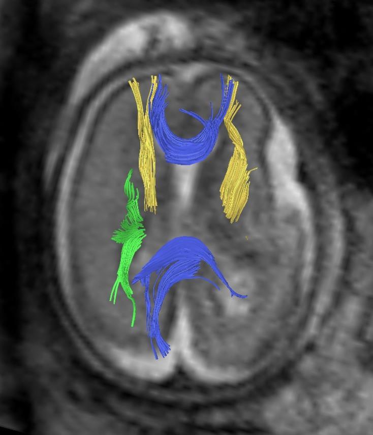
7 Fetal MRI: How? GW 20+4 Sequence = Choice of parameters influencing signals, resolution Continuous series of images In 20 sec


8 T2 T1 EPI Angiography FLAIR Diffusion- Thick slab Dynamic Metabolic Perfusion- information f-mri, 3D
 therapy required recurrence risk")
9 pathology present? no yes survival? no yes yes but (surgical) therapy required recurrence risk prenatal prognosis postnatal
10 pathology present? Frequent question: isolated ventriculomegaly on Ultrasound Ventricle in mm Classification Normal Borderline Mild VM <10mm 8.5mm 10mm 10mm 15mm Severe VM >15mm Pagani G, Thilaganathan B, Prefumo F. Neurodevelopmental outcome in isolated mild fetal ventriculomegaly: systematic review and meta-analysis. UOG. 2014;44(3):


11 pathology present? Premature gyri yes Cobble stone Liss encephaly GW 24 healthy GW 23+5 GW 29+3

12 pathology present? no MRI does not only show the surface of the brain but also the developing parenchyma GW 20+4 * * * * Most important structure: subplate (*) : integrity crucial for normal cortical devlopment Kostovic I: The Anatomical Record 267 ;1-6 (2002)


13 pathology present? no * * GW 23 * * FLAIR T2 GW 29 From GW 24 onwards Subplate (*) better visible on FLAIR than on T2


14 pathology present? no GW 26 GW 31 Borderline Ventriculomegaly delineation of subplate normal! 2


15 pathology present? Thin subplate yes Type I Liss encephaly Shallow insular cistern GW 22 GW 20 normal


16 survival? no Brainstem segmentation disorder Corticospinal tract absent Infratentorially* Cleft palate GW 22 Brainstem interrupted * Visualised by MRI -tractography


17 survival? no Trisomy 13 GW 33+5 Lobar holoprosencephaly Missing venous duct Referal because of cardiac malformation facial cleft and rocker bottom feet on US

18 survival? Yes. Females only but. Dysplastic cortex GW 25 female Corpus callosum agenesis with Probst Bundles* * Visualised by MRI-tractography
19 survival? Yes. Females only but. Fernández-Ramos JA et al. Rev Neurol.2013 Dec 1; (11): Aicardi syndrome presumably X-linked dominant, agenesis of the corpus callosum, chorioretinal lacunae infantile spasms, with lethality in males. Seizures severe neurological impairment.
")


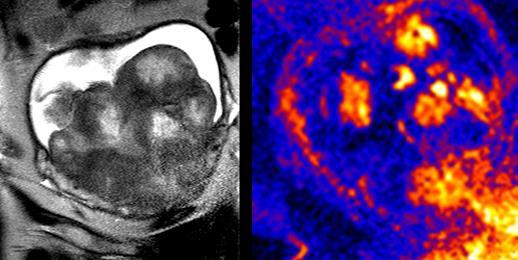
20 survival? yes but (surgical) therapy required prenatal Fetuses with open dystaphism improve after prenatal closure of the cele GW 32, Chiari II malformation
 therapy required prenatal GW 23 before Fetuses with open dysraphism improve")
21 survival? yes but (surgical) therapy required prenatal GW 23 before Fetuses with open dysraphism improve after prenatal closure of the cele GW 28 after Courtesy Gregor Kasprian Vienna/ Houston


22 open neural tube defects 1 Questions for fetal MRI Spina bifida? closed neural tube defects 1 potentially treatable! Prenatal surgery unnecessary Myelo- meningocele Myelocele Meningocele Myelo- 1: Tortori-Donati P, Rossi AMD, Biancheri R. Pediatric neuroradiology. Berlin ; [Great Britain]: Springer cystocele


23 GW 29+4 GW 31+5


24 survival? GW 28+5 yes but (surgical) therapy required postnatal Postnatal shunt with less impact on vermian herniation 2Mo Danzer E et al. Dev Med Child Neurol. 2012;54(1):8-14.
")

25 survival? yes but (surgical) therapy required GW 28+5 postnatal Late spinal complications 3a 12a


26 recurrence risk 2 month old with seizures Partial Callosal Agenesis? Callosal Dysgenesis? Schizencephalic clefts?


27 recurrence risk Fetal Thrombotic Vasculopathy GW 26 Demised Co-Twin Sato Y, Benirschke K. Pediatrics. 2006;117(1):113-7.


28 recurrence risk Subependymal heterotopia 1 st Fetus GW nd Fetus GW21+3 mother Filamin A gene mutation! Parrini et al. Brain Jul;129(Pt 7): , Brain 2006


.")
29 prognosis Spinal level in open defects? GW, 22 GW 20 GW, 22 Hipflection: L1/2 Kneeflection: Kneeextgension: L3 Foot dorsiflection: Foot plantarflection: sacral L4 L5 Lindseth RE. (1976) Treatment of the lower extremity in children paralyzed by myelomeningocele (birth to 18 months).aaosic Lectures 25:


30 prognosis Spinal level in open defects? Meningocele! L1 GW,22 GW 20 GW 22 assessments correlated ± 1 Level of anatomical defect Pedreira DA et al. Am J Obstet Gynecol. 2016


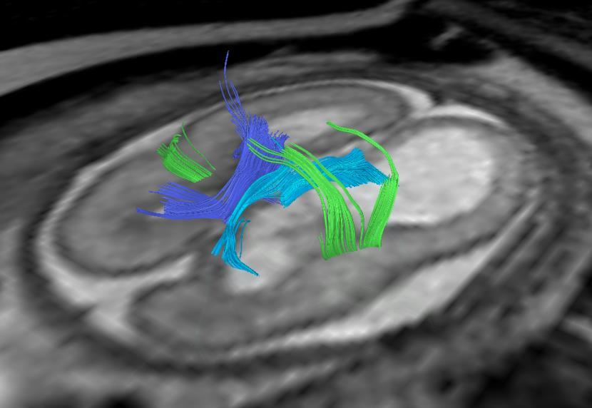
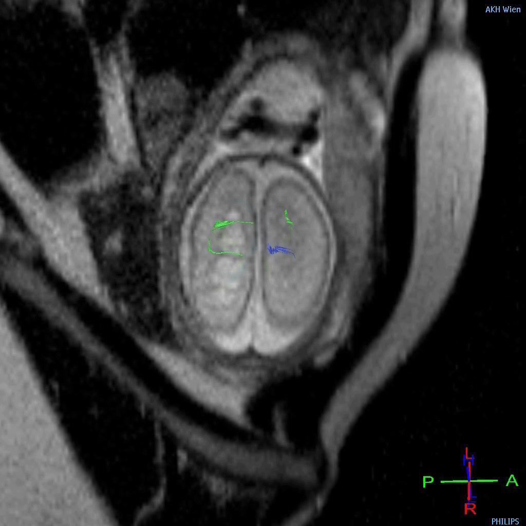
31 prognosis Use of diffusion- tensor imaging Diffusion weighted imaging measures degree and directionality of water motion in tissue Stochastic motion Brownian motion Isotropy Anisotropy


32 GW 25+6 Corpus callosum Questions for fetal MRI prognosis Anisotropic Diffusion-weighted Imaging GW 21+3

33 prognosis Connectivity at GW 23
34 prognosis Callosal Hypogenesis GW 22


35 prognosis GW36

36 prognosis GW 22


37 prognosis GW 22 5a

38 prognosis At 5 Years Normal intelligence Symmetrical use of arms Epilepsy with rare seizures, EEG focus left hemisphere


39 Normal CC agnesis CC agenesis with malformations Take home: Fetal MRI can help with more accurate prenatal diagnosis and prognosis and thus support the managment of a complicated pregnancy

40 Fetal MRI Precongress course September 15th
Fetal CNS MRI. Daniela Prayer. Division of Neuroradiology And Musculoskeletal Radiology. Medical University of Vienna, AUSTRIA
 Fetal CNS MRI Daniela Prayer Division of Neuroradiology And Musculoskeletal Radiology Medical University of Vienna, AUSTRIA Methods Normal development Malformations Acquired pathology MR- methods for assessment
Prenatal Prediction of The Neurologically Impaired Neonate By Ultrasound
 Prenatal Prediction of The Neurologically Impaired Neonate By Ultrasound Robert H. Debbs, D.O.,F.A.C.O.O.G. Professor of OB-GYN Perelman School of Medicine, University of Pennsylvania Director, Pennsylvania
Fetal Medicine. Case Presentations. Dr Ermos Nicolaou Fetal Medicine Unit Chris Hani Baragwanath Hospital. October 2003
 Case Presentations Dr Ermos Nicolaou Fetal Medicine Unit Chris Hani Baragwanath Hospital October 2003 Case 1 Ms A M 22year old P0 G1 Referred from Sebokeng Hospital at 36w for polyhydramnios On Ultrasound:
Supplementary Online Content
 Supplementary Online Content Honein MA, Dawson AL, Petersen E, et al; US Zika Pregnancy Registry Collaboration. Birth Defects Among Fetuses and Infants of US Women With Laboratory Evidence of Possible
Central nervous system. Obstetrics Content Outline Obstetrics - Fetal Abnormalities
 Obstetrics Content Outline Obstetrics - Fetal Abnormalities Many congenital malformations of the CNS result from incomplete closure of the neural tube Effective February 2007 10 16% the most common neural
Basic Training. ISUOG Basic Training Examining the Upper Lip, Face & Profile
 ISUOG Examining the Upper Lip, Face & Profile Learning objectives At the end of the lecture you will be able to: Describe how to obtain the 3 planes required to assess the anatomy of the fetal face Recognise
The Brain: Prenatal and Postnatal Effects of Congenital Heart Disease. Dianna M. E. Bardo, M D Swedish Cherry Hill Radia, Inc.
 The Brain: Prenatal and Postnatal Effects of Congenital Heart Disease Dianna M. E. Bardo, M D Swedish Cherry Hill Radia, Inc. Seattle, WA embryology We recognize the VACTERL association and frequency of
Spine and spinal cord
 NEURORADIOLOGY Spine and spinal cord Erika Vörös University of Szeged Department of Radiology SZEGED DISEASES OF SPINE AND SPINAL CORD I. Non-tumourous diseases developmental anomalies vascular disorders
CNS Embryology 5th Menstrual Week (Dorsal View)
") Imaging of the Fetal Brain; Normal & Abnormal Alfred Abuhamad, M.D. Eastern Virginia Medical School CNS Embryology 5th Menstrual Week (Dorsal View) Day 20 from fertilization Neural plate formed in ectoderm
Imaging of the Fetal Brain; Normal & Abnormal Alfred Abuhamad, M.D. Eastern Virginia Medical School CNS Embryology 5th Menstrual Week (Dorsal View) Day 20 from fertilization Neural plate formed in ectoderm
Spectrum of Cranio-facial anomalies during 2 Ultrasound. trimester on
 Spectrum of Cranio-facial anomalies during 2 Ultrasound nd trimester on Poster No.: C-0378 Congress: ECR 2015 Type: Scientific Exhibit Authors: K. Dave, S. Solanki; Ahmedabad/IN Keywords: Obstetrics (Pregnancy
Supplemental Information
 ARTICLE Supplemental Information SUPPLEMENTAL TABLE 6 Mosaic and Partial Trisomies Thirty-eight VLBW infants were identified with T13, of whom 2 had mosaic T13. T18 was reported for 128 infants, of whom
Basic Training. ISUOG Basic Training The 20 Planes Approach to the Routine Mid Trimester Scan
 ISUOG The 20 Planes Approach to the Routine Mid Trimester Scan Learning objective At the end of the lecture you will be able to: Explain how to perform a structured routine examination, including measurements,
Symposium: OB/GY US (Room B) CNS Anomalies
 CNS Anomalies") 82 Symposium: OB/GY US (Room B) 11 : 50 1 2 : 10 CNS Anomalies Brain area Midline structure S u p r a t e n t o r i a l ventricular system Cerebral hemisphere Posterior fossa Head size and shape Image
82 Symposium: OB/GY US (Room B) 11 : 50 1 2 : 10 CNS Anomalies Brain area Midline structure S u p r a t e n t o r i a l ventricular system Cerebral hemisphere Posterior fossa Head size and shape Image
intracranial anomalies
 Chapter 5: Fetal Central Nervous System 84 intracranial anomalies Hydrocephaly Dilatation of ventricular system secondary to an increase in the amount of CSF. Effects of hydrocephalus include flattening
Measurements of the Posterior Fossa in Normal Fetus MRI
 Measurements of the Posterior Fossa in Normal Fetus MRI Ber Roee, 3 rd year medical student, Sackler School of Medicine, Tel Aviv University Supervised by: Dr. Katorza Eldad, Antenatal Diagnostic Unit,The
Han-Sung Kwon M.D. Department of Obstetrics and Gynecology Konkuk University School of Medicine Seoul, Korea
 Han-Sung Kwon M.D. Department of Obstetrics and Gynecology Konkuk University School of Medicine Seoul, Korea Embryologic features of the developing hindbrain Embryologic features of the developing hindbrain
Central Nervous System Congenital Abnormalities
 Central Nervous System Congenital Abnormalities Eva Brichtova, M.D., Ph.D., Department of Pediatric Sugery, Orthopaedics and Traumatology, University Hospital Brno Neural tube defects Dysraphism uncomplete
Introduction to Neurosurgical Subspecialties:
 Introduction to Neurosurgical Subspecialties: Pediatric Neurosurgery Brian L. Hoh, MD 1 and Gregory J. Zipfel, MD 2 1 University of Florida, 2 Washington University Pediatric Neurosurgery Pediatric neurosurgeons
Obstetrics Content Outline Obstetrics - Fetal Abnormalities
 Obstetrics Content Outline Obstetrics - Fetal Abnormalities Effective February 2007 10 16% renal agenesis complete absence of the kidneys occurs when ureteric buds fail to develop Or degenerate before
Ultrasound Anomaly Details
 Appendix 2. Association of Copy Number Variants With Specific Ultrasonographically Detected Fetal Anomalies Ultrasound Anomaly Details Abdominal wall Bladder exstrophy Body-stalk anomaly Cloacal exstrophy
DWI assessment of ischemic changes in the fetal brain
 DWI assessment of ischemic changes in the fetal brain Dafi Bergman, 4 th year Medical student in the 4-year program, Sackler school of medicine B.Sc Life and Medical Sciences, Tel Aviv University Supervised
ISUOG Basic Training. Assessing the Neck & Chest Gihad Chalouhi, Lebanon
 ISUOG Basic Training Assessing the Neck & Chest Gihad Chalouhi, Lebanon Learning objectives 9 & 10 At the end of the lecture you will be able to: recognise the differences between the normal & most common
Developmental Neuropathology
 Developmental Neuropathology Pathology, Radiology, and Clinical Correlations Reid Heffner MD Distinguished Teaching Professor Department of Pathology and Anatomy I HAVE NO CONFLICTS OF INTEREST OR DISCLOSURES
ISUOG Basic Training Distinguishing Between Normal and Abnormal Appearances of the Fetal Anatomy. Basic Training
 ISUOG Distinguishing Between Normal and Abnormal Appearances of the Fetal Anatomy Learning Objective At the end of the lecture you will be able to: Compare the differences between the ultrasound appearances
ISUOG Basic Training Distinguishing Between Normal and Abnormal Appearances of the Fetal Anatomy
 ISUOG Basic Training Distinguishing Between Normal and Abnormal Appearances of the Fetal Anatomy Reem S. Abu-Rustum, Lebanon Learning Objective At the end of the lecture you will be able to: Compare the
Elliott Sherr, MD University of California, San Francisco
 University of California, San Francisco ACC AND A SSOCIATED F EATURES MRI features associated with ACC Clinical diagnoses found in individuals with ACC Clinical Syndromes in which ACC is a component or
SPLIT NOTOCHORD SYNDROME ASSOCIATION. DR. Hasan Nugud Consultant Paediatric Surgeon
 SPLIT NOTOCHORD SYNDROME ASSOCIATION DR. Hasan Nugud Consultant Paediatric Surgeon CASE PRESENTATION :- New born baby, boy, referred to the paediatric surgical team at the age of 14 hours. Birth History
Pregnancy and Epilepsy
 Pregnancy and Epilepsy Nowhere is the problem more evident or more complicated than in pregnancy. In the United States, epilepsy affects nearly one million women of childbearing potential. Alarm bells
Central nervous system
 Chapter 2 Central nervous system NORMAL SONOGRAPHIC ANATOMY The fetal brain undergoes major developmental changes throughout pregnancy. At 7 weeks of gestation, a sonolucent area is seen in the cephalic
Chapter 8. Pediatric Surgery
 Chapter 8 Pediatric Surgery 8.1 Hydrocephalus Hydrocephalus is a congenital disorder. There may be difficulties during normal vaginal delivery due large size of the head. In 1970s, when these pictures
A 50 Year Experience with Management of Spina Bifida Aperta : Myelomeningocele
 A 50 Year Experience with Management of Spina Bifida Aperta : Myelomeningocele David B. Shurtleff, M. D. Professor Department of Pediatrics University of Washington Seattle, Washington, USA Etiology of
A Case of Naso-Ethmoidal Meningoencephalocele
 A Case of Naso-Ethmoidal Meningoencephalocele Divyanshu Dubey, Sonjjay Pande, Pradeep Dubey, Anshudha Sawhney Vol. 3 No. 8 (August 2011) International Journal of Collaborative Research on Internal Medicine
Nationwide Survey of the Etiology and Associated Conditions of Prenatally and Postnatally Diagnosed Congenital Hydrocephalus in Japan
 Neurol Med Chir (Tokyo) 47, 448 452, 2007 Nationwide Survey of the Etiology and Associated Conditions of Prenatally and Postnatally Diagnosed Congenital Hydrocephalus in Japan Kouzo MORITAKE, HidemasaNAGAI,
The Fetal Cardiology Program
 The Fetal Cardiology Program at Texas Children s Fetal Center About the program Since the 1980s, Texas Children s Fetal Cardiology Program has provided comprehensive fetal cardiac care to expecting families
Lung sequestration and Scimitar syndrome
 Lung sequestration and Scimitar syndrome Imaging approaches M. Mearadji International Foundation for Pediatric Imaging Aid Rotterdam, The Netherlands Pulmonary sequestration Pulmonary sequestration (PS)
Appendix 3.5 Case Inclusion Guidance for Potentially Zika-related Birth Defects
 Appendix 3.5 Case Inclusion Guidance for Potentially Zika-related Birth Defects Appendix 3.5 A3.5-1 Case Definition Appendix 3.5 Case Inclusion Guidance for Potentially Zika-related Birth Defects Contents
Malformations of the Nervous System November 10, Dr. Peter Ostrow
 Malformations of the Nervous System November 10, 2016 Dr. Peter Ostrow Malformations of the Nervous System 1. Abnormal closure of the neural tube 1. Disorders of forebrain formation 1. Cortical anomalies
High spatial resolution reveals excellent detail in pediatric neuro imaging
 Publication for the Philips MRI Community Issue 46 2012/2 High spatial resolution reveals excellent detail in pediatric neuro imaging Achieva 3.0T with 32-channel SENSE Head coil has become the system
Neuropathology Specialty Conference
 Neuropathology Specialty Conference March 22, 2010 Case 2 Rebecca Folkerth, MD Brigham and Women s Hospital Children s Hospital Harvard Medical School Clinical History 18-gestational-week fetus found on
A Retrospective Analysis of Clinical Profile and Surgical Outcome in Patients with Spinal Dysraphism at Tertiary Care Center
 Original Research Article A Retrospective Analysis of Clinical Profile and Surgical Outcome in Patients with Spinal Dysraphism at Tertiary Care Center Premlal KV * Assistant Professor, Department of Neurosurgery,
Use of MRI in Evaluating Fetal Ventriculomegaly Lisa McLeod, Harvard Medical School Year III Gillian Lieberman, MD
 January 2004 Use of MRI in Evaluating Fetal Ventriculomegaly Lisa McLeod, Harvard Medical School Year III http://bidmc.harvard.edu/content/departments/radiology/files/fetalatlas/default.htm Objectives:
The Fetal Care Center at NewYork-Presbyterian/ Weill Cornell Medicine
 The Fetal Care Center at NewYork-Presbyterian/ Weill Cornell Medicine Prompt and Personalized Care for Women with Complex Pregnancies A Team of Experts additional training in maternal and fetal complications
Fetal Renal Malformations: The Role of Ultrasound in Diagnosis & Management
 Fetal Renal Malformations: The Role of Ultrasound in Diagnosis & Management 12 weeks Alfred Abuhamad, M.D. Eastern Virginia Medical School 13 weeks 2nd trimester Medullary pyramids Renal Sinus Cortex 2nd
Developmental Posterior Fossa Abnormalities with Associated Supratentorial Findings
 Developmental Posterior Fossa Abnormalities with Associated Supratentorial Findings Seattle Children s Hospital Christopher J Hurt, MD Gisele E Ishak, MD Dennis W Shaw, MD Introduction Barkovich has classified
Congenital Anomalies
 Congenital Anomalies Down Syndrome 7580 7580 DOWN''S SYNDROME Q900 Q90.0 : Trisomy 21, meiotic nondisjunction 7580 7580 DOWN''S SYNDROME Q901 Q90.1 : Trisomy 21, mosaicism (mitotic nondisjunction) 7580
NEURORADIOLOGY Part I
 NEURORADIOLOGY Part I Vörös Erika University of Szeged Department of Radiology SZEGED DISEASES OF CNS BRAIN Developmental anomalies Cerebrovascular disorders Tumours Inflammatory diseases Trauma DISEASES
Perinatal Neuroradiology
 Fabio Triulzi Cristina Baldoli Cecilia Parazzini Andrea Righini Perinatal Neuroradiology From the Fetus to the Newborn 123 Perinatal Neuroradiology Fabio Triulzi Cristina Baldoli Cecilia Parazzini Andrea
Posterior fossa malformations
 ANDREA ROSSI, MD Head, Department of Pediatric Neuroradiology G. Gaslini Children s Research Hospital Genoa Italy andrearossi@ospedale-gaslini.ge.it Posterior fossa malformations Cerebellar ataxia Hypotonia
MRI OF FETAL DEVELOPMENTAL ANOMALIES
 MRI OF FETAL DEVELOPMENTAL ANOMALIES Nadine J Girard, MD, PhD Department of Neuroradiology Timone Hospital 264 rue Saint Pierre, 13385 Marseille cedex 5, France nadine.girard@ap-hm.fr fax number : 33 4
International Journal of Pharma and Bio Sciences. Meckel-Gruber Syndrome Associated with CNS Malformations A Case Report
 International Journal of Pharma and Bio Sciences RESEARCH ARTICLE PATHOLOGY Meckel-Gruber Syndrome Associated with CNS Malformations A Case Report Corresponding Author DR. N. HIMA BINDU Assistant Professor,
Magnetic Resonance Imaging of the fetus
 Magnetic Resonance Imaging of the fetus Mary A Rutherford Perinatal Imaging Group, MRC Clinical Sciences Centre Imperial College m.rutherford@imperial.ac.uk The Moonbeam Trust Overview Practicalities and
JMSCR Vol 4 Issue 02 Page February 2016
 www.jmscr.igmpublication.org Impact Factor 3.79 Index Copernicus Value: 5.88 ISSN (e)-2347-176x ISSN (p) 2455-0450 DOI: http://dx.doi.org/10.18535/jmscr/v4i02.65 Septo-Optic Dysplasia: A Case Report Authors
PRENATAL DIAGNOSIS OF ARACHNOID CYSTS
 REVIEW ARTICLE PRENATAL DIAGNOSIS OF ARACHNOID CYSTS Chih-Ping Chen* Department of Obstetrics and Gynecology, Mackay Memorial Hospital, Taipei, and Department of Biotechnology, Asia University, Taichung,
ISUOG Basic Training. Examining Fetal Anatomy from Longitudinal Sections Titia Cohen-Overbeek, The Netherlands
 ISUOG Basic Training Examining Fetal Anatomy from Longitudinal Sections Titia Cohen-Overbeek, The Netherlands Learning objectives 2 & 3 At the end of the lecture you will be able to: describe how to obtain
Complex Hydrocephalus
 2012 Hydrocephalus Association Conference Washington, DC - June 27-July1, 2012 Complex Hydrocephalus Marion L. Walker, MD Professor of Neurosurgery & Pediatrics Primary Children s Medical Center University
Development of the Nervous System. Leah Militello, class of 2018
 Development of the Nervous System Leah Militello, class of 2018 Learning Objectives 1. Describe the formation and fate of the neural tube and neural crest including timing and germ layer involved. 2. Describe
Agenesis of the corpus callosum
 Agenesis of the corpus callosum What is a callosal disorder? Disorders of the corpus callosum are conditions in which the corpus callosum does not develop in a typical manner. Since these are disorders
Pediatric Spinal Anomalies
 Department of Radiology University of California San Diego Pediatric Spinal Anomalies John R. Hesselink, M.D. Spine Embryogenesis 1. Primitive streak 2. Proliferation of cells at primitive pit (Hensen's
ISUOG Basic Training. Distinguishing between Normal & Abnormal Appearances of the Urinary Tract. Seshadri Suresh, India
 ISUOG Basic Training Distinguishing between Normal & Abnormal Appearances of the Urinary Tract Seshadri Suresh, India Learning objectives 13 & 14 At the end of the lecture you will be able to: describe
Congenital Diaphragmatic Hernia information for parents. David M Notrica MD FACS FAAP Pediatric Surgeons of Phoenix
 Congenital Diaphragmatic Hernia information for parents David M Notrica MD FACS FAAP Pediatric Surgeons of Phoenix CDH Congenital absence of a portion of the diaphragm allowing abdominal contents to migrate
ISUOG Basic Training Distinguishing between Normal & Abnormal Appearances of the Long Bones & Extremities. Basic Training
 ISUOG Basic Training Distinguishing between Normal & Abnormal Appearances of the Long Bones & Extremities Basic Training Learning objectives At the end of the lecture you will be able to: Describe how
Genetic test for Bilateral frontoparietal polymicrogyria
 Genetic test for Bilateral frontoparietal polymicrogyria Daniela Pilz, Cardiff UKGTN Genetic testing for neurological conditions; London February 26 th 2013 Region-specific Polymicrogyria (PMG) bilateral
KYAMC Journal Vol. 8, No.-1, July Two Cases of Holoprosencephalies
 Case Report Two Cases of Holoprosencephalies Sharif M M 1, Parvin K 2, Rahman M T 3, Ullah N 4, Islam S 5 Abstract Two pregnant women with around 33-34 weeks of gestation were reported to Gynaecology and
Pediatric Neurointervention: Vein of Galen Malformations
 Pediatric Neurointervention: Vein of Galen Malformations Johanna T. Fifi, M.D. Assistant Professor of Neurology, Neurosurgery, and Radiology Icahn School of Medicine at Mount Sinai November 9 th, 2014
Torch Infections and Prenatal Ultrasound Findings
 Tutorial [1] August 09, 2011 By Eran Casiff, MD [2] TORCH INFECTIONS AND PRENATAL ULTRASOUND FINDINGS Eran Casiff M.D. Department of Obstetrics and Gynecology Kaplan Medical Center Rehovot 76100, Israel
NYEIS Version 4.3 (ICD) ICD - 10 Codes Available in NYEIS at time of version launch (9/23/2015)
 ICD - 10 Codes Available in NYEIS at time of version launch (9/23/2015)") D82.1 Di George's syndrome E63.9 Nutritional deficiency, unspecified E70.21 Tyrosinemia E70.29 Other disorders of tyrosine metabolism E70.30 Albinism, unspecified E70.5 Disorders of tryptophan metabolism
D82.1 Di George's syndrome E63.9 Nutritional deficiency, unspecified E70.21 Tyrosinemia E70.29 Other disorders of tyrosine metabolism E70.30 Albinism, unspecified E70.5 Disorders of tryptophan metabolism
Neuropathic bladder and spinal dysraphism
 Archives of Disease in Childhood, 1981, 56, 176-180 Neuropathic bladder and spinal dysraphism MALGORZATA BORZYSKOWSKI AND B G R NEVILLE Evelina Children's Department, Guy's Hospital, London SUMMARY The
Accuracy of the Fetal Echocardiogram in Double-outlet Right Ventricle
 Blackwell Publishing IncMalden, USACHDCongenital Heart Disease 2006 The Authors; Journal compilation 2006 Blackwell Publishing, Inc.? 200723237Original ArticleFetal Echocardiogram in Double-outlet Right
Ring 18. Our Mission: To help individuals. they face so they might lead healthy, happy, and productive lives. Normal Chromosomes
 The Chromosome 18 Registry & Research Society Ring 18 There are five major conditions involving large changes of chromosome 18. Each of these conditions has a wide variety of characteristics. Additionally,
Fetal MRI. Page 1 of 69
 Fetal MRI Poster No.: C-0878 Congress: ECR 2015 Type: Educational Exhibit Authors: Y. Kocaba# Köksel, M. A. Oztek, C. Y. Sanhal, S. Toru, #. Mendilcio#lu, M. #im#ek, K. Karaali; Antalya/TR Keywords: Obstetrics,
MALFORMATIONS OF CORTICAL DEVELOPMENT: A PICTORIAL REVIEW
 MALFORMATIONS OF CORTICAL DEVELOPMENT: A PICTORIAL REVIEW Padmaja K. Naidu, M.D. Usha D. Nagaraj, M.D. William T. O Brien, Sr., D.O. AOCR Annual Conference 2018 Scottsdale, Arizona @CincyKidsRad facebook.com/cincykidsrad
Neuro-Anatomical Study of a Rare Brain Malformation: Lissencephaly, A Report of Eleven Patients
 Original Research Article Neuro-Anatomical Study of a Rare Brain Malformation: Lissencephaly, A Report of Eleven Patients Kataria Sushma K 1, Gurjar Anoop S 2,*, Parakh Manish 3, Gurjar Manisha 4, Parakh
FETAL ICD-10 CODES QUICK REFERENCE GUIDE
 FETAL ICD-10 CODES QUICK REFERENCE GUIDE Page CONTENTS 1 Cardiac Anomalies 3 Chromosome Abnormalities 4 Central Nervous System Anomalies 5 Extremity Anomalies 6 Face / Neck Anomalies 7 Gastrointestinal
Imaging of Pediatric Epilepsy MRI. Epilepsy: Nonacute Situation
 Imaging of Pediatric Epilepsy Epilepsy: Nonacute Situation MR is the study of choice Tailor MR study to suspected epileptogenic zone Temporal lobe Extratemporal A. James Barkovich, MD University of California
Fetal Tetralogy of Fallot
 36 Fetal Tetralogy of Fallot E.D. Bespalova, R.M. Gasanova, O.A.Pitirimova National Scientific and Practical Center of Cardiovascular Surgery, Moscow Elena D. Bespalova, MD Professor, Director Rena M,
Major Forms of Congenital Heart Disease: Consultant Pediatric and Fetal Cardiology King Abdulaziz Cardiac Center, National Guard Hospital Riyadh
 Major Forms of Congenital Heart Disease: Impact of Prenatal Detection and Diagnosis Dr Merna Atiyah Consultant Pediatric and Fetal Cardiology King Abdulaziz Cardiac Center, National Guard Hospital Riyadh
Postmortem MR imaging has been used to evaluate the fetal
 ORIGINAL RESEARCH E. Widjaja S. Geibprasert S. Zarei Mahmoodabadi N.E. Brown P. Shannon Corroboration of Normal and Abnormal Fetal Cerebral Lamination on Postmortem MR Imaging with Postmortem Examination
CYANOTIC CONGENITAL HEART DISEASES. PRESENTER: DR. Myra M. Koech Pediatric cardiologist MTRH/MU
 CYANOTIC CONGENITAL HEART DISEASES PRESENTER: DR. Myra M. Koech Pediatric cardiologist MTRH/MU DEFINITION Congenital heart diseases are defined as structural and functional problems of the heart that are
PIAF study: Placental insufficiency and aortic isthmus flow Jean-Claude Fouron, MD
 Dear colleagues, I would like to thank you very sincerely for agreeing to participate in our multicentre study on the clinical significance of recording fetal aortic isthmus flow during placental circulatory
Basic Training. ISUOG Basic Training Distinguishing Between Normal & Abnormal Appearances of the Skull & Brain
 ISUOG Distinguishing Between Normal & Abnormal Appearances of the Skull & Brain Learning objectives At the end of the lecture you will be able to: Describe how to obtain the 3 planes required to assess,
ESP 755A SUMMER Multiple Choice Identify the choice that best completes the statement or answers the question. 1. Autosomal recessive disorders
 ESP 755A SUMMER 2017 Multiple Choice Identify the choice that best completes the statement or answers the question. 1. Autosomal recessive disorders a. affect only males c. are caused when the abnormal
Corporate Medical Policy
 Corporate Medical Policy Invasive Prenatal (Fetal) Diagnostic Testing File Name: Origination: Last CAP Review: Next CAP Review: Last Review: invasive_prenatal_(fetal)_diagnostic_testing 12/2014 3/2018
Neuroembryology II. Dr. Newton COPH G210
 Neuroembryology II Dr. Newton COPH G210 Anterior and posterior neuropore closure at E25 & E27, respectively, is essential for normal nervous system development. NTDs occur 1/1K births. Incidence can be
Isolated Choroid Plexus Cyst
 Isolated Choroid Plexus Cyst This guideline was updated in April 2015 by Dr Joana De Sousa, with input from members of the New Zealand Maternal Fetal Medicine Network. Background Midtrimester soft markers
Arnold Chiari Malformation - A hospital based autopsy study
 Rapotra Megha et al / International Journal of Biomedical Research 2017; 8(05): 250-254. 250 International Journal of Biomedical Research ISSN: 0976-9633 (Online); 2455-0566 (Print) Journal DOI: https://dx.doi.org/10.7439/ijbr
National follow-up program CPUP Pediatric Neurology paper form
 National follow-up program CPUP Pediatric Neurology paper form 110206 1 National Follow-Up program- CPUP Pediatric Neurology Personal nr (unique identifier): Last name: First name: Region child belongs
Technical advances in fetal MR imaging have made it an
 REVIEW ARTICLE O.A. Glenn J. Barkovich Magnetic Resonance Imaging of the Fetal Brain and Spine: An Increasingly Important Tool in Prenatal Diagnosis: Part 2 SUMMARY: Fetal MR imaging is an increasingly
SWISS SOCIETY OF NEONATOLOGY. Yunis-Varon syndrome
 SWISS SOCIETY OF NEONATOLOGY Yunis-Varon syndrome January 2003 2 Heyland K, Hodler C, Bänziger O, Neonatology, University Children s Hospital of Zurich, Switzerland Swiss Society of Neonatology, Thomas
GU Ultrasound in First Trimester
 Fetal Renal Malformations: The Role of Ultrasound in Diagnosis & Management Outline 1. Renal Anomalies Urinary Tract Dilation Aberrant Early Development Defects Terminal Maturation Alfred Abuhamad, M.D.
ETHICAL CHALLENGES IN PEDIATRIC PALLIATIVE CARE
 ETHICAL CHALLENGES IN PEDIATRIC PALLIATIVE CARE Disclosure I have no financial relationships to disclose. Glen Medellin, MD, FAAP, FAAHPM Learning Objectives Define the basic principles of ethical analysis
Grand Rounds Mullerian Anomalies. Sara Schaenzer, PGY-3 9/26/18
 Grand Rounds Mullerian Anomalies Sara Schaenzer, PGY-3 9/26/18 Background Congenital uterine anomalies occur in 2-4% of women Three times more common in women with recurrent pregnancy loss True incidence
Systematic approach to Fetal Echocardiography. Objectives. Introduction 11/2/2015
 Systematic approach to Fetal Echocardiography. Pediatric Echocardiography Conference, JCMCH November 7, 2015 Rajani Anand Objectives Fetal cardiology pre-test Introduction Embryology and Physiology of
How to recognise a congenitally infected fetus? Dr. Amar Bhide Consultant in Obstetrics and Fetal Medicine
 How to recognise a congenitally infected fetus? Dr. Amar Bhide Consultant in Obstetrics and Fetal Medicine Scope Cytomegalovirus Parvovirus Varicella Toxoplasma Rubella Clinical scenarios Maternal exposure
Chiari Malformations. Google. Objectives Seventh Annual NKY TBI Conference 3/22/13. Kerry R. Crone, M.D.
 Chiari Malformations Kerry R. Crone, M.D. Professor of Neurosurgery and Pediatrics University of Cincinnati College of Medicine University of Cincinnati Medical Center Cincinnati Children s Hospital Medical
An Unusual Kind Of Traumatic Intracranial Hemorrhage: Post Traumatic Bleed Into The Schizencephalic Cleft
 ISPUB.COM The Internet Journal of Radiology Volume 8 Number 2 An Unusual Kind Of Traumatic Intracranial Hemorrhage: Post Traumatic Bleed Into The Schizencephalic J T, V Rajendran, E Devarajan Citation
ISUOG Basic Training. Distinguishing Between Normal & Abnormal Appearances of the Skull & Brain. Seshadri Suresh, India
 ISUOG Basic Training Distinguishing Between Normal & Abnormal Appearances of the Skull & Brain Seshadri Suresh, India Learning objectives 4 & 5 At the end of the lecture you will be able to: Describe how
From Head to Toe Use of Advanced Dynamic Flow in prenatal ultrasound
 From Head to Toe Use of Advanced Dynamic Flow in prenatal ultrasound Without doubt, the B- Schwerdtfeger, R. tant diagnostic instrument. Furthermore, we use colour in feto- mode imaging is the most important
Summary. HVRA s Cardio Vascular Genetic Detailed L2 Obstetrical Ultrasound. CPT 76811, 76825, _ 90% CHD detection. _ 90% DS detection.
 What is the role of fetal echocardiography (2D 76825, cardiovascular color flow mapping 93325) as performed in conjunction with detailed fetal anatomy scan (CPT 76811) now that AIUM requires limited outflow
Neuro. Development. Judy Philbrook, NNP-BC. ! Primary neurulation! Prosencepahlic! Neuronal proliferation. ! 3-4 weeks! 2-3 months!
 Neuro Judy Philbrook, NNP-BC Microsoft clip art Development! Primary neurulation! Prosencepahlic! Neuronal proliferation! Neuronal migration! Organization! Myelination! 3-4 weeks! 2-3 months! 3-4 months!
Analysis of errors made on in utero MR studies of the foetal brain in the MERIDIAN study
 European Radiology (2019) 29:195 201 https://doi.org/10.1007/s00330-018-5508-x MAGNETIC RESONANCE Analysis of errors made on in utero MR studies of the foetal brain in the MERIDIAN study Ruth Batty 1 Mary