Prenatal Prediction of The Neurologically Impaired Neonate By Ultrasound
|
|
|
- Eleanor Lawson
- 5 years ago
- Views:
Transcription
1 Prenatal Prediction of The Neurologically Impaired Neonate By Ultrasound Robert H. Debbs, D.O.,F.A.C.O.O.G. Professor of OB-GYN Perelman School of Medicine, University of Pennsylvania Director, Pennsylvania Hospital MFM Network
2 Dr. Debbs has no Conflict of Interest to disclose Dr. Debbs has no Financial or Scientific disclosures Dr. Debbs has no Off- Label disclosures.
3 Introduction Maladaptation of Fetal Brain Structural Abnormalities Perinatal Infections Metabolic Diseases Newborn Encephalopathy Brain injury of the Very Low Birth Weight Preterm Neonate Mechanical in utero injury, compression
4 Objectives Identify most common CNS malformations on prenatal Ultrasound Develop an a protocol for Ultrasound of the CNS of a fetus Identify normal variant from pathology on prenatal CNS ultrasound


5 Psychosocial Maladaptation Delivery explains < 15% of Neurological injuries
6 Fetal Brain Development Neural tube differentiation Cellular differentiation Neuronal migration Neuroblast differentiation Brain growth l Dobbings & Sands, 1979 l Sherman et al., 1985
7 Structural Abnormalities Ventriculomegaly Agenesis of Corpus Collosum Dandy Walker Cyst Syringomelia Microcephaly Arachnoid Cyst Brain destructive lesions Choroid Plexus Cyst

8 Anencephaly..(A no brainer!)

9 Transventricuar &Transcerebellar Planes

10 Normal Fetal Brain
11 Borderline Lateral Ventriculomegaly 10-15mm 22% abnormal outcome, without MRI 4-8% abnormal with normal MRI 3.7% perinatal death 3.8% Chromosomal abnormality 11.5% cognitive =/- motor delay 22 v. 4 % abnormal neuro F>M Pilu et al: Us Obstet Gynecol 2002

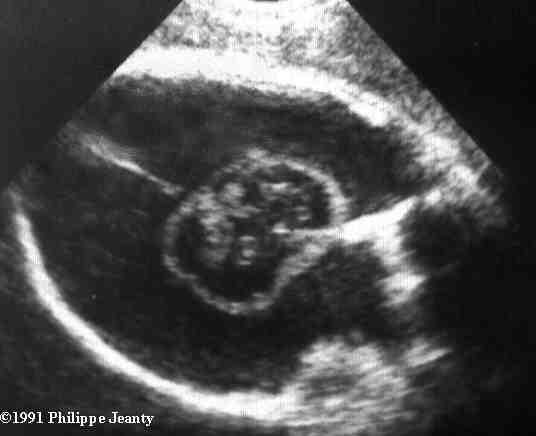
12 Ventriculomegaly atria Dangling choroid
13 Ventriculomegaly 1% at weeks >10mm 1-2/1000 develop hydrocephalus Borderline 10-15mm Morbidity related to other problems 3% chromosomal Syndromic/hemorrhage/infection 6-10% mild to moderate delay Maldevelopment or destructive lesion
14 Agenesis of Corpus Callosum Complete or partial 5/1,000 Maldevelopment or destructive Chromosomal(21/13/18),p deletion syndromes Syndromic Isolated

15 Corpus Callosum
16

17 Agenesis Of The Corpus Callosum

18 Power Angiography

19 MRI of Corpus Callosum
20 Agenesis of Corpus Callosum 90 % normal development if isolated Early intervention crucial Hard to R/O some syndromes(aicardi) l ACC, Epilepsy, Retinal dysplasia Developmental delay usually from associated anomalies Sporadic / X-linked
21 Dandy Walker Complex 1/30,000 births Complete or partial agenesis of vermis with enlarged posterior fossa Variant-partial agenesis with normal posterior fossa Mega Cisterna Magna Aneuploidy/syndromic/isolated Warfarin Half isolated

22 Dandy Walker Malformation Dandy Walker Malformation Variant Megacisternamagna

23 Dandy Walker Variant
24 Dandy Walker Complex 20% mortality 50% neuro abnormality with syndrome Much better prognosis is variant Megacisternamagna usually normal outcome
25 Syringomelia Fluid filled cavity in spinal canal l Usually cervical in origin l Associate with Chiari type II herniation l Can be asymptomatic for years l Can present with varying degree of neurologic impairment l Reported with in utero compression, tumor, arachnoiditis, PPROM, infection l Can be idiopathic
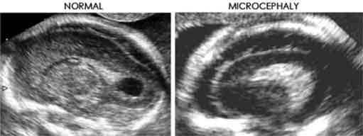
26 Syringomelia Mid forceps rotation, no longer done Can be developmental from in-utero injury l MVA l Mal-positioning (common) l Myomas with severe compression l Causes neurogenic atrophy of phrenic, intercostal, and spinaothalamic tract

27 Reed Group

28

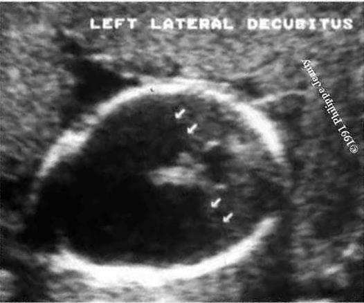
29
30

31 ATROPHY
32 Syringomelia Also seen with Spinal muscular atrophy mutation, Williams syndrome If no focal hemorrhage, edema, necrosis then likely prenatal injury Intrapartum injury usually from head entrapment in breech vaginal attempt In utero malposition plays larger role than once thought l Found at normal vertex delivery or cesarean section without trauma
33 Microcephaly 1 per 1,000 births Chromosomal/syndromic Fetal hypoxia Congenital infection Radiation.>10 rads Teratogen..ie warfarin/etoh Need progressive decrease > 2sd below mean > 50% severe mental retardation
34 Microcephaly
35 Microcephaly
36 Arachnoid Cyst Fluid filled cysts in arachnoid space Rare..<1 in 30,000 Cause unknown Sonolucent cysts with thin regular outline No blood flow No ventricular connection
37 Arachnoid Cyst Not associated with loss of tissue Most in midline Differential l Glioependymal cyst l Porencephalic cyst

38 Arachnoid Cysts

39 Arachnoid Cyst
40 Arachnoid Cysts Prognosis lintracranial HTN with large cysts...neuro surgery lnormal intellectual development in > 90%
41 Brain Destructive Lesions Hydranencephaly Porencephaly Schizencephaly l1 in 10,000 births
42 Hydranencephaly Sporadic l Bilateral carotid artery occlusion l Leukomalacia with confluence l Diffuse Hypoxic ischemic necrosis l Infection/necrotizing vasculitis l Trophoblastic material from twin
43
44

45 Hydranencephaly

46
47 Hydranencephaly Differential l Extreme hyrocephalus..important! l Holoprosencephaly l Porencephaly MRI most helpful Prognosis l Universally poor l Lethal by 1 year Jeanty/Romero/callan

48 Brain Destructive lesions
49 Porencephaly Infarction of cerebral arteries Hemorrhage into brain parenchyma Cystic areas in cortex communicating with the ventricle Within fissures or midline. compresses brain Prognosis variable..size/location.. l >50% severe handicaps
50 Schizencephaly Bilateral clefts Connects lateral ventricles to subarachnoid space Associated with absence of cavum septum pellucidum Prognosis poor with severe developmental delay and seizures

51 Choroid Plexus Cysts
52 Choroid Plexus Cysts Found in 2% at Anatomy scan All resolve usually by 26 weeks Cellular debris occludes capillaries causing cyst during normal development Cysts themselves of no pathologic significance
53 Choroid Plexus Cysts Must look for other abnormalities Isolated cysts may raise aneuploid risk by a factor of for trisomy 18 If normal biochemistry, no other markers and less than 35, relative risk is lower than risk of loss from amniocentesis Trisch et al, 2000

54 Thank You
intracranial anomalies
 Chapter 5: Fetal Central Nervous System 84 intracranial anomalies Hydrocephaly Dilatation of ventricular system secondary to an increase in the amount of CSF. Effects of hydrocephalus include flattening
CNS Embryology 5th Menstrual Week (Dorsal View)
") Imaging of the Fetal Brain; Normal & Abnormal Alfred Abuhamad, M.D. Eastern Virginia Medical School CNS Embryology 5th Menstrual Week (Dorsal View) Day 20 from fertilization Neural plate formed in ectoderm
Imaging of the Fetal Brain; Normal & Abnormal Alfred Abuhamad, M.D. Eastern Virginia Medical School CNS Embryology 5th Menstrual Week (Dorsal View) Day 20 from fertilization Neural plate formed in ectoderm
Central nervous system
 Chapter 2 Central nervous system NORMAL SONOGRAPHIC ANATOMY The fetal brain undergoes major developmental changes throughout pregnancy. At 7 weeks of gestation, a sonolucent area is seen in the cephalic
Symposium: OB/GY US (Room B) CNS Anomalies
 CNS Anomalies") 82 Symposium: OB/GY US (Room B) 11 : 50 1 2 : 10 CNS Anomalies Brain area Midline structure S u p r a t e n t o r i a l ventricular system Cerebral hemisphere Posterior fossa Head size and shape Image
82 Symposium: OB/GY US (Room B) 11 : 50 1 2 : 10 CNS Anomalies Brain area Midline structure S u p r a t e n t o r i a l ventricular system Cerebral hemisphere Posterior fossa Head size and shape Image
Central nervous system. Obstetrics Content Outline Obstetrics - Fetal Abnormalities
 Obstetrics Content Outline Obstetrics - Fetal Abnormalities Many congenital malformations of the CNS result from incomplete closure of the neural tube Effective February 2007 10 16% the most common neural
Fetal Medicine. Case Presentations. Dr Ermos Nicolaou Fetal Medicine Unit Chris Hani Baragwanath Hospital. October 2003
 Case Presentations Dr Ermos Nicolaou Fetal Medicine Unit Chris Hani Baragwanath Hospital October 2003 Case 1 Ms A M 22year old P0 G1 Referred from Sebokeng Hospital at 36w for polyhydramnios On Ultrasound:
Neurosonography: State of the art
 Neurosonography: State of the art Lisa H Lowe, MD, FAAP Professor and Academic Chair, University MO-Kansas City Pediatric Radiologist, Children s Mercy Hospitals and Clinics Learning objectives After this
Supplementary Online Content
 Supplementary Online Content Honein MA, Dawson AL, Petersen E, et al; US Zika Pregnancy Registry Collaboration. Birth Defects Among Fetuses and Infants of US Women With Laboratory Evidence of Possible
Neuropathology Specialty Conference
 Neuropathology Specialty Conference March 22, 2010 Case 2 Rebecca Folkerth, MD Brigham and Women s Hospital Children s Hospital Harvard Medical School Clinical History 18-gestational-week fetus found on
Use of MRI in Evaluating Fetal Ventriculomegaly Lisa McLeod, Harvard Medical School Year III Gillian Lieberman, MD
 January 2004 Use of MRI in Evaluating Fetal Ventriculomegaly Lisa McLeod, Harvard Medical School Year III http://bidmc.harvard.edu/content/departments/radiology/files/fetalatlas/default.htm Objectives:
Complex Hydrocephalus
 2012 Hydrocephalus Association Conference Washington, DC - June 27-July1, 2012 Complex Hydrocephalus Marion L. Walker, MD Professor of Neurosurgery & Pediatrics Primary Children s Medical Center University
Appendix 3.5 Case Inclusion Guidance for Potentially Zika-related Birth Defects
 Appendix 3.5 Case Inclusion Guidance for Potentially Zika-related Birth Defects Appendix 3.5 A3.5-1 Case Definition Appendix 3.5 Case Inclusion Guidance for Potentially Zika-related Birth Defects Contents
Spectrum of Cranio-facial anomalies during 2 Ultrasound. trimester on
 Spectrum of Cranio-facial anomalies during 2 Ultrasound nd trimester on Poster No.: C-0378 Congress: ECR 2015 Type: Scientific Exhibit Authors: K. Dave, S. Solanki; Ahmedabad/IN Keywords: Obstetrics (Pregnancy
Measurements of the Posterior Fossa in Normal Fetus MRI
 Measurements of the Posterior Fossa in Normal Fetus MRI Ber Roee, 3 rd year medical student, Sackler School of Medicine, Tel Aviv University Supervised by: Dr. Katorza Eldad, Antenatal Diagnostic Unit,The
Prenatal Diagnosis of Central Nervous System (CNS) Pathologies: does Fetal MRI help in their management?
 Pathologies: does Fetal MRI help in their management?") Prenatal Diagnosis of Central Nervous System (CNS) Pathologies: does Fetal MRI help in their management? Daniela Prayer, Division of Neuroradiology and Musculoskeletal Radiology Medical University Vienna/Austria
Prenatal Diagnosis of Central Nervous System (CNS) Pathologies: does Fetal MRI help in their management? Daniela Prayer, Division of Neuroradiology and Musculoskeletal Radiology Medical University Vienna/Austria
Han-Sung Kwon M.D. Department of Obstetrics and Gynecology Konkuk University School of Medicine Seoul, Korea
 Han-Sung Kwon M.D. Department of Obstetrics and Gynecology Konkuk University School of Medicine Seoul, Korea Embryologic features of the developing hindbrain Embryologic features of the developing hindbrain
PRENATAL DIAGNOSIS OF ARACHNOID CYSTS
 REVIEW ARTICLE PRENATAL DIAGNOSIS OF ARACHNOID CYSTS Chih-Ping Chen* Department of Obstetrics and Gynecology, Mackay Memorial Hospital, Taipei, and Department of Biotechnology, Asia University, Taichung,
Malformations of the Nervous System November 10, Dr. Peter Ostrow
 Malformations of the Nervous System November 10, 2016 Dr. Peter Ostrow Malformations of the Nervous System 1. Abnormal closure of the neural tube 1. Disorders of forebrain formation 1. Cortical anomalies
Basic Training. ISUOG Basic Training Distinguishing Between Normal & Abnormal Appearances of the Skull & Brain
 ISUOG Distinguishing Between Normal & Abnormal Appearances of the Skull & Brain Learning objectives At the end of the lecture you will be able to: Describe how to obtain the 3 planes required to assess,
NEURO IMAGING 2. Dr. Said Huwaijah Chairman of radiology Dep, Damascus Univercity
 NEURO IMAGING 2 Dr. Said Huwaijah Chairman of radiology Dep, Damascus Univercity I. EPIDURAL HEMATOMA (EDH) LOCATION Seventy to seventy-five percent occur in temporoparietal region. CAUSE Most likely caused
Isolated Choroid Plexus Cyst
 Isolated Choroid Plexus Cyst This guideline was updated in April 2015 by Dr Joana De Sousa, with input from members of the New Zealand Maternal Fetal Medicine Network. Background Midtrimester soft markers
ISUOG Basic Training. Distinguishing Between Normal & Abnormal Appearances of the Skull & Brain. Seshadri Suresh, India
 ISUOG Basic Training Distinguishing Between Normal & Abnormal Appearances of the Skull & Brain Seshadri Suresh, India Learning objectives 4 & 5 At the end of the lecture you will be able to: Describe how
Ultrasound examination of the neonatal brain
 Ultrasound examination of the neonatal brain Guideline for the performance and reporting of neonatal and preterm brain ultrasound examination, by the Finnish Perinatology Society and the Paediatric Radiology
Supplemental Information
 ARTICLE Supplemental Information SUPPLEMENTAL TABLE 6 Mosaic and Partial Trisomies Thirty-eight VLBW infants were identified with T13, of whom 2 had mosaic T13. T18 was reported for 128 infants, of whom
Neuro. Development. Judy Philbrook, NNP-BC. ! Primary neurulation! Prosencepahlic! Neuronal proliferation. ! 3-4 weeks! 2-3 months!
 Neuro Judy Philbrook, NNP-BC Microsoft clip art Development! Primary neurulation! Prosencepahlic! Neuronal proliferation! Neuronal migration! Organization! Myelination! 3-4 weeks! 2-3 months! 3-4 months!
Ultrasound Anomaly Details
 Appendix 2. Association of Copy Number Variants With Specific Ultrasonographically Detected Fetal Anomalies Ultrasound Anomaly Details Abdominal wall Bladder exstrophy Body-stalk anomaly Cloacal exstrophy
The Brain: Prenatal and Postnatal Effects of Congenital Heart Disease. Dianna M. E. Bardo, M D Swedish Cherry Hill Radia, Inc.
 The Brain: Prenatal and Postnatal Effects of Congenital Heart Disease Dianna M. E. Bardo, M D Swedish Cherry Hill Radia, Inc. Seattle, WA embryology We recognize the VACTERL association and frequency of
GLIOEPENDYMAL AND ARACHNOID CYSTS: UNUSUAL CAUSES OF EARLY VENTRICULOMEGALY IN UTERO
 PRENATAL DIAGNOSIS, VOL. 16: 729-733 (1996) SHORT COMMUNICATION GLIOEPENDYMAL AND ARACHNOID CYSTS: UNUSUAL CAUSES OF EARLY VENTRICULOMEGALY IN UTERO JAMIYAH HASSAN*, WALDO SEPULVEDA*, JERONIMA TEIXEIRA*
ECMUS The Safety Committee of EFSUMB : Tutorial
 Neonatal cranial ultrasound Safety Aspects (2013) Prepared for ECMUS by B.J. van der Knoop, M.D. 1, J.I.P. de Vries, M.D., PhD 1, I.A. Zonnenberg, M.D. 2, J.I.M.L. Verbeke, M.D. 3 R.J. Vermeulen, M.D.,
2 nd Trimester Anomaly Scan What you can see & What you must see
 2 nd Trimester Anomaly Scan What you can see & What you must see D.Paladini Fetal Medicine & Surgery Unit Gasllini Children s Hospital - Genoa dpaladini49@gmail.com All images in this lecture were taken
Developmental Neuropathology
 Developmental Neuropathology Pathology, Radiology, and Clinical Correlations Reid Heffner MD Distinguished Teaching Professor Department of Pathology and Anatomy I HAVE NO CONFLICTS OF INTEREST OR DISCLOSURES
Enhancement of Cranial US: Utility of Supplementary Acoustic Windows and Doppler Harriet J. Paltiel, MD
 Enhancement of Cranial US: Utility of Supplementary Acoustic Windows and Doppler Harriet J. Paltiel, MD Boston Children s Hospital Harvard Medical School None Disclosures Conventional US Anterior fontanelle
HEAD AND NECK IMAGING. James Chen (MS IV)
") HEAD AND NECK IMAGING James Chen (MS IV) Anatomy Course Johns Hopkins School of Medicine Sept. 27, 2011 OBJECTIVES Introduce cross sectional imaging of head and neck Computed tomography (CT) Review head
HEAD AND NECK IMAGING James Chen (MS IV) Anatomy Course Johns Hopkins School of Medicine Sept. 27, 2011 OBJECTIVES Introduce cross sectional imaging of head and neck Computed tomography (CT) Review head
Nationwide Survey of the Etiology and Associated Conditions of Prenatally and Postnatally Diagnosed Congenital Hydrocephalus in Japan
 Neurol Med Chir (Tokyo) 47, 448 452, 2007 Nationwide Survey of the Etiology and Associated Conditions of Prenatally and Postnatally Diagnosed Congenital Hydrocephalus in Japan Kouzo MORITAKE, HidemasaNAGAI,
Schizencephaly and Porencephaly Due to Fetal Intracranial Hemorrhage: A Report of Two Cases
 Yonago Acta Medica 2017;60:241 245 doi: 10.24563/yam.2017.12.005 Patient Report Schizencephaly and Porencephaly Due to Fetal Intracranial Hemorrhage: A Report of Two Cases Takashi Harada, Takashi Uegaki,
NYEIS Version 4.3 (ICD) ICD - 10 Codes Available in NYEIS at time of version launch (9/23/2015)
 ICD - 10 Codes Available in NYEIS at time of version launch (9/23/2015)") D82.1 Di George's syndrome E63.9 Nutritional deficiency, unspecified E70.21 Tyrosinemia E70.29 Other disorders of tyrosine metabolism E70.30 Albinism, unspecified E70.5 Disorders of tryptophan metabolism
D82.1 Di George's syndrome E63.9 Nutritional deficiency, unspecified E70.21 Tyrosinemia E70.29 Other disorders of tyrosine metabolism E70.30 Albinism, unspecified E70.5 Disorders of tryptophan metabolism
Normal fetal face and neck
 Normal fetal face and neck Maria A. Calvo-Garcia, MD. Associate Professor of Radiology Cincinnati Children s Hospital Medical Center Cincinnati, Ohio Disclosure I have no disclosures Goals & objectives
Index. aneurysm, 92 carotid occlusion, 94 ICA stenosis, 95 intracranial, 92 MCA, 94
 A ADC. See Apparent diffusion coefficient (ADC) Aneurysm cerebral artery aneurysm, 93 CT scan, 93 gadolinium, 93 Angiography, 13 Anoxic brain injury, 25 Apparent diffusion coefficient (ADC), 7 Arachnoid
NEURORADIOLOGY Part I
 NEURORADIOLOGY Part I Vörös Erika University of Szeged Department of Radiology SZEGED DISEASES OF CNS BRAIN Developmental anomalies Cerebrovascular disorders Tumours Inflammatory diseases Trauma DISEASES
REVIEW ARTICLE Egypt. J. Hum. Genet. Vol. 8, No. 2, Nov Dandy-Walker Malformation
 REVIEW ARTICLE Egypt. J. Hum. Genet. Vol. 8, No. 2, Nov. 2007 Medical Genetics Center, Ain Shams University INTRODUCTION Dandy-Walker malformation is a rare congenital malformation and involves the cerebellum
Transfontanelar Ultrasound Technique, Normal Anatomy, Anatomic Variants and Classification Review
 Transfontanelar Ultrasound Technique, Normal Anatomy, Anatomic Variants and Classification Review Poster No.: C-2615 Congress: ECR 2013 Type: Educational Exhibit Authors: S. E. Vazquez, R. E. Ochoa Albíztegui
ISUOG Basic Training. Assessing the Neck & Chest Gihad Chalouhi, Lebanon
 ISUOG Basic Training Assessing the Neck & Chest Gihad Chalouhi, Lebanon Learning objectives 9 & 10 At the end of the lecture you will be able to: recognise the differences between the normal & most common
Cavum velum interpositum cyst causing symptomatic trapped ventricle: A case report
 Cavum velum interpositum cyst causing symptomatic trapped ventricle: A case report Poster No: R-0286 Congress: 2014 CSM Type: Scientific Exhibit Authors: T Singh, S Dupre; NAMBOUR/AU Keywords: CNS, Neuroradiology
Ventricles, CSF & Meninges. Steven McLoon Department of Neuroscience University of Minnesota
 Ventricles, CSF & Meninges Steven McLoon Department of Neuroscience University of Minnesota 1 Coffee Hour Thursday (Sept 14) 8:30-9:30am Surdyk s Café in Northrop Auditorium Stop by for a minute or an
INTRACRANIAL ARACHNOID CYSTS: CLASSIFICATION AND MANAGEMENT. G. Tamburrini, Rome
 INTRACRANIAL ARACHNOID CYSTS: CLASSIFICATION AND MANAGEMENT G. Tamburrini, Rome Incidence 2% of occasional neuroradiological findings From clinical studies (1960 s): 0.4-1% of intracranial space occupying
Medical Conditions Resulting in High Probability of Developmental Delay and DSCC Screening Information
 Jame5. L.Jma5, ~reuiry Medical Conditions Medical Conditions Resulting in High Probability of Developmental Delay and DSCC Screening Information I Not Listed later Children with medical conditions which
Neuro-Anatomical Study of a Rare Brain Malformation: Lissencephaly, A Report of Eleven Patients
 Original Research Article Neuro-Anatomical Study of a Rare Brain Malformation: Lissencephaly, A Report of Eleven Patients Kataria Sushma K 1, Gurjar Anoop S 2,*, Parakh Manish 3, Gurjar Manisha 4, Parakh
Central Nervous System Malformations
 Review rticle Central 10.5005/jp-journals-10009-1472 Nervous System Malformations 1 D ddario Vincenzo, 2 Capuano Pasquale STRCT Ultrasound (US) is a useful tool to evaluate the normal morphology, the developmental
Posterior fossa malformations
 ANDREA ROSSI, MD Head, Department of Pediatric Neuroradiology G. Gaslini Children s Research Hospital Genoa Italy andrearossi@ospedale-gaslini.ge.it Posterior fossa malformations Cerebellar ataxia Hypotonia
AAP ZIKA ECHO (EXTENSION FOR COMMUNITY HEALTHCARE OUTCOMES)
") AAP ZIKA ECHO (EXTENSION FOR COMMUNITY HEALTHCARE OUTCOMES) HOUSEKEEPING ITEMS For educational and quality improvement purposes, this ECHO session will be recorded Project ECHO collects participation data
AAP ZIKA ECHO (EXTENSION FOR COMMUNITY HEALTHCARE OUTCOMES) HOUSEKEEPING ITEMS For educational and quality improvement purposes, this ECHO session will be recorded Project ECHO collects participation data
Differential diagnosis of intracranial cystic lesions.
 Differential diagnosis of intracranial cystic lesions. Poster No.: C-0215 Congress: ECR 2015 Type: Educational Exhibit Authors: S. P. G. Alandete, M. A. Meseguer, E. De la Via, D. Uceda, C. Poyatos; Valencia/ES
Prevalence of "Compressed" and Asymmetric Lateral Ventricles in Healthy Full Term Neonates: Sonographic
 Prevalence of "Compressed" and symmetric Lateral Ventricles in Healthy Full Term Neonates: Sonographic Study 149 Patricia Winchester 1 Paula W. rill1 Rebecca Cooper2 lfred N. Krauss 2 Hart dec Peterson
Original Articles NEUROSONOGRAPHIC ABNORMALITIES IN NEONATES WITH HYPOXIC ISCHEMIC ENCEPHALOPATHY
 Original Articles NEUROSONOGRAPHIC ABNORMALITIES IN NEONATES WITH HYPOXIC ISCHEMIC ENCEPHALOPATHY N.K. Anand A.K. Gupta I.M.S. Lamba ABSTRACT Pattern of neurosonographic (NSG) abnormalities in 150 term
SOP: Cerebral Ultrasound
 SOP: Cerebral Ultrasound Version Author(s) Date Changes Approved by 1.0 Cornelia Hagmann Manon Benders 29.5.2012 Initial Version Gorm Greisen 1.1 Cornelia Hagmann 18.6.2012 Minor changes Gorm Greisen 1.2
ISUOG Basic Training Distinguishing Between Normal and Abnormal Appearances of the Fetal Anatomy. Basic Training
 ISUOG Distinguishing Between Normal and Abnormal Appearances of the Fetal Anatomy Learning Objective At the end of the lecture you will be able to: Compare the differences between the ultrasound appearances
Developmental Posterior Fossa Abnormalities with Associated Supratentorial Findings
 Developmental Posterior Fossa Abnormalities with Associated Supratentorial Findings Seattle Children s Hospital Christopher J Hurt, MD Gisele E Ishak, MD Dennis W Shaw, MD Introduction Barkovich has classified
ISUOG Basic Training Distinguishing Between Normal and Abnormal Appearances of the Fetal Anatomy
 ISUOG Basic Training Distinguishing Between Normal and Abnormal Appearances of the Fetal Anatomy Reem S. Abu-Rustum, Lebanon Learning Objective At the end of the lecture you will be able to: Compare the
An Unusual Kind Of Traumatic Intracranial Hemorrhage: Post Traumatic Bleed Into The Schizencephalic Cleft
 ISPUB.COM The Internet Journal of Radiology Volume 8 Number 2 An Unusual Kind Of Traumatic Intracranial Hemorrhage: Post Traumatic Bleed Into The Schizencephalic J T, V Rajendran, E Devarajan Citation
JMSCR Vol 4 Issue 02 Page February 2016
 www.jmscr.igmpublication.org Impact Factor 3.79 Index Copernicus Value: 5.88 ISSN (e)-2347-176x ISSN (p) 2455-0450 DOI: http://dx.doi.org/10.18535/jmscr/v4i02.65 Septo-Optic Dysplasia: A Case Report Authors
Spine and spinal cord
 NEURORADIOLOGY Spine and spinal cord Erika Vörös University of Szeged Department of Radiology SZEGED DISEASES OF SPINE AND SPINAL CORD I. Non-tumourous diseases developmental anomalies vascular disorders
The "Keyhole": A Sign of
 473 The "Keyhole": A Sign of Herniation of a Trapped Fourth Ventricle and Other Posterior Fossa Cysts Barbara J. Wolfson' Eric N. Faerber' Raymond C. Truex, Jr. 2 When a cystic structure in the posterior
Genetic test for Bilateral frontoparietal polymicrogyria
 Genetic test for Bilateral frontoparietal polymicrogyria Daniela Pilz, Cardiff UKGTN Genetic testing for neurological conditions; London February 26 th 2013 Region-specific Polymicrogyria (PMG) bilateral
S Alandete, M Meseguer, CR Poyatos, D Uceda, E de la Via, J Sales, J Vilar. H.U. Dr Peset, Valencia (Spain)
") S Alandete, M Meseguer, CR Poyatos, D Uceda, E de la Via, J Sales, J Vilar. H.U. Dr Peset, Valencia (Spain) Introduction Cystic lesions are usually a common finding in clinical practice and you can find
S Alandete, M Meseguer, CR Poyatos, D Uceda, E de la Via, J Sales, J Vilar. H.U. Dr Peset, Valencia (Spain) Introduction Cystic lesions are usually a common finding in clinical practice and you can find
FETAL ICD-10 CODES QUICK REFERENCE GUIDE
 FETAL ICD-10 CODES QUICK REFERENCE GUIDE Page CONTENTS 1 Cardiac Anomalies 3 Chromosome Abnormalities 4 Central Nervous System Anomalies 5 Extremity Anomalies 6 Face / Neck Anomalies 7 Gastrointestinal
Attenuation value in HU From -500 To HU From -10 To HU From 60 To 90 HU. From 200 HU and above
 Brain Imaging Common CT attenuation values Structure Air Fat Water Brain tissue Recent hematoma Calcifications Bone Brain edema and infarction Normal liver parenchyma Attenuation value in HU From -500
Head CT Scan Interpretation: A Five-Step Approach to Seeing Inside the Head Lawrence B. Stack, MD
 Head CT Scan Interpretation: A Five-Step Approach to Seeing Inside the Head Lawrence B. Stack, MD Five Step Approach 1. Adequate study 2. Bone windows 3. Ventricles 4. Quadrigeminal cistern 5. Parenchyma
Lectures 4 Early fetal assessment, screening, ultrasound and treatment modalities during pregnancy. II. Asphyxia and Resuscitation (3 lectures)...
...") Outline of a 2 year Neonatology educational course (80 lectures) PLUS 2 graduate level courses (GENETICS and BIOSTATISTICS & EPIDEMIOLOGY Approximate Percent in Examination I. Maternal-Fetal Medicine (6
Outline of a 2 year Neonatology educational course (80 lectures) PLUS 2 graduate level courses (GENETICS and BIOSTATISTICS & EPIDEMIOLOGY Approximate Percent in Examination I. Maternal-Fetal Medicine (6
KYAMC Journal Vol. 8, No.-1, July Two Cases of Holoprosencephalies
 Case Report Two Cases of Holoprosencephalies Sharif M M 1, Parvin K 2, Rahman M T 3, Ullah N 4, Islam S 5 Abstract Two pregnant women with around 33-34 weeks of gestation were reported to Gynaecology and
Brain Meninges, Ventricles and CSF
 Brain Meninges, Ventricles and CSF Lecture Objectives Describe the arrangement of the meninges and their relationship to brain and spinal cord. Explain the occurrence of epidural, subdural and subarachnoid
MRI OF FETAL DEVELOPMENTAL ANOMALIES
 MRI OF FETAL DEVELOPMENTAL ANOMALIES Nadine J Girard, MD, PhD Department of Neuroradiology Timone Hospital 264 rue Saint Pierre, 13385 Marseille cedex 5, France nadine.girard@ap-hm.fr fax number : 33 4
HYDROCEPHALUS OF THE INFANT (ABOUT 86 CASES)
") HYDROCEPHALUS OF THE INFANT (ABOUT 86 CASES) K.EL KHOU;R.ANDALOUSSI;L.OUZIDANE Pediatric radiology department-chu Ibn Rochd Casablanca-Morroco Morroco. Introduction Hydrocephalus of infant is a progressive
HYDROCEPHALUS OF THE INFANT (ABOUT 86 CASES) K.EL KHOU;R.ANDALOUSSI;L.OUZIDANE Pediatric radiology department-chu Ibn Rochd Casablanca-Morroco Morroco. Introduction Hydrocephalus of infant is a progressive
Meninges and Ventricles
 Meninges and Ventricles Irene Yu, class of 2019 LEARNING OBJECTIVES Describe the meningeal layers, the dural infolds, and the spaces they create. Name the contents of the subarachnoid space. Describe the
Chapter 6: Genitourinary and Gastrointestinal Systems 93
 Chapter 6: Genitourinary and Gastrointestinal Systems 93 Chapter 6 Genitourinary and Gastrointestinal Systems Embryology Three sets of excretory organs or kidneys develop in human embryos: Pronephros:
Chapter 5: Fetal Central Nervous System 71
 71 Chapter 5 Fetal Central Nervous System Embryology NEURULATION begins with the formation of the neural plate, the neural folds and their ultimate fusion and closure as the NEURAL TUBE. NEURAL PLATE -
Pediatric Neurointervention: Vein of Galen Malformations
 Pediatric Neurointervention: Vein of Galen Malformations Johanna T. Fifi, M.D. Assistant Professor of Neurology, Neurosurgery, and Radiology Icahn School of Medicine at Mount Sinai November 9 th, 2014
Babies First and CaCoon Risk Factors (A Codes and B Codes)
") Babies First and Risk Factors (A Codes and B Codes) (Birth through 4 years of age) Medical Risk Factors A1. Drug exposed infant (See A29) A2. Infant HIV positive A3. Maternal PKU or HIV positive A4. Intracranial
Babies First and Risk Factors (A Codes and B Codes) (Birth through 4 years of age) Medical Risk Factors A1. Drug exposed infant (See A29) A2. Infant HIV positive A3. Maternal PKU or HIV positive A4. Intracranial
Slide 1. Slide 2. Slide 3. Tomography vs Topography. Computed Tomography (CT): A simplified Topographical review of the Brain. Learning Objective
: A simplified Topographical review of the Brain. Learning Objective") Slide 1 Computed Tomography (CT): A simplified Topographical review of the Brain Jon Wheiler, ACNP-BC Slide 2 Tomography vs Topography Tomography: A technique for displaying a representation of a cross
Slide 1 Computed Tomography (CT): A simplified Topographical review of the Brain Jon Wheiler, ACNP-BC Slide 2 Tomography vs Topography Tomography: A technique for displaying a representation of a cross
RESEARCH ARTICLE RELATIVE FREQUENCY OF HYDROCEPHALUS IN RASHT PEDIATRIC PATIENTS
 RESEARCH ARTICLE RELATIVE FREQUENCY OF HYDROCEPHALUS IN RASHT PEDIATRIC PATIENTS Elham BIDABADI MD Assistant Professor of Pediatric Neurology, Guilan University of Medical Sciences,Guilan,Iran Corresponding
International Journal of Case Reports and Images (IJCRI)
") www.edoriumjournals.com CLINICAL IMAGE PEER REVIEWED OPEN ACCESS Porencephalic cyst Mugtaba Alghazali, Ikhlas Abdelaziz, Hatim Zain Alabdeen ABSTRACT Abstract is not required for Clinical Images International
www.edoriumjournals.com CLINICAL IMAGE PEER REVIEWED OPEN ACCESS Porencephalic cyst Mugtaba Alghazali, Ikhlas Abdelaziz, Hatim Zain Alabdeen ABSTRACT Abstract is not required for Clinical Images International
Basic Training. ISUOG Basic Training Examining the Upper Lip, Face & Profile
 ISUOG Examining the Upper Lip, Face & Profile Learning objectives At the end of the lecture you will be able to: Describe how to obtain the 3 planes required to assess the anatomy of the fetal face Recognise
Diagnosis and Management of a Vein of Galen Malformation in a Neonate
 86 JDMS 26:86-90 March/April 2010 Diagnosis and Management of a Vein of Galen Malformation in a Neonate STACY L. FRENCH, RT(R), RDMS, RVT Vein of Galen arterial malformation (VGAM) is an arteriovenous
Chapter 3. Neonatal cranial ultrasonography: how to optimize its performance
 Chapter 3 Neonatal cranial ultrasonography: how to optimize its performance Sylke J. Steggerda Lara M. Leijser Frans J. Walther Gerda van Wezel-Meijler Early Human Development 2009; 85(2): 93-99 Chapter
No conflict of interest to report
 Ultrasound Findings in Fetal Infection No conflict of interest to report Kim A. Boggess MD Ob Gyn UNC at Chapel Hill Learning Objectives At conclusion, participants will Identify maternal infections that
Introduction to Neurosurgical Subspecialties:
 Introduction to Neurosurgical Subspecialties: Pediatric Neurosurgery Brian L. Hoh, MD 1 and Gregory J. Zipfel, MD 2 1 University of Florida, 2 Washington University Pediatric Neurosurgery Pediatric neurosurgeons
Torch Infections and Prenatal Ultrasound Findings
 Tutorial [1] August 09, 2011 By Eran Casiff, MD [2] TORCH INFECTIONS AND PRENATAL ULTRASOUND FINDINGS Eran Casiff M.D. Department of Obstetrics and Gynecology Kaplan Medical Center Rehovot 76100, Israel
EGI Clinical Data Collection Form Cover Page
 EGI Clinical Data Collection Form Cover Page Please find enclosed the EGI Clinical Data Form for my patient. This form was completed by: On (date): _ Page 1 of 14 EGI Clinical Data Form Patient Name: Date
Cribside Neurosonography:
 501 Cribside Neurosonography: Real-Time Sonography for Intracranial Investigation of the Neonate Mary K. Edwards 1 David L. Brown 1 Jans Muller Charles B. Grossman 1 Gonzalo T. Chua 1 prospective study
Neuroanatomy. Assistant Professor of Anatomy Faculty of Medicine The University of Jordan Dr Maha ELBeltagy
 Neuroanatomy Dr. Maha ELBeltagy Assistant Professor of Anatomy Faculty of Medicine The University of Jordan 2018 Development of the Central Nervous System Development of the nervous system Development
Quick practical guide to Cranial Ultrasound in the newborn
 Quick practical guide to Cranial Ultrasound in the newborn Introduction A standard set of views is taken to assist with consistent visualisation of structures and in the interpretation of possible abnormalities.
Correlation of Neurodevelopmental Outcome and brain MRI/EEG findings in term HIE infants
 Correlation of Neurodevelopmental Outcome and brain MRI/EEG findings in term HIE infants Ajou University School of Medicine Department of Pediatrics Moon Sung Park M.D. Hee Cheol Jo, M.D., Jang Hoon Lee,
Historical Perspectives on the Etiology of Cerebral Palsy 233 Tonse N.K. Raju
 PERINATAL CAUSES OF CEREBRAL PALSY Preface Marcus C. Hermansen xv Historical Perspectives on the Etiology of Cerebral Palsy 233 Tonse N.K. Raju This essay presents the early history on the evolution of
Neonatal Intracranial Ultrasound Imaging - A Pictorial Review from The Royal Liverpool Children's Hospital, Alder Hey, Liverpool.
 Neonatal Intracranial Ultrasound Imaging - A Pictorial Review from The Royal Liverpool Children's Hospital, Alder Hey, Liverpool. Poster No.: C-1115 Congress: ECR 2012 Type: Educational Exhibit Authors:
Basic Training. ISUOG Basic Training The 20 Planes Approach to the Routine Mid Trimester Scan
 ISUOG The 20 Planes Approach to the Routine Mid Trimester Scan Learning objective At the end of the lecture you will be able to: Explain how to perform a structured routine examination, including measurements,
International Journal of Pharma and Bio Sciences. Meckel-Gruber Syndrome Associated with CNS Malformations A Case Report
 International Journal of Pharma and Bio Sciences RESEARCH ARTICLE PATHOLOGY Meckel-Gruber Syndrome Associated with CNS Malformations A Case Report Corresponding Author DR. N. HIMA BINDU Assistant Professor,
Management of Pregestational and Gestational Diabetes Mellitus
 Background and Prevalence Management of Pregestational and Gestational Diabetes Mellitus Pregestational Diabetes - 8 million women in the US are affected, complicating 1% of all pregnancies. Type II is
CNS TUMORS. D r. Ali Eltayb ( U. of Omdurman. I ). M. Path (U. of Alexandria)
. M. Path (U. of Alexandria)") CNS TUMORS D r. Ali Eltayb ( U. of Omdurman. I ). M. Path (U. of Alexandria) CNS TUMORS The annual incidence of intracranial tumors of the CNS ISmore than intraspinal tumors May be Primary or Secondary
CNS TUMORS D r. Ali Eltayb ( U. of Omdurman. I ). M. Path (U. of Alexandria) CNS TUMORS The annual incidence of intracranial tumors of the CNS ISmore than intraspinal tumors May be Primary or Secondary
2. Subarachnoid Hemorrhage
 Causes: 2. Subarachnoid Hemorrhage A. Saccular (berry) aneurysm - Is the most frequent cause of clinically significant subarachnoid hemorrhage is rupture of a saccular (berry) aneurysm. B. Vascular malformation
Neonatal Hypotonia. Encephalopathy acute No encephalopathy. Neurology Chapter of IAP
 The floppy infant assumes a frog legged position. On ventral suspension, the baby can not maintain limb posture against gravity and assumes the position of a rag doll. Encephalopathy acute No encephalopathy
CSF. Cerebrospinal Fluid(CSF) System
 System") Cerebrospinal Fluid(CSF) System By the end of the lecture, students must be able to describe Physiological Anatomy of CSF Compartments Composition Formation Circulation Reabsorption CSF Pressure Functions
Cerebrospinal Fluid(CSF) System By the end of the lecture, students must be able to describe Physiological Anatomy of CSF Compartments Composition Formation Circulation Reabsorption CSF Pressure Functions
Sample page. Radiology. Cross Coder. Essential links from CPT codes to ICD-10-CM and HCPCS
 Cross Coder 2018 Radiology Essential links from CPT codes to ICD-10-CM and HCPCS POWER UP YOUR CODING with Optum360, your trusted coding partner for 32 years. Visit optum360coding.com. Contents Introduction...