Posterior fossa malformations
|
|
|
- Berenice Hall
- 5 years ago
- Views:
Transcription


1 ANDREA ROSSI, MD Head, Department of Pediatric Neuroradiology G. Gaslini Children s Research Hospital Genoa Italy andrearossi@ospedale-gaslini.ge.it Posterior fossa malformations Cerebellar ataxia Hypotonia Asthenia and fatigue Superior cognitive function deficits Cerebellar Syndrome Patients with posterior fossa malformations typically present with psychomotor/developmental delay Desmond JE et al., J Neurosci months, nystagmus, apnea 5-mm study 3D 1 mm 3-mm Molar tooth malformation normal Anterior vs posterior vermis: 1:2 Midbrain/medulla vs Pons 1:2 Primary fissure Primary fissure T Ex vacuo enlargement megacisterna magna! Inferior vermis hypoplasia Brainstem dysplasia (early AP patterning) 1


2 DWI b 700 T1 FGF8 Otx2 Isthmic organizer Cr10q4 Cr14q22 26 W 40 W Barkovich, Front Neuroanatomy W normal 21 W 28 W Be careful with early diagnoses of vermian hypoplasia/dwm! BPC 2


3 CEREBELLAR AGENESIS DIABETES MELLITUS, PERMANENT NEONATAL, WITH CEREBELLAR AGENESIS PTF1A: 10p12.3 RHOMBENCEPHALOSYNAPSIS Absence of the vermis with midline fusion of the cerebellar hemispheres, dentate nuclei, and cerebellar peduncles normal 22 W Case courtesy Daniela Prayer, Vienna, Austria PCH PONTOCEREBELLAR HYPOPLASIA PCH type 1 (with spinal anterior horn degeneration) (Barth, type 1) PCH type 2 (with extrapyramidal features) (Barth, type 2) PCH type 3 (with optic atrophy, 7q11-21) PCH type 4 (fatal infantile OPCH) PCH type 5 (with hypocellular vermis and fetal seizures) PCH type 6 (multiple respiratory chain defects and mutations in the mt RARS2 gene) MRI: the dragonfly sign Consequence of extreme prematurity with a birth weight <1500 g Proposed mechanisms: - Selective vulnerability at weeks - Presence of hemosiderin At birth (28 W) 3
")


4 DANDY-WALKER MALFORMATION 1) Complete or partial agenesis of the vermis with counter-clockwise rotation 2) Cystic dilatation of the fourth ventricle 3) Enlarged posterior fossa with upward displacement of lateral sinuses, tentorium, and torcular Affected individuals often have motor deficits such as delayed motor development, hypotonia, and ataxia; about half have mental retardation and some have hydrocephalus Who knows what a Dandy-Walker malformation really is? Case courtesy Anna Pichiecchio, Pavia, Italy 22 W 2 months 2 years 21 W 30 W 5 days 1 year 4


")


 D Dx:")


 Blake s Pouch Cyst")
")


5 Anterior Membranous Area choroidal fold Counter-clockwise rotation of a hypoplastic vermis with torcular elevation is the key feature indicating a developmental arrest of the anterior membranous area Flat fastigium Large ( > 45 ) tegmento-vermian angle Who knows what a Dandy-Walker malformation really is? My own opinion in 2014: There is no Dandy-Walker malformation, but rather a Dandy-Walker sign resulting from two hits: (i) inferior vermian hypoplasia plus (ii) a mechanical expansion of the fourth ventricle that causes rotation (perhaps due to overlying meningeal dysplasia, or even acquired defects) D Dx: Retrocerebellar cysts with intact cerebellum Posterior Membranous Area choroidal fold BP Blake s pouch Megacisterna Magna (No hydrocephalus, incidental finding) Blake s Pouch Cyst persistent > 25 W!! (macrocrania, raised ICP) Normal 25 W UNILATERAL CEREBELLAR HYPOPLASIA 3 years 7 days Affected patients are typically asymptomatic or minimally symptomatic and, typically, no associated abnormalities are found elsewhere in the brain Early disruption 29 W 5


6 CEREBELLAR CORTICAL DYSGENESIS HME MEB Z Rumboldt MOLAR TOOTH MALFORMATION MOLAR TOOTH MALFORMATION 20 W Joubert syndrome Abnormal breathing (hyperpnea/apnea) Ataxia Abnormal eye movements Developmental delay Joubert syndrome Abnormal breathing (hyperpnea/apnea) Ataxia Abnormal eye movements Developmental delay normal Joubert-related cerebello-oculo-renal syndromes In JS, the fibers of the pyramidal tract and the superior cerebellar peduncles do not cross, irrespective of the underlying mutation Poretti et al, AJNR 2007? JS Ophthalmologic and nephrologic evaluation Abdominal US 6


7 PTCD: PONTINE TEGMENTAL CAP DYSPLASIA inverted pons HGPPS HORIZONTAL GAZE PALSY WITH PROGRESSIVE SCOLIOSIS SPLIT PONS SIGN Rossi A, AJNR 2003 DORSAL TRANSVERSE AXONAL BAND Jissendi-Tchofo 2009, Barth 2007 PTCD NORMAL HGPPS HORIZONTAL GAZE PALSY WITH PROGRESSIVE SCOLIOSIS SPLIT PONS SIGN Rossi A, AJNR 2003 KAL1 L1 PROK2 ROBO3 PROKR2 CHN1 KIF21A NORMAL HGPPS Wahl M, 2010 Engle et al
Han-Sung Kwon M.D. Department of Obstetrics and Gynecology Konkuk University School of Medicine Seoul, Korea
 Han-Sung Kwon M.D. Department of Obstetrics and Gynecology Konkuk University School of Medicine Seoul, Korea Embryologic features of the developing hindbrain Embryologic features of the developing hindbrain
Measurements of the Posterior Fossa in Normal Fetus MRI
 Measurements of the Posterior Fossa in Normal Fetus MRI Ber Roee, 3 rd year medical student, Sackler School of Medicine, Tel Aviv University Supervised by: Dr. Katorza Eldad, Antenatal Diagnostic Unit,The
Developmental Posterior Fossa Abnormalities with Associated Supratentorial Findings
 Developmental Posterior Fossa Abnormalities with Associated Supratentorial Findings Seattle Children s Hospital Christopher J Hurt, MD Gisele E Ishak, MD Dennis W Shaw, MD Introduction Barkovich has classified
CNS Embryology 5th Menstrual Week (Dorsal View)
") Imaging of the Fetal Brain; Normal & Abnormal Alfred Abuhamad, M.D. Eastern Virginia Medical School CNS Embryology 5th Menstrual Week (Dorsal View) Day 20 from fertilization Neural plate formed in ectoderm
Imaging of the Fetal Brain; Normal & Abnormal Alfred Abuhamad, M.D. Eastern Virginia Medical School CNS Embryology 5th Menstrual Week (Dorsal View) Day 20 from fertilization Neural plate formed in ectoderm
Neuropathology Specialty Conference
 Neuropathology Specialty Conference March 22, 2010 Case 2 Rebecca Folkerth, MD Brigham and Women s Hospital Children s Hospital Harvard Medical School Clinical History 18-gestational-week fetus found on
MRI characteristics of posterior cranial fossa malformations.
 MRI characteristics of posterior cranial fossa malformations. Poster No.: C-0814 Congress: ECR 2015 Type: Educational Exhibit Authors: A. Cabaj, M. Bekiesinska-Figatowska, A. Duczkowska, M. Duczkowski;
Congenital abnormalities of the posterior fossa
 Zurich Open Repository and Archive University of Zurich Main Library Strickhofstrasse 39 CH-8057 Zurich www.zora.uzh.ch Year: 2015 Congenital abnormalities of the posterior fossa Bosemani, Thangamadhan;
intracranial anomalies
 Chapter 5: Fetal Central Nervous System 84 intracranial anomalies Hydrocephaly Dilatation of ventricular system secondary to an increase in the amount of CSF. Effects of hydrocephalus include flattening
CME Article Clinics in diagnostic imaging (118)
") Medical Education Singapore Med 1 2007, 48 (9) : 869 CME Article Clinics in diagnostic imaging (118) Subramanian S, Hari S, Santosh Kumar S Fig. I Axial TI -W image of the brain taken at the level of the
Medical Education Singapore Med 1 2007, 48 (9) : 869 CME Article Clinics in diagnostic imaging (118) Subramanian S, Hari S, Santosh Kumar S Fig. I Axial TI -W image of the brain taken at the level of the
REVIEW ARTICLE Egypt. J. Hum. Genet. Vol. 8, No. 2, Nov Dandy-Walker Malformation
 REVIEW ARTICLE Egypt. J. Hum. Genet. Vol. 8, No. 2, Nov. 2007 Medical Genetics Center, Ain Shams University INTRODUCTION Dandy-Walker malformation is a rare congenital malformation and involves the cerebellum
Brainstem. Steven McLoon Department of Neuroscience University of Minnesota
 Brainstem Steven McLoon Department of Neuroscience University of Minnesota 1 Course News Change in Lab Sequence Week of Oct 2 Lab 5 Week of Oct 9 Lab 4 2 Goal Today Know the regions of the brainstem. Know
INTRACRANIAL ARACHNOID CYSTS: CLASSIFICATION AND MANAGEMENT. G. Tamburrini, Rome
 INTRACRANIAL ARACHNOID CYSTS: CLASSIFICATION AND MANAGEMENT G. Tamburrini, Rome Incidence 2% of occasional neuroradiological findings From clinical studies (1960 s): 0.4-1% of intracranial space occupying
A STUDY OF POSTERIOR FOSSA MALFORMATIONS: MR IMAGING Ravi Ningappa 1, Vaishali D. M 2, Vijayaraghavachari T. V 3, Manjappa B. H 4
 A STUDY OF POSTERIOR FOSSA MALFORMATIONS: MR IMAGING Ravi Ningappa 1, Vaishali D. M 2, Vijayaraghavachari T. V 3, Manjappa B. H 4 HOW TO CITE THIS ARTICLE: Ravi Ningappa, Vaishali D. M, Vijayaraghavachari
Cerebellum. Steven McLoon Department of Neuroscience University of Minnesota
 Cerebellum Steven McLoon Department of Neuroscience University of Minnesota 1 Anatomy of the Cerebellum The cerebellum has approximately half of all the neurons in the central nervous system. The cerebellum
Cerebellum John T. Povlishock, Ph.D.
 Cerebellum John T. Povlishock, Ph.D. OBJECTIVES 1. To identify the major sources of afferent inputs to the cerebellum 2. To define the pre-cerebellar nuclei from which the mossy and climbing fiber systems
DEVELOPMENT OF BRAIN
 Ahmed Fathalla OBJECTIVES At the end of the lecture, students should: List the components of brain stem. Describe the site of brain stem. Describe the relations between components of brain stem & their
The view from the mastoid fontanel of the neonatal brain
 The view from the mastoid fontanel of the neonatal brain Poster No.: C-0974 Congress: ECR 2016 Type: Educational Exhibit Authors: Y. Pekcevik, F. C. Sarioglu, H. Sahin ; Karabaglar/Izmir/TR, 1 2 2 1 2
Fetal Medicine. Case Presentations. Dr Ermos Nicolaou Fetal Medicine Unit Chris Hani Baragwanath Hospital. October 2003
 Case Presentations Dr Ermos Nicolaou Fetal Medicine Unit Chris Hani Baragwanath Hospital October 2003 Case 1 Ms A M 22year old P0 G1 Referred from Sebokeng Hospital at 36w for polyhydramnios On Ultrasound:
Connection of the cerebellum
 CEREBELLUM Connection of the cerebellum The cerebellum has external layer of gray matter (cerebellar cortex ), & inner white matter In the white matter, there are 3 deep nuclei : (a) dentate nucleus laterally
Prenatal Prediction of The Neurologically Impaired Neonate By Ultrasound
 Prenatal Prediction of The Neurologically Impaired Neonate By Ultrasound Robert H. Debbs, D.O.,F.A.C.O.O.G. Professor of OB-GYN Perelman School of Medicine, University of Pennsylvania Director, Pennsylvania
Complex Hydrocephalus
 2012 Hydrocephalus Association Conference Washington, DC - June 27-July1, 2012 Complex Hydrocephalus Marion L. Walker, MD Professor of Neurosurgery & Pediatrics Primary Children s Medical Center University
Bio 3411 Midterm Review:
 Midterm Review: Structure/Development/Systems/ Plastics/Talents/Diseases/Genes Structure General Overview Wednesday 1( 2( THE BRAIN ATLAS 3 rd ed, p. 8! THE BRAIN ATLAS 3 rd ed, p. 9! Mid-line (sagittal)
Bio 3411 Midterm Review:
 Bio 3411 Midterm Review: Structure/Development/Systems/ Plastics/Talents/Diseases/Genes Structure General Overview Wednesday October 26, 2011 1 2 THE BRAIN ATLAS 3 rd ed, p. 8! THE BRAIN ATLAS 3 rd ed,
b. The groove between the two crests is called 2. The neural folds move toward each other & the fuse to create a
 Chapter 13: Brain and Cranial Nerves I. Development of the CNS A. The CNS begins as a flat plate called the B. The process proceeds as: 1. The lateral sides of the become elevated as waves called a. The
Copy Right- Hongqi ZHANG-Department of Anatomy-Fudan University. Systematic Anatomy. Nervous system Cerebellum. Dr.Hongqi Zhang ( 张红旗 )
") Systematic Anatomy Nervous system Cerebellum Dr.Hongqi Zhang ( 张红旗 ) Email: zhanghq58@126.com 1 The Cerebellum Cerebellum evolved and developed with the complication of animal movement. Key points about
Systematic Anatomy Nervous system Cerebellum Dr.Hongqi Zhang ( 张红旗 ) Email: zhanghq58@126.com 1 The Cerebellum Cerebellum evolved and developed with the complication of animal movement. Key points about
Science & Technologies. A CASE OF HYDROCEPHALUS AND DANDY-WALKER SYNDROME IN A CALF Murat ÇALIŞKAN 1, Soner ÇAĞATAY 1, Ömer BEŞALTI 1
 A CASE OF HYDROCEPHALUS AND DANDY-WALKER SYNDROME IN A CALF Murat ÇALIŞKAN 1, Soner ÇAĞATAY 1, Ömer BEŞALTI 1 1 Ankara University, Faculty of Veterinary Medicine, Department of Surgery, Ankara-Turkey Abstract
Neonatal Hypotonia Guideline Prepared by Dan Birnbaum MD August 27, 2012
 Neonatal Hypotonia Guideline Prepared by Dan Birnbaum MD August 27, 2012 Hypotonia: reduced tension or resistance to range of motion Localization can be central (brain), peripheral (spinal cord, nerve,
Ch 13: Central Nervous System Part 1: The Brain p 374
 Ch 13: Central Nervous System Part 1: The Brain p 374 Discuss the organization of the brain, including the major structures and how they relate to one another! Review the meninges of the spinal cord and
Dandy-Walker syndrome: different modalities of treatment and outcome in 42 cases
 Child s Nerv Syst (2001) 17:348 352 DOI 10.1007/s003810000425 ORIGINAL PAPER Raj Kumar Manoj Kumar Jain Devendra Kumar Chhabra Dandy-Walker syndrome: different modalities of treatment and outcome in 42
Symposium: OB/GY US (Room B) CNS Anomalies
 CNS Anomalies") 82 Symposium: OB/GY US (Room B) 11 : 50 1 2 : 10 CNS Anomalies Brain area Midline structure S u p r a t e n t o r i a l ventricular system Cerebral hemisphere Posterior fossa Head size and shape Image
82 Symposium: OB/GY US (Room B) 11 : 50 1 2 : 10 CNS Anomalies Brain area Midline structure S u p r a t e n t o r i a l ventricular system Cerebral hemisphere Posterior fossa Head size and shape Image
Mystery Case - Answers Due September 17, Neurology Resident & Fellow Mystery Case. A 2-month old girl with difficulty swallowing
 Mystery Case - Answers Due September 17, 2018 Neurology Resident & Fellow Mystery Case Page description: August 27, 2018 New Mystery Case! A 2-month old girl with difficulty swallowing Adapted from a NeuroImage
Brainstem. Amadi O. Ihunwo, PhD School of Anatomical Sciences
 Brainstem Amadi O. Ihunwo, PhD School of Anatomical Sciences Lecture Outline Constituents Basic general internal features of brainstem External and Internal features of Midbrain Pons Medulla Constituents
Department of Cognitive Science UCSD
 Department of Cognitive Science UCSD Verse 1: Neocortex, frontal lobe, Brain stem, brain stem, Hippocampus, neural node, Right hemisphere, Pons and cortex visual, Brain stem, brain stem, Sylvian fissure,
Neuroanatomy. Assistant Professor of Anatomy Faculty of Medicine The University of Jordan Dr Maha ELBeltagy
 Neuroanatomy Dr. Maha ELBeltagy Assistant Professor of Anatomy Faculty of Medicine The University of Jordan 2018 Development of the Central Nervous System Development of the nervous system Development
By Dr. Saeed Vohra & Dr. Sanaa Alshaarawy
 By Dr. Saeed Vohra & Dr. Sanaa Alshaarawy 1 By the end of the lecture, students will be able to : Distinguish the internal structure of the components of the brain stem in different levels and the specific
Chapter 3. Neonatal cranial ultrasonography: how to optimize its performance
 Chapter 3 Neonatal cranial ultrasonography: how to optimize its performance Sylke J. Steggerda Lara M. Leijser Frans J. Walther Gerda van Wezel-Meijler Early Human Development 2009; 85(2): 93-99 Chapter
NEURO IMAGING 2. Dr. Said Huwaijah Chairman of radiology Dep, Damascus Univercity
 NEURO IMAGING 2 Dr. Said Huwaijah Chairman of radiology Dep, Damascus Univercity I. EPIDURAL HEMATOMA (EDH) LOCATION Seventy to seventy-five percent occur in temporoparietal region. CAUSE Most likely caused
BRAIN STEM AND CEREBELLUM..
 Lecture Title: BRAIN STEM AND CEREBELLUM.. (CNS Block, Radiology) Dr. Hamdy Hassan Ass.Prof. Consultant Radiology Department KKHU King Saud University Lecture Objectives.. Students at the end of the lecture
Use of MRI in Evaluating Fetal Ventriculomegaly Lisa McLeod, Harvard Medical School Year III Gillian Lieberman, MD
 January 2004 Use of MRI in Evaluating Fetal Ventriculomegaly Lisa McLeod, Harvard Medical School Year III http://bidmc.harvard.edu/content/departments/radiology/files/fetalatlas/default.htm Objectives:
Slide 1. Slide 2. Slide 3. Tomography vs Topography. Computed Tomography (CT): A simplified Topographical review of the Brain. Learning Objective
: A simplified Topographical review of the Brain. Learning Objective") Slide 1 Computed Tomography (CT): A simplified Topographical review of the Brain Jon Wheiler, ACNP-BC Slide 2 Tomography vs Topography Tomography: A technique for displaying a representation of a cross
Slide 1 Computed Tomography (CT): A simplified Topographical review of the Brain Jon Wheiler, ACNP-BC Slide 2 Tomography vs Topography Tomography: A technique for displaying a representation of a cross
Enhancement of Cranial US: Utility of Supplementary Acoustic Windows and Doppler Harriet J. Paltiel, MD
 Enhancement of Cranial US: Utility of Supplementary Acoustic Windows and Doppler Harriet J. Paltiel, MD Boston Children s Hospital Harvard Medical School None Disclosures Conventional US Anterior fontanelle
Elliott Sherr, MD University of California, San Francisco
 University of California, San Francisco ACC AND A SSOCIATED F EATURES MRI features associated with ACC Clinical diagnoses found in individuals with ACC Clinical Syndromes in which ACC is a component or
Unit VIII Problem 5 Physiology: Cerebellum
 Unit VIII Problem 5 Physiology: Cerebellum - The word cerebellum means: the small brain. Note that the cerebellum is not completely separated into 2 hemispheres (they are not clearly demarcated) the vermis
Dissection of the Sheep Brain
 Dissection of the Sheep Brain Laboratory Objectives After completing this lab, you should be able to: 1. Identify the main structures in the sheep brain and to compare them with those of the human brain.
Abdullah AlZibdeh. Dr. Maha ElBeltagy. Maha ElBeltagy
 19 Abdullah AlZibdeh Dr. Maha ElBeltagy Maha ElBeltagy Introduction In this sheet, we discuss the cerebellum; its lobes, fissures and deep nuclei. We also go into the tracts and connections in which the
Located below tentorium cerebelli within posterior cranial fossa. Formed of 2 hemispheres connected by the vermis in midline.
 The Cerebellum Cerebellum Located below tentorium cerebelli within posterior cranial fossa. Formed of 2 hemispheres connected by the vermis in midline. Gray matter is external. White matter is internal,
Introducing the cerebellum. Mid-hindbrain morphogenesis. Famous paper towel model - CBL 1/19/2017
 Introducing the cerebellum Canary in the coal mine: the cerebellum as a sentinel for developmental brain disorders Highly conserved structure and function Foliation conserved across evolution Structure
Lecturer. Prof. Dr. Ali K. Al-Shalchy MBChB/ FIBMS/ MRCS/ FRCS 2014
 Lecturer Prof. Dr. Ali K. Al-Shalchy MBChB/ FIBMS/ MRCS/ FRCS 2014 Dorsal root: The dorsal root carries both myelinated and unmyelinated afferent fibers to the spinal cord. Posterior gray column: Long
Fig.1: A, Sagittal 110x110 mm subimage close to the midline, passing through the cingulum. Note that the fibers of the corpus callosum run at a
 Fig.1 E Fig.1:, Sagittal 110x110 mm subimage close to the midline, passing through the cingulum. Note that the fibers of the corpus callosum run at a slight angle are through the plane (blue dots with
The choroid plexus of the fourth ventricle and its arteries
 O R I G I N A L A R T I C L E Folia Morphol. Vol. 64, No. 3, pp. 194 198 Copyright 2005 Via Medica ISSN 0015 5659 www.fm.viamedica.pl The choroid plexus of the fourth ventricle and its arteries Mansoor
Supplemental Information
 ARTICLE Supplemental Information SUPPLEMENTAL TABLE 6 Mosaic and Partial Trisomies Thirty-eight VLBW infants were identified with T13, of whom 2 had mosaic T13. T18 was reported for 128 infants, of whom
Meninges and Ventricles
 Meninges and Ventricles Irene Yu, class of 2019 LEARNING OBJECTIVES Describe the meningeal layers, the dural infolds, and the spaces they create. Name the contents of the subarachnoid space. Describe the
Lecture 4 The BRAINSTEM Medulla Oblongata
 Lecture 4 The BRAINSTEM Medulla Oblongata Introduction to brainstem 1- Medulla oblongata 2- Pons 3- Midbrain - - - occupies the posterior cranial fossa of the skull. connects the narrow spinal cord
Genetic test for Bilateral frontoparietal polymicrogyria
 Genetic test for Bilateral frontoparietal polymicrogyria Daniela Pilz, Cardiff UKGTN Genetic testing for neurological conditions; London February 26 th 2013 Region-specific Polymicrogyria (PMG) bilateral
Brain ميهاربا لض اف دمح ا د The Meninges 1- Dura Mater of the Brain endosteal layer does not extend meningeal layer falx cerebri tentorium cerebelli
 .احمد د فاضل ابراهيم Lecture 15 Brain The Meninges Three protective membranes or meninges surround the brain in the skull: the dura mater, the arachnoid mater, and the pia mater 1- Dura Mater of the Brain
Supplementary Online Content
 Supplementary Online Content Honein MA, Dawson AL, Petersen E, et al; US Zika Pregnancy Registry Collaboration. Birth Defects Among Fetuses and Infants of US Women With Laboratory Evidence of Possible
Fetal CNS MRI. Daniela Prayer. Division of Neuroradiology And Musculoskeletal Radiology. Medical University of Vienna, AUSTRIA
 Fetal CNS MRI Daniela Prayer Division of Neuroradiology And Musculoskeletal Radiology Medical University of Vienna, AUSTRIA Methods Normal development Malformations Acquired pathology MR- methods for assessment
Obstetrics Content Outline Obstetrics - Fetal Abnormalities
 Obstetrics Content Outline Obstetrics - Fetal Abnormalities Effective February 2007 10 16% renal agenesis complete absence of the kidneys occurs when ureteric buds fail to develop Or degenerate before
Introduction to the Central Nervous System: Internal Structure
 Introduction to the Central Nervous System: Internal Structure Objective To understand, in general terms, the internal organization of the brain and spinal cord. To understand the 3-dimensional organization
Ultrasound Anomaly Details
 Appendix 2. Association of Copy Number Variants With Specific Ultrasonographically Detected Fetal Anomalies Ultrasound Anomaly Details Abdominal wall Bladder exstrophy Body-stalk anomaly Cloacal exstrophy
PHYSIOLOHY OF BRAIN STEM
 PHYSIOLOHY OF BRAIN STEM Learning Objectives The brain stem is the lower part of the brain. It is adjoining and structurally continuous with the spinal cord. 1 Mid Brain 2 Pons 3 Medulla Oblongata The
Internal Organisation of the Brainstem
 Internal Organisation of the Brainstem Major tracts and nuclei of the brainstem (Notes) The brainstem is the major pathway for tracts and houses major nuclei, that contain sensory, motor and autonomics
Brain and Cranial Nerves (Ch. 15) Human Anatomy lecture. caudal = toward the spinal cord)
 Human Anatomy lecture. caudal = toward the spinal cord)") Insight: Some cranial nerve disorders Brain and Cranial Nerves (Ch. 15) Human Anatomy lecture I. Overview (Directional terms: rostral = toward the forehead caudal = toward the spinal cord) A. 3 Major parts
Insight: Some cranial nerve disorders Brain and Cranial Nerves (Ch. 15) Human Anatomy lecture I. Overview (Directional terms: rostral = toward the forehead caudal = toward the spinal cord) A. 3 Major parts
Histology of the CNS
 Histology of the CNS Lecture Objectives Describe the histology of the cerebral cortex layers. Describe the histological features of the cerebellum; layers and cells of cerebellar cortex. Describe the elements
Cranial Nerve VIII (The Vestibulo-Cochlear Nerve)
") Cranial Nerve VIII (The Vestibulo-Cochlear Nerve) Please view our Editing File before studying this lecture to check for any changes. Color Code Important Doctors Notes Notes/Extra explanation Objectives
Cranial Nerve VIII (The Vestibulo-Cochlear Nerve) Please view our Editing File before studying this lecture to check for any changes. Color Code Important Doctors Notes Notes/Extra explanation Objectives
Biological Bases of Behavior. 3: Structure of the Nervous System
 Biological Bases of Behavior 3: Structure of the Nervous System Neuroanatomy Terms The neuraxis is an imaginary line drawn through the spinal cord up to the front of the brain Anatomical directions are
Central nervous system. Obstetrics Content Outline Obstetrics - Fetal Abnormalities
 Obstetrics Content Outline Obstetrics - Fetal Abnormalities Many congenital malformations of the CNS result from incomplete closure of the neural tube Effective February 2007 10 16% the most common neural
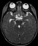
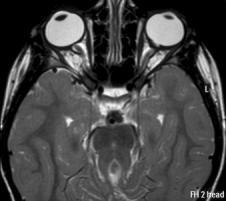
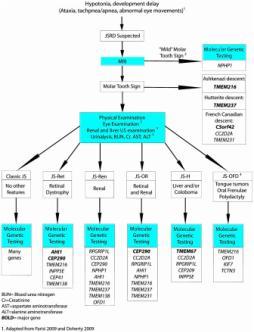
Joubert syndrome: report of 11 cases
 The Turkish Journal of Pediatrics 2012; 54: 605-611 Original Joubert syndrome: report of 11 cases Faruk İncecik 1, M. Özlem Hergüner 1, Şakir Altunbaşak 1, Joseph G. Gleeson 2 1 Department of Pediatric
Cerebellum 1/20/2016. Outcomes you need to be able to demonstrate. MHD Neuroanatomy Module
 This power point is made available as an educational resource or study aid for your use only. This presentation may not be duplicated for others and should not be redistributed or posted anywhere on the
Lecture X. Brain Pathways: Movement!
 Bio 3411 Readings (background only) Bio 3411 Monday Neuroscience 4 th ed Page(s) Feature 423-451Upper motor control of Brain Stem and Spinal Cord The Brain Atlas 3 rd ed Page(s) Feature 198-199 Vestibular
Developmental sequence of brain
 Cerebellum Developmental sequence of brain Fourth week Fifth week Location of cerebellum Lies above and behind the medullar and pons and occupies posterior cranial fossa Location of cerebellum External
Spectrum of Cranio-facial anomalies during 2 Ultrasound. trimester on
 Spectrum of Cranio-facial anomalies during 2 Ultrasound nd trimester on Poster No.: C-0378 Congress: ECR 2015 Type: Scientific Exhibit Authors: K. Dave, S. Solanki; Ahmedabad/IN Keywords: Obstetrics (Pregnancy
Lecture X. Brain Pathways: Movement!
 Bio 3411 Monday 1 Readings (background only) Neuroscience 5 th ed Page(s) Feature 353-398Upper motor control of Brain Stem and Spinal Cord Neuroscience 4 th ed Page(s) Feature 423-451Upper motor control
LOCALIZATION NEUROLOGY EPISODE VI HEARING LOSS AND GAIT ATAXIA
 LOCALIZATION NEUROLOGY EPISODE VI HEARING LOSS AND GAIT ATAXIA EPISODE VI HEARING LOSS APPROACH and DIAGNOSIS 2 Cochlea and Auditory nerve Pons (superior olive) lateral lemniscus Inferior colliculus Thalamus
Year 2003 Paper two: Questions supplied by Tricia
 question 43 A 42-year-old man presents with a two-year history of increasing right facial numbness. He has a history of intermittent unsteadiness, mild hearing loss and vertigo but has otherwise been well.
Prenatal Diagnosis of Central Nervous System (CNS) Pathologies: does Fetal MRI help in their management?
 Pathologies: does Fetal MRI help in their management?") Prenatal Diagnosis of Central Nervous System (CNS) Pathologies: does Fetal MRI help in their management? Daniela Prayer, Division of Neuroradiology and Musculoskeletal Radiology Medical University Vienna/Austria
Prenatal Diagnosis of Central Nervous System (CNS) Pathologies: does Fetal MRI help in their management? Daniela Prayer, Division of Neuroradiology and Musculoskeletal Radiology Medical University Vienna/Austria
Lab 2. we will look into several angled horizontal sections ( orbitomeatal plane ) i.e passing from the orbit into the ear
 i.e passing from the orbit into the ear") we will look into several angled horizontal sections ( orbitomeatal plane ) i.e passing from the orbit into the ear Figure I page 76 : looking at the key on the left side this section passed through the
we will look into several angled horizontal sections ( orbitomeatal plane ) i.e passing from the orbit into the ear Figure I page 76 : looking at the key on the left side this section passed through the
PHYSIOLOGY OF CSF AND PATHOPHYSIOLOGY OF HYDROCEPHALUS
 PHYSIOLOGY OF CSF AND PATHOPHYSIOLOGY OF HYDROCEPHALUS Introduction Dynamic component of CNS Invaluable tool to diagnosis Physiological reservoir of human proteome Reflects the physiologic state of CNS
Anatomy and Physiology (Bio 220) The Brain Chapter 14 and select portions of Chapter 16
 The Brain Chapter 14 and select portions of Chapter 16") Anatomy and Physiology (Bio 220) The Brain Chapter 14 and select portions of Chapter 16 I. Introduction A. Appearance 1. physical 2. weight 3. relative weight B. Major parts of the brain 1. cerebrum 2.
Anatomy and Physiology (Bio 220) The Brain Chapter 14 and select portions of Chapter 16 I. Introduction A. Appearance 1. physical 2. weight 3. relative weight B. Major parts of the brain 1. cerebrum 2.
Posterior fossa veins: Embryology, anatomy, variations and pathology
 Posterior fossa veins: Embryology, anatomy, variations and pathology Poster No.: C-2668 Congress: ECR 2010 Type: Educational Exhibit Topic: Neuro Authors: S. Nair, D. B. Sarkar, J. J. Bhattacharya, M.
Appendix 3.5 Case Inclusion Guidance for Potentially Zika-related Birth Defects
 Appendix 3.5 Case Inclusion Guidance for Potentially Zika-related Birth Defects Appendix 3.5 A3.5-1 Case Definition Appendix 3.5 Case Inclusion Guidance for Potentially Zika-related Birth Defects Contents
Characteristic features of CNS pathology. By: Shifaa AlQa qa
 Characteristic features of CNS pathology By: Shifaa AlQa qa Normal brain: - The neocortex (gray matter): six layers: outer plexiform, outer granular, outer pyramidal, inner granular, inner pyramidal, polymorphous
Kinks and Clefts : A Review of Congenital Brain Stem Abnormalities
 PEDIATRICS CME ABBREVIATION KEY DTI diffusion tensor imaging JS Joubert syndrome RL rhombic lip VZ ventricular zone Kinks and Clefts : A Review of Congenital Brain Stem Abnormalities B. Adams, D.J. Warren,
A&P 1 Brain & Cranial Nerves Guide - Lab Exercises
 A&P 1 Brain & Cranial Nerves Guide - Lab Exercises Please make sure you read the entire set of instructions on Dissection the Sheep Brain before beginning to cut. Also, please do not forget to go over
Septo-optic dysplasia (SOD) is a highly heterogeneous, usually. Midbrain-Hindbrain Involvement in Septo-Optic Dysplasia
 is a highly heterogeneous, usually. Midbrain-Hindbrain Involvement in Septo-Optic Dysplasia") Published April 24, 2014 as 10.3174/ajnr.A3959 ORIGINAL RESEARCH PEDIATRICS Midbrain-Hindbrain Involvement in Septo-Optic Dysplasia M. Severino, A.E.M. Allegri, A. Pistorio, B. Roviglione, N. Di Iorgi,
Published April 24, 2014 as 10.3174/ajnr.A3959 ORIGINAL RESEARCH PEDIATRICS Midbrain-Hindbrain Involvement in Septo-Optic Dysplasia M. Severino, A.E.M. Allegri, A. Pistorio, B. Roviglione, N. Di Iorgi,
RESEARCH ARTICLE RELATIVE FREQUENCY OF HYDROCEPHALUS IN RASHT PEDIATRIC PATIENTS
 RESEARCH ARTICLE RELATIVE FREQUENCY OF HYDROCEPHALUS IN RASHT PEDIATRIC PATIENTS Elham BIDABADI MD Assistant Professor of Pediatric Neurology, Guilan University of Medical Sciences,Guilan,Iran Corresponding
BIOL Dissection of the Sheep and Human Brain
 BIOL 2401 Dissection of the Sheep and Human Brain Laboratory Objectives After completing this lab, you should be able to: Identify the main structures in the sheep brain and to compare them with those
Functional Distinctions
 Functional Distinctions FUNCTION COMPONENT DEFICITS Start Basal Ganglia Spontaneous Movements Move UMN/LMN Cerebral Cortex Brainstem, Spinal cord Roots/peripheral nerves Plan Cerebellum Ataxia Adjust Cerebellum
The Cerebellum. The Little Brain. Neuroscience Lecture. PhD Candidate Dr. Laura Georgescu
 The Cerebellum The Little Brain Neuroscience Lecture PhD Candidate Dr. Laura Georgescu Learning Objectives 1. Describe functional anatomy of the cerebellum - its lobes, their input and output connections
Lecture - Chapter 13: Central Nervous System
 Lecture - Chapter 13: Central Nervous System 1. Describe the following structures of the brain, what is the general function of each: a. Cerebrum b. Diencephalon c. Brain Stem d. Cerebellum 2. What structures
Anatomy & Physiology Central Nervous System Worksheet
 1. What are the two parts of the CNS? 2. What are the four functions of the CNS Anatomy & Physiology Central Nervous System Worksheet 3. What are the four functions of the meninges? (p430) 4. Starting
The clinical spectrum of Blake s pouch cyst: report of six illustrative cases
 Childs Nerv Syst (2010) 26:1057 1064 DOI 10.1007/s00381-010-1085-2 ORIGINAL PAPER The clinical spectrum of Blake s pouch cyst: report of six illustrative cases Erwin M. J. Cornips & Geke M. Overvliet &
Brainstem. By Dr. Bhushan R. Kavimandan
 Brainstem By Dr. Bhushan R. Kavimandan Development Ventricles in brainstem Mesencephalon cerebral aqueduct Metencephalon 4 th ventricle Mylencephalon 4 th ventricle Corpus callosum Posterior commissure
Partial rhombencephalosynapsis and Chiari II malformation; 局部後腦窩先天性畸形和 II 型小腦篇桃體延隨聯合畸形
 Title Partial rhombencephalosynapsis and Chiari II malformation; 局部後腦窩先天性畸形和 II 型小腦篇桃體延隨聯合畸形 Author(s) Wan, SMY; Khong, PL; Ip, P; Ooi, GC Citation Hong Kong Medical Journal, 2005, v. 11 n. 4, p. 299-302;
 Spinal Cord Tracts DESCENDING SPINAL TRACTS: Are concerned with somatic motor function, modification of ms. tone, visceral innervation, segmental reflexes. Main tracts arise form cerebral cortex and others
Central nervous system
 Chapter 2 Central nervous system NORMAL SONOGRAPHIC ANATOMY The fetal brain undergoes major developmental changes throughout pregnancy. At 7 weeks of gestation, a sonolucent area is seen in the cephalic
Case Studies in Sella/Parasellar Region. Child thirsty, increased urination. Imaging. Suprasellar Germ Cell Tumor (Germinoma) No Disclosures
 No Disclosures") Case Studies in Sella/Parasellar Region No Disclosures 2018 Head and Neck Imaging Conference Child thirsty, increased urination Suprasellar Germ Cell Tumor (Germinoma) Midline Pineal >> Suprasellar > Other
Case Studies in Sella/Parasellar Region No Disclosures 2018 Head and Neck Imaging Conference Child thirsty, increased urination Suprasellar Germ Cell Tumor (Germinoma) Midline Pineal >> Suprasellar > Other
Functions. Traditional view: Motor system. Co-ordination of movements Motor learning Eye movements. Modern view: Cognition
 The Cerebellum Involved in motor coordination and timing Is simple and well documented Only has one type of output cell (Purkinje) The cerebellum influences motor activity through inhibition The Cerebellum
The Cerebellum. Outline. Lu Chen, Ph.D. MCB, UC Berkeley. Overview Structure Micro-circuitry of the cerebellum The cerebellum and motor learning
 The Cerebellum Lu Chen, Ph.D. MCB, UC Berkeley 1 Outline Overview Structure Micro-circuitry of the cerebellum The cerebellum and motor learning 2 Overview Little brain 10% of the total volume of the brain,