THE PALATOVAGINAL CANAL. Done by: Sultan alanazy. 30/3/2016
|
|
|
- Violet Juliana Goodwin
- 5 years ago
- Views:
Transcription
1 THE PALATOVAGINAL CANAL Done by: Sultan alanazy. 30/3/2016
2 Why? It is rarely mentioned in the medical literature and is even omitted in articles and textbook chapters describing the anatomy of the pterygopalatine fossa. Also a group of researchers concluded that the palatovaginal canal cannot be visualized because of its small size, and some describes it as duplication of vidian canal.
3 Importance: Major pathway for spread of malignant, tumors and infections. Could be cause of epistaxis. Landmark in base of skull surgery. Pharyngeal artery used for flap. Not to be mistaken with vidian.

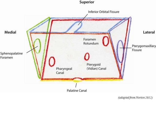
4 PPF:
5 Introduction: The palatovaginal canal (PVC) is a short bone tunnel that extends from the pterygopalatine fossa to the roof of the pharynx. PVC is found inferomedially on the posterior wall of the pterygopalatine fossa, in the roof of the nasopharynx.

6 PVC is formed by the application of: The sphenoid process of the palatine bone. The vaginal process of the sphenoid bone.(ant inf,med wall of sphenoid).


7
8 Since the palatine bone does not articulate with the vaginal process of the sphenoid bone, Pinheiro- Neto et al, in their anatomical study, suggested that the term palatovaginal is a misnomer and the canal containing the pharyngeal artery should be named palatosphenoidal canal.
9 The canal transmits: The palatovaginal artery. The pharyngeal nerve: from the pterygopalatine ganglion to the pharyngeal orifice of the auditory tube, to supply the mucosa of nasopharynx behind the auditory tube.
10 palatovaginal artery: The pterygovaginal artery (pharyngeal artery), is a posterior branch of the internal maxillary artery, in PPF. In some variation: arising early from the internal maxillary artery lateral to the pterygomaxillary fissure then coursed medially across the posterior wall of the pterygopalatine fossa and posterior to the plane of the nerves. In fact, the PA can be of considerable size, equal to that of the SPA.
11 Once in the canal, the artery courses posteriorly and emerges from the PVC in the nasopharynx to enter in an area of rich anastomoses in the region of the Eustachian tube.

12
13 Although rare, the pharyngeal artery may be an anastomotic artery between the distal IMA and the ascending pharyngeal artery (APhA). This anastomosis takes place in the region of the Eustachian tube, where both the (PhA) and (APhA). supply, and the blood is carried in a retrograde fashion from the APhA to the IMA through the PhA. Even rarer with the accessory meningeal artery, or the (APhA).

14 The average diameter of the palatovaginal canals was 0.9 mm (range, mm)

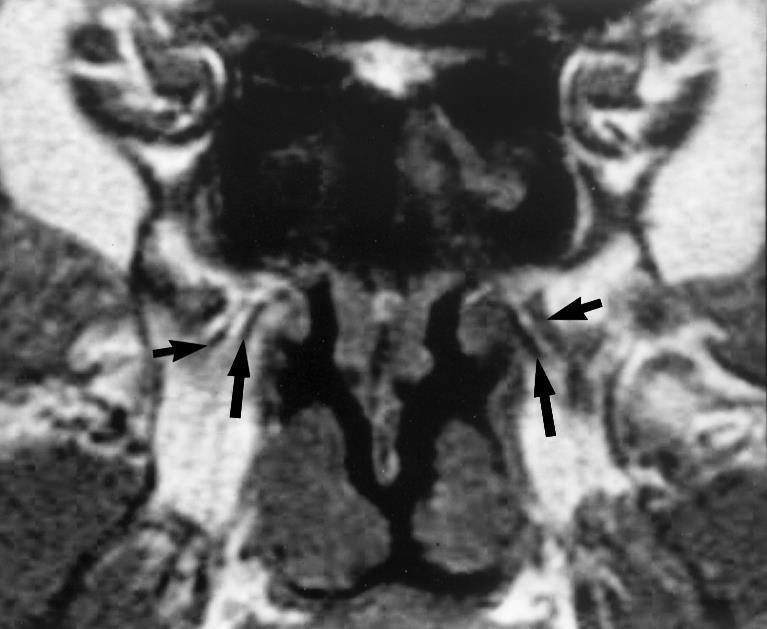
15 How can be identified on CT san?

16

17


18

19

20 Large PVC:
21 2 case reports: Borden NM, Dungan D, Dean BL, Flom RA. Posttraumatic epistaxis from injury to the pterygovaginal artery. AJNR Am J Neuroradiol 1996;17: Apostolos Karligkiotis,Luca Volpi,Vincenzo Abbate, Paolo Battaglia,Francesco Meloni, Mario Turri-Zanoni,Maurizio Bignami,Paolo Castelnuovo. Palatovaginal (pharyngeal) artery: clinical implication and surgical experience. Eur Arch Otorhinolaryngol (2014) 271:

22

23
24

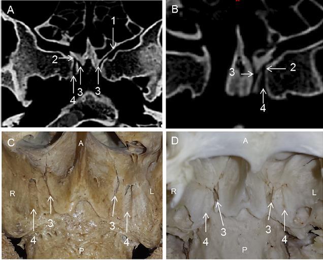
25 Using the posterior groove, the PV canal was correctly identified in 99 sides (70.7 %). Using the conventional method, the PV canal was correctly identified in 69 sides (49.3 %).

26

27


28

29
30 The PA was found to arise from the proximal portion of the SPA (or the distal portion of internal maxillary artery) within the pterygopalatine fossa. Inferomedial wall thickness in quite variable. In endoscopic procedures that involve drilling of the sphenoid floor, the PA was identified as a source of intraoperative bleeding.


31

32
PTERYGOPALATINE FOSSA
 PTERYGOPALATINE FOSSA Outline Anatomical Structure and Boundaries Foramina and Communications with other spaces and cavities Contents Pterygopalatine Ganglion Especial emphasis on certain arteries and
Dr.Ban I.S. head & neck anatomy 2 nd y جامعة تكريت كلية طب االسنان مادة التشريح املرحلة الثانية أ.م.د. بان امساعيل صديق 6102/6102
 جامعة تكريت كلية طب االسنان مادة التشريح املرحلة الثانية أ.م.د. بان امساعيل صديق 6102/6102 Pterygopalatine fossa: The pterygopalatine fossa is a cone-shaped depression, It is located between the maxilla,
Infratemporal fossa: Tikrit University college of Dentistry Dr.Ban I.S. head & neck Anatomy 2 nd y.
 Infratemporal fossa: This is a space lying beneath the base of the skull between the lateral wall of the pharynx and the ramus of the mandible. It is also referred to as the parapharyngeal or lateral pharyngeal
Mohammad Hisham Al-Mohtaseb. Lina Mansour. Reyad Jabiri. 0 P a g e
 2 Mohammad Hisham Al-Mohtaseb Lina Mansour Reyad Jabiri 0 P a g e This is only correction for the last year sheet according to our record. If you already studied this sheet just read the yellow notes which
The Palatovaginal Canal: Can It Be Identified on Routine CT and MR Imaging?
 Zoran Rumboldt 1,2 Mauricio Castillo 1 Jeffrey K. Smith 1 Received ugust 28, 2001; accepted after revision January 24, 2002. 1 Department of Radiology, C #7510, University of North Carolina at Chapel Hill,
Anatomic Relations Summary. Done by: Sohayyla Yasin Dababseh
 Anatomic Relations Summary Done by: Sohayyla Yasin Dababseh Anatomic Relations Lecture 1 Part-1 - The medial wall of the nose is the septum. - The vestibule lies directly inside the nostrils (Nares). -
Omran Saeed. Luma Taweel. Mohammad Almohtaseb. 1 P a g e
 2 Omran Saeed Luma Taweel Mohammad Almohtaseb 1 P a g e I didn t include all the photos in this sheet in order to keep it as small as possible so if you need more clarification please refer to slides In
Temporal fossa Infratemporal fossa Pterygopalatine fossa Terminal branches of external carotid artery Pterygoid venous plexus
 Outline of content Temporal fossa Infratemporal fossa Pterygopalatine fossa Terminal branches of external carotid artery Pterygoid venous plexus Boundary Content Communication Mandibular division of trigeminal
Major Anatomic Components of the Orbit
 Major Anatomic Components of the Orbit 1. Osseous Framework 2. Globe 3. Optic nerve and sheath 4. Extraocular muscles Bony Orbit Seven Bones Frontal bone Zygomatic bone Maxillary bone Ethmoid bone Sphenoid
Skull-2. Norma Basalis Interna Norma Basalis Externa. Dr. Heba Kalbouneh Associate Professor of Anatomy and Histology
 Skull-2 Norma Basalis Interna Norma Basalis Externa Dr. Heba Kalbouneh Associate Professor of Anatomy and Histology Norma basalis interna Base of the skull- superior view The interior of the base of the
Maxilla, ORBIT and infratemporal fossa. Neophytos C Demetriades MD, DDS, MSc Associate professor European University of Cyprus School of Medicine
 Maxilla, ORBIT and infratemporal fossa Neophytos C Demetriades MD, DDS, MSc Associate professor European University of Cyprus School of Medicine MAXILLA Superior, middle, and inferior meatus Frontal sinus
MAXILLA, ORBIT & PTERYGOPALATINE FOSSA. Neophytos C Demetriades MD, DDS, MSc Associate professor European University of Cyprus School of Medicine
 MAXILLA, ORBIT & PTERYGOPALATINE FOSSA Neophytos C Demetriades MD, DDS, MSc Associate professor European University of Cyprus School of Medicine Maxilla MAXILLA Superior, middle, and inferior meatus Frontal
SCHOOL OF ANATOMICAL SCIENCES Mock Run Questions. 4 May 2012
 SCHOOL OF ANATOMICAL SCIENCES Mock Run Questions 4 May 2012 1. With regard to the muscles of the neck: a. the platysma muscle is supplied by the accessory nerve. b. the stylohyoid muscle is supplied by
Dr.Ban I.S. head & neck anatomy 2 nd y. جامعة تكريت كلية طب االسنان املرحلة الثانية
 جامعة تكريت كلية طب االسنان التشريح مادة املرحلة الثانية أ.م.د. بان امساعيل صديق 6102-6102 1 The Palate The palate forms the roof of the mouth and the floor of the nasal cavity. It is divided into two
Trigeminal Nerve Worksheets, Distributions Page 1
 Trigeminal Nerve Worksheet #1 Distribution by Nerve Dr. Darren Hoffmann Dental Gross Anatomy, Spring 2013 We have drawn out each of the branches of CN V in lecture and you have an idea now for their basic
Dr. Sami Zaqout, IUG Medical School
 The skull The skull is composed of several separate bones united at immobile joints called sutures. Exceptions? Frontal bone Occipital bone Vault Cranium Sphenoid bone Zygomatic bones Base Ethmoid bone
The orbit-1. Dr. Heba Kalbouneh Assistant Professor of Anatomy and Histology
 The orbit-1 Dr. Heba Kalbouneh Assistant Professor of Anatomy and Histology Orbital plate of frontal bone Orbital plate of ethmoid bone Lesser wing of sphenoid Greater wing of sphenoid Lacrimal bone Orbital
Parotid Gland, Temporomandibular Joint and Infratemporal Fossa
 M1 - Anatomy Parotid Gland, Temporomandibular Joint and Infratemporal Fossa Jeff Dupree Sanger 9-057 jldupree@vcu.edu Parotid gland: wraps around the mandible positioned between the mandible and the sphenoid
Anatomy and Physiology. Bones, Sutures, Teeth, Processes and Foramina of the Human Skull
 Anatomy and Physiology Chapter 6 DRO Bones, Sutures, Teeth, Processes and Foramina of the Human Skull Name: Period: Bones of the Human Skull Bones of the Cranium: Frontal bone: forms the forehead and the
Parotid Gland. Parotid Gland. Largest of 3 paired salivary glands (submandibular; sublingual) Ramus of Mandible. Medial pterygoid.
 Ramus of Mandible. Medial pterygoid.") Parotid region Parotid Gland Largest of 3 paired salivary glands (submandibular; sublingual) Ramus of Mandible Medial pterygoid Cross section of mandible Masseter D S SCM Parotid Gland Mastoid Process
Parotid region Parotid Gland Largest of 3 paired salivary glands (submandibular; sublingual) Ramus of Mandible Medial pterygoid Cross section of mandible Masseter D S SCM Parotid Gland Mastoid Process
Structure Location Function
 Frontal Bone Cranium forms the forehead and roof of the orbits Occipital Bone Cranium forms posterior and inferior portions of the cranium Temporal Bone Cranium inferior to the parietal bone forms the
The Ear The ear consists of : 1-THE EXTERNAL EAR 2-THE MIDDLE EAR, OR TYMPANIC CAVITY 3-THE INTERNAL EAR, OR LABYRINTH 1-THE EXTERNAL EAR.
 The Ear The ear consists of : 1-THE EXTERNAL EAR 2-THE MIDDLE EAR, OR TYMPANIC CAVITY 3-THE INTERNAL EAR, OR LABYRINTH 1-THE EXTERNAL EAR Made of A-AURICLE B-EXTERNAL AUDITORY MEATUS A-AURICLE It consists
Skull Base Forgotten Foramina: A CT Pictorial Review
 Skull Base Forgotten Foramina: A CT Pictorial Review Poster No.: C-1224 Congress: ECR 2015 Type: Educational Exhibit Authors: N. Trifa, K. Fakhfakh; Sfax/TN Keywords: Trauma, Cancer, elearning, Computer
Gross Anatomy of the. TEMPORAL BONE, EXTERNAL EAR, and MIDDLE EAR. Assignment: Head to Toe Temporomandibular Joint (TMJ)
") Gross Anatomy the TEMPORAL BONE, EXTERNAL EAR, and MIDDLE EAR M1 Gross and Developmental Anatomy 9:00 AM, December 11, 2008 Dr. Milton M. Sholley Pressor Anatomy and Neurobiology Assignment: Head to Toe
Gross Anatomy the TEMPORAL BONE, EXTERNAL EAR, and MIDDLE EAR M1 Gross and Developmental Anatomy 9:00 AM, December 11, 2008 Dr. Milton M. Sholley Pressor Anatomy and Neurobiology Assignment: Head to Toe
Gross Anatomy of the. TEMPORAL BONE, EXTERNAL EAR, and MIDDLE EAR
 Gross Anatomy of the TEMPORAL BONE, EXTERNAL EAR, and MIDDLE EAR M1 Gross and Developmental Anatomy 9:00 AM, December 11, 2008 Dr. Milton M. Sholley Professor of Anatomy and Neurobiology Assignment: Head
Bisection of Head & Nasal Cavity 頭部對切以及鼻腔. 解剖學科馮琮涵副教授 分機
 Bisection of Head & Nasal Cavity 頭部對切以及鼻腔 解剖學科馮琮涵副教授 分機 3250 E-mail: thfong@tmu.edu.tw Outline: The structure of nose The concha and meatus in nasal cavity The openings of paranasal sinuses Canals, foramens
Juvenile Angiofibroma
 Juvenile Angiofibroma Disclaimer The pictures used in this presentation have been obtained from a number of sources. Their use is purely for academic and teaching purposes. The contents of this presentation
Bones Ethmoid bone Inferior nasal concha Lacrimal bone Maxilla Nasal bone Palatine bone Vomer Zygomatic bone Mandible
 splanchnocranium - Consists of part of skull that is derived from branchial arches - The facial bones are the bones of the anterior and lower human skull Bones Ethmoid bone Inferior nasal concha Lacrimal
Skull-2. Norma Basalis Interna. Dr. Heba Kalbouneh Assistant Professor of Anatomy and Histology
 Skull-2 Norma Basalis Interna Dr. Heba Kalbouneh Assistant Professor of Anatomy and Histology Norma basalis interna Base of the skull- superior view The interior of the base of the skull is divided into
Dr. Sami Zaqout Faculty of Medicine IUG
 The Nose External Nose Nasal Cavity External Nose Blood and Nerve Supplies of the External Nose Blood Supply of the External Nose The skin of the external nose Branches of the ophthalmic and the maxillary
Dr.Noor Hashem Mohammad Lecture (5)
") Dr.Noor Hashem Mohammad Lecture (5) 2016-2017 If the mandible is discarded, the anterior part of this aspect of the skull is seen to be formed by the hard palate. The palatal processes of the maxillae
Dr.Noor Hashem Mohammad Lecture (5) 2016-2017 If the mandible is discarded, the anterior part of this aspect of the skull is seen to be formed by the hard palate. The palatal processes of the maxillae
Anatomy #1; Respiratory Nose and the Nasal Cavity December 1st, 2013
 Note #1: the doctor skipped some slides in the lecture. Those slides are not included in this sheet and so you will have to review the slides to study them. The reason they were not included is because
Cranial Nerve VII - Facial Nerve. The facial nerve has 3 main components with distinct functions
 Cranial Nerve VII - Facial Nerve The facial nerve has 3 main components with distinct functions Somatic motor efferent Supplies the muscles of facial expression; posterior belly of digastric muscle; stylohyoid,
Skull and Axial Skeleton
 Published on Second Faculty of Medicine, Charles University (http://www.lf2.cuni.cz ) Skull and Axial Skeleton Description of the test The examination of the skull skeleton is in oral format. It consists
Temporal region. temporal & infratemporal fossae. Zhou Hong Ying Dept. of Anatomy
 Temporal region temporal & infratemporal fossae Zhou Hong Ying Dept. of Anatomy Temporal region is divided by zygomatic arch into temporal & infratemporal fossae. Temporal Fossa Infratemporal fossa Temporal
ORIGINAL ARTICLE. The Value of Preoperative Computed Tomographic Guidance. most common form of chronic nonallergic rhinitis,
 ORIGINAL ARTICLE Endoscopic Vidian Neurectomy The Value of Preoperative Computed Tomographic Guidance Shao-Cheng Liu, MD; Hsing-Won Wang, MD; Wan-Fu Su, MD Objective: To explore the vidian nerve anatomy
University of Palestine. Midterm Exam 2013/2014 Total Grade:
 [ Course No: DNTS2208 Course Title: Head and Neck Anatomy Date: 17/11/1024 No. of Questions: (52) Time: 2hours Using Calculator (No) University of Palestine Midterm Exam 2013/2014 Total Grade: Instructor
NEXT STOP : Central Station "Pterygopalatine fossa"
 NEXT STOP : Central Station "Pterygopalatine fossa" Poster No.: C-1359 Congress: ECR 2015 Type: Educational Exhibit Authors: I. Alba de Caceres, A. Paniagua, L. Ibañez, J. A. Blanco ; 1 1 1 1 2 2 Madrid/ES,
Neuroradiology Case of the Day
 Neuroradiology Case of the Day 76 th CAR Annual Meeting, Montreal, Quebec April 27, 2013 Eugene Yu, MD Assistant Professor of Radiology and Otolaryngology-Head and Neck Surgery Head and Neck Imaging Princess
Boundaries Septum Turbinates & Meati Lamellae Drainage Pathways Variants
 The Fastest 20 Minutes in Michelle A. Michel, MD Professor of Radiology and Otolaryngology Medical College of Wisconsin, Milwaukee Overview Nasal cavity Anterior skull base Ostiomeatal complex Frontal
Structure and Nerve Supply of The Larynx
 Kingdom of Bahrain Arabian Gulf University College of Medicine and Medical sciences Structure and Nerve Supply of The Larynx This presentation was originally prepared by: Dr. Kumar Notes were added by:
The Anatomical Relationship Between the Eustachian Tube and Petrous Internal Carotid Artery
 The Laryngoscope VC 2012 The American Laryngological, Rhinological and Otological Society, Inc. The Anatomical Relationship Between the Eustachian Tube and Petrous Internal Carotid Artery Kayhan Ozturk,
Introduction to Local Anesthesia and Review of Anatomy
 5-Sep Introduction and Anatomy Review 12-Sep Neurophysiology and Pain 19-Sep Physiology and Pharmacology part 1 26-Sep Physiology and Pharmacology part 2 Introduction to Local Anesthesia and Review of
The Pharynx. Dr. Nabil Khouri MD. MSc, Ph.D
 The Pharynx Dr. Nabil Khouri MD. MSc, Ph.D Introduction The pharynx is the Musculo-fascial halfcylinder that links the oral and nasal cavities in the head to the larynx and esophagus in the neck Common
ARTICLE. Imaging the cranial nerves in cancer
 Cancer Imaging (2004) 4, S1 S5 DOI: 10.1102/1470-7330.2004.0006 CI ARTICLE Vincent Chong Department of Diagnostic Radiology, Singapore General Hospital, Outram Road, Singapore 169608, Singapore Corresponding
Dr.Ban I.S. head & neck anatomy 2 nd y. جامعة تكريت كلية طب االسنان املرحلة الثانية أ.م.د. بان امساعيل صديق 6102/6102
 جامعة تكريت كلية طب االسنان التشريح مادة املرحلة الثانية أ.م.د. بان امساعيل صديق 6102/6102 Parotid region The part of the face in front of the ear and below the zygomatic arch is the parotid region. The
The sebaceous glands (glands of Zeis) open directly into the eyelash follicles, ciliary glands (glands of Moll) are modified sweat glands that open
 open directly into the eyelash follicles, ciliary glands (glands of Moll) are modified sweat glands that open") The Orbital Region The orbits are a pair of bony cavities that contain the eyeballs; their associated muscles, nerves, vessels, and fat; and most of the lacrimal apparatus upper eyelid is larger and more
The Orbital Region The orbits are a pair of bony cavities that contain the eyeballs; their associated muscles, nerves, vessels, and fat; and most of the lacrimal apparatus upper eyelid is larger and more
Lec [8]: Mandibular nerve:
![Lec [8]: Mandibular nerve:](/thumbs/94/121295776.jpg "Lec [8]: Mandibular nerve:") Lec [8]: Mandibular nerve: The mandibular branch from the trigeminal ganglion lies in the middle cranial fossa lateral to the cavernous sinus. With the motor root of the trigeminal nerve [motor roots lies
Lec [8]: Mandibular nerve: The mandibular branch from the trigeminal ganglion lies in the middle cranial fossa lateral to the cavernous sinus. With the motor root of the trigeminal nerve [motor roots lies
Nose & Mouth OUTLINE. Nose. - Nasal Cavity & Its Walls. - Paranasal Sinuses. - Neurovascular Structures. Mouth. - Oral Cavity & Its Contents
 Dept. of Human Anatomy, Si Chuan University Zhou hongying eaglezhyxzy@163.com Nose & Mouth OUTLINE Nose - Nasal Cavity & Its Walls - Paranasal Sinuses - Neurovascular Structures Mouth - Oral Cavity & Its
Chapter 7: Head & Neck
 Chapter 7: Head & Neck Osteology I. Overview A. Skull The cranium is composed of irregularly shaped bones that are fused together at unique joints called sutures The skull provides durable protection from
THE INTERIOR OF THE PHARYNX. By Dr. Muhammad Imran Qureshi
 THE INTERIOR OF THE PHARYNX By Dr. Muhammad Imran Qureshi The Cavity The cavity of the pharynx is divided into: 1. The Nasal part (called Nasopharynx) 2. The Oral part (called the Oropharynx), 3. And the
Vascular supply with angio-ct for superselective intra-arterial chemotherapy in advanced maxillary sinus cancer
 The British Journal of Radiology, 83 (2010), 171 178 PICTORIAL REVIEW Vascular supply with angio-ct for superselective intra-arterial chemotherapy in advanced maxillary sinus cancer 1 N KASHIWAGI, MD,
Read Me. We are the Learning Lab. to look
 Respiratory Tract Anatomy Lab In-Lab Exercises Read Me We are going to look at models and slides. Much of this can be done in the Learning Lab on your own time. The steps do not have to be done in order,
The Skull and Temporomandibular joint II Prof. Abdulameer Al-Nuaimi. E. mail:
 The Skull and Temporomandibular joint II Prof. Abdulameer Al-Nuaimi E-mail: a.al-nuaimi@sheffield.ac.uk E. mail: abdulameerh@yahoo.com Temporal fossa The temporal fossa is a depression on the temporal
University of Palestine. Midterm Exam 2013/2014 Total Grade:
 Course No: DNTS2208 Course Title: Head and Neck Anatomy Date: 09/11/2013 No. of Questions: (50) Time: 1hour Using Calculator (No) University of Palestine Midterm Exam 2013/2014 Total Grade: Instructor
Bones of the skull & face
 Bones of the skull & face Cranium= brain case or helmet Copyright The McGraw-Hill Companies, Inc. Permission required for reproduction or display. The cranium is composed of eight bones : frontal Occipital
Anatomicosurgical References for Endoscopic Localization of Sphenopalatine /jp-journals A Cadaveric Study AIJCR ABSTRACT
 AIJCR Anatomicosurgical References for Endoscopic Localization of Sphenopalatine 10.5005/jp-journals-10013-1282 Foramen: A Cadaveric Study Original Article Anatomicosurgical References for Endoscopic Localization
Trigeminal Nerve Anatomy. Dr. Mohamed Rahil Ali
 Trigeminal Nerve Anatomy Dr. Mohamed Rahil Ali Trigeminal nerve Largest cranial nerve Mixed nerve Small motor root and large sensory root Motor root Nucleus of motor root present in the pons and medulla
Tikrit University collage of dentistry Dr.Ban I.S. head & neck anatomy 2 nd y. Lec [5] / Temporal fossa :
![Tikrit University collage of dentistry Dr.Ban I.S. head & neck anatomy 2 nd y. Lec [5] / Temporal fossa :](/thumbs/88/115294566.jpg "Tikrit University collage of dentistry Dr.Ban I.S. head & neck anatomy 2 nd y. Lec [5] / Temporal fossa :") Lec [5] / Temporal fossa : Borders of the Temporal Fossa: Superior: Superior temporal line. Inferior: gap between zygomatic arch and infratemporal crest of sphenoid bone. Anterior: Frontal process of the
Lec [5] / Temporal fossa : Borders of the Temporal Fossa: Superior: Superior temporal line. Inferior: gap between zygomatic arch and infratemporal crest of sphenoid bone. Anterior: Frontal process of the
Exposure techniques in endoscopic skull base surgery: Posterior septectomy, medial maxillectomy, transmaxillary and transpterygoid approach
 European Annals of Otorhinolaryngology, Head and Neck diseases (2012) 129, 284 288 Available online at www.sciencedirect.com TECHNICAL NOTE Exposure techniques in endoscopic skull base surgery: Posterior
Prevertebral Region, Pharynx and Soft Palate
 Unit 20: Prevertebral Region, Pharynx and Soft Palate Dissection Instructions: Step1 Step 2 Step 1: Insert your fingers posterior to the sternocleidomastoid muscle, vagus nerve, internal jugular vein,
The Pterygopalatine Fossa: Postoperative MR Imaging Appearance
 AJNR Am J Neuroradiol 21:1315 1319, August 2000 The Pterygopalatine Fossa: Postoperative MR Imaging Appearance Ling-Ling Chan, June Chong, Ann M. Gillenwater, and Lawrence E. Ginsberg BACKGROUND AND PURPOSE:
Nasal region. cartilages: septal cartilage (l); lateral nasal cartilage (2); greater alar cartilages (2); lesser alar cartilages (?
; lateral nasal cartilage (2); greater alar cartilages (2); lesser alar cartilages (?") Nasal region skull bones: nasal and frontal processes of maxilla cartilages: septal cartilage (l); lateral nasal cartilage (2); greater alar cartilages (2); lesser alar cartilages (?) 1 Nasal cavity Roof
Nasal region skull bones: nasal and frontal processes of maxilla cartilages: septal cartilage (l); lateral nasal cartilage (2); greater alar cartilages (2); lesser alar cartilages (?) 1 Nasal cavity Roof
MR and CT anatomy and the pathology of skull base focusing on pterygopalatine fossa
 MR and CT anatomy and the pathology of skull base focusing on pterygopalatine fossa Poster No.: C-1688 Congress: ECR 2010 Type: Educational Exhibit Topic: Head and Neck Authors: S. Kandatsu, R. Kishimoto,
Bony orbit Roof The orbital plate of the frontal bone Lateral wall: the zygomatic bone and the greater wing of the sphenoid
 Bony orbit Roof: Formed by: The orbital plate of the frontal bone, which separates the orbital cavity from the anterior cranial fossa and the frontal lobe of the cerebral hemisphere Lateral wall: Formed
Head&Neck Imaging. ssregypt.com. Parapharyngeal Spaces. Mamdouh mahfouz MD
 Head&Neck Imaging Parapharyngeal Spaces ssregypt.com Mamdouh mahfouz MD mamdouh.m5@gmail.com Definitio n Fat filled triangular space lateral the pharynx Extends from the skull base to the oropharynx Parapharyngeal
APRIL
 APRIL - 2003 OCTOBER - 2003 February 2009 [KU 652] Sub. Code : 4131 FIRST B.D.S DEGREE EXAMINATION (Modified Regulations III) Paper I HUMAN ANATOMY, HISTOLOGY AND EMBRYOLOGY Time : Three hours
Embryology of the Ophthalmic Artery: a Revived Concept
 www.centauro.it Interventional Neuroradiology 15: 363-368, 2009 Letter to the Editor Embryology of the Ophthalmic Artery: a Revived Concept M. KOMIYAMA Department of Neurosurgery, Osaka City General Hospital;
Distribution of the maxillary artery related to sinus graft surgery for implantation
 42 Distribution of the maxillary artery related to sinus graft surgery for implantation LvingWell Dental Hospital LivingWell Institute of Dental Research Jang-yeol Lee, Hyoun-chull Kim, Il-hae Park, Sang-chull
OPEN ACCESS ATLAS OF OTOLARYNGOLOGY, HEAD & NECK OPERATIVE SURGERY
 OPEN ACCESS ATLAS OF OTOLARYNGOLOGY, HEAD & NECK OPERATIVE SURGERY INFERIOR MAXILLECTOMY Tumours of the hard palate and superior alveolus may be resected by inferior maxillectomy (Figure 1). A Le Fort
Anatomical Study of Critical Features on the Posterior Wall of the Maxillary Sinus: Clinical Implications
 The Laryngoscope VC 2014 The American Laryngological, Rhinological and Otological Society, Inc. Anatomical Study of Critical Features on the Posterior Wall of the Maxillary Sinus: Clinical Implications
Research Article Length and Geometric Patterns of the Greater Palatine Canal Observed in Cone Beam Computed Tomography
 International Dentistry Volume 2010, Article ID 292753, 6 pages doi:10.1155/2010/292753 Research Article Length and Geometric Patterns of the Greater Palatine Canal Observed in Cone Beam Computed Tomography
Patterns of perineural spread of head and neck malignancies.
 Patterns of perineural spread of head and neck malignancies. Poster No.: C-1234 Congress: ECR 2014 Type: Educational Exhibit Authors: C. Martins Jarnalo, G. Lycklama à Nijeholt, E. Sanchez-Aliaga, 1 1
Perineural Tumor Spread (PNS) Perineural Tumor Spread (PNS) PNS Anatomic Considerations. Perineural Tumor Spread-Imaging
 Perineural Tumor Spread (PNS) PNS Anatomic Considerations. Perineural Tumor Spread-Imaging") Imaging of Perineural Tumor Spread in Head and Neck Cancer Lawrence E. Ginsberg, MD Departments of Diagnostic Radiology and Head and Neck Surgery University of Texas M.D. Anderson Cancer Center Houston,
Imaging of Perineural Tumor Spread in Head and Neck Cancer Lawrence E. Ginsberg, MD Departments of Diagnostic Radiology and Head and Neck Surgery University of Texas M.D. Anderson Cancer Center Houston,
The PHARYNX. Dr. Nabil Khouri MD Ph.D
 The PHARYNX Dr. Nabil Khouri MD Ph.D PHARYNX Fibromuscular tube lined with mucous membrane extends from base of skull to lower border of cricoid cartilage (C-6). 12-14 cm long At the lower border of cricoid
Image-Guided Endoscopic Endonasal Transmaxillary Transpterygoid Approach to Meckel s Cave
 DOI: 10.5137/1019-5149.JTN.6430-12.0 Received: 20.04.2012 / Accepted: 07.06.2012 Case Report Image-Guided Endoscopic Endonasal Transmaxillary Transpterygoid Approach to Meckel s Cave Xuejian WANG, Xiaobiao
Transnasal Endoscopic Sinonasal Surgery
 Reda kamel, Cadaveric dissection 1 Transnasal Endoscopic Sinonasal Surgery Cadaver Dissection Guide For Endoscopic Sinus Surgery Cairo University Egypt Reda Kamel Professor of Rhinology Cairo University
locomotice system Plastinated specimensⅠ: Silicone specimens Regional specimens and organs
 locomotice system Plastinated specimensⅠ: Silicone specimens Regional specimens and organs Art-No. Name Description The locomotor system SL001 Two hundred pieces of plastinated bones (without six The bones
Anatomy Sheet: Oral cavity Done by: rasha Rakan edited by: khansaa Mahmoud
 Anatomy Sheet: Oral cavity Done by: rasha Rakan edited by: khansaa Mahmoud The oral cavity has 2 parts: 1. Oral vestibule: outer part that consists of outside the teeth, between the teeth, the cheeks and
AUDITORY APPARATUS. Mr. P Mazengenya. Tel 72204
 AUDITORY APPARATUS Mr. P Mazengenya Tel 72204 Describe the anatomical features of the external ear Describe the tympanic membrane (ear drum) Describe the walls of the middle ear Outline the structures
*in general the blood supply of the nose comes from branches of the internal and external carotid arteries.
 In the previous lecture we talked about the anatomy of the nasal cavity, today we will talk about its blood supply, venous drainage, innervations, and finally about the paranasal sinuses. When we describe
Candidate s instructions Look at this cross-section taken at the level of C5. Answer the following questions.
 Section 1 Anatomy Chapter 1. Trachea 1 Candidate s instructions Look at this cross-section taken at the level of C5. Answer the following questions. Pretracheal fascia 1 2 5 3 4 Questions 1. Label the
Anatomy of the Trigeminal Nerve
 19 Anatomy of the Trigeminal Nerve.1 Introduction 0. The Central Part of the Trigeminal Nerve 1..1 Origin 1.. Trigeminal Nuclei.3 The Peripheral Part of the Trigeminal Nerve 4.3.1 Ophthalmic Nerve 4.3.
THE SURGEON S LIBRARY
 THE SURGEON S LIBRARY THE HISTORY AND SURGICAL ANATOMY OF THE VAGUS NERVE Lee J. Skandalakis, M.D., Chicago, Illinois, Stephen W. Gray, PH.D., and John E. Skandalakis, M.D., PH.D., F.A.C.S., Atlanta, Georgia
We are IntechOpen, the world s leading publisher of Open Access books Built by scientists, for scientists. International authors and editors
 We are IntechOpen, the world s leading publisher of Open Access books Built by scientists, for scientists 4,100 116,000 120M Open access books available International authors and editors Downloads Our
THYROID & PARATHYROID. By Prof. Saeed Abuel Makarem & Dr. Sanaa Al-Sharawy
 THYROID & PARATHYROID By Prof. Saeed Abuel Makarem & Dr. Sanaa Al-Sharawy 1 OBJECTIVES By the end of the lecture, the student should be able to: Describe the shape, position, relations and structure of
Skeletal System: Skull.
 Skeletal System: Skull www.fisiokinesiterapia.biz Bones of the Skull SPLANCHNOCRANIUM Nasal (2) Maxilla (2) Lacrimal (2) Zygomatic (2) Palatine (2) Inferior concha (2) Vomer Mandible NEUROCRANIUM Frontal
Anatomy images for MSS practical exam- 2019
 Anatomy images for MSS practical exam- 2019 Ilium Ischium Pubis Acetabulaum Iliac crest Iliac tubercle ASIS (muscle and ligament attached) AIIS (muscle attached) PSIS PIIS Ischial spine Ischial tuberosity
14.25 UBC SINUS & SKULL BASE COURSE 2018 STANDARD COURSE : AUG (FRI-SAT) SKULL BASE COURSE : AUG 12 (SUN) ubccpd.ca CESEI CENTRE VANCOUVER, BC
 SKULL BASE COURSE : AUG 12 (SUN) ubccpd.ca CESEI CENTRE VANCOUVER, BC") 14.25 MOC SECTION 1 UBC SINUS & SKULL BASE COURSE 2018 STANDARD COURSE : AUG 10 11 (FRI-SAT) SKULL BASE COURSE : AUG 12 (SUN) CESEI CENTRE VANCOUVER, BC WHO SHOULD ATTEND Surgeons Advanced Trainees Fellows
14.25 MOC SECTION 1 UBC SINUS & SKULL BASE COURSE 2018 STANDARD COURSE : AUG 10 11 (FRI-SAT) SKULL BASE COURSE : AUG 12 (SUN) CESEI CENTRE VANCOUVER, BC WHO SHOULD ATTEND Surgeons Advanced Trainees Fellows
Congenital Neck Masses C. Stefan Kénel-Pierre, MD
 Congenital Neck Masses C. Stefan Kénel-Pierre, MD SUNY-LICH Medical Center Department of Surgery Case Presentation xx year old male presents with sudden onset left lower neck swelling x 1 week Denies pain,
Unit 18: Cranial Cavity and Contents
 Unit 18: Cranial Cavity and Contents Dissection Instructions: The calvaria is to be removed without damage to the dura mater which is attached to the inner surface of the calvaria. Cut through the outer
Subdivided into Vestibule & Oral cavity proper
 Extends from the lips to the oropharyngeal isthmus The oropharyngeal isthmus: Is the junction of mouth and pharynx. Is bounded: Above by the soft palate and the palatoglossal folds Below by the dorsum
JPMER ABSTRACT. INTRODUCTION The sphenopalatine ganglion (SPG) is a parasympathetic ganglion in the course of greater petrosal nerve and is
 is a parasympathetic ganglion in the course of greater petrosal nerve and is") ORIGINAL ARTICLE 10.5005/jp-journals-10028-1093 Anatomic Landmarks and Morphometric Measurements for Accurate Localization of the Sphenopalatine Ganglion via the Transnasal and Infrazygomatic Approaches:
ORIGINAL ARTICLE 10.5005/jp-journals-10028-1093 Anatomic Landmarks and Morphometric Measurements for Accurate Localization of the Sphenopalatine Ganglion via the Transnasal and Infrazygomatic Approaches:
Human Anatomy and Physiology - Problem Drill 07: The Skeletal System Axial Skeleton
 Human Anatomy and Physiology - Problem Drill 07: The Skeletal System Axial Skeleton Question No. 1 of 10 Which of the following statements about the axial skeleton is correct? Question #01 A. The axial
External carotid blood supply to acoustic neurinomas
 External carotid blood supply to acoustic neurinomas Report of two cases HARVEY L. LEVINE, M.D., ERNEST J. FERmS, M.D., AND EDWARD L. SPATZ, M.D. Departments of Radiology, Neurology, and Neurosurgery,
REVIEW OF HEAD AND NECK CRANIAL NERVES AND EVERYTHING ELSE
 REVIEW OF HEAD AND NECK CRANIAL NERVES AND EVERYTHING ELSE OLFACTORY NERVE CN I ANTERIOR CRANIAL FOSSA CRISTA GALLI OF ETHMOID OLFACTORY FORAMINA IN CRIBIFORM PLATE OF ETHMOID BONE CN I OLFACTORY NERVE