Appropriate Imaging Tests Lead to Meaningful Results. Dr. Richard Wasley May 2011
|
|
|
- Deborah Johnston
- 5 years ago
- Views:
Transcription

1 Appropriate Imaging Tests Lead to Meaningful Results Dr. Richard Wasley May 2011

2 Summarize the advantages and limitations of specific imaging tests and why clinical information is so important to radiologists Generate an understanding of how to select the most appropriate test for a patient in a specific scenario.
3 The ACR Appropriateness Criteria are evidence-based guidelines to assist in making the most appropriate imaging or treatment decision for a specific clinical condition. Available online at ACR.org Enhance quality of care and contribute to the most efficacious use of radiology. At SMMC, we also take into account radiation dose to the patient and overall cost
4 ACR Appropriateness Criteria Clinical Condition: Acute Abdominal Pain and Fever or Suspected Abdominal Abscess. Postoperative patient with fever. Radiologic Procedure Appropriateness Rating CT abdomen and pelvis with contrast 8 CT abdomen and pelvis without contrast 7 US abdomen 6 MRI abdomen and pelvis with contrast 6 X-ray abdomen 5 MRI abdomen and pelvis without contrast 5 X-ray contrast enema 4 Ga-67 scan abdomen 4 X-ray upper GI series with small bowel 3 In-111 WBC scan abdomen and pelvis 3
5 X-ray Ultrasound Computed Tomography MRI Angiography and nuclear medicine PET Fluoroscopy
6 X-ray Ultrasound Computed Tomography MRI Angiography and nuclear medicine PET Fluoroscopy
7 Which imaging modality to use? What is the clinical question to be answered? Do I need to ask for special imaging protocols, IV contrast or oral contrast?
8 Abdomen Chest Neuro/spine

9 Abdomen Chest Neuro/spine
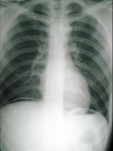
10 Advantages Rapid, generally easily available inexpensive Relatively low radiation dose Appropriate for: Ingested foreign body Suspected perforation SBO

11


12


13 Supine positioning Upright positioning


14 ? constipation Massive stomach distention Secondary to Gastro paresis


15 fat? Renal stones Tooth in a teratoma
16 Ultrasound No radiation exposure Good for biliary tract disease Abdominal organ survey Ascites Computed Tomography Bowel disease More complete view overall


17 Skin surface fluid shadow


18 Stone in Common Bile Duct

19 hydronephrosis Dilated renal pelvis
20 Good for: Fluid Gallbladder, dilated collecting system, bladder Ascites, pleural effusions Superficial structures Lymphadenopathy seromas Vascular imaging Female pelvis
21 Limited for: Masses of liver, spleen and kidneys Characterization of superficial masses Renal stones Not good for: Gas obscured structures Pancreas Deep vasculature Bowel Fat
22 Limited for:
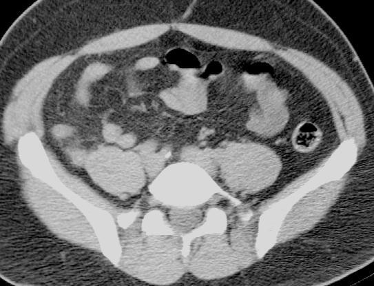
23 Good for: Fat obscured organs/large patients Gas-obscured structures Abdominal masses, deep or superficial Renal stones Bowel Not so good for: Uterus/adnexal pathology Gallstones/choledocolithiasis
24 Fat is the friend of CT In the acute setting, oral contrast is not generally required IV contrast helpful, especially in the thin patient Studies have shown CT to be the most costeffective imaging modality, >96% sensitive Appendicitis Diverticulitis ischemia

25 Renal stones 1) 32 y/o man with right flank pain hydronephrosis stranding

26 stone

27 Sensitivity Uric acid stone

28 nflamed ppendix


29 diverticula Inflammation of fat

30 ? stool

31

32 Portal venous gas
33 What are you looking for? Interpretation of findings can be dependent upon type of patient, history of current situation, duration of symptoms, etc More information is better.
34 Free fluid

35 Free fluid Adnexal cyst rupture Free fluid

36 Free fluid Liver laceration Spleen laceration

37 Free fluid carcinomatosis
38 Fat is a natural contrast Thin patients need IV contrast High suspicion for renal stones probably does NOT require IV contrast If unsure, the use of IV contrast helps in finding other diagnoses
39 Hydronephrosis? stone

40 Pyelonephritis in 32 y/o woman with fevers and UTI

41 Renal vein thrombosis in 27 y/o with SLE

42 Renal artery occlusion in Patient with Atrial Fibrillation Hydronephrosis? stone Unenhanced kidney
43 Contraindications to iodinated contrast Otherwise has relatively low risk Will not obscure findings such as stones Help elucidate disease process Short Answer: probably will not hurt to use IV Contrast
44 Not all abdominal CT s are created equal Rapid multiphase imaging possible Pancreatic protocol Liver protocol CT angiography

45 Pancreas Protocol Hepatic artery Pancreatic CA Celiac axis
46 Liver protocol Question lesion? Standard CT abdomen
47 Liver protocol Standard CT abdomen Arterial phase Hepatocellular CA

48 Liver protocol - Portal vein thrombosis
49 AAA/PVD Renal artery stenosis Mesenteric artery stenosis Sensitivity >95% (equal to conventional angiography)


50 Right renal artery Aortic Dissection Left renal artery

51 Abdominal Aortic Aneurysm Right kidney renal artery Flow lumen Calcified wall


52 Oral contrast is helpful for: Chronic symptoms Post operative patient in the evaluation for leak or abscess Tumor staging Sigmoid colon CA Oral contrast leak
53 MRI Generally not for acute abdominal complaints? Appendicitis in pregnant patients Good for: Incidental adrenal mass workup MRCP Adnexal/uterine pathology Certain tumors of liver and kidneys


54
55 Abdominal x-ray series SBO, free air, FB Cheap, fast, but limited Ultrasound Thin or young patients Biliary disease, pelvic disease in women or children CT Large body habitus. IV (not oral) in thinner patients and women Bowel disease, deep organs, gas Complete evaluation. Very sensitive


56 Ultrasound


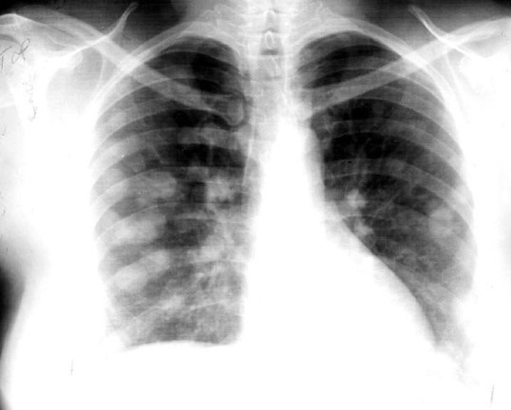
57 CT patient


58

59 What are you looking for? indication for exam? CT, US, XRAY or MRI If CT, with IV or Oral contrast? Special procedures CTA, multiphase studies of liver or pancreas
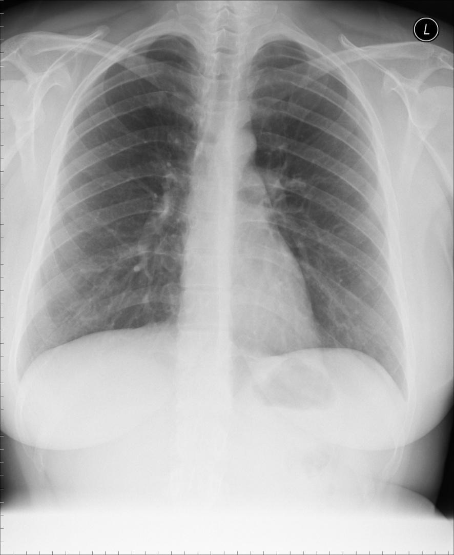
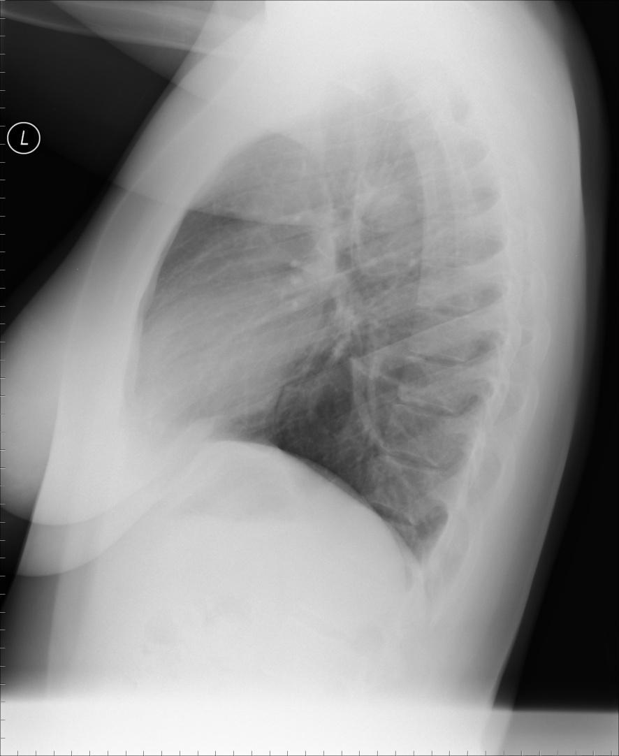
60 CXR is still the most valuable player Infiltrate and effusion Pneumothorax Detection of masses CT Pulmonary embolism Workup/follow-up of tumor Vascular abnormalities Nuclear medicine VQ scan
61
62
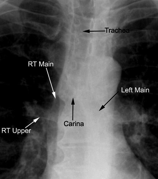
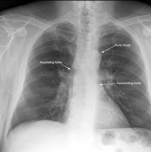
63 Based on perception of density change Water, cells or debris in normally aerated structures Air in normally dense structures Configuration/mass effect Bones, lines, other


64 Normal lung silhouetted against the dense heart or diaphragm Infiltrate obscures the normal interface RLL infiltrate obscures diaphragm RUL infiltrate obscures mediastinum

65 Distended azygous vein Parahilar prominence effusion Kerley lines cardiomegaly

66 Curves around lung Mass effect Decreased lung volume

67 Density within lung Volume loss Trachea shifted


68 See pleural line with aerated lung central and air in the pleural space peripherally

69
70 Which to order? CT or VQ scan VQ is slightly cheaper VQ gives relative probability for PE CT is very accurate. nearly 100% positive predictive value Very high negative predictive value for significant PE CT slightly LESS radiation dose best for pregnancy VQ for elevated creatinine, unstable patient


71 Saddle embolus Main PA

72 target appearance is 100% diagnostic of pulmonary embolism

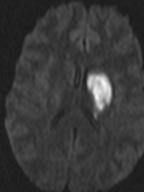
73 Timing is important
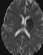
74 CXR is king. Great first step most of the time Inexpensive and often diagnostic Be sure to provide appropriate indication No rule out. Needs symptoms, signs or affecting history CT pulmonary angiogram vs VQ CTPA more specific, accurate for PE and can find alternative diagnoses VQ for contraindications to CTPA
75 For the brain: CT is the preferred imaging for: Trauma to skull or face. Sinus dz Acute, high clinical suspicion, headache Worst headache of life. Thunderclap headache Acute stroke symptoms. CTA of intracranial and extracranial vasculature MRI probably best for all else. More expensive, but no radiation. Often CT is non-diagnostic or an MRI is required for further workup anyway.

76 trauma Subdural hematoma Epidural hematoma

77


78 >90% stenosis External Carotid Artery ulceration


79 aneurysm searches sensitive for aneurysms >= 3mm pre-operative planning Left MCA aneurism


80 Thrombosed MCA Acute symptoms Repeat CT in 24 hours
81 Really is CT plus MRI for stroke CT is for the acute presentation. Excludes hemorrhage Treatment stratification Rapid test code stroke MRI is very valuable in stroke imaging

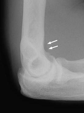
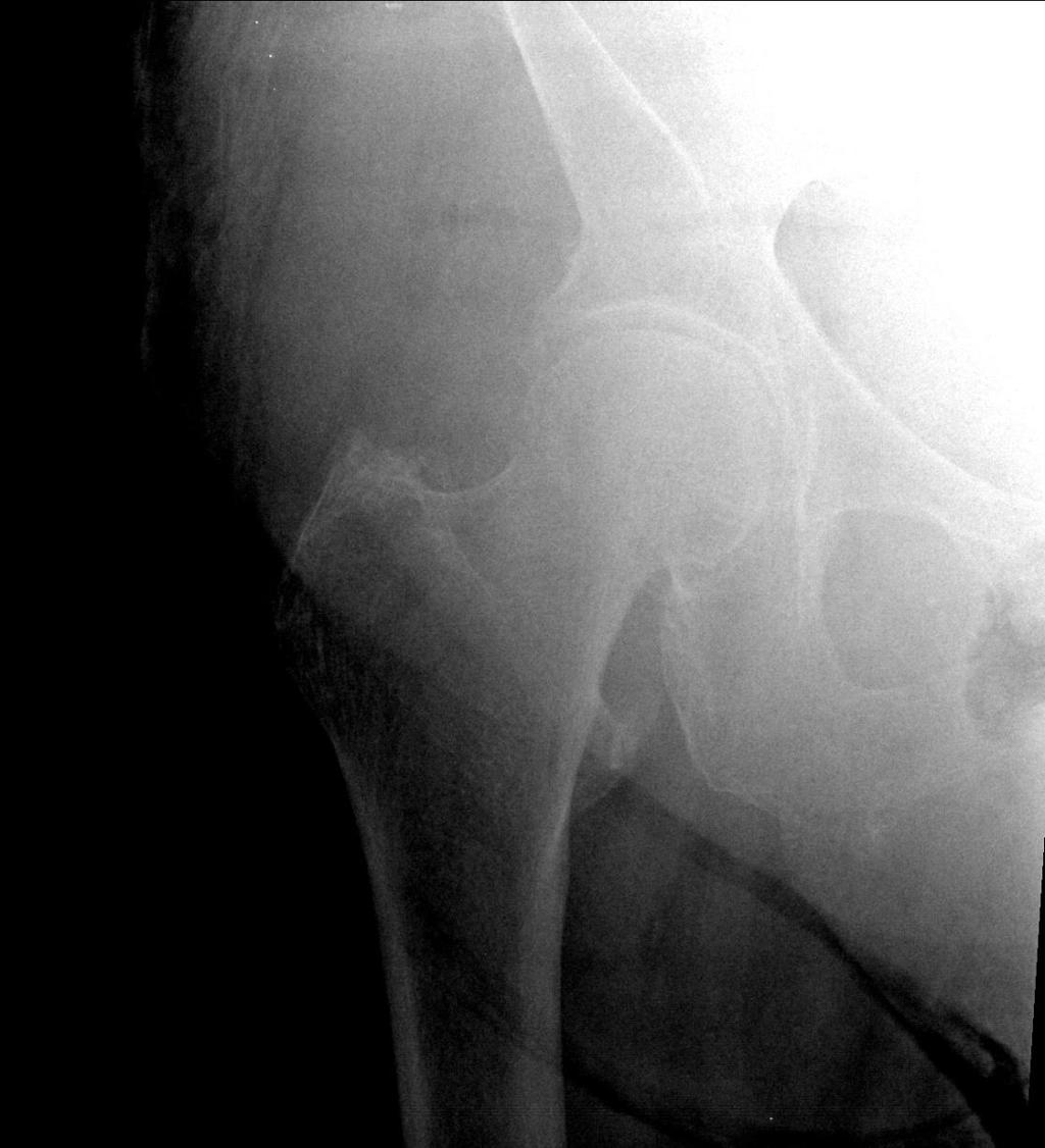
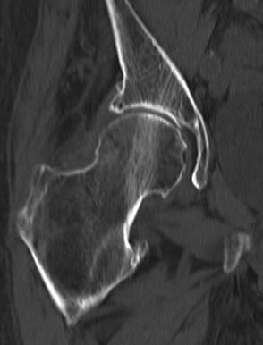
82
83 Choice between X-ray and CT is clinically driven for C spine. If low/moderate suspicion, recommend X-ray If high suspicion recommend CT Cspine. If cannot see C1 thru T1, recommend CT LS spine, X-rays or MRI. CT with high radiation exposure. Little information not seen on standard X-ray series


84 Jefferson fracture

85


86 C2, Hangman s type fracture

87
88 X-rays important in the acute setting CT for high clinical suspicion C spine MRI for almost all others Assess edema, acuity Assess cord compromise IV gadolinium if post operative
89 X-rays still are usually the best imaging modality in the acute setting Inexpensive and often diagnostic MRI for orthopedic issues CT Internal derangements of joints, soft tissue, muscle AVN Assess non-union Orthopedic assessment of fractures Occult fracture of hip
90 Most fractures visualized by x-ray and do not require further imaging with more expensive tests May require follow-up film in days to look for the occult fracture. There are a few fractures which can be tricky

91 normal effusion


92 normal effusion

93 Fractures often seen only on one view Series should include 3 views of most body parts
94 Depending on study, <90% fractures are visible on standard X-ray series CT>95% accurate, but not 100% MRI final determination Nuclear medicine lesser value as does not show details of fracture for surgical planning Also has a false negative rate in acute fracture less than 3-4 days old
95

96
97 X rays detect most fractures Prefer 3 views to 2 views as some fractures are only seen on one view of a series CT for facial fractures, occult hip fracture or orthopedic follow-up. MRI for orthopedic follow-up, workup of soft tissue or joint abnormalities. MRI for AVN or trauma sequellae.
98
99 Osteomyelitis prefer MRI, If suspect abscess, perform with IV gadolinium Bone scan cheaper, but not as accurate Hearing loss Conductive or middle ear symptoms CT Sensor neural MRI CT angiography CTA for renal, carotid, aortic dissection, runoff Circle of Willis MR for screening, CT for detail coronary
100 Imaging test selection guidelines in radiology aim to: determine the single best imaging test for specific symptomatology/diagnoses Ultimately contribute to cost-effective strategies Radiation dose reduction Overall accuracy and timeliness to diagnosis Clinical information is invaluable to: determine which imaging modality is best to use; decide if IV or oral contrast or specific protocols are required; improve the relevancy of interpretations. Do not hesitate to ask for help in choosing the best test
n Make tremendous difference in patients lives: n Diagnosing or excluding disease and injury n Evaluating response to therapy
 Imaging: Choosing the Appropriate Exam Rob Milman, MD Austin Radiological Association What is a Radiologist? A physician who specializes in diagnosing and treating disease and injury by using medical imaging
Contrast Guidelines for Common CT/CTA & MRI/MRA
 Contrast Guidelines for Common /A & /MRA Body Imaging Gastrointestinal CLINICAL GUIDELINES EXAM DESCRIPTION /A CPT CODES EXAM DESCRIPTION /MRA CPT CODES Abdominal mass Abdomen & Pelvis w 74177 Abdomen
Effective Utilization of Imaging. John V. Roberts, M.D. Premier Radiology Abdominal Imaging
 Effective Utilization of Imaging John V. Roberts, M.D. Premier Radiology Abdominal Imaging Safety Contrast and Radiation What to order Abdomen/Pelvis Brain/Spine Chest Musculoskeletal Ob/Gyn Head and Neck
Request Card Task ANSWERS
 Request Card Task ANSWERS Medical Student Workbook Author: Dr Sam Leach, SpR Case 1 What differential diagnoses are most likely? Which investigation is most appropriate? Case 1 The most likely diagnosis
Radiology of the abdomen Lecture -1-
 Radiology of the abdomen Lecture -1- Objectives To know radiology modalities used in abdomen imaging mainly GI tract. To know advantages and disadvantages of each modality. To know indications and contraindications
CLINICAL PRESENTATION AND RADIOLOGY QUIZ QUESTION
 Donald L. Renfrew, MD Radiology Associates of the Fox Valley, 333 N. Commercial Street, Suite 100, Neenah, WI 54956 6/23/2012 Radiology Quiz of the Week # 78 Page 1 CLINICAL PRESENTATION AND RADIOLOGY
General Imaging. Imaging modalities. Incremental CT. Multislice CT Multislice CT [ MDCT ]
![General Imaging. Imaging modalities. Incremental CT. Multislice CT Multislice CT [ MDCT ]](/thumbs/76/74079340.jpg "General Imaging. Imaging modalities. Incremental CT. Multislice CT Multislice CT [ MDCT ]") General Imaging Imaging modalities Conventional X-rays Ultrasonography [ US ] Computed tomography [ CT ] Radionuclide imaging Magnetic resonance imaging [ MRI ] Angiography conventional, CT,MRI Interventional
General Imaging Imaging modalities Conventional X-rays Ultrasonography [ US ] Computed tomography [ CT ] Radionuclide imaging Magnetic resonance imaging [ MRI ] Angiography conventional, CT,MRI Interventional
Communication of Critical and/or Discrepant Findings. Stephen Alden, Lorie Cavalli and Bobby Kalb, MD Effective Date: 08/01/08
 DEPARTMENT OF MEDICAL IMAGING POLICIES, PROCEDURES AND PROTOCOLS Subject: Policy: Communication of Critical and/or Discrepant Findings RR003 Author: Stephen Alden, Lorie Cavalli and Bobby Kalb, MD Effective
Radiology. Undergraduate Radiology Sample Questions
 Radiology Undergraduate Radiology Sample Questions April 2012 The following examples are offered of questions that might be used to assess undergraduate radiology. There are 3 different styles: An OSCE
Plain abdomen The standard films are supine & erect AP views (alternative to erect, lateral decubitus film is used in ill patients).
.") Plain abdomen The standard films are supine & erect AP views (alternative to erect, lateral decubitus film is used in ill patients). The stomach can be readily identified by its location, gastric rugae
Plain abdomen The standard films are supine & erect AP views (alternative to erect, lateral decubitus film is used in ill patients). The stomach can be readily identified by its location, gastric rugae
CT PROCEDURE REFERENCE GUIDE 2017
 Head CT PROCEDURE REFERENCE GUIDE 2017 Procedure Contrast Scan Field Preparatio n Base of Skull to Vertex Sinuses Orbits Mastoids/IAC/ Temporal Bones Facial Bones ST Neck Low Dose Lung Screening Routine
Abdomen and Pelvis CT (1) By the end of the lecture students should be able to:
 By the end of the lecture students should be able to:") RAD 451 Abdomen and Pelvis CT (1) By the end of the lecture students should be able to: State the common indications for Abdomen and pelvis CT exams Identify possible contra indications for Abdomen and
RAD 451 Abdomen and Pelvis CT (1) By the end of the lecture students should be able to: State the common indications for Abdomen and pelvis CT exams Identify possible contra indications for Abdomen and
HONG KONG COLLEGE OF RADIOLOGISTS. Higher Training (Radiology) Subspecialty Training in Computed Tomography
 Subspecialty Training in Computed Tomography") HONG KONG COLLEGE OF RADIOLOGISTS Higher Training (Radiology) Subspecialty Training in Computed Tomography [The following guidelines should be read in conjunction with the General Guidelines on Higher
HONG KONG COLLEGE OF RADIOLOGISTS Higher Training (Radiology) Subspecialty Training in Computed Tomography [The following guidelines should be read in conjunction with the General Guidelines on Higher
ASSESSING THE PLAIN ABDOMINAL RADIOGRAPH M A A M E F O S U A A M P O F O
 ASSESSING THE PLAIN ABDOMINAL RADIOGRAPH M A A M E F O S U A A M P O F O Introduction The abdomen (less formally called the belly, stomach, is that part of the body between the thorax (chest) and pelvis,
Contrast Materials Patient Safety: What are contrast materials and how do they work?
 Contrast Materials Patient Safety: What are contrast materials and how do they work? Which imaging exams use contrast materials? How safe are contrast materials? How should I prepare for my imaging procedure
Pediatric Abdomen Trauma
 Pediatric Abdomen Trauma Susan D. John, MD, FACR Pediatric Trauma Trauma is leading cause of death and disability in children and adolescents Causes and effects vary between age groups Blunt trauma predominates
JUSTIFICATION PROTOCOLS FOR CT SCANNING ALBURY WODONGA HEALTH WODONGA CAMPUS
 JUSTIFICATION PROTOCOLS FOR CT SCANNING ALBURY WODONGA HEALTH WODONGA CAMPUS JUSTIFICATION PROTOCOLS FOR CT SCANNING INTRODUCTION: In accordance with the Victorian Radiation Act 2005 Wodonga Medical Imaging,
General Data. 王 X 村 78 y/o 男性
 General Data 王 X 村 78 y/o 男性 Chief Complaint Vomiting twice this early morning Fever up to 38.9ºC was noted Present Illness (1) Old CVA with left side weakness for more than 10 years and with bed ridden
Learning Objectives for Rotations in Vascular Surgery Year 3 Basic Clerkship
 Learning Objectives for Rotations in Vascular Surgery Year 3 Basic Clerkship CLINICAL PROBLEMS IN VASCULAR SURGERY 1. ABDOMINAL AORTIC ANEURYSM A 70 year old man presents in the emergency department with
Radiological Investigations of Abdominal Trauma
 76 77 Investigations of Abdominal Trauma Introduction: Trauma to abdominal organs is a common cause of patient morbidity and mortality among trauma patients. Causes of abdominal trauma include blunt injuries,
Radiology Rotation Educational Goals & Objectives for Internal Medicine
 Radiology Rotation Educational Goals & Objectives for Internal Medicine Internists provide continuing care for patients with a myriad of medical and psychosocial problems. During many patient encounters,
RADPrimer Curriculum Breast Topics Covered Basic Intermediate 225
 Breast Anatomy & Normal Variants 11 Breast Imaging Modalities 13 BI RADS Lexicon 3 Mammography: Masses 9 Mammography: Calcifications 17 Mammography: Additional Findings 8 Ultrasound Features 10 Ultrasound
73725x2 MRA Pelvis Runoff (to ankle) CTA Abdomen with & without CTA Cardiac Brain without 70551
 CTA Abdomen with & without CTA Cardiac Brain without 70551") CT CT Myelogram MRI Abdomen without 74150 Cervical 62302 Abdomen / MRCP 74181 Abdomen with 74160 Thoracic 62303 Abdomen / MRCP with & without 74183 Abdomen with & without 74170 Lumbar 62304 Abdomen / Pelvis
CT CT Myelogram MRI Abdomen without 74150 Cervical 62302 Abdomen / MRCP 74181 Abdomen with 74160 Thoracic 62303 Abdomen / MRCP with & without 74183 Abdomen with & without 74170 Lumbar 62304 Abdomen / Pelvis
ACUTE ABDOMEN. Dr. M Asadi. Surgical Oncology Research Center MUMS. Assistant Professor of General Surgery
 ACUTE ABDOMEN Dr. M Asadi Assistant Professor of General Surgery Surgical Oncology Research Center MUMS Definition I. The term Acute Abdomen refers to signs & symptoms of abdominal pain and tenderness,
Imaging abdominal vascular emergencies. V.Stoynova
 Imaging abdominal vascular emergencies V.Stoynova Abdominal vessels V. Stoynova 2 Acute liver bleeding trauma anticoagulant therapy liver disease : HCC, adenoma, meta, FNH, Hemangioma Diagnosis :CT angiography
Job Task Analysis for ARDMS Abdomen Data Collected: June 30, 2011
 Job Task Analysis for ARDMS Abdomen Data Collected: June 30, 2011 Reported: Analysis Summary for: Abdomen Examination Survey Dates 06/13/2011-06/26/2011 Invited Respondents 6,000 Surveys with Demographics
PREAMBLE GENERAL DIAGNOSTIC RADIOLOGY
 PREAMBLE The General Diagnostic Radiology category is intended to cover the body of knowledge a practicing board certified Diagnostic Radiologist should know. Since the range of content relevant to the
Lab Monitor Images Dissection of the Abdominal Vasculature + Lower Digestive System
 Lab Monitor Images Dissection of the Abdominal Vasculature + Lower Digestive System Stomach & Duodenum Frontal (AP) View Nasogastric tube 2 1 3 4 Stomach Pylorus Duodenum 1 Duodenum 2 Duodenum 3 Duodenum
Dr. Rami M. Adil Al-Hayali Assistant Professor in Medicine
 Dr. Rami M. Adil Al-Hayali Assistant Professor in Medicine Venous thromboembolism: pulmonary embolism (PE) deep vein thrombosis (DVT) 1% of all patients admitted to hospital 5% of in-hospital mortality
Diagnostic Imaging
 www.fisiokinesiterapia.biz Diagnostic Imaging Diagnostic Imaging is no longer limited to radiography. Major technological advancements have lead to the use of new and improved imaging technologies. The
MAKING THE BEST USE OF CLINICAL RADIOLOGY SERVICES. Dr Martina Paetzel Consultant Radiologist
 MAKING THE BEST USE OF CLINICAL RADIOLOGY SERVICES Dr Martina Paetzel Consultant Radiologist LEARNING OBJECTIVES To be aware of guidelines regulating ionising radiation & radiation dose To introduce how
Appendix 5. EFSUMB Newsletter. Gastroenterological Ultrasound
 EFSUMB Newsletter 87 Examinations should encompass the full range of pathological conditions listed below A log book listing the types of examinations undertaken should be kept Training should usually
Pediatric radiology. Varga Edit. Semmelweis University MR Research Center Semmelweis University 2nd Department of Pediatrics
 Pediatric radiology Varga Edit Semmelweis University MR Research Center Semmelweis University 2nd Department of Pediatrics Modalities radiograpy (x-ray, fluoroscopy) ultrasound computer tomography (CT)
Case Discussion Splenic Abscess
 Case Discussion Splenic Abscess Personal Data Gender: male Birth Date: 1928/Mar/06th Allergy: Mefenamic Smoking: 0.5 PPD for 55 years Alcohol: negative (?) 4 Months Ago Abdominal pain: epigastric area
Back to Basics: What Imaging Test should I order? Jeanne G. Hill, M.D. Pediatric Radiology Medical University of South Carolina
 Back to Basics: What Imaging Test should I order? Jeanne G. Hill, M.D. Pediatric Radiology Medical University of South Carolina Disclosure Neither I nor any member of my immediate family has a relevant
Special Instructions
 FDA and ACR guidelines are as follows: Special Instructions Safety concerning NSF and gadolinium-based contrast agents (GBCA) Prior to administering MRI contrast (GBCA), any patient who answers yes to
Advances in Emergency Imaging
 Hampton Symposium,, October 16 th, 2010 Advances in Emergency Imaging Robert A. Novelline, MD Professor of Radiology, Harvard Medical School Director of Emergency Radiology, Massachusetts General Hospital
Looking Outside the Box: Incidental Extracardiac Finding in Echo
 Looking Outside the Box: Incidental Extracardiac Finding in Echo Dr. Aijaz Shah Head of Division, Adult Echocardiography Laboratory Prince Sultan Cardiac Centre Riyadh Case 1 17 year old boy presented
Spine MRI and Spine CT Test Request Tip Sheet
 Spine MRI and Spine CT With/Without Contrast CT, MRI Studies should NOT be ordered simultaneously as dual studies (i.e., with and without contrast). Radiation exposure is doubled and both views are rarely
Learning Radiology: Recognizing the Basics. Text with Student Consult Online Access Code
 Learning Radiology: Recognizing the Basics. Text with Student Consult Online Access Code Herring, W ISBN-13: 9780323074445 Table of Contents 1. Recognizing Anything The "colorful" world of radiology A
Radiology Update: Balancing Diagnostic Needs with Cost Effectiveness
 Radiology Update: Balancing Diagnostic Needs with Cost Effectiveness October 2018 Ramin Khorasani, MD, MPH Professor of Radiology, Harvard Medical School Director, Center for Evidence-Based Imaging Vice
Spine MRI and Spine CT Test Request Tip Sheet
 Spine MRI and Spine CT With/Without Contrast CT, MRI The study considered best for a specific clinical scenario should be ordered. The second study should be done ONLY if the first study does not provide
Deep Vein Thrombosis and Pulmonary Embolism: Patient Information
 Deep Vein Thrombosis and Pulmonary Embolism: Patient Information A Deep Vein Thrombosis (DVT) and a Pulmonary Embolism (PE) are both disorders of unwanted blood clotting. Unwanted blood clots can occur
Incidental findings: A retrospective analysis of management
 Incidental findings: A retrospective analysis of management Authors and disclosures Authors: Steven Boe, Dana Boe, Jeffrey Kaye, Anu Bansal, Marc Glickstein Disclosures: None Purpose Determine if appropriate
Evidence Process for Abdominal Pain Guideline Research 11/16/2017. Guideline Review using ADAPTE method and AGREE II instrument 11/16/2017
 Evidence Process for Abdominal Pain Guideline Research Guideline Review using ADAPTE method and AGREE II instrument Approximately 139 Potentially relevant guidelines identified in various resources* 59
GUIDELINES FOR. Advanced. Imaging Studies
 GUIDELINES FOR Advanced Imaging Studies Locations Herrin Hospital Memorial Hospital of Carbondale St. Joseph Memorial Hospital 201 S 14th Street Herrin, IL 62948 618.942.2171 ext 35400 General Radiography*
W/ (2) (3) (4) (5) (5) (6) (6) CTA
 (3) (4) (5) (5) (6) (6) CTA") Index Abdomen W/ and W/Out (2) Abdomen Pelvis W/Out (3) Abdomen Pelvis W/ (4) Pelvis W/ (5) Chest W/Out (5) Chest/Abdomen/Pelvis W/ (6) Chest W/ (6) CTA ( 7-8) Neuro (8-9) Musculoskeletal (10) Trauma (11)
Index Abdomen W/ and W/Out (2) Abdomen Pelvis W/Out (3) Abdomen Pelvis W/ (4) Pelvis W/ (5) Chest W/Out (5) Chest/Abdomen/Pelvis W/ (6) Chest W/ (6) CTA ( 7-8) Neuro (8-9) Musculoskeletal (10) Trauma (11)
CT & PET/CT Scheduling Guidelines LVHN (5846) Fax:
 Fax:") www.lvhn.org CHEST CT (x-ray is almost always required) 71250 Nodules Abnormal cxr, ancillary finding, f/u study WO No 71250 HRCT: Interstitial lung disease, i.e. asbestosis, sarcoidosis, TB SOBOE, dry
www.lvhn.org CHEST CT (x-ray is almost always required) 71250 Nodules Abnormal cxr, ancillary finding, f/u study WO No 71250 HRCT: Interstitial lung disease, i.e. asbestosis, sarcoidosis, TB SOBOE, dry
Role of imaging in the evaluation of the acute abdomen
 Prof. András Palkó MD, PhD Role of imaging in the evaluation of the acute abdomen Faculty of General Medicine University of Szeged Hungary 1 Definition Sudden onset of severe symptoms requiring emergency
Spine MRI and Spine CT Test Request Tip Sheet
 Spine MRI and Spine CT MRI is almost always preferred over CT scan; if ordering CT, CLEARLY document why MRI is not appropriate. In cases of back pain without red flags, six weeks of multimodality supervised
Alice Fung, MD Oregon Health and Science University
 Alice Fung, MD Oregon Health and Science University Disclosure Comments The speaker Alice Fung, MD Has relevant financial relationships to disclose. Received honorarium from (Guerbet). This individual
Contributors. Thanks to Peter Miller, MD; LCDR Kevin Preston, MD; and Keith Newbrough, MD for their generous contribution of images:
 Contributors Thanks to Peter Miller, MD; LCDR Kevin Preston, MD; and Keith Newbrough, MD for their generous contribution of images: Peter Miller, MD, Indiana University School of Medicine Chapter 1: Figure
Guidelines, Policies and Statements D5 Statement on Abdominal Scanning
 Guidelines, Policies and Statements D5 Statement on Abdominal Scanning Disclaimer and Copyright The ASUM Standards of Practice Board have made every effort to ensure that this Guideline/Policy/Statement
Vascular Imaging in the Pediatric Abdomen. Jonathan Swanson, MD
 Vascular Imaging in the Pediatric Abdomen Jonathan Swanson, MD Goals and Objectives To understand the imaging approach, appearance, and clinical manifestations of the common pediatric abdominal vascular
Pulmonary Embolism. Thoracic radiologist Helena Lauri
 Pulmonary Embolism Thoracic radiologist Helena Lauri 8.5.2017 Statistics 1-2 out of 1000 adults annually are diagnosed with deep vein thrombosis (DVT) and/or pulmonary embolism (PE) About half of patients
Clinician s Guide To Ordering NeuroImaging Studies
 Clinician s Guide To Ordering NeuroImaging Studies MRI CT South Jersey Radiology Associates The purpose of this general guide is to assist you in choosing the appropriate imaging test to best help your
The role for contrast-enhanced ultrasonography outside of focal liver lesions
 The role for contrast-enhanced ultrasonography outside of focal liver lesions Paul S. Sidhu King s College Hospital, London, UK Introduction Contrast-enhanced ultrasonography (US) of focal liver lesions
Spine MRI and Spine CT Test Request Tip Sheet
 Spine MRI and Spine CT With/Without Contrast CT, MRI The study considered best for a specific clinical scenario should be ordered. The second study should be done ONLY if the first study does not provide
My Patient Has Abdominal Pain PoCUS of the Biliary Tract and the Urinary Tract
 My Patient Has Abdominal Pain PoCUS of the Biliary Tract and the Urinary Tract Objectives PoCUS for Biliary Disease PoCUS for Renal Colic PoCUS for Urinary Retention Biliary Disease A patient presents
Basic Abdominal and Pelvic Imaging Concepts. David L. Smith, MD Assistant Professor of Radiology
 Basic Abdominal and Pelvic Imaging Concepts David L. Smith, MD Assistant Professor of Radiology Basic Imaging Concepts Contrast Resolution vs Spacial Resolution Spacial Resolution......refers to the ability
Abdomen Sonography Examination Content Outline
 Abdomen Sonography Examination Content Outline (Outline Summary) # Domain Subdomain Percentage 1 2 3 Anatomy, Perfusion, and Function Pathology, Vascular Abnormalities, Trauma, and Postoperative Anatomy
Newcastle HPB MDM updated radiology imaging protocol recommendations. Author Dr John Scott. Consultant Radiologist Freeman Hospital
 Newcastle HPB MDM updated radiology imaging protocol recommendations Author Dr John Scott. Consultant Radiologist Freeman Hospital This document is intended as a guide to aid radiologists and clinicians
11/1/2014. Radiologic incidentalomas Ordering pitfalls Newer technology and applications
 Bilal Tahir, MD Gitasree Borthakur, MD Indiana University School of Medicine Department of Radiology & Imaging Sciences October 31, 2014 ACP 2014 Dr. V. Aaron Nuclear (vaaron@iupui.edu) Dr. S. Westphal
Abdominal radiology 腹部放射線學
 Abdominal radiology 腹部放射線學 台北醫學大學 - 市立萬芳醫院 留偉順 laowilson@hotmail.com The Normal Abdominal Series Chest Supine abdomen Erect abdomen Left lateral decubitus abdomen Learning objectives Understanding normal
Measuring What Students Know: Writing Effective MCQ Questions
 Measuring What Students Know: Writing Effective MCQ Questions 1 What Can Student Assessments Do? Communicate important content and skills Provide basis for grading, passing, graduation Identify students
FOR CMS (MEDICARE) MEMBERS ONLY NATIONAL COVERAGE DETERMINATION (NCD) FOR COMPUTED TOMOGRAPHY:
 MEMBERS ONLY NATIONAL COVERAGE DETERMINATION (NCD) FOR COMPUTED TOMOGRAPHY:") National Imaging Associates, Inc. Clinical guidelines CHEST CTA Original Date: September 1997 Page 1 of 5 CPT Codes: 71275 Last Review Date: August 2014 NCD 220.1 Last Effective Date: March 2008 Guideline
National Imaging Associates, Inc. Clinical guidelines CHEST CTA Original Date: September 1997 Page 1 of 5 CPT Codes: 71275 Last Review Date: August 2014 NCD 220.1 Last Effective Date: March 2008 Guideline
Department of Radiology Teaching Hospital, Kandy
 Department of Radiology Teaching Hospital, Kandy Welcome to the Department of Radiology Department of Radiology is one of the oldest departments to be established in Teaching hospital Kandy and, involved
CT Scan Reference Guide
 Diagnostic Imaging A Department of Rutland Regional Medical Center CT Scan Reference Guide 10/01/2017 Safety Questions for Exams with IV Contrast...Page 3 CPT Code Scan Ranges: Head...Pages 4-5 Neck/Spine...Pages
Bladder Trauma Data Collection Sheet
 Bladder Trauma Data Collection Sheet If there was no traumatic injury with PENETRATION of the bladder DO NOT proceed Date of injury: / / Time of injury: Date of hospital arrival: / / Time of hospital arrival:
Appendix 9: Endoscopic Ultrasound in Gastroenterology
 Appendix 9: Endoscopic Ultrasound in Gastroenterology This curriculum is intended for clinicians who perform endoscopic ultrasonography (EUS) in gastroenterology. It includes standards for theoretical
Computed Tomography (CT) - Abdomen and Pelvis
 - Abdomen and Pelvis") Scan for mobile link. Computed Tomography (CT) - Abdomen and Pelvis Computed tomography (CT) of the abdomen and pelvis is a diagnostic imaging test used to help detect diseases of the small bowel, colon
Scan for mobile link. Computed Tomography (CT) - Abdomen and Pelvis Computed tomography (CT) of the abdomen and pelvis is a diagnostic imaging test used to help detect diseases of the small bowel, colon
INVESTIGATIONS OF GASTROINTESTINAL DISEAS
 INVESTIGATIONS OF GASTROINTESTINAL DISEAS Lecture 1 and 2 دز اسماعيل داود فرع الطب كلية طب الموصل Radiological tests of structure (imaging) Plain X-ray: May shows soft tissue outlines like liver, spleen,
Perforation of a Duodenal Diverticulum. Elective Student S. C.
 Perforation of a Duodenal Diverticulum 2008 4 Elective Student S. C. Case History An elderly male presented to the Emergency Department with abdominal pain. Chief Complaint: Worsening, diffuse abdominal
General Surgery Service
 General Surgery Service Patient Care Goals and Objectives Stomach/Duodenum and Bariatric assessed for a) Obesity surgery b) Treatment of i) Adenocarcinoma of the stomach ii) GIST iii) Carcinoid 2) Optimize
From 2015/2016 Batch
 Department of & Nuclear February 7, 2018 Medical Imaging Module 04 th Year 1 st and 2 nd Semesters From 2015/2016 Batch Topic Objectives Time Dept. T / L Activity Comments Understand the principles of
Interventional Radiology Curriculum for Medical Students
 Cardiovascular and Interventional Radiological Society of Europe Interventional Radiology Curriculum for Medical Students C RSE Introduction It has been recognized that the teaching of radiology in medical
Vascular Technology Examination Content Outline
 Vascular Technology Examination Content Outline (Outline Summary) # Domain Subdomain Percentage 1 Normal Anatomy, Perfusion, and Function Evaluate normal anatomy, perfusion, function 2 Pathology, Perfusion,
Abdomen and Retroperitoneum Ultrasound Protocols
 Abdomen and Retroperitoneum Ultrasound Protocols Reviewed By: Anna Ellermeier, MD Last Reviewed: March 2018 Contact: (866) 761-4200, Option 1 **NOTE for all examinations: 1. If documenting possible flow
Computerized Tomography of the Acute Left Upper Quadrant Pain
 Computerized Tomography of the Acute Left Upper Quadrant Pain Authors 1. Temel Tirkes, M.D. (1,2) Associate Professor of Radiology 2. Zachary Ballenger, M.D. (1) 3. Scott D. Steenburg, M.D. (1) Associate
Body MRI from the Liver to the Bladder
 Body MRI from the Liver to the Bladder I Want You! Audience Participation Methodist Hospital Continuing Education Seminar Jordan Swensson, MD November 7, 2015 Objectives Observe the uses of MRI for organs
Medical Review Guidelines Magnetic Resonance Angiography
 Medical Review Guidelines Magnetic Resonance Angiography Medical Guideline Number: MRG2001-05 Effective Date: 2/13/01 Revised Date: 2/14/2006 OHCA Reference OAC 317:30-5-24. Radiology. (f) Magnetic Resonance
Shedding Light on Neonatal X-rays. Objectives. Indications for X-Rays 5/14/2018
 Shedding Light on Neonatal X-rays Barbara C. Mordue, MSN, NNP-BC Neonatal Nurse Practitioner LLUH Children s Hospital, NICU Objectives Utilize a systematic approach to neonatal x-ray interpretation Identify
Radiology of hepatobiliary diseases
 GI cycle - Lecture 14 436 Teams Radiology of hepatobiliary diseases Objectives 1. To Interpret plan x-ray radiograph of abdomen with common pathologies. 2. To know the common pathologies presentation.
Introduction to Radiology
 Introduction - Lecture 1 436 Teams Introduction to Radiology Objectives Introduce the various Medical Imaging Modalities. Understand the basics of image generation. Relate imaging to gross anatomy. Appreciate
FIRST COAST SERVICE OPTIONS FLORIDA MEDICARE PART B LOCAL COVERAGE DETERMINATION
 FIRST COAST SERVICE OPTIONS FLORIDA MEDICARE PART B LOCAL COVERAGE DETERMINATION CPT/HCPCS Codes 70544 Magnetic resonance angiography, head; without contrast material(s) 70545 with contrast material(s)
Ct Of The Acute Abdomen Medical Radiology
 We have made it easy for you to find a PDF Ebooks without any digging. And by having access to our ebooks online or by storing it on your computer, you have convenient answers with ct of the acute abdomen
CT abdomen and pelvis
 CT abdomen and pelvis General indications: Assessment of vague abdominal symptoms (pain, colics,distenstion,...) Varifecation of a lesion discovered by other diagnostic modalities as US, barium,ivp, Staging
9/21/15. Joshua Pruitt, MD, FAAEM Medical Director, LifeGuard Air Ambulance Iowa PA Society Fall CME Conference September 29, 2015
 Unless they prove otherwise. ~Every ED attending ever Joshua Pruitt, MD, FAAEM Medical Director, LifeGuard Air Ambulance Iowa PA Society Fall CME Conference September 29, 2015 AAA with rupture Mesenteric
Stroke / CVA TIA Trauma Dizziness Headaches. Acoustic Neuroma Syrinx Visual Change Vascular Lesions (AVM) Elevated Prolactin Vertigo Bell s palsy
 Elevated Prolactin Vertigo Bell s palsy") Head Brain Alzheimer s Mental Status Change Confusion Dementia Memory Loss Dizziness Headaches MRI Brain w/o 70551 Tumor / Mass / Cancer Cranial Nerve Lesions HIV Infection Suspected MS Neurofibromatosis
Head Brain Alzheimer s Mental Status Change Confusion Dementia Memory Loss Dizziness Headaches MRI Brain w/o 70551 Tumor / Mass / Cancer Cranial Nerve Lesions HIV Infection Suspected MS Neurofibromatosis
Alexander A Schult, M.D., FCCP. October 21, 2017 Revised 1/10/18
 Alexander A Schult, M.D., FCCP October 21, 2017 Revised 1/10/18 Identifying normal anatomy Identifying various pathologic states Identifying placement of hardware Identifying limitations of portable CXR
Is Structured Reporting More Accurate Than Conventional Reporting in CT Reporting of the Abdomen and Pelvis?
 Is Structured Reporting More Accurate Than Conventional Reporting in CT Reporting of the Abdomen and Pelvis? A M Almuslim, MBBS; J G Ryan, MD; A Murtaza, MD Purpose The purpose of this research is to determine
In any operation. Indications. Anaesthesia. Position of the patient. Incision. Steps of the operation. Complications.
 In any operation Indications. Anaesthesia. Position of the patient. Incision. Steps of the operation. Complications. Abdominal operation I position for operation Supine Abdominal operation I position for
Imaging for Pediatric Trauma and the Acute Patient What to Order When
 Imaging for Pediatric Trauma and the Acute Patient What to Order When Paula Shultz, MD Pediatric Radiologist Central Oregon Radiology Associates November 8, 2013 5 yr old male involved in multicar accident,
ACUTE ABDOMEN IN OLDER CHILDREN. Carlos J. Sivit M.D.
 ACUTE ABDOMEN IN OLDER CHILDREN Carlos J. Sivit M.D. ACUTE ABDOMEN Clinical condition characterized by severe abdominal pain developing over several hours ACUTE ABDOMINAL PAIN Common childhood complaint
MANAGEMENT RECOMMENDATIONS
 1 MANAGEMENT RECOMMENDATIONS 1. Adrenal masses!!!!!!! page 2 2. Liver Masses!!!!!!! page 3 3. Obstetric US Soft Markers for Aneuploidy!! pages 4-6 4. Ovarian and Adnexal Cysts!!!!! pages 7-10 5. Pancreatic
Acquired pediatric esophageal diseases Imaging approaches and findings. M. Mearadji International Foundation for Pediatric Imaging Aid
 Acquired pediatric esophageal diseases Imaging approaches and findings M. Mearadji International Foundation for Pediatric Imaging Aid Acquired pediatric esophageal diseases The clinical signs of acquired
Safe Answers For The American Board of Surgery Certifying Exam & Recertifying Exam
 Safe Answers For The American Board of Surgery Certifying Exam & Recertifying Exam By Sarmad Aji, MD., FACS. A comprehensive review of the most commonly asked questions on the American Board of Surgery
Abdominal Ultrasound
 Abdominal Ultrasound What is Ultrasound Imaging of the Abdomen? What are some common uses of the procedure? How should I prepare? What does the equipment look like? How does the procedure work? How is
Case 1. A 35-year-old male presented with fever, cough, and purulent sputum for one week. This was his CXR (Fig. 1.1). What is the diagnosis?
. What is the diagnosis?") 1 Interpreting Chest X-Rays CASE 1 Fig. 1.1 Case 1. A 35-year-old male presented with fever, cough, and purulent sputum for one week. This was his CXR (Fig. 1.1). What is the diagnosis? CASE 1 Interpreting
1 Interpreting Chest X-Rays CASE 1 Fig. 1.1 Case 1. A 35-year-old male presented with fever, cough, and purulent sputum for one week. This was his CXR (Fig. 1.1). What is the diagnosis? CASE 1 Interpreting
Imaging of Biliary Tract Emergencies in Jorge A. Soto, MD Professor of Radiology Boston University Medical Center.
 Imaging of Biliary Tract Emergencies in 2011 Jorge A. Soto, MD Professor of Radiology Boston University Medical Center Introduction Biliary emergencies are: Common Come in many flavors Deceiving: frequent