A Dissertation SUBMITTED TO THE FACULTY OF THE UNIVERISITY OF MINNESOTA BY. Dereck L. Salisbury
|
|
|
- Dwight Mathews
- 5 years ago
- Views:
Transcription
1 The Effects of Two Modes of Exercise Training on Plasma Biomarkers of Inflammation and Oxidative Stress in Patients with Symptomatic Peripheral Artery Disease A Dissertation SUBMITTED TO THE FACULTY OF THE UNIVERISITY OF MINNESOTA BY Dereck L. Salisbury IN PARTIAL FULFILLMENT OF THE REQUIREMENTS FOR THE DEGREE OF DOCTOR OF PHILOSOPHY Arthur S. Leon, MD, MS, FAHA, FACC, FACSM and Ulf G. Bronas, PhD, ATC, ATR, FSVM, FAHA August 2014!
2 Dereck L Salisbury, 2014!
3 Acknowledgements I would first like to thank my advisors Dr. Leon and Dr. Bronas, in regards to time, effort, and advice that you have given me in my pursuit of this degree. Your guidance has been invaluable and your passion for helping students is second to none. Without your help and guideance, this would not have been possible. I would also like to thank Dr. Treat- Jacobson and Dr. Ruth Lindquist for giving me a chance in the field of research, I truly appreciate the opportunities that you have given me. In addition, thanks must be mentioned to all of my former professors, coaches, and parents who have put in substantial time and effort in developing me as a student, athlete, and human being. Thanks must also be given to the University of Minnesota Cytokine Laboratory for their work in processing all of the lab specimens required for the completion of this dissertation and lastly to all of the 75 participants that completed the 12 weeks of intervention within this ancillary study. i!
4 Dedication Page This dissertation is dedicated to my wonderful and patient wife Madi. You have endured and sacrificed so much during these last 5 years, which has allowed me to complete this dissertation and associated degree. This degree says as much about you as it does myself because none of this would have been possible without you. Thank you for your understanding and support as I continue to strive for my educational and career goals. I always can turn to you in times of self doubt and difficulty with the reassureance in knowing that you support me 100%, a blessing I know not possessed by many. As we always say, life is about the journey and not necessarily the destination, so here is to the next step of our journey. ii!
5 Abstract Introduction: Peripheral Artery Disease (PAD) is a manifestation of progressive atherosclerosis involving the main conduit arteries supplying the lower extremities. It is well known that atherosclerotic cardiovascular disease including PAD, is related partly to vascular inflammation and oxidative stress. Treadmill walking exercise to moderate claudication pain is considered the gold standard for improving walking distance in patients with PAD and claudication. Our group had previously reported that nonischemia inducing upper body ergometry exercise training improves pain-free and maximal walking distance similar to ischemic inducing treadmill exercise training in patients with claudication. The influence of ischemic and non-ischemic inducing exercise training on systemic inflammation and vascular oxidative stress remains to be fully elucidated. Methods: A total of 75 patients (59 male and 16 female) with symptomatic PAD from the randomized controlled trial, Exercise Training to Reduce Claudication (EXERT), were used in a secondary analysis of inflammation and oxidative stress. Analysis of plasma for TNFα, IL-10, and F 2 Isoprostane were performed at baseline and following 12 weeks of moderate intensity, claudication inducing treadmill training (T), upper body ergometry training (UBE), or usual care (C). Analysis of covariance was used to evaluate changes among groups for all biomarkers following intervention, using baseline level as a covariate. Pearson s correlation coefficient was used to assess correlation among baseline plasma biomarkers and physical and physiological variables. Results: After 12 weeks of intervention, all patients, regardless of the group increased TNFα levels. In particular, patients randomized to the UBE group significantly increased TNFα levels compared to the control groups after adjusting for iii!
6 baseline TNFα and allopurinol (a significant covariate). Participants in the treadmill group had non-significant increases in IL-10, while all groups showed non-significant decreases in F 2 Isoprostanes. Additionally there was no significant correlation between baseline plasma inflammatory and oxidative stress biomarkers, with physical and physiological variables such as ankle-brachial index, pain-free walking distance, and maximal walking distance at baseline. However, body mass index was significantly correlated to baseline TNFα levels (r=0.228, p=0.05). Conclusion: Moderate intensity UBE training appears to significantly increase the proinflammatory cytokine TNFα compared to a control group in patients with symptomatic PAD. However, all groups increased TNFα after 12 weeks of intervention, which contradicts the deemed antiinflammatory effect of aerobic exercise training. It is clear that further study is required to establish if exercise training in patients with claudication is anti-inflammatory. iv!
7 Table of Contents List of Tables...vi List of Figures viii List of Definitions..x Chapter I 1 Chapter II...7 Chapter III. 63 Chapter IV. 76 Chapter V...80 Illustrations (Tables & Figures).93 Bibliography..134 v!
8 List of Tables Table 1: Inflammation, Oxidative Stress, and Antioxidant Activity in PAD..93 Table 2: Mitochondrial Dysfunction: Impaired Oxygen On-kinetics Studies in PAD 95 Table 3: Mitochondrial Dysfunction: Impaired Oxidative Phosphorylation in PAD...96 Table 4 : Mitochondrial Dysfunction: Calf Muscle Enzymatic Studies in PAD.97 Table 5: Acute Effects of Exercise on Biomarkers of Inflammation and Oxidative Stress in PAD.98 Table 6: The Effects of Exercise Training on the Biomarkers of an Acute Inflammatory Response in PAD 99 Table 7: The Effects of Exercise Training on Resting Biomarkers of Inflammation and Oxidative Stress 100 Table 8: Exercise Training Effects on Enzymatic Function in PAD..101 Table 9: Exercise Training Effects on Endothelial Functioning in Patients with PAD..102 Table 10:Potential Mechanisms of Anti-inflammatory and Antioxidant Effects of Exercise Training.103 Table 11: Baseline Demographic and Medical Variables Table 12: Baseline Physiological and Physical Variables..107 Table 13a: Plasma levels of TNFα, IL-10, and F 2 Isoprostane (unadjusted values) 108 Table 13b: Plasma levels of TNFα, IL-10 and F 2 Isoprostane (adjusted for significant covariates and baseline plasma levels of biomarkers)..109 vi!
9 Table 13c: Plasma levels of TNFα, IL-10 and F 2 Isoprostane (adjusted for significant covariates within this population, commonly expressed counfounding variables in regards to inflammation found in the literature, and baseline plasma levels of biomarkers) Table 13d. Pairwise Comparison among Groups for TNF Change.111 Table 14: Effects of Exercise Compliance on Inflammatory Biomarkers..112 Table 15: Pearson Correlation Coefficients for Baseline Inflammatory Biomarkers and Walking Performance Variables.113 Table 16: Pearson Coefficients among Baseline Inflammatory and Oxidative Stress Biomarkers.113 Table 17: Pearson Correlation Coefficients among Baseline Inflammatory and Oxidative Stress Biomarkers on Various Physical and Physiological Variables 114 Table 18: Effects of Exercise Dose on Change in Plasma Inflammatory Biomarkers Table 19: Pearson Correlation Coefficients among Exercise Dose and Change in Plasma Inflammatory and Oxidative Stress Biomarker Change 116 Table 20: Effects of Exercise Training on Body Weight and BMI 116 vii!
10 List of Figures Figure 1: Inflammation and Oxidative Stress in the Progression of Atherosclerosis Figure 2: Theoretical Framework for the Anti-inflammatory Effect of Exercise in PAD Figure 3: Effects of Inflammation and Oxidative Stress on Endtothelial Dysfunction.119 Figure 4: Mechanisms of Inflammmation and Oxidative Stress Leading to Vascular Dysfunction 120 Figure 5: Anti-inflammatory Effects of IL Figure 6a: Pre and Post Test Plasma TNFα Levels Following 12 Weeks of Therapy in Patients with PAD 122 Figure 6b: Effect of Exercise Training on Plasma IL Figure 6b: Pre and Post Test Plasma IL-10 Levels Following 12 Weeks of Therapy in Patients with PAD..124 Figure 6c: Pre and Post Test Plasma IL-10 Levels Following 12 Weeks of Therapy in Patients with PAD..124 Figure 6d: Effects of Exercise Training on Plasma F2 IsoP 125 Figure 6e: Pre and Post Test Plasma F 2 -Isoprostane Levels Following 12 Weeks of Therapy in Patients with PAD 126 Figure 7a: The Effects of Exercise Intervention on Mean TNFα Change 127 Figure 7b: The Effects of Exercise Intervention on Mean IL-10Change.128 Figure 7c: The Effects of Exercise Intervention on Mean F2-Isoprostane Change.129 Figure 8a: The Effects of Exercise Compliance on Mean TNFα Change in Patients viii!
11 with PAD Following 12 Weeks of Intervention.130 Figure 8b: The Effects of Exercise Compliance on Mean IL-10 Change in Patients with PAD Following 12 Weeks of Intervention.131 Figure 8c: The Effects of Exercise Compliance on Mean F 2 -Isoprostane Change in Patients with PAD Following 12 Weeks of Intervention Figure 9: Correlation between Baseline Plasma TNFα Levels and Body Mass Index in Patients with PAD..133 ix!
12 Definitions & Abbreviations Acute Phase Reactant Proteins that are secreted into the blood in increased or decreased quantities by hepatocytes in response to trauma, inflammation, or disease. These proteins can serve as inhibitors or mediators of the inflammatory processes. Certain acute-phase proteins have been used to diagnose and follow the course of diseases or as tumor markers. Asymmetric dimethylarginine (ADMA) A protein found in plasma that is a known competitive inhibitor of enos, thereby reducing the bioavailability of nitric oxide and the promotion of endothelial dysfunction. C-Reactive Protein - A acute phase reactant plasma protein that circulates in increased amounts during inflammation and after tissue damage. Catalase - An oxidoreductase that catalyzes the conversion of hydrogen peroxide to water and oxygen. Cellular Adhesion Molecule Surface ligands, usually glycoproteins, that mediate cellto-cell adhesion. Their functions include the assembly and interconnection of various vertebrate systems, as well as maintenance of tissue integration, wound healing, morphogenic movements, cellular migrations, and metastasis. Chronic (Systemic) inflammation - A pathological process characterized by injury or destruction of tissues caused by a variety of cytologic and chemical reactions mediated by cytokines and immunoregulators. x!
13 Claudication - Claudication (symptomatic PAD) comes from the Latin word "to limp and can be classified as classic or atypical. Classic claudication is often described as crampy leg pain (typically in the calf) that occurs during exercise, especially walking due to a mismatch between oxygenated blood supply and skeletal muscle mitochondrial demand. About a third to a half of patients with PAD have this symptom. Atypical symptoms (often described in non-calf muscle groups) may be felt as pain, achiness, a sense of fatigue, or nonspecific discomfort that occurs with exercise. Symptoms go away only with rest, within several minutes. Symptoms may only initially be present when walking uphill, walking faster, or walking for longer distances. Copper/Zinc Superoxide Dismutase (Cu/ZnSOD) - An oxidoreductase that catalyzes the reaction between superoxide anions and hydrogen to yield molecular oxygen and hydrogen peroxide. The isoform of this enzyme, which is found in the cytosol, protects the cell against dangerous levels of superoxide. Cyclooxygenase (COX) - Enzyme complexes that catalyze the formation of prostaglandins (such as prostacyclins, thromboxanes, and prostanoids) from the appropriate unsaturated fatty acid, molecular oxygen, and a reduced acceptor. COX is a well-known enzymatic source of superoxide. Cytokine - Non-antibody proteins secreted by inflammatory leukocytes and some nonleukocytic cells that act as intercellular mediators. They differ from classical hormones in that they are produced by a number of tissue or cell types rather than by specialized xi!
14 glands. They generally act locally in a paracrine or autocrine rather than endocrine manner. E-Selectin - Cell adhesion molecule and CD antigen that mediates neutrophil, monocyte, and memory T-cell adhesion to cytokine-activated endothelial cells. Endothelial Dysfunction a disorder of the inner lining of the artery that is detected as the presence of reduced vasodilating response to endothelial stimuli, and has been observed to be associated with major cardiovascular risk factors, such as aging, hyperhomocysteinemia, postmenopause state, smoking, diabetes, hypercholesterolemia, inflammation, oxidative stress and hypertension. Endothelial Nitric Oxide Synthase (enos) - A calcium-dependent enzyme that catalyzes the conversion of L-arginine and oxygen to produce citrulline and nitric oxide. This particular isoform of NOS is located in the endothelium and is also referred to in the literature as the type III form. Fibrinogen Plasma glycoprotein clotted by thrombin, composed of a dimer of three non-identical pairs of polypeptide chains (alpha, beta, gamma) held together by disulfide bonds. Fibrinogen clotting is a sol-gel change involving complex molecular arrangements: whereas fibrinogen is cleaved by thrombin to form polypeptides A and B, the proteolytic action of other enzymes yields different fibrinogen degradation products Free Radical - Highly reactive molecules with an unsatisfied electron valence pair. Free radicals are produced in both normal and pathological processes. They are proven or xii!
15 suspected agents of tissue damage in a wide variety of circumstances including radiation, damage from environment chemicals, and aging Glutathione - A tripeptide with many roles in cells. It conjugates to drugs to make them more soluble for excretion, is a cofactor for some enzymes, is involved in protein disulfide bond rearrangement and reduces peroxides Glutathione Peroxidase (GPx) - An enzyme catalyzing the oxidation of 2 moles of glutathione in the presence of hydrogen peroxide to yield oxidized glutathione and water Hydrogen Peroxide (H 2 O 2 ) Compound that is classified as a reactive species (due to its strong ability to oxidize cellular components), but is not considered a free radical. Hydrogen peroxide is essential in redox signaling, particularly in pathways the elicit mitochondria biogenesis, and is therefore essential for human life. Hydroxyl Radical (OH) Free radical that is a potent oxidizing agent, particularly of phospholipids. Hydroxyl radical is strongly linked to the lipid peroxidation cascade of cell membranes. Inducible Nitric Oxide Synthase (inos) - A calcium-independent subtype of nitric oxide synthase that may play a role in immune function. It is an inducible enzyme whose expression is transcriptionally regulated by a variety of proinflammatory cytokines such as TNF alpha, results in the formation of superoxide. Interleukin (IL) Soluble factors which stimulate growth-related activities of leukocytes as well as other cell types. They enhance cell proliferation and differentiation, DNA xiii!
16 synthesis, secretion of other biologically active molecules and responses to immune and inflammatory stimuli. Interleukin 1 Beta (IL-1B) A soluble factor produced by monocytes; macrophages, and other cells which activates T-lymphocytes and potentiates their response to mitogens or antigens. The IL-1 beta subtype is known as a proinflammatory cytokine and intermediate of the inflammatory cascade. Interleukin 6 (IL-6) A cytokine that stimulates the growth and differentiation of B- lymphocytes and is also considered a proinflammatory cytokine. IL-6 is produced by many different cells including T-lymphocytes; monocytes; and fibroblasts, however, as a myokine, IL-6 is believed to exhibit anti-inflammatory properties. Interleukin 10 (IL-10) A cytokine produced by a variety of cell types, including T- lymphocytes; monocytes; dendritic cells; and epithelial cells that exerts a variety of antiinflammatory and immunoregulatory. Ischemic Preconditioning (IPC) A technique in which tissue is rendered resistant to the deleterious effects of prolonged ischemia and reperfusion by prior exposure to brief, repeated periods of vascular occlusion Ischemia/Reperfusion Injury (I/RI) - Adverse functional, metabolic, or structural changes in ischemic tissues resulting from the restoration of blood flow to the tissue (reperfusion), including swelling; hemorrhage; necrosis; and damage from free radicals. The most common instance is myocardial I/RI. xiv!
17 Lipoxygenase (LOO) - An enzyme of the oxidoreductase class that catalyzes reactions between linoleate and other fatty acids and oxygen to form hydroperoxy-fatty acid derivatives. LOO is a common enzymatic source of ROS. Manganese Superoxide Dismutase (MnSOD) An oxidoreductase that catalyzes the reaction between superoxide anions and hydrogen to yield molecular oxygen and hydrogen peroxide. The isoform of this enzyme, which is found in the mitochondria, protects the cell against dangerous levels of superoxide Maximal Walking Distance (MWD) Distance walked (in meters) that elicits a level of claudication resulting in the termination of walking. Mitochondrial Dysfunction (Myopathy) - Any of a group of myopathies associated with an increased number of abnormal mitochondria in muscle fibers with diminished enzyme activity and manifested by exercise intolerance, weakness, lactic acidosis, and cardiac abnormalities. Muscle biopsy tests for cellular respiration or DNA deletions may be used as invasive tests to diagnosis presences of disease. NADPH oxidase (NOX) A flavoprotein enzyme that catalyzes the univalent reduction of oxygen using NADPH as an electron donor to create superoxide anion. The enzyme is dependent on a variety of cytochromes. Oxidative Stress - A disturbance in the prooxidant-antioxidant balance in favor of the former, leading to potential damage. Indicators of oxidative stress include damaged DNA bases, protein oxidation products, and lipid peroxidation products. xv!
18 P-Selectin - Cell adhesion molecule and CD antigen that mediates the adhesion of neutrophils and monocytes to activated platelets and endothelial cells Pain Free Walking Distance (PFWD) Distance walked (in meters) that elicited the first symptoms of claudication. Plasminogen Activator Inhibitor-1 (PAI-1) A member of the serpin family of proteins. It inhibits both the tissue-type and urokinase-type plasminogen activators. Prostacyclin (PGI 2 ) - A prostaglandin that is a powerful vasodilator and inhibits platelet aggregation. It is biosynthesized enzymatically from prostaglandin endoperoxides in human vascular tissue. Reactive Oxygen Species (ROS) Molecules or ions formed by the incomplete oneelectron reduction of oxygen. These reactive oxygen intermediates include singlet oxygen; superoxides; peroxides; hydroxyl radical; and hypochlorous acid. They contribute to the microbicidal activity of phagocytes, regulation of signal transduction and gene expression, and the oxidative damage to nucleic acids; proteins; and lipids. Soluble Intracellular Adhesion Molecule 1 (sicam-1) - A cell-surface ligand involved in leukocyte adhesion and inflammation. Its production is induced by gamma-interferon and it is required for neutrophil migration into inflamed tissue Soluble Vascular Adhesion Molecule 1 (svcam-1) Cytokine-induced cell adhesion molecule present on activated endothelial cells, tissue macrophages, dendritic cells, bone xvi!
19 marrow fibroblasts, myoblasts, and myotubes. It is important for the recruitment of leukocytes to sites of inflammation. Superoxide (O - 2 ) - Highly reactive compounds produced when oxygen is reduced by a single electron. In biological systems, they may be generated during the normal catalytic function of a number of enzymes and during the oxidation of hemoglobin to methemoglobin. Thromboxane A 2 (TXA 2 ) - An unstable intermediate between the prostaglandin endoperoxides and thromboxane B 2. TXA 2 is a potent inducer of platelet aggregation and causes vasoconstriction. Tissue Plasminogen Activator (tpa) A proteolytic enzyme in the serine protease family found in many tissues, which converts plasminogen to fibrinolysin. It has fibrin-binding activity and is immunologically different from urokinase-type plasminogen activator. Tumor Necrosis Factor Alpha (TNFα) - Serum glycoprotein and proinflammatory cytokine produced by activated macrophages and other mammalian mononuclear leukocytes. It has necrotizing activity against tumor cell lines and is highly involved in the pathogenesis of atherosclerosis. vonwillebrand Factor (vwf) - A high-molecular-weight plasma protein, produced by endothelial cells and megakaryocytes, that is part of the factor VIII/von Willebrand factor complex. The von Willebrand factor has receptors for collagen, platelets, and ristocetin activity as well as the immunologically distinct antigenic determinants. It functions in adhesion of platelets to collagen and hemostatic plug formation. xvii!
20 Xanthine Oxidase (XO) - An iron-molybdenum flavoprotein containing flavin-adenine dinucleotide that oxidizes hypoxanthine, some other purines and pterins, and aldehydes. XO is a potent source of superoxide that is produced during I/RI xviii!
21 Chapter 1 Background/Introduction Peripheral Artery Disease (PAD) is a manifestation of progressive atherosclerosis involving the main conduit arteries supplying the lower extremities. Major risk factors include aging, smoking, type 2 diabetes mellitus, hypertension, and dyslipidemias, 1 which are associated with increased rates of cardiovascular ischemic events and death. 2 PAD affects approximately 8 million people in the United States, including 12-20% of individuals older than age 65. 3,4 Symptomatic PAD, (also referred to as claudication) is associated with reduced functional capacity, 5-7 walking ability, 8-10 and quality of life Claudication, which is Latin for limping, and can be classified as classic or atypical, results from a mismatch between oxygenated blood supply and demand, that culminates in leg pain during walking-type activity. Following a short duration of rest (typically 3-5 minutes), the ischemia-induced leg pain ceases and walking is typically resumed. Research over the past two decades has established that the pathophysiology and progression of atherosclerotic cardiovascular diseases including PAD, is related partly to vascular inflammation and oxidative stress, and is shown in Figure 1. Walking exercise has been considered for the past decade as a primary treatment for symptomatic PAD to improve walking distance, and is considered a class I, level A evidence recommendation by the American Heart Association for improving walking capacity in patients with claudication. 22 The three-fold goals of exercise as a treatment for 1!
22 claudication as reviewed by Hamburg and colleagues, include: 1) to decrease the occurrence of cardiovascular events, 2) reduce limb symptoms: and 3) prevent or lessen physical disability, and enhance walking ability and functional capacity. 23 Exercise also is generally considered to have anti-inflammatory and antioxidant 33,34 properties, which are postulated to be part of its wide array of cardioprotective effects. 35 Indeed, randomized, controlled, clinical trials have demonstrated that moderate intensity, aerobic exercise training significantly reduce levels of proinflammatory cytokines in both healthy individuals 36 and in patients with overt cardiovascular diseases (CVD) albeit with some variability in findings in other populations Treadmill walking has been the most studied and utilized form of exercise as a treatment for PAD. In 2008, a Cochrane review assessed randomized controlled trials (n=22) that focused on various exercise regimens (walking, ergometry, polestriding, and resistance training), medical, and surgical therapy with the primary goal to compare their relative effectiveness in improving walking ability. The findings within this review provided additional support for the use of exercise therapy as a primary treatment for symptomatic PAD, and suggest that patients with claudication may see improvements in maximal walking distance (MWD) of % after participation of long-term treadmill training. 40 Although the Cochrane review 40 did not make conclusions in regards to exercise training vs. surgical interventions due to lack of head to head trials, studies published after this publication suggest that exercise training in the form of supervised walking results in superior improvements in maximal walking distance (MWD) compared to surgical intervention. 41,42 2!
23 Although the primary mechanism underlying exercise-induced improvements in walking distance and functional capacity in patients with PAD is not fully understood, the evidence is robust, and is the topic of several reviews. 23,43-45 The traditional treadmill rehabilitation protocol involves the PAD patient to walk to a moderate level of claudication at which point the walking bout is terminated, and the patient is to sit down and rest until the leg symptoms are alleviated, at which point walking exercise is once again performed. 22 This pattern of work/rest intervals continues throughout the typical sixty-minute exercise training session. 22 However, the effects of the repeated ischemiareperfusion cycle brought on by treadmill exercise may have on systemic inflammation and oxidative stress in this population are not well understood. Currently two theories exists: 1) skeletal muscle ischemia-inducing treadmill walking, to a level of moderate claudication, is postulated to elicit a response known as ischemic preconditioning (IPC) with the subsequent reduction of proinflammatory cytokines and oxidative damage, 46,47 and 2) ischemia-inducing treadmill exercise may promote ischemia/reperfusion injury (I/RI), which can be destructive to the vascular endothelium and skeletal muscle, 48 and is demonstrated in our theoretical framework (Figure 2). Although the concept of exercise induced IPC is well-documented in cardiac muscle, 49,50 it has been far less studied and understood in skeletal muscle, particularly with the induction of claudication. 51,52 In regards to I/RI, although highly plausible, patients with claudication are exposed to this phenomenon on a daily basis yet clinical trials show significantly improved walking performance after treadmill training, 6,40,42,53-57 which suggests that inflammation and oxidative stress may not be a mediator of these outcome in PAD patients. Regardless in the past decade, an alternative, non-ischemic mode of exercise training, in the form of 3!
24 upper body ergometry (UBE) also has been shown to improve walking ability in patients with claudication. 52,58-60 Although the exact mechanisms that promote the increased walking capacity in patients with claudication, following chronic UBE training are not fully understood, adaptations are postulated to be at least partially related to systemic anti-inflammatory and antioxidant effects. 52,61 4!
25 Statement of Purpose The primary objective of this secondary analysis of data from the controlled, randomized study entitled Exercise Training to Reduce Claudication (EXERT) (NCT ) is to compare the relative efficacy of 12 weeks of UBE training vs. treadmill walking as compared to a control group (receiving standard medical care) on plasma biomarkers of systemic inflammation and oxidative stress in patients with symptomatic PAD through the following specific aims. Another objective was to determine if an association between baseline levels of inflammation and walking performance exist in patients with PAD. Primary Aim. The primary aim was to compare the effect of 12 weeks of supervised, nonischemic upper body ergometry training (n=30) to ischemic-inducing treadmill exercise training (n=30) versus a control group (n=15) receiving usual care, on inflammation as measured by plasma levels of TNFα and IL-10 and oxidative stress via circulating levels of F 2 Isoprostanes (a marker of lipid peroxidation). Secondary Aim. The secondary (exploratory) aim was to evaluate the association among systemic inflammation, maximal walking distance (MWD) and pain free walking distance (PFWD) in patients with claudication (n=75) prior to initiation of a moderate intensity aerobic exercise training program. 5!
26 Hypotheses Primary Hypotheses. It was hypothesized that: 1. After 12 weeks of supervised exercise training, participants randomized to UBE training would show a greater reduction in plasma biomarkers of inflammation (TNFα) and oxidative stress (F 2 Isoprostanes) compared to individuals in the treadmill training and control groups. 2. After 12 weeks of supervised, exercise training participants randomized to UBE training would show a greater improvement in the plasma levels of antiinflammatory cytokines (IL-10) as compared to individuals in the treadmill training and control groups. Secondary Hypothesis. It was postulated that patients with greater baseline plasma biomarkers of inflammation (TNFα) will exhibit shorter MWD and pain free walking distance (PFWD) compared to individuals with less baseline inflammation. 6!
27 Chapter II Background & Literature Review This chapter is divided into the following sections: 1. Pathophysiology of Atherosclerosis: From Atherogenesis to Thrombosis with Emphasis on Inflammation and Oxidative Stress Contributors 2. Inflammation & Oxidative Stress in PAD: Pathophysiology, Sources, and Consequences a. Cytokine Biomarkers of Inflammation in PAD b. Sources of Reactive Oxygen and Nitrogen Species (RONS) i. Nicotinamine Adenine Dinucleotide Phosphate (NADPH) oxidase ii. Xanthine Oxidoreductase (XO) iii. Mitochondria iv. Uncoupling of endothelial nitric oxide synthase (enos) v. Cyclooxygenase (COX) vi. Myeloperoxidase (MPO) vii. Lipoxygenase (LOO) & angiotensin II (AII) viii. Homocysteine c. Description of RONS Cascade d. Endogenous Defenses against Inflammation & Oxidative Stress i. Endogenous Antioxidant Defenses against Inflammation and RONS in the presence of Claudication 7!
28 e. Consequences of Chronic Inflammation and Oxidative Stress Pertaining to the Etiology of PAD i. Mitochondrial Dysfunction due to Chronic Inflammation and Oxidative Stress 1. Mitochondrial Dysfunction in PAD 2. Analyses of Oxygen Kinetics in PAD 3. Analyses of Oxidative Phosphorylation in PAD 4. Analyses of Skeletal Muscle Enzyme Activity in PAD 5. Conclusions from Mitochondrial Dysfunction Studies in PAD ii. Pathophysiology of Endothelial Dysfunction related to Inflammation and Oxidative Stress in PAD 3. Therapeutic Effects of Exercise on Inflammation, Oxidative Stress, Antioxidant Capacity, and Mitochondrial and Endothelial Dysfunction, pertaining to PAD a. Aerobic Exercise & Indices of Inflammation b. Aerobic Exercise as an Antioxidant c. Aerobic Exercise and Skeletal Muscle Metabolic Parameters in PAD d. Aerobic Exercise and Endothelial Dysfunction (PAD studies) 8!
29 Review of Literature Pathophysiology of Atherosclerosis: From Atherogenesis to Thrombosis with Emphasis on Inflammation and Oxidative Stress Contributors. Endothelial dysfunction has been proposed to be the first step in the initiation of atherosclerosis, which is often preceded by several cardiovascular risk factors including smoking, oxidized low density lipoprotein (LDL), hyperglycemia, hypertension, dyslipidemias, inflammation and elevated free radicals or oxidative stress. 19 Endothelial dysfunction results in a cascade of events highlighted by increased membrane permeability, leukocyte, neutrophil, and platelet adhesion, and loss of anticoagulant and vasodilating capacity. A majority of these processes are regulated by endothelial-derived cytokines, which are also elevated as a response to this proinflammatory state. Damage to the endothelium and the increased membrane permeability results in the infiltration of LDL into the artery intima and its subsequent oxidation. This oxidized LDL is postulated to be a primary stimulus for monocyte-endothelial interactions. Once adhered to the endothelium, monocytes enter the intima (diapedesis) and differentiate into macrophages. Pattern recognition receptors including scavenger receptor A, cluster of differentiation 36 (CD-36, and toll-like receptors (TLR) on the macrophage recognize oxidized LDL leading to their uncontrolled phagocytosis. If the LDL particles cannot be mobilized from the cell in a sufficient extent, it accumulates and ultimately the lipidengorged macrophages form the foam cells in the artery intimas. 18 In addition to innate immune cells, cells of the adaptive immune system such as CD 4+ (helper) T cells and to 9!
30 a lesser extent B cells are also found in the initial sites of the atherosclerotic lesion. 62 T- cells are reactive to oxidized LDL and heat shock protein 60 (HSP-60), which serve as antigens which are bound to major-histocompatibility-complex class II molecules. 62 This ligation results in a cascade that elicits a type 1 T helper response (Th1) characterized by the production of interferon γ, a cytokine that has the following inflammatory and atherosclerotic effects: macrophage activation, augmentation of the synthesis of TNFα, IL-1ß, and increased efficiency of antigen presentation. 62 Activated macrophages, Th1, and foam cells are all potent generators of proinflammatory cytokines such as TNFα, IL-1ß, and interleukin 6 (IL-6), 19,63 which result in a positive feedback loop that promote the following vascular maladaptations: increased LDL uptake and oxidation (via increased expression of acetyl-coa acetyltransferase-1), increased expression of cellular adhesion molecules, and the attraction, adhesion, activation, and infiltration of neutrophils. 17,64 Reduced Nicotinamide adenine dinucleotide phosphate (NADPH) oxidase (from activated neutrophils), inducible nitric oxide synthase (inos), and xanthine oxidoreductase (XO), all of which are well-known producers of the potent radical superoxide (O - 2 ), and are all upregulated by the proinflammatory cytokine TNFα. 65,66 These processes demonstrate the intricate relationship of inflammation and oxidative stress to the generation and accumulation of foam cells in the artery intimas. Initial accumulation of foam cells results in the proliferation of smooth muscle cells, migration from the arteries media, and culminates with the walling off of the fatty streaks with connective tissue, known as the fibrous cap. This atheroma then is 10!
31 considered an advanced, complicated lesion, and at some point when the artery can no longer compensate by dilation, the lesion may intrude into the lumen to reduce blood flow. 19 Arterial occlusions of about 70% in coronaries may result in angina pectoris, in iliac and femoral arteries in claudication, or in cerebral arteries in a transient ischemic attack, hallmarks of coronary heart disease, PAD, and stroke respectively. The advanced lesion is also subject to remodeling, a process primarily regulated by inflammatory cytokines. In this proinflammatory environment, cytokines such as TNFα both impairs the smooth muscle cell production of collagen (required for fibrous cap repair) and directly promotes the disruption of a thin, vulnerable fibrous cap in a lipid laden lesion. 18 C-Reactive Protein (CRP), an acute phase protein whose release is promoted by TNFα and IL-1ß, 67 can exacerbate the plaque disruption via the upregulation of matrix metalloproteinase-1, an enzyme known to degrade the basement layer of the endothelium and the fibrous cap of the atheroma. 68 Disruption of a vulnerable plaque culminates in thromboembolism, which potentially can cause an acute coronary event, progression to ischemic rest pain in the limbs, major stroke, and if untreated, sudden death. TNFα and CRP both play an active role in the thrombogenic process following plaque rupture through the upregulation of tissue factor (TF) 69 and plasminogen activator inhibitor-1 (PAI-1). 70 TF is a protein that initiates the extrinsic clotting cascade through the formation of the TF-factor VII complex, while PAI-1 inhibits the action of tissue plasminogen activator (tpa), which ultimately prevents the removal of fibrin by the protein plasmin !
32 Inflammation & Oxidative Stress in PAD: Pathophysiology, Sources and Consequences Contributors to Inflammation & Cytokine Biomarkers in Patients with PAD As mentioned in the introduction PAD is an atherosclerotic occlusive disease involving major arteries supplying the lower extremities. PAD is associated with high levels of inflammation, as indicated by elevated proinflammatory biomarkers, including TNFα, IL-1, CRP, IL-6, soluble vascular cellular adhesion molecule-1 (svcam-1), soluble intercellular adhesion molecule-1 (sicam-1), and fibrinogen Recent data from the National Health and Nutrition Examination Survey (NHANES) published in 2005 (n=4787) suggests that inflammation is independently associated with PAD. This was indicated by a multivariate adjusted odds ratios of PAD associated with the highest versus the lowest quartile of CRP, fibrinogen, and leukocyte count were 2.14 (95% CI 1.41 to 3.25), 2.49 (95% CI 1.27 to 4.85), and 1.67 (95% CI 0.84 to 3.31) respectively (each p trend 0.05 across quartiles). 77 This multivariate model was adjusted for age, gender, race/ethnicity, education, smoking status, diabetes, physical inactivity, total cholesterol, body mass index, and systolic blood pressure. 77 Inflammation, in addition to its role in the etiology of PAD, is postulated to play a major role in the atherosclerotic-related progression, severity 79 and clinical outcomes such as death. 19,80 In particular, CRP and fibrinogen are highly correlated with markers of endothelial dysfunction, a hallmark of PAD, independent of the traditional risk factors 12!
33 discussed above. 72 Further, levels of CRP, sicam-1, svcam-1 are negatively correlated to the severity of circulatory impairment or atherosclerotic burden as assessed by the ankle-brachial index (ABI), in patients with claudication, but not asymptomatic PAD. 79 High levels of inflammation, particularly associated with elevated levels of TNFα, are also known to weaken the thin fibrous cap that surrounds the lipid rich atheromata. 81 Erosion or disruption of a vulnerable plaque in the periphery may result in a thromboembolism, and thereby acutely increases the risk for an acute coronary event, pulmonary embolism, or progression of PAD to critical limb ischemia, all conditions known to significantly elevate the risk of death. Inflammation can both initiate atherosclerotic PAD and exacerbate atherosclerotic burden through several mechanisms which may include; production of cytokines, recruitment of macrophages within the vascular wall, smooth muscle and leukocyte proliferation and chemoattraction, and increased endothelial membrane permeability. 77 These mechanisms may be a direct result of the atheroma or potentially due to I/RI that occurs with both ambulatory activities of daily living and acute bouts of exercise. In the former condition it is believed that the macrophages that have infiltrated the atherosclerotic plaque (stimulated by the presence of oxidized low density lipoprotein) cause the secretion of proinflammatory cytokines including TNFα, IL-1ß or IL-6 from the lesion. 82 IL-6, whose production may be mediated by TNFα, which also a known stimulator for the release of CRP and fibrinogen from the liver. All three inflammatory biomarkers likely reflect the environment of atherosclerotic burden in patients with claudication as mentioned above in the NHANES cohort 77 above. 13!
34 A second source of inflammation is I/RI, a phenomenon that exists in the daily life of those with claudication. 83,84 During walking bouts the energy requirements of the lower extremity myocytes in patients with claudication exceeds the capabilities of the vascular system to deliver oxygen, due primarily to atherosclerotic plaque occlusion but also to impaired vasodilatory capabilities of the arterial system due to endothelial dysfuction. The mismatch between skeletal muscle oxygen demand and oxygen delivery causes ischemia and claudication, which is exacerbated until the point where ambulation must cease. Upon rest, the metabolic demands of the lower extremity skeletal muscles return to baseline, and a slow return blood (and therefore oxygen) returns to the previously ischemic muscle (i.e. reperfusion), in an attempt to regain cellular homeostasis. This return of blood and oxygen, known as the ischemic window, which is dependent upon disease severity and amount of exercise accumulated, may take anywhere from five to ten minutes. 85 However, the return of oxygen to the previously ischemic muscle has the potential to intensify damage to myocytes. Park and colleagues 86 refer to this as reperfusion injury or the metabolic, functional, and structural consequences of restoring arterial flow, which theoretically can be avoided by modifying the conditions of reperfusion. Major characteristics of I/RI are an acute inflammatory phase, most notably attributed to the release of proinflammatory cytokines (including, but not limited to TNFα and IL-6) and the development of reactive oxygen and nitrogen species (RONS) developed through enzymatic and non-enzymatic mechanisms. 87 Primary implemented enzymatic sources include: NADPH oxidase (NOX), xanthine oxidase (XO), 14!
35 cyclooxygenase (COO), and lipoxygenase (LOO), while the leaky mitochondria are the primary non-enzymatic sources of RONS. The initiation of inflammation with I/RI is an event that culminates with neutrophil respiratory burst and oxidative cell damage that once again illustrates the intricate relationship between inflammation and RONS. 88 The mechanisms behind the oxidative damage promoted by the activated neutrophil (neutrophil attraction, adhesion, rolling, and respiratory burst) are complex and beyond the scope of this thesis. For a more detailed depiction of these processes the reader is encouraged to read works by Blankenberg and colleagues. 17 The resulting I/RI, regardless of the source causes damage to both the skeletal and cardiac muscle 86,89 and microvasculature. 87 The main manifestation of I/RI to the vasculature is an impaired endothelial dependent NO-mediated relaxation of smooth muscle to all receptor-dependent vasodilators, likely due to the overproduction of superoxide anions (O - 2 ) by post-ischemic endothelial cells. 90 Another hallmark of I/RI, increased leukocyte-endothelial adhesion and activation, results in capillary malperfusion (and tissue hypoxia) primarily due to the plugging of capillaries by the stiffer, activated leukocytes. 91 Capillary malperfusion (due to I/RI) also may arise from the accumulation of interstitial fluid (oedema) caused by a leukocyte-dependent enhancement of protein and fluid leakage in post-ischemic venules that promotes the compression of the microvasculature, thereby impeding the movement of oxygenated red blood cells and other blood elements within the capillaries. 91 This thereby limits the amount of oxygen available to diffuse into the myocyte. Lastly, some research supports skeletal muscle injury secondary to increased production of ROS by 15!
36 proinflammatory cytokines and dysfunctional mitochrondria; both associated with ischemia and I/RI. 92 Skeletal muscle injury may range from peripheral neuropathy 93,94 to damage resulting in impaired aerobic metabolic capabilities The forms of endothelial and mitochrondial dysfunction (discussed in detail later in this review) are major contributors to claudication. Sources of RONS NADPH Oxidase (NOX) NOX is a very prevalent source of O 2 that is activated primarily via inflammatory (cytokines) and vasoactive (e.g. angiotensin II or AII) factors. 99 During I/RI and other types of muscle injury, myocytes and other cells produce and release a number of proinflammatory cytokines, such as IL-1, IL-6, IL-8, and TNFα, which promote the chemoattraction of the neutrophil to the endothelium. 100 These proinflammatory cytokines are the stimulus for the upregulation of adhesion factors (selectins) on the endothelial surface required for neutrophil adhesion. 101 E-Selectin is found on the activated endothelium while P & L-Selectin reside on the surface of the neutrophil. 24,64,102 Upon capturing or loose binding to the endothelium, the selectins, primarily P-selectin, mediate neutrophil rolling necessary for activation of NOX. 103,104 Firm adhesion then is mediated by E-selectin and cellular adhesion molecules (ICAM-1 and VCAM) on the endothelium, integrins and CD11/CD18 complex on the neutrophils, 105,106 the latter being upregulated by P-Selectin and platelet activating factor (PAF). Once fully adhered to the endothelium, the now activated neutrophil can 16!
37 transmigrate through the activated endothelium and into the source of activation. 105 At the site of inflammation, the activated neutrophils undergo a respiratory burst, an autoimmune response characterized by the rapid release of ROS (primarily O 2 and other oxidants such as myeloperoxidase) (MPO), designed to rid the area of cellular damage and debris. Furthermore the O 2 generation from respiratory bursts may promote further neutrophil attraction, resulting in a positive feedback loop, characterized oxidative stressinduced cellular damage. 107 Although the exact mechanisms of the leukocyte cascade are not universally accepted, it is generally agreed upon that inflammation is a necessary stimulus for the adhesion and activation of the neutrophils and subsequent oxidative burst. Several studies have found significantly elevated plasma or serum levels of selectins, 78,108 cytokines, 109,110 adhesion molecules 75,78,110,111 in PAD patients with claudication compared to healthy age-matched controls. Further, resting levels of activated neutrophils are elevated in patients with PAD. 112 This suggests that even at rest, patients with PAD likely are exposed to an internal environment that promotes neutrophil activation, which is not surprising since atherosclerosis is generally recognized as an inflammatory disease. Xanthine Oxidase (XO) Xanthine dehydrogenase (XD) and xanthine oxidase (XO) are two isoforms of xanthine oxidoreductase, with the former implemented in oxidative stress, 113 particularly with coronary artery disease. 114,115 Both isoforms are known most notably for their roles in purine metabolism, with uric acid as the end product. During exercise, particularly 17!
38 exercise inducing ischemia, oxygen deprivation leads to the initiation of anaerobic glycolysis, resulting in lactate production and acidosis. As a result of the inefficiency of glycolysis to maintain ATP production, ATP levels fall causing the adenine nucleotides to be catabolized into adenosine, inosine, and hypoxanthine. 116,117 A buildup of these aforementioned metabolites is due primarily to the ischemic environment that prevents the necessary oxidative phosphorylation to replenish myocyte ATP levels. In addition, the ischemic environment leads to the protease-induced transformation of xanthine oxidoreductase from the dehydrogenase to oxidase form. Upon reperfusion, the necessary substrate for XO, oxygen, returns and leads to the generation of O Respiring Mitochondria During normal cellular respiration, oxygen is utilized by the mitochondria and at the terminal step of oxidative phosphorylation it is reduced to water. During this process, electrons from reduced nicotinamide adenine dinucleotide (NADH) or flavin adenine dinucleotide (FADH 2 ) via the Krebs Citric Acid Cycle (isocitrate dehydrogenase, alpha ketoglutarate dehydrogenase, succinic dehydrogenase, and malate dehydrogenase) and glycolytic (glyceraldehyde 3 phosphate dehydrogenase) reactions with the help of the malate-aspartate shuttle, are transported to the ETC (NADH to complex I and FADH 2 to complex II). Electrons travel along the respiratory chain transferring electrons from complex to complex (NADH ubiquinone oxidoreductase, succinate ubiquinone oxidoreductase, ubiquinone-cytochrome c oxidase, and cytochrome c oxidase) establishing proton gradients at complexes I, III, and IV which power complex V (ATP synthase) which results in oxidative phosphorylation !
39 However, this is an imperfect process since the electron chain is leaky, resulting in a loss of electrons into the matrix, and potentially in the cytosol of the cell, resulting in not only a decrease in energy production, but also the reduction of oxygen into ROS including O 2. It is estimated that at rest 90% of ROS occurs due to the escape of electrons from the ETC and that 1-4% of oxygen is reduced to superoxide during oxidative phosphorylation. 120 Additionally, during ischemia, the moderate ROS generation of O 2 is most likely from a mitochondria source. 99,121 The general consensus is that complexes I and III are primarily the sites of electron leakage and generation of the ROS involved in healthy aging, 122,123 but complex IV also may be involved as well in the etiology of certain diseases including PAD. 97 An increase in metabolic rate with exercise results in macronutrient breakdown and flux of substrate (NADH, FADH 2 ) into the mitochondria respiratory chain. Although plausible, state 4 respiration may not result in greater electron leakage compared to state 3, and O 2 production, but this is not conclusive. 124 Furthermore, O 2 production from the respiring mitochondria will first damage the complexes of the ETC, which are in close proximity, and can lead to a viscous cycle with the potential of the development of mitochondrial dysfunction. Uncoupled enos Uncoupled enos refers to the oxidation of enos cofactor tetrahydrobiopterin (BH 4 ) by O 2 or peroxynitrite (ONOO - ), which results in the production of O 2 from enos to a greater extent than that of nitric oxide (NO) The pathophysiology of the uncoupling of enos and O 2 is a common cause of endothelial dysfunction, and is discussed later in this review. 19!
40 Myeloperoxidase (from within the atheroma)-releases hypochlorous acid MPO is a peroxidase enzyme that is expressed within the neutrophil granulocyte and secretes a number of oxidants, primarily the production of hypochlorous acid and hydrogen peroxide (H 2 O 2 ), that directly contribute to lipid peroxidation and posttranslational modifications of proteins. 128 MPO is also expressed within the atheroma and may directly promote atherogenesis. 129 MPO may directly be involved in the etiology of atherosclerosis through several mechanisms which include: oxidation of LDL, promotion of endothelial dysfunction, and development of vulnerable plaques. 128,130 Lipoxygenase & AII AII is the principal product of the renin-angiotensin-aldosterone system and is recognized primarily for its promotion of vasoconstriction and peripheral vascular resistance. Additionally AII is also for maintenance of salt and water homeostasis and vascular remodeling through aldosterone secretion and angiogenic mechanisms. AII is highly implicated in the development of hypertension, a condition that also has proinflammatory and pro-oxidant properties, which promotes the formation of free radicals, such as O 2, H 2 O 2, and hydroxyl radicals (OH-). 65,131,132 In vascular smooth muscle and endothelial cells it is wellknown that AII activates NADPH oxidase to generate ROS, 133 however their production may be further mediated by multiple enzymatic sources. 134 Homocysteine 20!
41 Homocysteine is a non-protein homologue of the amino acid cysteine and is biosynthesized from methionine. Originally, observational epidemiological studies suggested that elevated homocysteine was a risk factor for cardiovascular disease, particularly myocardial infarction, 135 but subsequently clinical trials with the amelioration of plasma homocysteine with B-Vitamins, have proven unsuccessful in the reduction of cardiovascular events However, homocysteine may directly alter oxidative stress by generation of H 2 O 2 and suppression of glutathione peroxidase (GPx), believed to contribute to a reduction in the bioavailability of NO. 139 Additionally homocysteine elevation may also result in the elevation of asymmetric dimethylarginine (ADMA), which may be a consequence of oxidative induced damage to dimethylarginine dimethylaminohydrolase (DDAH) as discussed later in this review. RONS Cascade Oxygen is essential for eukaryotic life as it is used by the vast majority of organisms to extract energy from organic macromolecules. Further, oxygen plays a key role in the protection of cellular life as a necessary component of the immune system, particularly the neutrophil as previously mentioned. Because of oxygen s readily bioavailability and ease of distribution, it is advantageous for these cellular processes, however oxygen also can accept single electrons to form unstable derivatives (i.e. free radicals). As previously mentioned, O 2, the primary ROS, is a product of uncoupled electron transport, and other enzymatic reactions within the cell. O 2 due to its relatively 21!
42 long half-life, which optimizes diffusion capabilities within the cell, has a number of potential targets of oxidative damage and is therefore implicated in the pathophysiology of several diseases. 140 Some of the damaging reactions include, but are not limited to: mitochondrial DNA (MtDNA), 141,142 NO (forming ONOO - ), 127 and lipid peroxidation in cell membranes. 143,144 In healthy populations, most of the O 2 produced during cellular respiration is dismutated into the secondary ROS H 2 O 2 by manganese superoxide dismutase (MnSOD), 145 and in the mitochondrial inner membrane space by copperzinc/cytosolic isoform, Cu/ZnSOD 146 while O 2 produced during enzymatic (NOX, XO, LOO) release in the cytosol is dismutated by Cu/ZnSOD. 147,148 Since H 2 O 2 does not have any unpaired electrons it is technically not a free radical, but is still considered a ROS, because it is able to cross the mitochondrial membranes and enter the cytosol where in the presence of a transition metal (primarily iron) it is able to form the highly reactive OH. This is referred to as the Fenton reaction by which two molecules of H 2 O 2 are converted to two OH plus water. The enzyme GPx catalyzes the reaction of H 2 O 2 with the reduced glutathione (GSH) yielding water and oxidized glutathione (GSSH). 124,149 GSSH is converted back to its reduced form (GSH) by glutathione reductase and the addition of substrate NADPH generated in the pentose phosphate pathway. 150 H 2 O 2 also can be reduced in the cytosol by catalase to water and oxygen. 124,151 H 2 O 2 in the presence of chloride and MPO can be converted to hyperchlorite, a ROS that can be particularly damaging to cellular proteins. 140 OH, generally considered the most reactive of the ROS, reacts with whatever biomolecules it collides with. Phospholipids in cell membranes and proteins are 22!
43 generally attacked by OH, with the former resulting in a radical chain reaction. The process of radical chain reactions can be particularly damaging to cell membranes. Lipid peroxidation, largely accredited to OH 152 is initiated by OH scavenging a hydrogen atom from an unsaturated fatty acid component of phospholipids. This reaction alters the membrane structure and rigidity resulting in the loss of key characteristics of the cell membrane: 119 semi permeability, that results in cell dysfunction and death. The exogenous antioxidant molecules vitamins E, C, as well as beta carotenes help prevent OH induced cell membrane damage. 153,154 Vitamin E (especially alpha tocopherol) is a lipid soluble vitamin, which is in close proximity to the polyunsaturated fatty acids of the cell membrane phospholipids, scavenge OH thereby preventing the lipid peroxidation cascade. 155 Ascorbic acid (vitamin C), although not lipid soluble can also protect OH induced membrane damage by 2 mechanisms: react with peroxide radicals (produced in lipid chain reaction) formed before they reach the membrane and can also enhance overall antioxidant activity of vitamin E by regenerating reduced alpha tocopherol. 153,154 Reactive nitrogen species (RNS) primarily consist of NO and ONOO -. The former is primarily known for its role in vascular health, particularly endothelial function and vasodilation. NO, previously referred to as endothelial relaxing factor, 156 is derived from L-arginine and tetrahydrobiopterin (BH 4 ) in a reaction catalyzed by nitric oxide synthase, an enzyme with 3 isoforms. Although the presence of NO promotes a vasoprotective environment, it also can act as a weak reducing agent by its reduction to nitric dioxide. However, more importantly, NO can act as a scavenger of O - 2 to form ONOO -, 157 another free radical that can elicit cellular damage. NO reacts with superoxide 23!
44 three times more rapidly than the dismutation of O - 2 to H 2 O Further, the continuous formation of ONOO - can be detrimental nitric oxide generation, as this process is implicated in the development of enos uncoupling, a major step in the development of endothelial dysfunction, 127 as discussed below in greater detail. Endogenous Antioxidant Defenses against Inflammation and RONS are Impaired in Patients with Claudication The study of antioxidant biomarker levels in PAD at rest and immediately following exercise are a source of interest in PAD research, since antioxidants scavenge the ROS that lead to cellular damage and dysfunction. However, as is the case with the direct assessment of oxidative products, due to the large number of markers and testing techniques, concrete conclusions may be difficult to derive. Investigational biomarkers analyzed include serum levels of endogenous and exogenous antioxidants and/or their capacities (especially GPx, L-ascorbic acid, alkaline phosphatase, and total antioxidant capacity or TAC) 74,83,158 and mineral cofactors (e.g. selenium). 158 Biopsied skeletal muscle also can be analyzed to directly measure antioxidant enzyme activity and is considered a gold standard. However, biopsy of muscle from the lower extremity in patients with claudication is very controversial due to the increased risk of poor healing, resulting in ulcer formation at the excision site. Despite these risks, muscle biopsy techniques for the analysis of oxidative products and skeletal muscle antioxidant levels have been assessed. 97 A relative consistent finding in the literature from such studies is that PAD results in a decrease in both endogenous enzymatic antioxidant and serum exogenous antioxidant levels. In biopsied gastrocnemius muscle from a mix of functional and 24!
45 critical limb ischemia subjects, Pipinos and colleagues 97 found deficiencies in metalloenzymatic antioxidants. Compared to healthy, age matched control subjects, MnSOD levels were significantly reduced in resting skeletal muscle, which likely reflects an impaired antioxidant system that is unable to respond to abnormal levels of ROS. 97 However, catalase and GPx levels (measured from excised gastrocnemius) were elevated suggesting that the impaired antioxidant defenses in PAD patients may be isolated to the mitochondria. This is despite some research that suggests that PAD is associated with increased mitochondrial density, which implies a greater concentration of MnSOD. In contrast to findings by Pipinos et al, 97 research by Edwards and colleagues 158 suggest that serum GPx levels are significantly diminished in PAD patients. Additionally, the essential cofactor of GPx (selenium) is also significantly lowered in PAD as compared to healthy age-matched controls. 158 Support to the diminished SOD defenses has been demonstrated by Belch et al 159 in 20 patients with PAD. However, in their study GSH was elevated in the PAD patients compared to healthy age-matched controls. It should be noted that, enzymatic antioxidant testing in the study by Belch and colleagues 159 focused on the red blood cell, which is different from that by Pipinos and colleagues 97 which utilized biopsied gastrocnemius muscle and therefore may be more representative of the antioxidant deficiencies in PAD patients with claudication. Research by Langois and colleagues 74 has shown that vitamin C levels as assessed by alkaline phosphatase activity in PAD are reduced compared to both healthy control subjects and non-pad patients with hypertension. This study adds evidence that high 25!
46 oxidative stress associated with PAD, promotes the suppression of antioxidant defenses, which also is related to low grade inflammation. Findings of the diminished vitamin C levels were independent of potential confounding variables including: hypertension, hyperlipidemia, gender, physical activity status, and anti-hypertensive drugs. Lastly, Langois and colleagues 74 showed that lower serum vitamin C levels were related to disease severity as assessed by ABI, MWD, and inflammation (CRP). In contrast to the above findings, Turton et al 160 and Khaira et al 83 reported that baseline levels of plasma TAC, measured by enhanced chemiluminescent assay (horseradish peroxidase), is similar in PAD patients in comparison to controls. Khaira and colleagues 83 suggest that this assay may be a superior method of testing antioxidant status compared to the assessment of individual antioxidants and cofactors as many exist and their relative importance may vary with different situations. Disparate baseline postischemic exercise (via treadmill walking) findings were present between the two above studies. Khaira et al 83 found a significant post-exercise decrease in TAC in patients with PAD, while this biomarker increased post-exercise in healthy subjects. These researchers concluded that the non-significant baseline findings may have been due to the small sample size (n=20), and would probably be abolished with an increase in study participants. 83 However, Turton et al 160 demonstrated post exercise TAC levels were slightly elevated in three separate assays taken at 5, 30, and 50 minutes post-exercise. Although these were non-significant findings, the TAC levels in those participants with claudication were higher than healthy control subjects measured at each of the time points. Turton and colleagues 160 also note that the TAC may not encompass the entire 26!
47 picture of the antioxidant system. Changes to the levels of the intercellular antioxidant enzymes are not reflected by the horseradish assay method, so antioxidant deficiencies may still exist in chronically ischemic skeletal muscle. 97 For a summary of inflammation and oxidative stress levels in patients with claudication, refer to Table 1. Consequences of Chronic Inflammation and Oxidative Stress in patients with PAD Mitochondrial Dysfunction associated with PAD (summarized in Tables 2-4) Several researchers have postulated that chronic RONS exposure contributes to mitochondrial damage in patients with claudication, 56,97,141,142, which makes this disease particularly troublesome, as this process may further lower oxygen delivery and its utilization. 97,98 Damage to the mitochondria that results in deficiencies in ATP production has been dubbed in the literature as a form of mitochondrial myopathy, 56,97 and has been listed as a comorbidity in several other diseases, including type 2 diabetes mellitus and renal failure. Mitochondrial dysfunction is considered a disease of the mitochondria characterized because of the following: 1) slowed oxygen kinetics at the onset of exercise, 2) impaired oxidative phosphorylation, 3) lowered TCA activity, leading to the accumulation of acylcarnitines), and 4) lowered cellular respiration or decreased activity of the electron transport chain. Oxygen on-kinetics with claudication Impaired oxygen on-kinetics, or a delay in the rate of change of oxygen uptake, is a cornerstone of several diseases that are characterized by the inability of the cardiopulmonary system or skeletal muscle to deliver or utilize oxygen. Whole body 27!
48 VO 2 analysis during a constant workload, cardiopulmonary treadmill test has been utilized by researchers to demonstrate significantly slowed oxygen uptake kinetics in patients with claudication Such constant load, low intensity protocols require the patient to achieve steady state oxygen consumption, be below their anaerobic threshold, and cannot be experiencing claudication. Bauer et al 164,167 failed to find a relationship between disease severity (ABI criterion) and VO 2 on-kinetics leading the researchers to conclude that this was a reflection of altered skeletal muscle metabolism, independent of hemodynamic limitations that occur with claudication. A follow-up study by the same group of researchers 166 compared the VO 2 on-kinetics during upper vs. lower body exercise with endothelial function as measured by vascular response to reactive hyperemia, and also gave support to local skeletal muscle dysfunction. Compared to control subjects, VO 2 on-kinetics were significantly prolonged during treadmill exercise in PAD patients, but not with arm exercise, and neither were correlated to measures of reactive hyperemia. Since VO 2 on-kinetics were not related to vascular reactivity, impaired cardiac or pulmonary function (exclusionary criteria), the researchers therefore concluded that local abnormalities distal to the arterial occlusion in the presence of PAD is likely a major contributing factor in the pathophysiology of the disease. The viewpoint of local skeletal muscle dysfunction as a major determinate of altered VO 2 kinetics is not expressed by all investigators. In agreement with previous studies Barker and colleagues 168 did find impaired VO 2 on-kinetics with onset of exercise in patients with claudication as compared to control subjects, and this delay in the PAD group was significantly negatively correlated to walking time (r=-0.72) and 28!
49 peak VO 2 (r=-0.66, P=0.05). However the VO 2 on-kinetics were significantly correlated with resting ABI (r =-0.63, P=0.05) in patients with claudication, which suggests that blood flow limitations are a causative factor. In general, it is a universal finding of impaired VO 2 kinetics in patients with claudication, however due to methodological limitations, researchers have come to disparate conclusions on whether this is caused by metabolic 164,166 or hemodynamic deficiencies in PAD patients. 168 Since blood flow was not directly analyzed during cardiopulmonary testing, the mechanism for these findings can only be speculative. Additional research from Bauer et al 167 has attempted to alleviate the above limitation with the use of near infrared spectroscopy (NIRS) to assess blood flow and tissue oxygen saturation (StO 2 ). Using concurrent NIRS analysis the researchers demonstrated impaired oxygen extraction by the mitochondria during the initial during the initial phase of a constant workload cardiopulmonary treadmill test, indicative of a mitochondrial defect. In patients with claudication, StO 2 was slowed at the onset of exercise as compared to matched control subjects as indicated on the NIRS as a delay in hemoglobin desaturation. These investigators postulate that the findings are consistent with a metabolic not hemodynamic limitation. Their reasoning for the above conclusion is as followed: if at the onset of exercise the delivery is limited by arterial disease, there should be a rapid desaturation of hemoglobin and myoglobin present in the muscle as oxygen is utilized, which was not found in this cohort. In contrast, if the delivery is adequate to meet the initial demands of exercise, but muscle oxygen use is impaired, then hemoglobin desaturation would be delayed. The assumption that the researchers are 29!
50 making is based on the fact that at the onset of exercise, blood flow is not impaired, a finding in early research by Sorlie and Myhre. 169 However, this viewpoint is not unanimously endorsed in the field today. Impaired Oxidative Phosphorylation in Patients with Claudication Phosphorus 31 magnetic resonance spectroscopy ( 31 P MRS) is a non-invasive technique that allows for the continuous analysis of phosphorylated compounds involved in muscle energetics. 170,171 This technique is used primarily for the analysis of oxidative phosphorylation primarily with the marker of phosphocreatine (PCr) and adenosine diphosphate (ADP) resynthesis, processes largely dependent upon aerobic metabolism P MRS has been utilized frequently in claudication studies demonstrating delayed calf muscle PCr recovery after fatiguing isometric, isotonic, 98,170,176 and non-ischemic plantar flexion protocols. 173 All studies to date, covered in this review, all showed significantly impaired PCr recovery in patients with PAD compared to matched controls 98,170, as well as only in the symptomatic leg of persons with unilateral PAD. 176 Support to the degree of large vessel stenosis causing impaired ATP production have been presented and are based on strong and significant correlations of PCr recovery to ABI or angiography readings. 177 However, these correlations to hemodynamic parameters have not been a universal finding. 98,170 Conclusions on the cause of the skeletal muscle dysfunction cannot be made in this population unless simultaneous measurements of blood flow are made, which is a limitation in some, 170,173,176 but not all studies. 98 In addition to the simultaneous measurement of tissue perfusion with PCr recovery, studies 30!
51 which focus on such measurements pre and post vascularization have aided in the understanding of the pathophysiology of PAD. 178 Kemp and affiliates 174 used a combination of NIRS and MRS in an attempt to differentiate hemodynamic and metabolic factors in the pathophysiology present in the altered oxidative phosphorylation capabilities in those with claudication. In addition to finding impaired NIRS and PCr recovery after the performance of both 50% and 75% maximal voluntary contraction isometric exercise that was interpreted as a reflection in inadequate oxygen supply. This was determined in particular by the data of NIRS recovery, which was interpreted as the oxygen use outpacing the delivery. As with the mechanisms leading to impaired oxygen kinetics with exercise, conflicting evidence with PCr recovery exists. Anderson and colleagues, 98 in an elegant study, incorporated a protocol that simultaneously measured atherosclerotic plaque burden, tissue perfusion with MRI, and PCr recovery after maximal isotonic plantar flexion exercise. The researchers found that the severity of macrovascular obstruction, plaque burden, tissue perfusion, and aerobic metabolism parameters all relate to the functional impairments in PAD. However the most important findings giving credence to impaired aerobic metabolism, independent of hemodynamic parameters, was that PCr recovery was uncoupled from tissue perfusion (tissue blood flow in calf corrected for arterial inflow, thereby measuring microvascular perfusion). Support for impaired skeletal muscle bioenergetics before and after revascularization has been demonstrated in patients with claudication in studies by Zatina et al 178 and West et al. 179 In the former, all participants with claudication (ABI ranges !
52 0.9) showed prolonged PCr recovery after isotonic plantar flexion exercise, but more importantly those with severe ischemia (ABI 0.4) were also shown to have prolonged PCr recovery rates before and after revascularization and did not improve until several months after surgery. This finding suggests that in severe ischemia, impaired oxidative phosphorylation may be due to mitochondria dysfunction independent of altered hemodynamics associated with PAD. In contrast, a recently published pilot study by West and colleagues 179 did find significant improvement in PCr recovery after percutaneous lower extremity revascularization in ten patients with claudication (although not to the degree of healthy, age and sex-matched controls). Although certain limitations existed, primarily in subject number and certain endpoints were underpowered, revascularization and subsequent PCr recovery did not alter tissue perfusion or improve exercise parameters such as 6 minute walk test, maximal walking distance on graded exercise test and peak oxygen consumptions. West and colleagues 179 hypothesized that with large vessel revascularization, bulk blood flow to the calf improves, which can alter post exercise PCr recovery primarily through positive influences of shear stress on endothelial functioning. Enzyme activity analyses in claudication Analyses of muscle enzyme content and activity in patients with PAD have produced heterogeneous results; however, muscle biopsy of the calf muscle has helped in the elucidation of the etiology of PAD. This invasive technique allows measurements to be performed in a controlled setting, and more importantly, negates the confounding factor of blood flow, a limitation in many techniques. Factors that directly influence 32!
53 oxidative phosphorylation independent of blood flow include mitochondrial density and enzymatic function of the electron transport chain within the mitochondria, and substrate availability resulting in the generation of NADH and FADH 2. It is a common finding that mitochondrial content per unit of muscle as assessed by mitochondrial DNA content, 141 non-collagen protein, 161 citrate synthase, 56,95, and cytochrome c oxidase, 95,141,184 does not differ or may be slightly elevated in those with claudication compared to healthy control subjects. However, this may not be true in patients with critical limb ischemia (ABI 0.4), 185 as both citrate synthase and cytochrome c oxidase levels are suppressed compared to healthy individuals. 97,184 In addition, findings by Pipinos et al, 161 show that mitochondrial content per gram of wet weight muscle as demonstrated by citrate synthase and cytochrome c oxidase are strongly related to VO 2 peak in healthy controls, but not in PAD participants, which further suggests that the density of mitochondria in the ischemic skeletal muscle may not the problem. With lack of substrate availability 56,96,180,186 and low mitochondrial density being unlikely, 56,95,97,141, impaired cellular metabolism downstream of glycolytic and B- oxidation pathways, such as enzyme activity within the pyruvate dehydrogenase complex (PDCa), TCA cycle and electron transport chain, may be implicated in the etiology of claudication. PAD metabolic abnormalities, particularly in the sedentary patient with PAD, may start with deficiencies in PDCa which have been postulated by several researchers. 186 Hou and colleagues 186 showed that gastrocnemius muscle from trained eight PAD individuals showed an increased ability to oxidize CHO substrate, such as pyruvate and 33!
54 malate compared to gastrocnemius muscle from seven untrained PAD patients and eleven healthy control subjects. However, the metabolism of esterified fatty acid substrate palmitoyl-l-carnitine (by the biopsied gastrocnemius) was similar in control subjects and those with PAD regardless of training status. This suggests that carbohydrate metabolism and the rate of PDCa may be of importance in providing flux through the TCA cycle. It is important to note that these results may also show the importance of exercise, which is discussed later in this review. Additional support to unaltered fatty acid oxidation is provided by Lundgren et al, 180 who showed that the activity of the B-oxidation enzyme, 3- hydroxyacyl-coa dehydrogenase is similar between patients with PAD and healthy controls subjects. Barker and colleagues, 168 in addition to measuring VO 2 on-kinetics, directly measured the activity of PDCa for deficiencies in carbohydrate oxidation. Although PDCa was not significantly lower in patients with PAD compared to matched control subjects, activity was significantly correlated to VO 2 on-kinetics, a variable that inversely correlates with MWD and functional capacity. This shows that although carbohydrate metabolism is not significantly inhibited, those with lower PDCa will likely have diminished exercise capacity, which may be improved with aerobic exercise training. The culmination of fatty acid and glucose metabolism is acetyl CoA, a 2 carbon intermediate that enters the TCA cycle for subsequent oxidation to CO 2 and H 2 O, and byproducts NADH and FADH 2, the fuel for the electron transport chain. Hiatt et al, 96, have demonstrated that patients with claudication often have increased levels of acylcarnitines, suggestive of compromised aerobic metabolism due to decreased TCA 34!
55 cycle intermediate concentration or enzyme activity. Adams summarizes the process and provides a hypothetical paradigm by suggesting that there is 1) an inadequate capacity for the TCA cycle relative to fuel delivery due in part to reduced anaplerotic propionyl CoA pools (reflected in lower propionylcarnitine levels and, 2) a mismatched acetyl CoA generation vs. entry into the TCA cycle leading to increased accumulation of acetyl CoA. In the former, it is hypothesized that inadequate substrate produced in pleiotropic pathways such as propionyl CoA (reflected in reduced propionylcarnitine levels can slow down TCA activity. However this may also reflect an enzymatic problem of propionyl CoA carboxylase or methylmalonyl-coa mutase, and may be a likely source of the problem as amino acids can be used in the generation of propionyl CoA. 190 Additionally, free radical induced suppression of enzyme activity, particularly of key regulatory steps such as citrate synthase, isocitrate dehydrogenase, and α ketogluterate dehydrogenase can depress the overall activity of the cycle and subsequent production of NADH and FADH As a result of the inhibition of the TCA cycle and the accumulation of acetyl CoA, negative feedback loops would suppress activity of enzymes involved in the allosteric regulation of carbohydrate and fatty acid metabolism including; PDCa, pyruvate kinase, and thiolase. The formation of acylcarnitines, often referred to also as the buffering of acetyl CoA by carnitine, is an attempt to maintain metabolic homeostasis through the prevention of acetyl CoA build up and subsequent pathway inhibitions. Although acylcarnitine levels are an indicator of an altered metabolic state, this marker has been significantly higher in some, 56,96,187 but not all cohorts 168 with claudication compared to matched control subjects. In a major study by Hiatt et al 96 PAD patients with the greatest accumulation of acylcarnitines, particularly in those with 35!
56 unilateral disease, had poorer treadmill performance, and this accumulation of acylcarnitines in the skeletal muscle was a greater predictor of walking performance in comparison to hemodynamic measures such as ABI. In addition to the proposed dysfunction of the TCA cycle, aberrant mitochondrial functioning within the electron transport chain may also slow ATP production and additionally promote the generation of ROS that may result in enzymatic inactivation and mtdna oxidation. Brass and colleagues 95 demonstrated a link between impaired ATP synthesis and diminished mitochondrial respiration in seventeen patients with claudication. Using biopsied muscle from the gastrocnemius and substrates to the ETC, it was found that NADH dehydrogenase (a proximal enzyme in complex I of the ETC) activity was 27% and 26% lower in PAD patients compared to that of biopsied gastrocnemius muscle excised from nine age-matched control subjects when normalized per citrate synthase and cytochrome c oxidase respectively. 95 Additionally, when expressed per unit of cytochrome c oxidase, activity of ubiquinol cytochrome c oxidoreductase was decreased by 38% in PAD patients in comparison to the healthy control subjects. 95 Similar findings have been documented by Pipinos et al, 97 in PAD patients as both mitochondrial complex I & III activity and respiration are significantly lower compared to matched control subjects when normalized per citrate synthase. An additional and novel finding from the study by Pipinos and colleagues 97 was that complex IV (cytochrome c oxidoreductase) also showed a significant reduction in activity. The diminished activity of complexes I, III, and IV will lead to a diminished transfer of electrons resulting in a decrease in the quantity of protons pumped from the 36!
57 mitochondrial matrix into the inner membrane. The proton gradient and pumping of protons back through ATP synthase into the matrix is the catalyst of oxidative phosphorylation. A reduced flow of protons across the proton gradient due to a lower complex activity will directly hamper oxidative energy production. 192 Not only does the dysfunctional ETC cause diminished energy production, the leakage of electrons from the complexes will oxidize oxygen in the mitochondria leading to superoxide production. 122 O - 2 that evades dismutation via mitochondrial SOD will directly cause damage to the electron transport change via oxidation. 122 Increased oxidative-induced damage to complexes I, III, and IV will cause both a diminished flow of electrons leading to further impaired energy production, and an increased generation of O - 2. This positive feedback loop also further intensifies mitochondrial dysfunction. Furthermore, the autocrine action of O - 2 can damage mtdna, which is highly susceptible to injury due to its close proximity to the electron transport chain. The belief is that repetitive mtdna damage due to chronic oxidative stress may result in deletion, particularly a 4977-base pair deletion spanning mtdna nucleotide pairs 8469 to ,141,142 Deletions in the mtdna genome would likely impair the ability of the cell to repair the damaged enzyme complexes caused by repetitive oxidative stress and accelerate O - 2 production leading to or exaccerbating mitochondrial dysfunction. Bhat and colleagues 141 indeed found mtdna deletion in those with PAD vs. healthy agedmatched control subjects (0.88% vs. 0.43%). Although the difference was significant between groups, the researchers concluded that due to the very low percentage of deletions that constitute the entire mtdna genome, the absolute number of mtdna 37!
58 deletions makes little difference in regards to mitochondrial dysfunction. Brass et al 142 also investigated the same mtdna deletion pattern in conditions of unilateral PAD. Although a large number of deletions were found in the biopsied gastrocnemius samples, the deletions were not limited to the symptomatic limb. The findings led the researchers to conclude that since the mtdna deletions were not limited to the ischemic limb, they are unlikely to contribute to the pathophysiology of PAD. 142 Conclusions from mitochondrial dysfunction studies in PAD 1. Rates of glycolytic (PFK, LDH) and beta oxidation (3-hydroxyacyl CoA dehydrogenase) enzymes appear to be similar at rest between patients with claudication and healthy age-matched controls. However aerobic treadmill training appears to upregulate the glycolytic enzymes, but not those involved in beta oxidation. 2. Build-up of acylcarnitines at rest and after exercise demonstrates a decrease in flux of acetyl-coa entering the TCA cycle. This has the potential to decrease the amount of NADH and FADH 2 available to enter the electron transport chain for ATP production. 3. Impaired bioenergetics are evident in those with PAD as evidenced by impaired PCr recovery after exercise suggesting deficient oxidative metabolism. 4. A chronic deficiency of oxygenated blood to the ischemic skeletal muscle is the one of the drivers behind the development of dysfunctional mitochondria or mitochondrial inertia. 38!
59 5. Despite normal markers of mitochondrial content in claudicating muscle as evidenced by citrate synthase and cytochrome c oxidase dysfunction still exists. 6. The electron transport chain is a main site of cellular dysfunction as mitochondrial respiration is hindered particularly at complexes I, III, and IV. The loss of electrons and diminished proton gradients reduce the amount of protons entering complex V (ATP synthase), which leads to reduced ATP production. The reduced ATP production by ATP synthase will lead to the diminished oxidative driven recovery of PCr. 7. As a result of electron loss during accelerated mitochondrial respiration, a higher generation of O 2 - is produced as oxygen accepts these electrons. 8. Additional sources of O - 2 such as NADPH oxidase due to inflammatory induced neutrophil attraction, adhesion, transmigration, and endothelial activation may occur. XO, also generated in conditions of ischemia and subsequent reperfusion, is also a likely major factor in O - 2 generation during bouts of claudication. 9. O - 2 released from a leaky or dysfunctional mitochondria will act in an autocrine manner and further damage mtdna and complexes I, III, and IV of the electron transport chain. This has to potential to limit complex repair that may ultimately lead to a feed forward cycle of complex damage and O - 2 production. Although damage to mtdna is significant, it is relatively small in terms of the overall mtdna genome. 39!
60 10. O - 2 may be dismutated to H 2 O 2 by the mitochondrial antioxidant MnSOD. H 2 O 2 is membrane permeable and can enter the cytoplasm where it may be converted to OH - in the presence of a metal catalyst such as Fe. OH - can cause further damage to the cell membrane of the mitochondria, making it more susceptible to damage. 11. Endogenous antioxidants, particularly MnSOD are deficient in PAD, which largely protect the mitochondria from O - 2 induced damage. An impaired antioxidant system promotes a cellular pro-oxidant state and likely promotes further cellular dysfunction. Endothelial Dysfunction and PAD The vascular endothelium is the inner layer of cells that line the blood vessels of our circulatory and lymphatic system. Endothelial cells play a major role in vascular biology as key regulators of blood flow via autocrine, paracrine, and hormonal-like mechanisms by the control of endothelial derived relaxing factors (EDRF) nitric oxide (NO) and prostacyclin (PGI 2 ) and vasoconstrictors; endothelin (ET-1) and thromboxane (TXA 2 ). In addition to the control of dilation and constriction, endothelial cells are major regulators of vascular homeostasis through the following mechanisms; maintenance of vascular tone, prevention of vascular smooth muscle proliferation, reduction in leukocyte adhesion and activation, inhibition of platelet aggregation, and thrombus formation. 73 All of the aforementioned risk factors are implemented in the development and evolution of atherosclerotic plaques that ultimately lead to the development and progression of PAD and may be caused by chronic inflammation and oxidative stress. 40!
61 Endothelial dysfunction 73,75,79,193 is one component of the pathophysiology of PAD and is associated with its disease severity 72,79 cardiovascular risk, and exerciseinduced claudication. Several biomarkers exist such as vonwillenbrand factor (vwf), ADMA, cellular adhesion molecules (CAMS), and selectins, which are commonly used to indicate endothelial dysfunction in patients with claudication, however, these are not universally agreed upon. Tests of post occlusive reactive hyperemia using strain gauge plethysmography or flow mediated dilation (FMD) techniques represent endothelial dependent vasodilation offer support to dysfunctional endothelium, a trademark of PAD. 197 Tests of post occlusive reactive hyperemia and even maximal hyperemia (exercise plus arterial occlusion) also prove valuable in the assessment of the therapeutic interventions such as exercise. 198,199 Although strain gauge plethysmography can assess hyperemic flow to both the forearm and calf, FMD is limited to the use of endothelial function in the brachial artery, but is still the gold standard in the systemic assessment of vascular reactivity. 200 The following is a brief depiction of the roles of inflammation and oxidative stress in the development of endothelial dysfunction. In the healthy endothelium, the amino acid L-arginine and cofactor BH 4 are converted to NO and L-citrulline in a reaction catalyzed by enos, 156,201 in response to receptor agonists such as bradykinin, adenosine, acetylcholine, and physiological stimuli such as laminar shear stress. 202 The resulting NO can diffuse to the adjacent smooth muscle where it promotes the G-protein-mediated activation of a membrane-bound guanylate cyclase (GC), 203 the formation of second messenger cgmp, 204 and subsequent activation of a cgmp dependent protein kinase. 204 The activation of the of cgmp and 41!
62 protein kinase promotes the opening of calcium dependent potassium channels thereby inducing a state of membrane hyperpolarization in the smooth muscle cell and thereby the inhibition of cytosolic calcium influx and reuptake of calcium into the sarcoplasmic reticulum. The regulation of calcium is therefore essential in maintenance of vascular tone and contractile activity in smooth muscle. Free calcium in the cytosol binds to a calcium-binding protein (i.e. calmodulin), an interaction that activates myosin light chain kinase, an enzyme that is capable of phosphorylating myosin light chains in the presence of ATP. 205 Myosin light chain phosphorylation leads to cross-bridge formation between the myosin heads and the actin filaments and hence, smooth muscle contraction. 205 The aforementioned process of smooth muscle vasoconstriction, is a normal physiological process, but may become pathological in chronic presence of chemical stimuli such as AII, TXA 2, ET-1, norepinephrine, and vasopressin. Each of these chemical stimuli will act on signal transduction pathways that culminate in the release of calcium. As mentioned above, the removal of calcium from the cytosol, activation of the second messengers metabolites (i.e. camp, cgmp) by vasodilatory agents (NO, PGI 2 ) and stimulation of myosin phosphatase (all essential in calcium regulation) initiate the process of smooth muscle relaxation. Endothelial dysfunction is described and characterized by the decrease in production and bioavailability of NO by the endothelium that predisposes the artery to conditions of vasoconstriction and thrombosis. 206 The reduction in the bioavailability of NO with endothelial dysfunction cannot be attributed to one single factor; in fact it may be due to several contributors 73 such as decreased enos expression, insufficient L- 42!
63 arginine 207 or BH 4, 208,209 presence of enos antagonists such as ADMA, and increased degradation of NO. 125 Other potential causes that may affect NO bioavailability include decreased DDAH (catalyst for ADMA degradation), 210,216 increased arginase, 217 and the uncoupling of enos and GC Munzel et al states that endothelial dysfunction is largely due to an increased production of reactive oxygen species such as O - 2, which leads to the reduction in the vascular bioavailability of NO. 125 Support to this claim is provided by Ohara and colleagues 126 who conclude that in pathogenic conditions, the vascular endothelium is a potent source of O - 2 that can inactivate enos. RONS are a culprit in the cellular maladaptations, such as the inhibition of DDAH and uncoupling of enos, GC, and BH 4, which suggests that RONS may be considered a major cause of endothelial dysfunction. 127,210,218,219,221 Protection from RONS is provided by endogenous antioxidants, however this protective effect may be inefficient as O - 2 reacts with NO at a rate 3 times as fast as the dismutation reaction catalyzed by SOD. 214 The reaction of O - 2 and NO yields ONOO -, 222 and therefore diminishes the short term bioavailability of NO, 223 potentially lowering arterial dilatory capabilities and protection for atherogenic stimuli. Additionally the chronic production of ONOO - can have serious consequences that lead to the development of endothelial dysfunction. Although ONOO - has been implicated in the oxidation of DNA and lipids, 222 this RNS has direct effects on the dilatory capabilities of the artery. Chronic ONOO - formation can lead to endothelial dysfunction via several mechanisms on BH 4 and enos itself. BH 4 is essential for enos functioning as it 43!
64 stabilizes the enzyme and increases the affinity of enos to the substrate L-arginine. ONOO - has been shown to directly oxidize BH 4 to the non-enos cofactor BH ,224 Without this cofactor in its reduced form, NO production is extremely limited, and the uncoupling of enos ensues. 224 This has been demonstrated by Zou and colleagues 220 as RNS directly oxidize the zinc-sulfur clusters present in the enos enzyme. Uncoupled enos will now generate O - 2 rather than NO, leading to a twofold problem of increased oxidative stress to the cell, and the exacerbation of the NO deficiency. ONOO - not only effects the endothelium, it also has damaging consequences in the vascular smooth muscle. 218 In the healthy endothelium, NO is released and binds to the smooth muscle surrounding the blood vessel activating GC and through signal transduction, inhibits the release of calcium into the cytosol therby promoting smooth muscle cell relaxation and therefore vasodilation. 203 GC is inhibited by ONOO -, which leads to a disruption in the signal transduction pathway where the relaxation message carried by NO is not received by the smooth muscle. 218 In addition to its inactivation of enos, ONOO - is a radical that is very capable in damaging the cell structures that may come in contact with. Similar to the OH -, ONOO - may initiate lipid peroxidation that is devastating to the endothelial membrane through an event known as arachadonic acid cascade characterized by the acute formation of eicosanoids. 211 It is believed that ischemia, ONOO - and OH - are potent activators of phospholipase A 2 (PLA 2 ), an enzyme that cleaves arachadonic acid from the cell membrane, which initiates the release of arachadonic acid from the membrane. 225 Arachadonic acid can be metabolized by several enzymes, most notably prostaglandin 44!
65 endoperoxidase cyclase or COX and LOO, which as previously mentioned are two more contributors to O - production. The COX and LOO reactions yield eicosanoids (prostacyclins, prostaglandins, thromboxanes, and leukotrienes), which have various effects on the vasculature. Most notably TXA 2 and leukotriene C 4 (LTC 4 ) contract smooth muscle, while TXA 2 induces platelets to adhere to one another in an event referred to as platelet aggregation. Furthermore these arachadonic acid metabolites further contribute to the inflammatory state induced by ischemia by increasing capillary permeability, leukocyte infiltration, and the production of RONS during their synthesis via LOO and COX. Inflammation may also result in endothelial desquamation, resulting in the loss of functional endothelium, and thereby reducing enos concentration and overall activity within the endothelium. 18 This process of desquamation may be the result of apoptosis, which is often regulated or promoted by chronic elevation of proinflammatory cytokines such as TNFœ. 81 Additionally inflammatory mediators and oxldl can stimulate the expression and activation of matrix metalloproteinases specialized in degrading components of the subendothelial basement membrane. 18 Therapeutic Effects of Exercise: Mechanisms Pertaining to Inflammation, Oxidative Stress, Antioxidant Capacity, Mitochondrial and Endothelial Dysfunctions Aerobic Exercise & Indices of Inflammation in Patients with PAD 45!
66 As discussed earlier, ischemia-inducing exercise may induce a state of inflammation, augmenting neutrophil-endothelial adhesion and endothelial activation, 61,160 microalbuminuria, 112 and ROS generation, and therefore promote endothelial dysfunction and atherosclerosis. Early research on this topic focused on the effects of acute treadmill exercise in patients with claudication, with several studies showing a resulting condition of post exercise neutrophilia. 61,160,226 Turton et al 226 showed that in individuals with PAD that walked to their maximal levels of claudication, showed both neutrophilia and a significant rise in CD11b expression compared to healthy agematched control subjects. Later (in a different study), Turton and colleagues 160 found that after an acute bout of exercise, untrained participants with PAD experienced a significant 66% increase in activated neutrophils (CD-18b) and this change was significant compared to age-matched control subjects. However, this inflammatory effect of acute exercise was abolished following the completion of 3 months of supervised exercise involving a combination of static and treadmill training. 160 Likewise, Nawaz et al 61 found significant increases in activated neutrophils (CD11-b & CD66-b) after an acute bout of treadmill exercise before and after 6 weeks of training among 3 groups (treadmill, armergometry and standard of care). However the group randomized to arm-ergometry had the lowest levels of activated neutrophils following treadmill exercise after 6 weeks of training. (Studies regarding the inflammatory response to acute exercise in patients with PAD are summarized in TABLES 5 & 6). The above findings suggest that acute bouts of exercise that induces ischemia may exacerbate systemic markers of inflammation and oxidative stress (in the short-term), but 46!
67 this response may be alleviated with prolonged endurance training. This response may be mediated by an IPC mechanism as discussed by Capecchi et al 51 as repeated submaximal, ischemia-inducing walking exercise improved PFWD and MWD in patients of PAD. Furthermore this effect was evident in a very short training intervention (1-7 days), which suggests a rapidly acting physiological mechanism. The augmented toleration to exercise was also associated with a decrease in neutrophil reactivity and therefore a decrease in ROS generation (likely from NADPH oxidase). 51 Lastly, the researchers also documented an increase in adenosine levels (from blood immediately drawn at the end of the symptom-limited treadmill test), which was ameliorated with 1 week of training. 51 The initial rise in adenosine was attributed to an internal mechanism to protect cells from I/RI, and the lower adenosine levels after training may represent a reduced requirement for the cell protective metabolite or IPC. 51 It should be mentioned that the accumulation of the metabolite adenosine suggests an energy deficient state resulting in the upregulation of rate limiting enzymes of several metabolic enzymes and is also a potent stimulus for enos. These results were in contrast to findings by Saxton et al, 52 who did not find any change in heat-shock protein 70 (HSP70), a surrogate biomarker for IPC, in PAD patients undergoing upper or lower body ergometry training. Several researchers have investigated the effects of exercise training on inflammatory biomarkers, and neutrophils in PAD, 52,61,158,160,227 which are summarized in Table 7. Biomarkers of inflammation pre and post exercise intervention in patients with PAD have produced disparate results. This may be due to several factors, which include; differences in exercise prescription (frequency, intensity, duration, and type), timing of 47!
68 blood extraction, and length of the intervention protocol, all of which make it difficult to compare results of studies. Studies by Nawaz et al 61 (n=52) and Saxton et al 52 (n=92) have both found that neither 6 nor 24 weeks of arm ergometry or leg ergometry significantly lowered E-Selectin levels in patients with claudication, which was in contrast to an 8 week treadmill training intervention by Saetre and colleagues. 227 In addition Saetre and colleagues 227 (n=29) also found significant reductions in ICAM-1, which was not a significant finding by Saxton et al. 52 Lastly, svcam did not change significantly in either study after exercise intervention. 52,227 Although the findings in these studies are disparate, it may be due to different biomarkers used, and suggests that additional research is needed to elucidate a potential IPC mechanism associated with treadmill training in PAD patients. Taken together, the failure for treadmill training to significantly exacerbate inflammatory biomarkers and the potential for IPC 51 (although not conclusive), likely alleviates any concern that ischemia-inducing treadmill training is detrimental to endothelial and skeletal muscle health, and further progresses atherosclerosis. However, the full anti-inflammatory effects of exercise training in this population are not yet well understood. Exercise as an Antioxidant Several variables exist in factoring the antioxidant capabilities of skeletal muscle, such as muscle fiber type, altered blood flow, and energy status. 230 Several researchers have demonstrated that the specific activities of SOD, catalase, and GPX are greater in skeletal muscles composed primarily of oxidative fiber types of both sedentary and 48!
69 exercise-trained rats. 231,232 Both slow-twitch oxidative (soleus) and fast-twitch oxidative (red gastrocnemius) have greater antioxidant enzyme activity than type II glycolytic (white gastrocnemius) muscle fibers. 231 As mentioned previously, there is growing evidence that the continued presence of a low concentration of RONS is necessary to induce the expression of antioxidant enzyme. This is explained by the concept of hormesis, which describes a state of eustress after low exposure to a toxin (in this case RONS), however this may turn to damage, if the exposure is high. 34 Chronic exercise training appears to be important in the regulation of oxidative stress in healthy individuals, likely through the concept of hormesis. 34 Skeletal muscle antioxidant enzyme adaptations to exercise training appear highly tissueand muscle fiber-specific, and are dependent on training variables such as overall volume and session duration. 232,233 The following is a brief review of studies investigating the effects of endurance training on the effects of individual endogenous antioxidants (in non-ischemic populations). In general, a majority of the studies investigating the chronic effects of exercise involved rats and can be considered high duration, high volume protocols, with differences existing in intensity. 231,232, SOD has been shown to increase in a majority of the reviewed studies, 232, but not in all. 231 Increases in SOD activity have been limited to oxidative fibers such as the soleus (type I) 232,234,236,237 and deep vastus lateralis (type IIa), 234,235 while superficial vastus lateralis (type IIb) fibers have not experienced these adaptations. 232,234 Powers and colleagues 232 have shown that SOD levels were increased in the soleus muscle, but this was only found in the high intensity, high 49!
70 duration group (75%VO2 max, 90 minutes per session). This finding was in agreement with previous research by the same group of investigators, who found that both high and moderate intensity significantly increased SOD in the myocardium. 236 However, Laughlin et al failed to find that 12 weeks of aerobic endurance treadmill training (2 hours/session) alters SOD activity in rats. 231 Concurrent findings in each of the aforementioned studies were that levels of citrate synthase activity increased (a well-known mitochondrial adaption to endurance training), and that these increases were in excess to those of antioxidant enzyme activity adaptations. 231,232,236 Similar training endurance training adaptations are present for GPX as compared to SOD. Increases in GPX activity have been shown in the soleus by both Criswell et al 237 and Laughlin et al, 231 but not in other studies. 232,234 However a majority of the research suggests that type IIa fiber types may be the source of the enzymatic adaptations as all reviewed studies showed significant increases in GPX activity following endurance training. 231,232,234,235,237 Furthermore manipulation of training variables may have an effect on the extent of these cellular adaptations. Criswell et al 237 found that 5 minute bouts of interval training (80-95% VO 2 max) was superior to 45 minutes of continuous, moderate intensity (70% VO 2 max), aerobic exercise for improving GPX activity. This improvement was also greater compared to SOD, which was significantly elevated in both training groups. Powers and colleagues 232 also showed that exercise durations in excess of 60 minutes significantly increased GPX levels, with high (75% VO 2 max) and moderate (65% VO 2 max) intensities to a greater extent than low (55% VO 2 max) intensity, although it may be argued the definition of high intensity in the latter study. 50!
71 Another common finding in the literature is that catalase activity seems to remain unaltered after endurance training 231,232,236 regardless of muscle type, although this finding is not universal. 234 Exercise does appear to be an adequate tool to counter free radical-induced oxidative stress, as RONS are the main promoters of antioxidant upregulation (hormesis). 34,238 Discrepancies between studies particularly in animal age, assay methods, and exercise variables do make comparisons challenging. 234 However, all reviewed methodologies used 5 day/week treadmill protocols with exercise durations ranging from 1-2 hours, which may be considered high volume and high duration, but make comparisons between studies easier. For exercise to be effective at upregulating antioxidant enzyme activity, special emphasis must be put on exercise prescription as it is apparent that sufficient intensity, volume, and duration of activity are essential for optimal adaptations. Exercise prescriptions, particularly that of moderate to high intensity (75% VO 2 or greater), and of sufficient duration minutes appear to be optimal for GPX and SOD upregulation. Furthermore antioxidant enzyme upregulation appears to be tissue and muscle fiber specific. Only oxidative (type I and type IIa) fiber types, which are primarily recruited during moderate intensity aerobic exercise, experience an increase in antioxidant activity, while this is not observed in higher threshold muscle fibers such as type IIb. 231,232, ,239,240 However, Ji et al 33,230 cautions that although chronic higher volume and duration endurance exercise may be optimal for up regulation of antioxidant enzymes, it may decrease intracellular vitamin antioxidant 51!
72 stores, with the potential to decrease the overall protective abilities of the cell and make the muscle more susceptible to oxidative stress. Research is lacking in regards to the effects of aerobic exercise training on enzymatic antioxidant adaptations in PAD populations, likely because of the risk associated with the biopsy of lower extremity skeletal muscle in this population. As mentioned previously in this review, antioxidant enzyme activity (SOD, and GPX) 97 and serum ascorbic acid levels 74 are highly diminished in patients with claudication, which makes research regarding exercise and antioxidant enzymatic adaptations of significant importance. However, exercise duration and intensity are limited during treadmill training in this population due to the presence of claudication, and it is known that both variables are important in antioxidant enzyme upregulation (as discussed above). It may be that I/RI and the build-up of RONS are the stimuli for upregulation of these enzymes, but the chronic pro-oxidative state may overwhelm the antioxidant capacity of the skeletal muscle in the PAD population. Although the effects of exercise training on systemic biomarkers of oxidative stress in patients with PAD is lacking, there have been several randomized trials investigating the effects of exercise training on systemic isoprostanes. These studies suggest that exercise may strongly reduce F 2 isoprostanes among young women, whereas this beneficial effect may be attenuated among older individuals and those with dyslipidemic, non-insulin depedent diabetes mellitus. 244,245 All of these studies noted similar reductions in body mass and increased aerobic fitness following exercise intervention, despite wide ranges of intervention periods (8 to 52 weeks) and age groups 52!
73 studied. Campbell et al 245 concluded that the gains in aerobic fitness (assessed by VO2 peak) improve oxidative stress levels, probably because of exercise-induced adaptations of the antioxidant defense system, and that the effect occurs independent of general or abdominal adiposity. Aerobic Exercise and Skeletal Muscle Metabolic Parameters in PAD Exercise training results in numerous biochemical and physiological adaptations within the skeletal muscle in both healthy and diseased persons. One major adaptation is the increased utilization of fatty acids and sparing of muscle glycogen and blood glucose as fuel sources. 246,247 This improved economy is partially the result of mitochondrial adaptations, as aerobic exercise is also a well-known stimulus for enhanced mitochondrial biogenesis and elevated function of individual mitochondrial enzymes. 248 Improvements in aerobic metabolism may result in a delay in onset of blood lactate accumulation and removal of lactic acid, which may be important particularly for patients with claudication. Although plausible, this has yet to be shown in high quality studies involving patients with symptomatic PAD, likely because many PAD patients do not reach their lactate threshold during treadmill exercise before claudication leads to exercise termination. A majority of metabolic adaptations to exercise training in patients with claudication have focused on products of metabolism such as carbon dioxide and lactate during exercise testing, 6,249,250 utilization of acylcarnitines, 56,251 and alterations in mitochondrial uptake of oxygen (a-vo 2 difference) 249 as indirect indices of improved 53!
74 mitochondrial functioning. Although the results of exercise training on aerobic metabolism parameters such as PCr recovery using MRS methods have yet to be utilized, analyses of aerobic and glycolytic enzymes have yielded results in support of the belief that lower extremity exercise training alters skeletal muscle metabolism in patients with PAD. However, it also should be noted that a major limitation in reviewing literature on the effects of exercise training on enzymatic activity from biopsied skeletal muscle from PAD patients involves subject numbers and characteristics. Several of the studies that are subsequently discussed are limited by low subject number and homogenicity, small inclusionary criteria, and poor external validity, and as a result, may lead to inconclusive findings. Indirect measures of oxidative metabolism in patients with claudication, measured by lactate concentration and respiratory exchange ratio (RER) during constant load treadmill testing, have shown positive adapations in response to prolonged treadmill exercise training. 6,252 Reductions in RER and lactate production after treadmill training were superior compared to PAD patients randomized to strength training as well as nonexercise control gropus. 6 Research by Sorlie and Myhre 249 also demonstrated a 26% increase in exercise capacity in participants with PAD that participated in 3 to 4 months of aerobic exercise training. In addition to reductions in lactate production or accumulation, improvements in aerobic metabolism were attributed to lower poplitealvenous oxygen saturation at exhaustion, suggestive of increased oxygen extraction. Although a-vo 2 difference may represent increased oxygen extraction and utilization, this may be a result of increased capillary density, a hemodynamic variable that is suggestive 54!
75 of enhanced oxygen delivery and diffusion to the mitochondria. 248 Recently, research by Duscha et al 253 showed that aerobic exercise training increased capillary density in 35 patients with claudication, and this improvement precedes increases in VO 2 peak. Although certain limitations exist in the analysis of improved a-vo 2 difference, as suggested above, muscle biopsy studies have aided in the understanding of metabolic adaptations to exercise. Mixed evidence exists in the effects of exercise training on mitochondrial biogenesis, as some studies, but not all 56,254 have indicated elevated levels of citrate synthase in patients with PAD. Hiatt and colleagues 56 showed that 3 months of supervised treadmill training resulted in a 26% decrease in resting short chain acylcarnitines and significant improvements in walking abilities and functional capacity as indicated by increases in MWD (123%) and VO 2 peak (30%) in 10 patients with symptomatic PAD as compared to 8 PAD patients that received standard care. Additionally, the 26% decrease in acylcarnitines was negatively correlated to change in maximal walking time (r =-0.71, p=0.05). 251 A major study by the same group confirmed these findings in patients randomized to treadmill training as improvement in exercise performance was correlated with a decrease in plasma (r=-0.67) and muscle (r=-0.59) short chain acylcarnitine concentrations. The lowering of acylcarnitine concentration likely is attributable to enhanced acetyl CoA oxidation, and an indirect marker of improved TCA cycle functioning. Investigations which pertain to the analysis of both glycolytic and oxidative enzymes prior to and following aerobic exercise training intervention have given support 55!
76 to enzymatic adaptations in patients with PAD. Hiatt et al, 56 in addition to the measurement of acylcarnitines, found that treadmill participants significantly increased PFK activity by 25%, while citrate synthase and LDH activity was unaltered. This glycolytic response may be suggestive of a greater dependence on glucose as a fuel source under conditions of ischemia. The improvement in glycolytic response has also been reported in an exercise training study in patients with claudication by Holm and colleagues. 250 In addition to the glycolytic enzyme training adaptation, the 4-month unspecified protocol, resulted in improvements suggestive of adaptations in oxidative metabolism including; an enhanced ability to oxidize fatty acids, increased succinic oxidase activity, and elevated production of carbon dioxide. Each aforementioned variable was believed to be a primary contributor to the improved walking abilities in the training group compared to controls subjects. 250 Lundgren and colleagues, 180 in an elaborate study, compared effects of revascularization and exercise on outcome variables such as ABI, hyperemic blood flow, walking ability, and the activities of glycolytic and oxidative enzymes. Participants randomized to the exercise training group (n=11) did increase cytochrome c oxidase activity, which was positively correlated to improved PFWD. This enzymatic adaptation was not found in the surgical (n=11) and surgical plus exercise training groups (n=11), which prompted the investigators to suggest that both the ischemia and physical activity were needed for enzymatic adaptations to occur. Summaries of studies involving enzymatic adaptations to exercise training in patients with PAD are found in Table 8. Aerobic Exercise and Endothelial Dysfunction (PAD studies) (Summarized in Table 9) 56!
77 A 1995 review and meta-analysis by Gardner and Poehlman of 21 exercise rehabilitation studies in patients with PAD suggest that improvements in walking ability are part of the plethora of adaptations which accompany chronic treadmill training. 255 Among the studies within the meta-analysis, nine measured maximal calf blood flow through a variety of techniques including venous occlusion plethysmography, Xenon-133 clearance, and thermodilution evaluation, and reported an average increase of 19%. 255 However, early studies that were included in this meta-analysis have limitations as several were non-randomized, lacked controls, or contained small sample sizes. After this review, several clinical trials performed after this analysis, have shown that aerobic exercise training results in improved endothelial function similar to those found in the aforementioned meta-analysis. 57, Research by Brendle and Gardner 259 showed enhanced post occlusive reactive hyperemia, as measured by FMD and strain gauge plethysmography, in 19 patients with claudication following 6 months of treadmill training characterized by walking bouts of near maximal claudication. Although no control groups were present in this study, patients increased max calf blood flow (as assessed strain gauge plethysmography) by 35% (p=0.04) and also exhibited a 15% (non-significant, p=0.1) increase in blood flow after 1 minute post occlusion reactive hyperemia. 259 (It should be noted that max calf blood flow or maximal hyperemia was measured following plantar flexion exercise with cuff-induced arterial occlusion). After exercise training, patients also demonstrated a significant 61% increase in flow-mediated brachial diameter (p<0.005) as measured by FMD. However, the results of this study should be used with caution as this study was 57!
78 not a randomized, controlled trial. In a large clinical trial, Gardner et al 258 showed significant increases in maximal hyperemia of 30% and MWD by 77%, where the PAD patients (n=31 exercise training group, n=30 standard of care group) used the same training protocol as prescribed earlier by Brendle and Gardner. 259 Unlike previous findings, the improvements in endothelial function (as suggested by maximal hyperemia) were significantly related to improvements in maximal walking distance (r=0.38, p=0.47). 258 Follow-up clinical trials by the same group of researchers, resulted in similar findings as 17 patients with claudication that completed 6 months of treadmill training showed an 80% average improvement in maximal walking distance, which was sustained after an additional year of training (frequency 2 days per week). 257 Both reactive and maximal hyperemic flow (measured by strain gauge plethysmography at the calf) significantly increased after the first 6 months of intervention compared to baseline levels and although did not improve during the subsequent 12 month maintenance period, were maintained. 257 Improvements in vascular reactivity by the exercise group were also significant in comparison to the change experienced by that of the non-exercise control group (n=14). 257 As with the effects of exercise training duration on the effects of endothelial function in patients with PAD, investigations pertaining to the outcomes of protocol intensities have yielded similar findings. 256 Gardner et al 256 compared the patients randomized two groups, 1) low intensity group (n=31): exercised at 40% maximal exercise capacity; 2) high intensity group (n=33): exercised at 80% maximal capacity, where each group was matched for total 58!
79 work performed as indicated by total caloric expenditure per session. (It should also be noted that percentage of maximal exercise capacity refers to percentage of grade achieved during the baseline maximal effort treadmill test, for example, a patient that achieved a level of maximal claudication at 10% on the maximal effort treadmill test and was randomized into the low intensity group, training intensity would ensue at 4% treadmill grade.) After 6 months of training, both groups significantly increased both maximal and pain free walking distance (p 0.05) and reactive and maximal hyperemia (as measured by strain gauge plethysmography at the calf). Furthermore change in maximal walking distance was correlated to improvements in maximal hyperemia (r=0.25, p=0.46), while change in maximal hyperemia was an independent predictor of PFWD. 256 Findings by McDermott et al, 57 Hiatt et al, 251 and Nowak et al 229 have produced similar findings as those from the Gardner group. 256,257 Hiatt and colleagues 251 showed that 3 months of supervised treadmill training resulted in a 38% increase in maximal hyperemia and 123% improvement in maximal walking time, however these changes proved not to be correlated. McDermott et al, 57 also found significant increases in hyperemic blood flow in participants randomized to treadmill training, a first to our knowledge that demonstrates that exercise improves brachial artery flow mediated dilation in patients with PAD. For brachial artery FMD, PAD patients in the treadmill group had a mean improvement of 1.53% (95% CI, 0.35%-2.70%; P=0.02) compared to control subjects. In addition patients with claudication increased maximal walking time by 3.44 minutes (95% CI, minutes: P 0.001) compared to patients in the control group. 57 The former suggests that supervised treadmill exercise training promotes 59!
80 a favorable systemic vascular effect (improved endothelial function) with the potential to result in a reduction in cardiovascular events in those with claudication. Lastly, research from Novak and colleagues, 229 showed that 12 weeks of both pain-free and moderate (walking bouts to moderate levels of claudication) treadmill training improved FMD levels, albeit non-significantly (actual results not shown). The authors stated that the findings would have likely reached significant levels if the study had more participants (n=12). Due to this limitation and a lack of a control group, results of this study should also be viewed and interpreted with caution. A summary of the effects of exercise training on endothelial functioning in patients with PAD are found in Table 9. Aerobic exercise is a potent activator of the NO/cGMP pathway that supports the vasodilatory capabilities of the arteriolar system. 260 The stimulus for this exerciseinduced activation of the NO/cGMP pathway is primarily due to an increase in the bioavailability of NO. Exercise intermittently changes the magnitude and frequency of physical forces (including laminar shear stress) that upregulates several atheroprotective genes, including enos. 202 Additionally, laminar shear stress can increase the formation of another atheroprotective antioxidant (MnSOD) within the endothelium. 147 The increased expression of MnSOD lowers the O - 2 induced formation of ONOO - from NO, which is essential in reducing the uncoupling of enos, and the subsequent suppression in NO bioavailablity. Furthermore, the resulting formation of H 2 O 2 from the dismutation of O - 2, leads to the increased expression and activity of enos through transcriptional and post-transcriptional modifications !
81 Chronic aerobic exercise training can positively alter NO bioavailability through its anti-inflammatory mechanisms. During rhythmic skeletal muscle contraction, IL-6 is produced by activated muscle fibers, which is believed to stimulate the appearance of other anti-inflammatory cytokines, such as IL-10 and IL-1ra. 24,27 Aerobic exercise training is also known to have potent immunomodulatory effects. In addition to the IL-6- induced production of IL-10, T-helper cells (type 2) and T-regulatory cells, also contribute to the systemic elevation of IL-10 after exercise training. 262 IL-10 and IL-1ra, inhibit the production of proinflammatory cytokines, most notably TNFα. 24,262 TNFα, as previously discussed, reduces the bioavailability of NO through several mechanisms including: the direct downregulation of enos mrna, suppression of DDAH, and production of O 2 - from enzymatic sources such as NADPH, XO, and inos. 81 IL-10, in addition to its inhibitory effects on TNFα production, suppresses macrophage chemokine production and inhibits leukocyte-endothelial interactions, which can lower O 2 production at the vascular endothelium. This link between anti-inflammatory cytokines and oxidative stress is supported by research Kodja et al 260 who concludes that the down regulation of NADPH oxidase following aerobic exercise training is partially due to concurrent systemic elevation of IL-10, and is therefore a major vasoprotective effect of exercise training. Lastly, ischemia-inducing exercise training has been shown to stimulate an increase in heat shock proteins (HSP). 49 HSP-90 may be especially important as it acts as a scaffolding for enos, thereby promoting enzymatic stability (one of the detrimental effects of a proinflammatory environment). In addition to adenosine, 51 HSP s are 61!
82 reported to be important mechanisms in IPC, and may prevent enos degradation in periods of ischemia and hypoxia. 264 An increase in HSP s therefore have the potential to positively influence NO bioavailability, in patients with ischemic diseases such as PAD. The potential mechanisms of exercise, inflammation, and endothelial dysfunction are shown in Figures 3 & 4 and Table !
83 Chapter III Methods & Materials Description of Study Design This dissertation is a secondary analysis of blood samples obtained from the parent study titled EXERT (Exercise Training to Reduce Claudication). EXERT, is a NIH-funded study (NCT ), led by Dr. Diane Treat-Jacobson, the primary investigator. The protocol for the EXERT study relevant to my dissertation topic is discussed below. In addition to the general protocol, the methodology for the biomarker analyses on stored plasma in current and past participants are discussed below (Blood Biomarkers of Inflammation and Oxidative Stress). EXERT Protocol The EXERT study is a randomized, controlled trial. Subjects were stratified by gender, and randomly assigned to one of 3 groups: (1) Control or Usual Care (n=30), (2) Treadmill Walking (n=60), and (3) Arm Ergometry (n=60) for a 12 weeks of intervention with a subsequent 12 week follow-up period. Permuted block randomization using blocks of 5 and 10 was used to balance treatment groups over time. Data was collected at baseline, 6, 12, and 24 weeks. For this disseration, the first 75 participants who completed the initial 12 weeks of the study, and had blood drawn (at baseline and 12 weeks) were included for analysis. The primary study endpoints are change in inflammation and oxidative stress as indicated by plasma cytokines (TNFα and IL-10), and oxidative stress (F2-isoprostanes) biomarkers. 63!
84 Study Participants Criteria for Inclusion. To be eligible for the study, individuals had to be >18 years of age, and had exercise and lifestyle limiting claudication due to PAD with an ankle brachial index (ABI) of <0.90 and a further 10% decrement in ABI following a symptom-limited treadmill exercise. To be eligible for the study, individuals had to provide written informed consent, be able to walk at 2.0 mph on a treadmill, and be willing to perform a 12-week exercise program. Individuals with either type 1 or type 2 diabetes mellitus were eligible for participation provided that they had fasting glucose levels within the acceptable range for exercise according to American College of Sports Medicine guidelines. 265 Criteria for Exclusion. Individuals were excluded if they had uncontrolled hypertension (>200 mmhg systolic blood pressure and/or diastolic blood pressure >100mmHg), since this generally is a contraindication for exercise participation. Other exclusionary criteria included ischemic rest leg pain and/or leg/foot ulceration, or impending gangrene. Further exclusionary criteria included exercise capacity limited by health problems other than claudication (e.g., angina pectoris, severe arthritis, extreme dyspnea on exertion, or unstable coronary heart disease); lower extremity or coronary revascularization within the past 3 months prior to enrollment; and currently taking medications to reduce claudication (i.e. cilostazol and pentoxyfylline) unless these medications were initiated at 64!
85 least three months before study enrollment. Additionally, participants were excluded from analysis if these pharmacological agents were initiated or discontinued during the study duration. The use of prescribed cardiac and or blood pressure control medications, were also not exclusionary. The rationale for this was to achieve a balance between being broadly inclusive while ensuring clinical stability of the study participants at the time of study enrollment. Any changes in prescription medications that occurred during the study were documented in the participant s record. Lastly, all dietary supplements with known anti-inflammatory and antioxidant effects such as multivitamins/antioxidants, omega 3 fatty acids, etc. did not result in exclusion from EXERT and from this ancillary study. Study Methodology. Graded Cardiopulmonary Treadmill Tests for determination of MWD. A graded exercise test (GXT) was performed, at each of the study periods, in the post-absorptive state with no smoking or caffeinated beverages permitted in the previous 3 hours. Participants were instructed to take their usual medications with the exception of insulin or oral hypoglycemic agents for subjects with diabetes, which were withheld until after the GXT. All GXTs were supervised by a physician to ensure patient safety. Heart rate and rhythm was monitored by continuous 12 lead ECG during the GXT. Blood pressure was assessed before the GXT, at three minute intervals during the test, and post-exercise at 1, 3, and 6 minutes. The GXT was initiated with participants walking on the treadmill at a speed of 2 mph and at a 0% grade (flat). The grade of the treadmill was progressively increased 3.5% every 3 minutes until a grade of 10.5% was 65!
86 achieved, at which time the speed subsequently was increased by 0.5 mph every 3 minutes, while the grade was maintained at 10.5%. This is approximately equal to an increase of one MET per stage of the test. Participants were only permitted to touch or hold the handrail lightly in order to maintain balance. During the treadmill exercise test, participants were asked to rate the severity of their claudication pain every 30 seconds, using the following claudication pain scale: 0=no pain, 1=initial onset of mild claudication pain, 2 and 3=moderate pain, 4=submaximal, and 5=maximal claudication pain, the point at which the participant could no longer tolerate the pain. The distance walked prior to initial onset of pain was defined as pain free walking distance (PFWD) and the distance at which the participant must stop due to claudication pain was defined as the maximal walking distance (MWD). EXERT Study Intervention Protocol Exercise groups (arm ergometry or treadmill walking). EXERT participants randomized to an exercise group could select 1 of 6 exercise rehabilitation sites to complete their exercise training. Exercise sessions were held 3 times/week for 12 weeks for a total of 36 sessions. Sessions were 70 minutes in length, including 5 minutes of warm-up, 60 minutes of exercise and 5 minutes of cool down. Exercise intensity was prescribed to maintain a rating of perceived exertion (RPE) of using the Borg RPE scale Exercise intensity was further modified to 65-80% of achieved VO 2 reserve as calculated from the achieved VO 2 peak during the baseline GXT (VO 2 reserve = VO 2 peak VO 2 rest ), VO 2 reserve is approximately equal to heart rate reserve. Intensity was initiated at 65% and progressed by 5% increments as tolerated until it reached 80%. Exercise intensity was further adjusted to allow the participant's heart 66!
87 rate not to exceed 85% of peak heart rate achieved during the baseline GXT. These modifications of the exercise training programs were done to provide both groups with similar intensity and volume of exercise training. An exercise therapist provided individualized attention and directed the progression of exercise training in accordance with the protocol. For each individual, the total amount of work performed was calculated and recorded for each session and for the total exercise program. Heart rate, RPE, and claudication pain were recorded by the exercise staff every 3-5 minutes to assess the physiological response to exercise and to determine how the participants response changed with increased workload. Blood pressure was assessed at rest, during, and after each exercise session. Documentation of the speed, grade and/or workload in watts, in addition to time exercising and time resting, allowed for quantification of the work performed at each session. Exercise adherence was monitored continuously, and participants were encouraged to make up any missed sessions. Participants were also questioned regarding any health-related events that may have occurred since the previous visit. Participants were not permitted to train more than 4 sessions per week. Treadmill Walking. The treadmill walking group participants began exercising at the grade and speed that elicited onset of claudication during their initial baseline treadmill test. They walked until the claudication pain became moderate to moderately-severe (3 to 4 out of 5 on the claudication scale), and then rested, steped off the treadmill and sat in an adjacent chair until the pain subsided. Exercise/rest periods were repeated throughout the exercise training session (60 minutes). The amount of work performed was estimated from the speed and grade of the treadmill and was recorded in metabolic equivalents (MET x 67!
88 minutes) using a standard formula. After the participant was able to walk 8 minutes at the initial workload without stopping, the grade was increased by 1.0% grade at the beginning of the next session. Increases in grade continued until the treadmill was at a 10% grade, at which time the speed was increased by 0.1 increments beginning at the start of the next session. After the participant was able to walk at a 10% grade and 3 mph for 8 minutes without reaching a 3 to 4 on the claudication scale, the grade was increased at 1% increments until a 15% grade was reached. Most participants were not expected to be able to reach this intensity during the 12 weeks of exercise training. If a participant was able to walk for 8 minutes at 15% grade and 3 mph without reaching 3 to 4 on the claudication scale, the speed was again increased by increments of 0.1 mph. Limiting the initial grade to no more than 10% was intended to reduce the risk of back, knee, and hip orthopedic injuries, and to reduce the tendency of participants from holding too tightly to the handlebars, thereby decreasing the intensity of the exercise. Initial pilot experience indicated that exercise training above 10% grade may cause some participants to stop before moderate claudication pain was achieved. Arm Ergometry Training. Each participant began exercising at one work level (in watts) below the maximal watt-level achieved during the arm ergometry test. Participants worked against this load intermittently in cycles of 2 minutes of exercise, followed by 2 minutes of rest for a total of 60 minutes (this equates to 30 minutes of exercise and 30 minutes of rest). After 3 weeks of training, the intensity was increased to the work level (in watts) achieved during the arm ergometer test. Exercise increments were increased by 1 minute every 2-3 weeks during the training period and the rest periods decreased to 1 minute for a maximal 68!
89 volume of intervals of 5 minutes of exercise and 1 minute of rest per cycle for a total of 60 minutes. Control Group Receiving Usual Care. Individuals who are randomized to the control group were instructed to continue their usual care" for their claudication, as recommended by their primary care provider. They also were provided with specific, standardized, written exercise instructions (specific for patients with PAD) by the American Heart Association. 22 Further, they were seen weekly by study personel for blood pressure and heart rate assessment. The weekly visits and specific walking exercise instructions provided attention to the control group with the goal of increasing their retention. Blood Biomarkers of Inflammation and Oxidative Stress. Measures of oxidative stress (F 2 isoprostanes) and inflammation (TNFα and IL- 10), were obtained according to standard laboratory protocols. Blood samples were collected following an overnight fast via venipuncture in the morning between 8-11AM to control for diurnal variability in biomarker levels. A small gauge needle was used to withdraw a small sample (approximately 3 teaspoons) of blood from a vein in the participant s arm. Blood was collected using EDTA as an anticoagulant for tubes to be analyzed for cytokines and isoprostanes respectively. Blood samples were immediately centrifuged for 10 minutes at 2700RPM s, and plasma aliquots in duplicate were collected and stored at -80 C. Analyses were performed in the University of Minnesota Medical Center s cytokine laboratory of Dr. Angela Panoskaltsis-Mortari (CLIA license number: 24D ). Plasma samples were assayed by an experienced lab techinician 69!
90 blinded to study group assignment to reduce the potential for bias, using the commercially available Fluorokine MAP Multiplex Human High Sensitivity Cytokine Panel (3 plex from R&D systems, Minneapolis, MN) for inflammatory cytokines and F 2 isoprostane EIA kit (from Cayman Chemical, Ann Arbor, MI) for F 2 isoprostanes according to manufacturers instruction. Measurement of plasma biomarkers of inflammation and oxidative stress were made at rest at baseline and after completion of the 12-week training program. The following is a brief description of the sensitivity, variability, and cross reactivity for each of the individual cytokines for the multiplex assay, and for the single analyte for F 2 isoprostanes. 1. TNFα: Sensitivity (0.54 pg/ml); Range ( pg/ml for serum, heparin plasma, citrate plasma, EDTA plasma); Cross reactivity (< 0.5% cross-reactivity observed with available related molecules. < 50% crossspecies reactivity observed with species tested). 2. IL-1ß: Sensitivity (0.18 pg/ml); Range ( pg/ml for serum, heparin plasma, citrate plasma, EDTA plasma); Cross reactivity (< 0.5% cross-reactivity observed with available related molecules. < 50% crossspecies reactivity observed with species tested). 3. IL-10: Sensitivity (0.24 pg/ml); Range ( pg/ml for serum, heparin plasma, citrate plasma, EDTA plasma); Cross reactivity (< 0.5% 70!
91 cross-reactivity observed with available related molecules. < 50% crossspecies reactivity observed with species tested). 4. F 2 Isoprostanes: Sensitivity (1.0 pg/ml); Range ( pg/ml for serum, heparin plasma, citrate plasma, EDTA plasma); Cross reactivity (< 0.5% cross-reactivity observed with available related molecules. < 50% cross-species reactivity observed with species tested) F 2 isoprostanes. F 2 Isoprostanes are the isomers of cyclooxygenase (COX)-derived prostaglandins formed in vivo during the free radical-induced peroxidation of arachadonic acid, 266,267 a major component of cell membranes (a commonly sited location of oxidative induced cellular damage). 268,269 F 2 IsoP s are valid and reliable markers of lipid peroxidation and afford the most accurate biomarker of oxidative stress, 268,270 particularly in the presence of atherosclerotic diseases 267,271 (including PAD 272 ) thus, they are the most studied class of isoprostanes. Specifically, isoprostanes are generally considered the gold standard for the assessment of oxidative stress in atherosclerotic cardiovascular disease pathophysiology 267,271 and this is because of the following: 270 1) the in-vivo formation of isoprostanes increase as a function of oxidative stress; 2) they can be measured accurately down to picomolar concentrations with analytical techniques including ELISA; 3) their measured values do not exhibit diurnal variation and likewise are not effected by the lipid content of the diet; 4) they are specific markers of lipid peroxidation; and 5) they are 71!
92 present in detectable amounts in biological fluids, thus allowing a reference value. Further, plasma F 2 isoprostanes have been measured successfully following periods of exercise training, 273 an essential part of this protocol. Cytokine panel. The Fluorokine MAP Multiplex Human High Sensitivity Cytokine Panel is compatible with the following analyte-specific bead sets: TNFα, IL-1ß, and IL-10. TNFα is a proinflammatory cytokine produced and released from several cell types including; endothelial cells, macrophages, neutrophils, smooth muscle cells, fibroblasts, T-lymphocytes and mast cells in response to inflammatory stimuli from bacterial or paracitic infections, or following acute and chronic vascular injuries such as in atherosclerosis and during ischemia/reperfusion. 274,275 The circulating TNFα concentrations are easily detectible in plasma or serum in disease states including PAD, 78,276 while the increase in levels associated with healthy aging is far from what is seen with ischemic states. 277 Anti-inflammatory cytokines are a relatively new area of study in the link of inflammation, atherosclerosis, and exercise training. The most commonly investigated anti-inflammatory cytokine (IL-10) is produced and released from monocytes, Th2 and T-regulatory T-lymphocytes, and is stimulated by the myokine IL-6 following acute bouts of exercise. IL-10, also referred to as human cytokine synthesis inhibitory factor, is most commonly known for its inhibitory effects on proinflammatory cytokines including TNFα. 27,30 Significant increases in IL-10 have been previously demonstrated following 72!
93 the participation in exercise training in patients with chronic inflammatory conditions, 278 but has yet to be investigated in cohorts with PAD. Statistical Analysis. SPSS version 20.1 statistical software was used for all discussed analyses. Descriptive statistics were used to identify normal distribution of data. If data was not normally distributed, an appropriate log transformation was incorporated into the statistical model in order to normalize the distribution. This log transformation was done on baseline and 12 week plasma levels of IL-10 and F 2 Isoprostanes, and the subsequent data is expressed on both scales. Group differences in baseline demographics and potential confounders were assessed by 1 Way ANOVA to determine if means were equivalent between the 3 training groups. Statistical Analysis for Specific Aim #1. Dependent variables were first tested for normal distribution using Shapiro-Wilk test of normality. As systemic biomarkers IL-10 and isoprostanes were positively skewed, they were normalised by logarithmic transformation before further analysis. These data are expressed as geometric mean values, with 95% confidence intervals in parentheses. Normally distributed data (including TNF) are reported as means and standard deviations. Changes among groups in the primary endpoints (markers of 73!
94 inflammation and oxidative stress) at baseline and 12 weeks were assessed using analysis of covariance (ANCOVA), with baseline measures being used as the covariate to improve the sensitivity of the analysis. ANCOVA evaluates whether population means of a dependent variable (inflammatory biomarkers) are equal across levels of a categorical independent variable (exercise training intervention), while statistically controlling for the effects of other continuous variables (covariates) that are not of primary interest, but may affect outcome. Additionally, adjustments for other confounding variables such as age, gender, BMI, smoking status, comorbidities, medications, and supplements that may alter the antioxidant status of the body were made for those measures that were not equivalent among the 3 treatment allocations. If the overall F test indicated there was a difference between groups in the ANCOVA, post hoc pair-wise comparisons, using the Bonferroni corrections to control for overall alpha level, was used to identify which groups were significantly different. The Bonferroni adjustment was selected because it is generally considered the safest post hoc test for preventing type I error (that increases with the number of tests made), however this is at expense of statistical power. Statistical Analysis for Specific Aim #2. For the secondary aim, bivariate relationships among levels of circulating inflammatory mediators, and walking performance were assessed using correlation. Based on the distribution of variables, a Pearson s correlation coefficient was used. Control of Confounders. 74!
95 In order to control for the influence of medications that may affect the primary outcome (walking distances), the following precautions were made: cilostazol and pentoxyfylline if taken, must have been initiated 3 months prior to study enrollment and all medication and supplement changes were documented as they occured during the study. All participants randomized into the study were at a minimum 3 months removed from any vascularization procedure. Although isoprostanes have been typically analyzed in urine samples, we did not have access to urine, however, analysis of isoprostanes in plasma is a reliable and valid marker for the determination of oxidative stress. 75!
96 Chapter IV Results Demographic, medical, physiological, and physical variables at baseline Seventy-five PAD patients with lifestyle-limiting claudication were assessed in this secondary analysis of the subjects in the parent study (EXERT). In Table 11 are the demographic and medical data of this patient subgroup, and baseline physiological and physical variables are shown in Table 12. A vast majority of this cohort presented with major risk factors for CVD, including past or present smoking, dyslipidemia and hypertension (93.3%, 80%, and 82.7% respectively), while diabetes mellitus was present in a smaller percentage of this cohort (25.33%). Medications used to treat these and other CVD risk factors (also presented in Table 11) and smoking status were unaltered during the first 12 weeks of the exercise training phase of the EXERT study. A majority of the patients within this cohort are classified as overweight (mean BMI = 28.7) and by their baseline ABI as having mild PAD (mean ABI = 0.75) with of these variables being similar across intervention groups, thus demonstrating successful randomization. Additionally 42% of this patient cohort had documented evidence of concurrent atherosclerotic coronary artery disease, while 50% of the cohort had undergone previous revascularization procedures. Baseline plasma levels of IL-10 and F 2 isoprostanes, but not TNFα, showed large degrees of variability, and as a result all subsequent results of these inflammatory and oxidative stress biomarker data are presented as is and also by their geometric means 76!
97 (Table 13a-c and Figure 6b-c). Lastly, there were no significant differences among groups in regards to any baseline demographic, medical, physiological, and physical variables. Intention to Treat Analyses for the Effect of Exercise Training on Inflammatory and Oxidative Stress Biomarkers (Tables 13a-d and Figures 6a-f) Following 12 weeks of exercise training therapy or a control group receiving usual care, patients randomized to the treadmill training group significantly increased plasma IL-10 levels (unadjusted) as compared to the control group (assessed via ANOVA). However, this finding lost significance using ANCOVA analysis after adjusting for relevant covariates such as baseline IL-10 levels and diabetes status. After 12 weeks of intervention, patients in the control group demonstrated a significantly lower TNFα compared to the UBE group as demonstrated by ANOVA and pairwise compairisons (p=0.04). This difference remained significant after adjusting for significant covariates such as baseline TNFα levels and allopurinol (p=0.022). Lastly exercise training had no significant effect on circulating F 2 isoprostanes, before and after relevant adjustments such as baseline F 2 Isoprostane levels and body weight. The magnitude of the change in circulating IL-10 (adjusted for baseline IL-10 and diabetes) relative to the control group was 0.04 for the treadmill group and for the UBE group. Although circulating TNFα levels (after adjusting for baseline TNFα and therapeutic use of allopurinol) was increased in all 3 groups after 12 weeks of treatment, patients in the treadmill group showed the lowest rise (0.05 pg/ml) compared to 0.19 pg/ml and 0.25 pg/ml increases by the control and UBE groups respectively. Analysis 77!
98 after removing 2 patients within the treadmill and UBE groups from the statistical models (due to influenza and exacerbation of chronic inflammatory disorders), resulted in no additional change in any biomarkers was observed across the 3 groups. Exercise Compliance Effects of Inflammation and Oxidative Stress Table 14 and Figures 8a-c depict the effect of exercise compliance on inflammation and oxidative stress biomarkers. When controlling for exercise compliance, patients who adhered to exercise (n=56) (defined as participating in aerobic exercise at least 3 days per week, 30 minutes per session, with 70% adherence) did not significantly increase TNFα or F 2 isoprostanes, as compared to those who did not meet criteria for exercise training adherence. Correlations Between Inflammation and Oxidative Stress with Walking Performance, Physical, and Physiological Variables Table 15 summarizes the Pearson s correlations among baseline plasma levels of TNFα, IL-10 and F 2 isoprostanes and baseline walking performance variables. None of these 3 biomarkers were significantly associated with baseline PFWD and MWD. Additionally baseline plasma levels of TNFα, IL-10, or F 2 isoprostanes were not significantly associated with age, smoking status or ABI (Table 17). However, F 2 isoprostanes were significantly and positively correlated with baseline body weight (r=0.311, p=0.007). TNFα also was positively associated with BMI (r=0.228, p=0.05) (Table 17 and Figure 9). 78!
99 Effects of Exercise Dose on Inflammation, Oxidative Stress and Weight Change We examined the effects of exercise training dose (total minutes exercised during the 12 weeks of exercise training intervention) within the treadmill and UBE training groups. The mean TNFα change adjusted for baseline TNFα and allopurinol did not significantly differ from the 4 th quartile of exercise dose (most exercise minutes performed) to the 1 st, 2 nd, or 3 rd quartiles. Additionally, a lack of significant statistical findings in regards to exercise dose/response was also found for change in plasma levels of IL-10 and F 2 isoprostanes. Pearson s correlations between change in plasma levels of TNFα, IL-10, and F 2 isoprostanes and exercise dose (across the 4 quartiles) also failed to yield any significant correlations (Table 19). 79!
100 Chapter V Discussion Walking therapy is routinely prescribed as the initial therapy for the treatment of symptomatic PAD, 22 however in the last decade, an alternative, pain-free, mode of aerobic UBE training has shown promising results in regards to improving walking ability in patients with PAD The purpose of this study was to determine whether UBE or treadmill training (to moderate levels of claudication) influenced the levels of plasma markers of inflammation and oxidative stress that are associated with the pathophysiology of the atherosclerotic process. In particular, we believe this is the first study to investigate this concept using a perceived non-ischemia producing mode of exercise training, i.e. UBE, in direct comparison ischemia inducing treadmill walking. In PAD patients with claudication, the findings in this study suggest that treadmill training does not significantly increase inflammatory or oxidative stress biomarkers (TNFα and F 2 isoprostanes), which is in agreement with previous findings. 227,228 In the past, it was postulated that although treadmill training can improve walking capacity in patients with symptomatic PAD, walking exercise has the potential to cause recurrent I/RI, resulting in chronic skeletal muscle and endothelial damage due to the deleterious effects of inflammation and oxidative stress. 48 If this was indeed the case in our patient cohort we would have expected to have seen a significant rise in inflammatory and oxidative stress biomarkers such as TNFα and F 2 isoprostanes, which was not the case in the patients who completed 12 weeks of treadmill exercise training. However, neither mode of exercise training appeared to reduce circulating plasma levels of TNFα or F 2 isoprostanes in 80!
101 patients with symptomatic PAD. This finding is in contrast to the commonly purported anti-inflammatory and anti-oxidant effects of exercise training. 24,27, After adjusting for exercise compliance, it was found that those who exercised 3 days/week (30 minutes per session) throughout the study, did not significantly increase inflammation to a greater extent than those who did not exercise (Table 14, Figures 8a-c). This is not the first study that has demonstrated a significant increase in plasma TNFα levels following aerobic exercise training (as found also in our UBE group). Castellano et al 284 showed that patients with multiple sclerosis (n=11) significantly increased TNFα following cycle ergometry training compared to healthy, age, and gender matched controls (n=11). The exercise prescription used in this study was very similar to that used in EXERT in terms of intensity (60% of VO 2 peak) and frequency (3 days), however the study duration (8 weeks) and total volume of exercise (720 minutes) were lower in that study. The significance of inflammatory response due to exercise training in patients with multiple sclerosis was unknown. The authors reported that the perceived disability decrease and expected improvement in VO 2 peak following exercise training suggested that changes in proinflammatory cytokines may not be linked to negative short-term disease outcomes. However, the possibility that elevated TNFα could have deleterious effects on multiple sclerosis over time could not be excluded. Although the biomarkers reported in this study have not been used previously to evaluate the anti-inflammatory and antioxidant effects of exercise training in patients with PAD, other blood biomarkers including CRP, CAMS, HSP s, Selectins, CD11b, CD66b, neutrophil elastase, total antioxidant capacity, malondaldehyde have been utilized, with varying results. 52,61, ,285 81!
102 Currently, to our knowledge, six major studies have investigated the postulated anti-inflammatory effects of aerobic exercise training in patients with symptomatic PAD. However, direct comparisons among studies are difficult because of different modes of exercise training variables and systemic inflammatory biomarkers used in these studies. Tisi et al 285 observed a significant decrease in CRP from 5.3 mg/l to 4.4 mg/l following 3 months of a home-based exercise program performed every day to the limit of claudication with a subsequent rise to 6.6 mg/l after additional 3 months of such training. Saxton et al, 52 in a randomized controlled trial also reported only a trend, without statistical significance, in lowering hs-crp level after 24 weeks of UBE and lower body ergometry (LBE) exercise training. However, in this study, exercise training had no effect on other inflammatory markers, including levels of VCAM-1 and ICAM-1. Likewise, Nawaz et al 61 reported no change in E-Selectin following both UBE and LBE exercise training. Both Saetre et al 227 and Mika et al 228 investigated the effects of treadmill training exercise (without control groups) on inflammatory biomarkers and had disparate results. Mika et al 228 showed CRP did not change in the exercise group randomized to a group performing moderate claudication inducing treadmill training, however in the treadmill group performing pain-free walking exercise showed a nonsignificant increase (0.17 mg/l) in CRP after 12-weeks of training. Lastly, Nowak 229 reported that after 12 weeks of treadmill training, patients with PAD experienced a lower level of IL-6 (p=0.03), but a higher level of MCP-1 (p=0.04) in the blood as compared to baseline levels. Furthermore, exercise training resulted in non-significant increases in plasma levels of TNFα, IL-1, VCAM-1, ICAM-1, MMP-9, and non-significant decreases in E-Selectin, IL-10, and IL-8. For a thorough review of the design and results of these 82!
103 six studies that investigated the effects of exercise training on inflammation in patients with PAD, the reader is referred to Table 7. Exercise training has been reported in several excellent reviews as having anti-inflammatory effects. 27,279,282,286 However after a thorough review of the data from our study as well as other relevant exercise training studies in PAD populations (discussed above), there is little research to support the general consensus view that exercise training is anti-inflammatory in patients with symptomatic PAD. The biomarker panel used in the present study (TNFα, IL-10, F 2 isoprostanes), were chosen for the following reasons; 1) TNFα and IL-10 are strictly proinflammatory and antiinflammatory cytokines, respectively, 81,287 2) similar to this cohort, TNFα and F 2 isoprostanes are elevated in patients with symptomatic PAD, 78 3) this biomarker panel was used successfully to determine the anti-inflammatory effects of aerobic exercise training in patients with chronic heart failure, 281 and 4) F 2 isoprostanes are considered the gold standard for measuring systemic oxidative stress in plasma. 270 Thus, the results of this study suggesting that both UBE and treadmill training increase inflammation in patients with symptomatic PAD is surprising, due to the commonly reported antiinflammatory effect of exercise as discussed above. Perhaps, if we had chosen to measure other indices of inflammation, we would not come to the same conclusion. The anti-inflammatory effect of exercise training has generally been ascribed to two possible mechanisms: increased production and release of anti-inflammatory cytokines from contracting skeletal muscle; 24,27,281 and 2) reduced expression of toll-like receptors on monocytes and macrophages. 279,282 However, the proposed anti- 83!
104 inflammatory effects of exercise have been postulated to be due not only from these two mechanisms but also from other effects of exercise. These include the inhibition of monocyte/macrophage infiltration into adipose tissue, 288 phenotypic switching of macrophages within adipose tissue, 288 a reduction in proinflammatory monocytes, 286 an increase in T-regulatory and Th2 lymphocytes, 289 and other factors. Patients in our cohort showed an elevation of the anti-inflammatory IL-10 following 12 weeks of treadmill training, however, this change was not significantly different compared to the UBE and control group. (FIGURE 6b and 6c, and 7b) The increases in plasma IL-10 are postulated to be caused by a preceding increase in the myokine IL-6, 24 particularly following exercise training, and also from an increase in both Th2 cells and regulatory T-cells. 290 However, it must be noted that the increase in IL-10 attributed to a concurrent rise in regulatory T-cells and Th2 cells are less established in clinical trials compared to the stimulatory effects of IL-6. Irrespective of the stimuli for the training-induced increase in IL-10 within our patient cohort, the principal role of IL-10 appears to be containment and suppression of inflammatory responses so as to downregulate adaptive immune effector responses. 287 IL-10 induces the downregulation of major histocompatibility complex antigens, CAMS, and compromises the capacity of effector T-cells to maintain the inflammatory response. Moreover, this cytokine has been postulated as the main molecule responsible for the orchestra of inflammatory reactions, especially the inhibition of the changes mediated by TNFα !
105 This is in direct contrast to our findings in that although treadmill training resulted in non-significant increases in plasma IL-10, TNFα also increased nonsignificantly. In exercise training studies in chronic heart failure, 37,291 plasma TNFα levels are consistently decreased and are attributed to a concurrent rise in IL The failure to find similar results may be due to the training protocol used. The treadmill exercise prescription involved patients walking to a 3 or 4/5 on the EXERT leg pain scale, where the patients would sit down and rest until the claudication subsided, and would start their next walking bout. Therefore participants in the treadmill training group were performing interval training, with varying degrees of continuous duration of exercise. Similarly, patients in the UBE group performed progressive interval exercise training at work/rest ratios of 2:2, 3:2, 3:1, 4:1, and 5:1. A limitation of the hypothesis that exercise-induced elevation of the myokine IL-6 and associated increase in IL-10 are responsible for the anti-inflammatory effect of regular exercise is that substantial increases in circulating IL-6 do not occur with short durations of low/moderate intensity exercise. 292 It is possible that the interval nature of the exercise programs in our study was not of sufficient continous duration to elicit a strong enough stimulus to significantly increase IL-10 to an extent that may be required to alleviate the proinflammatory environment, as shown an increase in TNFα. However, it is also important to note that we failed to find a dose-response relationship between minutes of exercise accumulated and change in plasma inflammatory and oxidative stress biomarkers (Table 19). Another major source of inflammation in patients with vascular disease is postulated to be excess adipose tissue. 24,62 The production of pro-inflammatory cytokines 85!
106 is increased with adipose tissue expansion, where as the amounts of anti-inflammatory cytokines produced are reduced. 293 Although, no direct measurements of truncal obesity such as by DEXA, or surrogate markers, such as waist circumference and waist to hip ratio were obtained at any point within this study, height and body weight were assessed and thereby BMI was calculated. Hence, we observed a significant positive correlation between baseline BMI and TNFα (r=0.228; p=0.05), a finding also noted in other cohorts. 294 After 12 weeks of intervention, patients in the UBE group showed an average weight gain of 3kg, while patients in the treadmill and control groups lost 0.7kg and 1.7kg of weight respectively (TABLE 20). However this weight change across groups did not reach statistical significance (p=0.678). It is unknown how this quantity of weight gain in the UBE group may have influenced adiposity and inflammation, although weight change was not significantly correlated with change in assessed inflammatory biomarkers (Table 17). Therefore it is cannot be shown in this study that the increase in plasma TNFα found in those in the exercise training groups was related to increases in adiposity, since we did not measure body fat or waist/hip ratio. We can only speculate the possibility that these body composition variables increased in the UBE group, and the influences they may have had on the plasma biomarkers of inflammation assessed. However, several studies have noted a substantial weight loss may be required to significantly reduce plasma levels of TNFα and CRP. 295,296 The study protocol of the EXERT study was designed to improve walking capacity in patients with PAD and not targeted at weight loss, so if indeed weight loss is the most important factor in the potential anti-inflammatory effect of exercise, we would not necessarily expect inflammation to decrease in our patient cohort after 12 weeks of intervention. 86!
107 Inflammation, and its associated cytokines or adipokines, derived from adipose tissue is complex and involves the infiltration of macrophages and T-cells. 297 It is thought that the size of the adipocyte triggers the macrophage infiltration and the magnitude of the inflammation. 298 For instance, monocytes can be differentiated into various macrophage subtypes upon contact with specific stimuli, such as Th1 cytokines (IFN and IL-1B) or Th2 cytokines (IL-10 and IL-4). Th1 cytokines induce differentiation into M1 macrophage, which produces proinflammatory cytokines (TNFα, IL-1B, and IL-6), and are believed to be a major contributor to systemic inflammation in overweight and obese individuals. 299,300 Aerobic exercise training is a potent stimuli for decreasing the size of the adipocyte as well as inducing a phenotypic switch of the macrophage in adipose tissue from M1 to M A recent study showed that low intensity exercise, consisting of walking 10,000 steps three times/week, upregulated markers of M2 macrophages and down regulated M1 markers. 301 Although markers of M1 and M2 macrophages and anthropometric measurements (body fat and waist/hip ratio) were not measured, the increase in TNFα paired with the concurrent maintainence of weight and BMI suggest exercise training likely did not positively modulate the inflammatory environment of the adipose tissue. As with systemic biomarkers of inflammation, exercise training did not significantly decrease plasma levels of F 2 isoprostanes, which is direct contrast to research from several randomized trials. 242,243,245 It is interesting to note that in one randomized trial, exercise training failed to significantly reduce F 2 isoprostanes in patients with dyslipidemic, non-insulin dependent diabetes mellitus, another chronic 87!
108 inflammatory disease. 244 We believe that this is the first study investigating the effects of exercise training on systemic biomarkers of oxidative stress using F 2 isoprostanes in patients with symptomatic PAD, therefore direct comparisons to this population are not possible. Currently it is believed that the gains in aerobic fitness that are associated with aerobic exercise training, improve oxidative stress levels, likely because of exerciseinduced adaptations of the antioxidant defense system. 238 However, we were unable to investigate this hypothesis, as we could not compare the change in VO 2 peak after 12 weeks of intervention to change in systemic F 2 isoprostanes levels. In contrast to other studies, in this study, baseline levels of inflammation were not correlated with physical performance variables. Saxton et al 52 reported that svcam-1 levels at baseline were moderately and negatively associated with the change in training limb-specific peak oxygen consumption in these patients with symptomatic PAD. We did not find any such physical performance correlations as baseline TNFα, IL- 10, and F 2 isoprostanes were not correlated to PFWD or MWD (Table 15). Saxton et al 52 also reported that exercising patients in the highest quartile for circulating svcam-1 at baseline exhibited a significantly poorer improvement with training in limb-specific peak oxygen consumption as compared to patients in the lowest svcam-1 quartile. As previously mentioned, we failed to find any significant correlation among baseline TNFα, IL-10, F 2 isoprostanes with PFWD or MWD, and is in accordance to research findings by Nylaendar et al, 302 who failed to show a significant correlation between plasma TNFα levels and MWD (r=-0.083). 88!
109 Likewise, baseline plasma inflammatory biomarkers in this patient cohort were not associated with disease severity, as assessed by baseline ABI (Table 17). This also is in direct contrast with findings by Silvestro et al 79 who found that 38 PAD patients, that the ABI correlated negatively with plasma levels of CRP (p < 0.05), sicam-1 (p < 0.05) and svcam-1 (p < 0.05). Thus, in PAD, inflammatory status, indicated by CRP, ICAM-1, and VCAM-1 is associated with the severity of the circulatory impairment, but not levels of TNFα (r=-0.05; p=0.658), IL-10 (r=-0.20; p=0.10), and F2IsoP (r=-0.085; p=0.467) (as shown within our patient cohort). Likewise, Beckman and colleagues, 303 using Pearson s correlation coefficients found significant correleations between ABI and several systemic inflammatory biomarkers including TNFα (r=0.36; p=0.001), CRP (r=0.32; p=0.001), IL-6 (r=0.29; p=0.001), and VCAM-1 (r=0.22; p=0.025). These findings by Silvestro et al 79 and Beckman et al, 303 suggest that inflammation may contribute to the severity of atherosclerotic burden, and associated morbidity. However, the former is not reflected by baseline correlations within our patient cohort. For a thorough review on the aforementioned studies the reader is referred to Table 1. Study Limitations Although the results of this study suggest that both ischemia inducing treadmill training and non-ischemic UBE training both increase inflammation, it does not necessarily mean that exercise training accelerates atherosclerosis in patients with PAD. Exercise training is well-known to positively modulate other important mediators of atherosclerosis including endothelial function, 257,258,304,305 glucose control, 306,307 89!
110 dyslipidemia, 35,308 and hypertension, 309,310 independent of its effect on systemic inflammation. However, this study and 6 other previously reported studies 52,61, ,285 show that more research is required in order to truly call exercise training antiinflammatory in patients with symptomatic PAD, regardless of the exercise training mode. Using the proinflammatory biomarker TNFα and the anti-inflammatory biomarker IL-10 to assess inflammation within this patient cohort with symptomatic PAD, suggest that UBE and treadmill training are not anti-inflammatory. However, if we had tested a wider array of plasma inflammatory biomarkers (CRP, CAMS, etc), toll-like receptors, leukocyte counts, local (muscle) effects in skeletal muscle, and direct analysis of plaque inflammation (via 18-F Fluorodeoxyglucose Positron Emission Tomography/ Computed Tomography) we might have reached a different conclusion. Likewise, exercise training may induce local anti-inflammatory effects in skeletal muscle that may not be reflected by biomarkers in the systemic circulation. 283 This shows that the utilization of a wider array of testing might have allowed for a more complete picture and analysis of the effects of exercise training on inflammation in patients with PAD. Although using TNFα and IL-10 allow for a basic understanding of the inflammatory environment, it only allows for the focus of one potential anti-inflammatory effect of exercise (albeit the most common hypothesized mechanism). Inability to utilize other testing techniques for the analysis of the potential anti-inflammatory effects of exercise was the major limitation within this study. Lastly, the last limitation was the use of multiple statistical comparisons following a significant F-test for the ANCOVA (for the primary aim). It is wellknown that when multiple statistical tests are performed, some fraction will be false positives (i.e. an increase in type I error rate). Although, we chose 90!
111 to utilize the Bonferroni correction as it is effective at controlling for type I error, this may have been at the cost of statistical power. Future Directions To further elucidate a potential anti-inflammatory effect of exercise training in this population, a more complete analysis of testing techniques must be performed. Systemic biomarkers of inflammation including cytokines/chemokines (TNFα, IL-1, IL-8, MCP-1, IL-10, IL-1ra, stnfr, IFN), CAMS, HSP-70, and the acute phase reactant CRP, and tolllike receptor 4 should be utilized to ensure all categories of systemic inflammation are analyzed. In addition, local biomarkers from biopsied skeletal muscle (TNFmRNA, IL- 6mRNA, IL-1mRNA, SOD, GPx, and catalase), should be investigated to ensure that a local anti-inflammatory and antioxidant effect of exercise training did not occur (if not reflected by systemic circulation). Measures of body composition, such as DEXA and surrogate markers such as waist circumference should also be performed to help determine if change in body composition is required for reductions in inflammation to occur. If the atherosclerotic plaque is perceived to be the major source of inflammation, novel testing techniques such as 18-Fluorodeoxyglucose Positron Emission Tomography/ Computed Tomography may be utilized to determine the degree of glucose uptake by macrophages within the atherosclerotic plaque, and therefore reflects macrophage activation and inflammation. A large scale, clinical trial should be performed with the aforementioned testing techniques to validate findings within this trial and to fully determine the effects of exercise training (that is designed to improve walking capacity) on inflammation in patients with symptomatic PAD. 91!
112 CONCLUSIONS UBE training, but not treadmill training appears to significantly increase plasma TNFα compared to a control group in patients with symptomatic PAD. However, it should be noted that all groups increased TNFα after 12 weeks of intervention, including the control group receiving the standard of care. There are several postulated mechanisms in regards to a lack of an anti-inflammatory effect of exercise training found in this patient cohort which include: 1) exercise does not decrease inflammation in patients with PAD (contrary to common belief), 2) the aerobic exercise prescription was not continuous and inadequate to stimulate significant IL-6 and IL-10 production to lower TNFα, 3) weight loss may be required for an anti-inflammatory effect (which was not generated by our exercise program), and 4) the exercise training may have induced a local anti-inflammatory effect that was not reflected in the systemic circulation. Inflammatory biomarkers were not correlated with PFWD and MWD as previously reported, nor were they correlated with disease severity (ABI) in patients with PAD. Much more research is required to call exercise training in patients with PAD as antiinflammatory. To further elucidate a potential anti-inflammatory effect of moderate intensity aerobic exercise training in this population, a more complete analysis with testing techniques designed to assess inflammation systemically and locally (i.e. atherosclerotic plaque, adipose tissue, and lower extremity skeletal muscle) must be performed. 92!
113 Table 1: Inflammation, Oxidative Stress, Antioxidant Activity in PAD STUDY Subjects (PAD/Controls) MARKERS SAMPLE; TESTING METHODS FINDINGS Edwards 158 N=38 (19/19) ABI: 0.82 (Median) Neutrophil Count, TXA2 vwf, GPX, and Se Plasma and Leukocytes; ELISA; GPX & Se significantly in PAD vs. Controls Pipinos 97 N=41 (25/16) ABI: 0.34 (PAD & CLI patients) Protein carbonyls Lipid hydroperoxides 4-hydroxy-2-nonenal Muscle biopsy; ferrous oxidation/xylenol orange technique (LHP), EIA (PC), chemiluminescence (HNE) PC, LHP, and HNE sig PAD vs. Controls Khaira 83 N =29 (20/9) ABI: 0.58 TAC Venous blood (AO) enzyme horseradish peroxidase and urine (ACR) after 90 min rest No difference in TAC between PAD and Controls at rest (attributed to small n) Pipinos 97 N=41 (25/16) ABI: 0.34 (PAD & CLI patients) Activities of Mn & Cu- Zn SOD GPX, CAT Muscle biopsy; Method of McCord and Fridovich (MnSOD and Total SOD), Total SOD MnSOD (CuSOD), Method of Flohe and Gunzler (GPX), Mn SOD sig PAD CAT & GPx sig PAD No differences in Cu-Zn SOD Langois 74 N=198 (85/113) ABI: 0.61 L-ascorbic acid, Fibrinogen, CRP Serum; ascorbate oxidase method (Vitamin C), high-sensitive latexenhanced immunonephelometry (CRP) Vitamin C significantly and CRP significantly in PAD vs. control Turton 160 N= 68 (46/22) ABI = 0.73 TAC Whole blood and Serum; ELISA, Chemilluminescence Assay No difference in TAC b/w groups 93#
114 STUDY Subjects (PAD/Controls) MARKERS SAMPLE; TESTING METHODS FINDINGS Signorelli 78 N=40 (20/20) ABI = 0.72 TNF, IL-6, E, P, L- Selectin, VCAM, ICAM Mononuclear cells and plasma; ELISA TNF and IL-6 in PAD patients significantly vs. controls Tisi 285 N=82 (67/15) CRP, Fibrinogen, SAA Serum; ELISA CRP, SAA, and Fibrinogen all significantly in PAD vs. Controls Fiotti 276 N=24 (8/8/8) PAD/Control/CLI ABI IL-1B, TNF, IL-6 TNFr (I & II), IL-1r, IL-6r, IL- 1ra, CRP Plasma; quantitative sandwich enzyme immunoassay technique TNFr (I &II), IL-1r, IL-6r, IL-1ra significantly in PAD vs. controls Brevetti 311 N=48 (34/14) ICAM, VCAM Plasma; ELISA ICAM significantly in PAD vs. Control. No difference for VCAM. Beckman 303 N=110 (60/50) ABI=0.68 CRP, ICAM, VCAM, TNF, IL-6 Plasma; ELISA TNF, CRP, IL-6 significantly in PAD vs. Controls. No differences for VCAM or ICAM. All significantly correlated to ABI (Except ICAM) McDermott 10 N=549 (346/203) ABI =0.65 CRP, SAA, Fibrinogen Plasma; High Sensitivity immunotechniques Fibrinogen and CRP significantly in PAD vs. Controls Nylaendae 302 N=127 (127/0) VCAM, ICAM, E & P- Selectin, TNF, IL-6, IL- 10, CD-40, MCP-1 McDermott 312 N=956 (107/848) CRP, Fibrinogen, TNF, IL-1B, IL-6, IL-10 Serum; ELISA Serum and Plasma; ELISA TNF, IL-6, CD-40, MCP-1 significantly correlated to angiographic score (unadjusted). CD-40, MCP-1 (remained significant after adjustment) IL-6, CRP, Fibrinogen, IL-1ra significant in PAD vs. Control 94#
115 Table 2: Mitochondrial Dysfunction: Impaired Oxygen On-kinetics Studies in PAD STUDY Subjects (PAD/Controls) PROTOCOL TESTING METHODS FINDINGS Bauer 167 N= 12 (6/6) ABI=0.62 Constant Load GXT NIRS (St O 2 or Hgb Desaturation) Kinetic time of StO 2 desaturation was prolonged in PAD vs. C (21.9 vs. 4.9 sec (p 0.01) Barker 168 N=18(10/8) ABI =0.73 GXT VO 2 kinetics & Biopsy: PDCa) Impaired VO 2 kinetics with onset of exercise correlated with walking time (r = -0.72), peak VO2 (r = -0.66), and ABI in PAD, but not controls. PDCa not correlated with PAD, but with impaired VO 2 kinetics during exercise (r = -0.56). Bauer 164 N=24(15/9) Constant Load GXT VO 2 kinetics VO 2 kinetics significantly slowed for PAD group vs. controls, no correlation with disease severity (ABI) Bauer 167 N=12 (6/6) ABI= Arms vs. Legs VO 2 kinetics, Reactive Hyperemia StO 2 desaturation kinetics were slowed at the onset of treadmill exercise in PAD vs. Controls 95#
116 Table 3: Mitochondrial Dysfunction: Impaired Oxidative Phosphorylation in PAD STUDY Keller 177 SUBJECTS (PAD/Controls) N=18(11/7) ABI= PROTOCOL TESTING METHODS FINDINGS Ischemic exercise PCr, ABI, angiography PCr recovery correlated to ABI (r=0.74, P=0.019) and angiography (r=0.89, P=0.005) Williams 313 N=25 (16/9) Isotonic PCr, ABI, angiography PCr recovery correlated to angiography, but not ABI ABI= 0.53 Anderson 98 N = 85 (85/0) ABI = 0.69 Isotonic plantar flexion P31 MRS & MRI calf muscle perfusion & PCr recovery No correlation between tissue perfusion and PCr recovery suggests that blood flow and metabolism are uncoupled Isbell 170 N =34 (20/14) ABI = 0.62 Isotonic plantar flexion P31 MRS, PCr recovery Median recovery time of PCr was 37.4 sec in normal vs seconds in PAD (significant). No correlation of PCr recovery to ABI (p=0.68) Kemp 174 N= 20 (11/9) ABI = 0.73 Isometric plantar flexion 50-75% MVC PCr recovery & NIRS Slower PCr recovery (47%) compared to controls, PAD had larger PCr changes during exercise (shortfall of OXPHOS) Pipinos 173 N = 26 (12/14) ABI= sec submax Isometric plantar flexion P31 MRS PCr and ADP recovery times PCr & ADP recovery time 137 sec (PAD) and 60 sec (control) were significantly different Greiner 176 N= 26 (17/9) ABI= 0.72 Isotonic plantar flexion 30/min (power output 2W) P31 MRS PCr recovery Compared to normal controls only the symptomatic legs in PAD showed significant PCr recovery (time) following exercise at 5 W. Time correlated to ABI in PAD group 96#
117 Table 4: Mitochondrial Dysfunction: Calf Muscle Enzymatic Studies in PAD STUDY SUBJECTS (PAD/Controls) PROTOCOL TESTING METHODS FINDINGS Hiatt 187 N=18 (11/7) ABI= 0.72 Resting & Peak treadmill test Plasma long & shortchain acylcarnitines and total carnitine Post exercise long chain and total acylcarnitines were significantly from resting values (+0.6 & +2.1 respectively): values dropped in controls Hiatt 96 N=16 (10/6) ABI (unilateral) Resting & Peak treadmill test Biopsy & plasma carnitine and acylcarnitine At rest carnitine levels significantly lower in symptomatic legs vs. both legs and long chain acylcarnitines were significantly higher in symptomatic legs vs. controls. LCAC were significantly elevated in PAD vs. controls post exercise Pipinos 97 N = 41 (25/16) ABI = 0.34 Pipinos 161 N = 18 (9/9) ABI = Brass 95 N= 26 (17/9) ABI= 0.64 NA Biopsy & Respirometry Complexes I (0.067 vs ), III (2.33 vs. 2.72), IV (0.042 vs ) have significant activity vs. controls (normalized to citrate synthase) NA Biopsy & Respirometry Mitochondrial respiratory rate is deficient after addition of malate & glutamate (substrate) in PAD calf vs. controls NA Biopsy & Respirometry NADH dehydrogenase (I) activity and cytochrome c oxidoreductase (III) were significantly in PAD vs. controls (2.45 vs. 3.35) & (7.35 vs. 9.34) * normalized for citrate synthase Bhat 141 N= 18 (8/10) ABI= 0.5 NA DNA extraction via biopsy; assay: 4797-bp deletion freq. % mtdna with 4797-bp deletion was significantly in PAD (both symptomatic and asymptomatic legs vs. controls (0.88 vs vs. 0.05) Brass 142 N= 9 (9/0) NA Bhat et al 141 PAD is associated with mtdna deletions 97#
118 Table 5: Acute Effects of Exercise on Biomarkers of Inflammation and Oxidative Stress in PAD STUDY* SUBJECTS*(PAD*/* Control)* DESIGN*/*PROTOCOL* SAMPLE/*TESTING* METHODS*/*MARKERS* FINDINGS* Signorelli 78 # N=40#(20/20)# ABI#=#0.72# Cross>Sectional;#5# minute#marching# Mononuclear#cells#and# plasma;#elisa;#tnf,#il>6,#e,# P,#L>Selectin,#VCAM,#ICAM# Significant# #in#tnf#and#il>6#in#pad#vs.#controls# following#acute#exercise# Fiotti 276 # N=16#(8/8)# Cross>Sectional;#30# minute#walk#(samples# taken#ipe#hr.#post# exercise)# Plasma;#quantitative# sandwich#enzyme# immunoassay#technique;# IL>1B,#TNF,#IL>6#TNFr#(I#&# II),#IL>1r,#IL>6r,#IL>1ra,#CRP# No#significant#differences#between#groups#at#IPE#or#4# hour#plasma#biomarkers# Edwards 158 # N=18#(11/7)#for# Leukocyte#and# Inflammation;#N=38# (19/19)#for#ROS# Scavengers# Cross>Sectional;#5# minute#treadmill#test# Plasma#and#Leukocytes;# ELISA;#Neutrophil#Count,# TXA 2 #vwf,#gpx,#and# Selenium# Resting#Neutrophil#count#significantly# #in#pad#vs.# Controls#while#GPX#and#Selenium#levels#significantly# # in#pad#vs.#controls#post#exercise.# Khaira 314 # N#=29#(20/9)# ABI:#0.58# Cross>Sectional;# Treadmill#exercise# Plasma;# Chemilluminescence# Assay;#TAC# TAC#significantly #in#pad#vs.#controls#post#exercise# Andreozzi 315 # N=18#(11/10/8)# PAD/CLI/Control# Cross#Sectional;# Maximal#Treadmill# Test# Plasma;#ELISA;#IL>6,#IL>1B# PAD#and#CLI#patients#significantly #IL>1B#and#IL>6# immediately#post#exercise#compared#to#controls## 98#
119 Table 6: The Effects of Exercise Training on the Biomarkers of an Acute Inflammatory Response in PAD STUDY SUBJECTS DESIGN / PROTOCOL Turton 160 N=66 (46/22) Cross Sectional + Intervention; Static RT and Treadmill walking (+5 Claudication): Duration: 12 weeks SAMPLE/ TESTING METHODS / MARKERS Lipid Peroxidation (MDA), Neutrophil Activation (CD-18b) TAC, (Measured: Pre & 30 min post-exercise) FINDINGS Baseline: Acute exercise significantly CD-18b & MDA from resting levels in PAD vs. Controls Post Training Intervention: Non-significant rise in CD-18b and MDA following acute exercise in PAD group Nawaz 61 N=67 (26/26/15) (UBE/LBE/C) Cross-Sectional + Intervention; UBE vs. LBE vs. C; Duration 6 weeks Plasma; EIA Neutrophil Activation (CD11b, CD66b), E-Selectin, vwf Baseline: LBE significantly CD-11b & CD66b from resting levels in PAD vs. Controls, not evident with UBE exercise Post Training Intervention: LBE significantly CD- 11b & CD66b from resting levels in PAD vs. Controls (regardless of training group), not found with acute UBE exercise test Nowak 229 N=12 (12/0) Cross-Sectional + Intervention; Treadmill Training; Duration 12 weeks Plasma; Multiplex EIA Assay; E-Selectin, VCAM, ICAM, MPO, IL- 1B, IL-6, IL-10, IL-12, IL- 17, MCP-1, TNF, IFN and SOD-1 (Leukocyte) Baseline: Acute exercise significantly TNF alpha and significantly SOD1. No significant change in other biomarkers Post Training Intervention: Acute exercise significantly SOD1. No significant change in other biomarkers 99#
120 Table 7: The Effects of Exercise Training on Resting Biomarkers of Inflammation and Oxidative Stress STUDY& SUBJECTS& DESIGN&/& PROTOCOL&& SAMPLE/&TESTING& METHODS&/&MARKERS& FINDINGS& Nawaz 61 N=67 (26/26/15) (UBE/LBE/C) Cross-Sectional + Intervention; UBE vs. LBE vs. C; Duration 6 weeks Plasma; EIA; Neutrophil Activation (CD11b, CD66b), E-Selectin, vwf No significant change in E-Selectin, CD-11b, or CD- 66b in either training group was present Saxton 52 N=92 (32/30/30) (UBE/LBE/C) Intervention; UBE vs. LBE vs. C; Duration 24 weeks Serum, EIA; VCAM, ICAM, CRP, HSP-60, HSP-70 Exercise had no significant effect on any biomarker. UBE group CRP (approached significance). Tisi 285 N=39 (22/17) Interventional; Active & Passive Leg Exercise + Daily Walking vs. Controls; Duration; 4 weeks Serum; ELISA; Fibrinogen, CRP, SAA Exercise training group had significantly CRP (at 3 month follow-up) and SAA (at 6 month follow-up vs. Control subjects. Mika 228 N=60 (30/30) 2 Exercise groups Interventional; PFTT vs. MTT; Duration: 12 weeks Serum; Method of Clauss & Nephelometric Method; Fibrinogen and CRP No significant changes in Fibrinogen or CRP following training in either group Nowak 229 N=12 (no control) Interventional; TM training; Duration 12 weeks Plasma; Multiplex EIA Assay; E-Selectin, VCAM, ICAM, MPO, IL-1B, IL-6, IL-10, MCP-1, TNF, IFN Exercise training significantly IL-6, but significantly MCP-1. No significant changes in any other biomarkers. 100#
121 Table 8: Exercise Training Effects on Enzymatic Function in PAD STUDY SUBJECTS DESIGN*/* PROTOCOL* TESTING METHODS FINDINGS Hou 186 N=26 (15/11) Trained and untrained PAD ABI=0.68 Training: 16 weeks walking or cycling (3 x week, min) Biopsy: 3 combinations of substrate added, max ATP production taken Trained PAD muscle has significantly better capabilities to oxidize CHO, but not esterfied FA Lundgren 180 N=58(38/20) Surgery, Surgery + Exercise, Exercise only Biopsy: PFK, LDH, CS, 3HCoA-DH, Cyt-c-ox No change in 3-hydroxy-CoA DH from baseline among groups, no change post exercise intervention at 12 weeks Hiatt 56 N=26 (26/0) ABI= weeks TM, RT or C Biopsy: LDH, CS, PFK, carnitine, TAC, LCAC, SCAC NS decrease in SCAC, LCAC, TC after 12wk TM training (no change vs. control no exercise), TM training had no effect on CS & LDH, but sig PFK (15 to 18.8) 101#
122 Table 9: Exercise Training Effects on Endothelial Functioning in Patients with PAD STUDY SUBJECTS DESIGN/ PROTOCOL TESTING METHODS FINDINGS Brendle 259 N=19 (no control) 6 months near max claudication (15-30 min per session) Venous Occlusion Plethysmography 30% increase in max blood flow, 15% in RH No correlation to improvements in MWD Gardner 258 N=61 6 months - near max claudication (15-30 min per session) Gardner 257 N=31 6 months supervised near max claudication (15-30 min per session 12 months 2 x per week Venous Occlusion Plethysmography Venous Occlusion Plethysmography 30% increase in max hyperemia (P=0.001) 77% increase in MWD: Improved Max hyperemia to MWD correlation ( r = 0.38, p=0.47) 18% increase in max hyperemia (p 0.001) 80% increase in MWD Both changes significantly greater than controls Gardner 256 N=64 (no control) 2 exercise groups 6 months (low vs. high intensity) Venous Occlusion Plethysmography 24% significant increase in MH in exercise group. MWD significant increase of 61-63% change in MWD correlated to improvements in MH (r=0.25, p=0.46) Hiatt 251 N=19 (10/9) 3 months Venous Occlusion Plethysmography 38% (p 0.05) change in MH; change not correlated to change in MWT McDermott 57 N = 156 (51/52/53) 6 months treadmill vs. strength training vs. control Brachial Artery Flow Mediated Dilation Treadmill group mean improvement RH 1.53% (95 CI, 0.35%-2.70%; P=0.2) and MWT (3.44 min; 95%CI) 102#
123 Table 10: Potential Mechanisms of Anti-inflammatory and Antioxidant Effects of Exercise Training Source Muscle Contraction Laminar Shear Stress 263 Hormesis 33,34,238,316 Weight Loss & Altered Phenotype of Adipose Tissue 288 Mobilization of T- Regulator Cells 289,290 Downregulation of Toll-Like Receptors (TLR) 279,282 Mechanism Rhythmic muscular contraction induces the production of the anti-inflammatory myokine IL-6, which has direct inhibitory effects on TNF production. Myokine derived IL-6 increases systemic production of other anti-inflammatory cytokines including IL-10 and IL-1ra. The anti-inflammatory effects of IL-10 are shown in Figure 4 An increase in laminar shear stress on the arterial endothelium (present during aerobic exercise) induces the upregulation of several atheroprotective genes that lower inflammatory and oxidative stress biomarkers. Genes that are increased are NO and MnSOD. An increase in NO decreases CAM production, while SOD lowers ONOO - formation thereby decreasing oxidative cell damage. O 2 - is a well-known activatior of NFkB, a transcription factor that stimulates proinflammatory cytokine production. Oxidative stress and O 2 - production also are potent upregulators of enzyzmatic antioxidants, particularly during periods of cellular stress (such as during the acute bouts of exercise). An increase in enzymatic antioxidants will therefore likely decrease oxidative induced cellular damage and NFkB activation by RONS. Chronic aerobic exercise training results in the maintenance of the anti-inflammatory phenotype of adipose tissue, which is marked by small adipocyte size and the presence of anti-inflammatory immune cells such as T Reg cells. A positive energy balance and physical inactivity lead to an accumulation of visceral fat and adipose tissue infiltration by proinflammatory macrophages and TH 1 cells. Chronic aerobic exercise training results in the mobilization of T Reg cells (which are potent sources of IL-10 Exercise training reduces the expression of TLR-4 on monocytes, and thereby induce an anti-inflammatory response marked by lower levels of proinflammatory cytokines and reduced adipose tissue infiltration. 103#
124 Source Ischemia 49,51 HDL 317,318 Reduction in CD14+CD16+ Monocytes 286,319 Cortisol and Catecholamines 320,321 Mechanism Ischemia-inducing exercise training increases the production of SOD, HSPs and adenosine, and are potential mediators in the IPC response (and therefore a cardioprotective effect of exercise). HSP-90 can increase the stability of enos (thereby augmenting the bioavailability of NO), while cells expressing greater concentrations of HSP-72 have greater resistance to hypoxic stress and will have a resulting decrease in inflammation and oxidative stress. Exercise can have variable effects on HDL, which has anti-inflammatory and antioxidant properties including; the inhibition of endothelial expression of CAMS and inhibition of LDL oxidation (a important mediator to atherosclerotic foam cell formation). CD14+CD16+ monocytes have 2.5 fold increase in TLR-4 expression on surface compared to the classic CD14+ monocytes. Exercise training has been documented to reduce the percentage of the proinflammatory monocyte subtype and have a further reduction in TNFα production after stimulation by endotoxin Activation of the hypothalamic-pituitary-adrenal axis and the sympathetic nervous system leads to the release of cortisol and catecholamines from the adrenal cortex and medulla respectively. These hormones can reduce the circulating number of CD14+CD16+ monocytes and may additionally inhibit the release of TNFα. 104#
125 Table 11: Baseline Demographic and Medical Variables VARIABLE TM (n=30) UBE (n=29) Control (n=16) Total (N=75) P-Value Mean (SD) or n, % Mean (SD) or n, % Mean (SD) or n, % Mean (SD) or n, % Age (11.58) (8.33) (6.04) (9.39) Gender: Male 24, 80% 24, 82.80% 11, 68.80% 59, 78.67% 0.55 Female 6, 20% 5, 17.20% 5, 31.3% 16, 21.33% 0.59 Smoking 12, 16% 4, 5.33% 2, 2.67% 18, 24% Total Pack Years (27.92) (22.48) (21.66) (24.58) Revascularization 16, 53.3% 11, 37.9% 11, 68.8% 38, 50.67% Diabetes 9, 30% 8, 27.6% 2, 12.5% 19, 25.33% Hypertension 22, 73.3% 25, 86.2% 15, 93.8% 62, 82.67% Dyslipidemia 22, 73.3% 24, 82.8% 14, 87.5% 60, 80% Coronary Heart Disease Chronic Heart Failure 11, 36.7% 15, 51.7% 6, 37.5% 32, 42.67% , 6.7% 1, 3.4% 0 3, 4% #
126 Arthritis 2, 6.7% 5, 17.2% 5, 31.3% 12, 16% COPD 3, 10% 2, 6.9% 0 5, 7% Medications Cilostazol 1, 3.4% 1, 3.4% 1, 6.3% 3, 4% Pentoxyfylline Statin 22, 73.3% 24, 82.8% 13, 81.3% 59, 84% ACE Inhibitor 14, 46.7% 13, 44.8% 10, 62.5% 37, 49.32% B-Blocker 15, 50% 14, 48.3% 11, 68.8% 40, 53.33% ASA 24, 80% 21, 72.4% 13, 81.2% 58, 77.33% Allopurinol 0 3, 10.3% 2, 12.5% 5, 6.67% Supplements Antioxidant 9, 30% 13, 44.8% 5, 31.3% 27, 36% Omega 3 7, 23.3% 3, 10.3% 0 10, 13.33% All continuous data are reported as mean & standard deviation. Categorical variables are reported as number and percentage. All patients with diabetes mellitus were taking oral glycemics or insulin. (* denotes significance at 0.05 level) # 106#
127 Table 12: Baseline Physiological and Physical Variables VARIABLE Treadmill (n=30) Mean (SD) UBE (n=29) Mean (SD) Control (n=16) Mean (SD) Total (N=75) Mean (SD) P-Value TNF (pg/ml) 3.41 (1.24) 3.56 (1.35) 3.03 (1.22) 3.39 (1.28) IL-10 (pg/ml) 0.29 (0.25) 0.28 (0.16) 0.22 (0.15) 0.27 (0.20) F2IsoP (pg/ml) (30.57) (17.83) (38.32) (28.29) PFWD (m) (63.39) (99.68) (79.71) (87.41) MWD (m) (234.08) (224.31) (177.41) (224.52) ABI 0.71 (0.19) 0.77 (0.17) 0.83 (0.22) 0.75 (0.19) Weight (kg) (16.48) (21.69) (16.96) (18.81) BMI (4.16) (5.01) (4.70) (4.56) All continuous data reported as mean & (standard deviation). Ankle-Brachial Indices (ABI) are presented for the most symptomatic limb. BMI calculated as mass in kg / height in meters squared. (* denotes significance at 0.05 level) 107#
128 Table 13a: Plasma levels of TNF, IL-10, and F2 Isoprostanes (unadjusted values) BIOMARKER Baseline 12 wk WGD F-Test P-value TNF (pg/ml) Treadmill 3.47 (1.24) 3.56 (1.25) * UBE 3.57 (1.35) 3.94 (1.62) 0.37 Control 3.03 (1.22) 2.79 (1.31) IL-10 (pg/ml) Treadmill (0.36) (0.35) * UBE (0.30) (0.26) 0.01 Control (0.34) (0.34) F2-IsoP (pg/ml) Treadmill 1.42 (0.28) 1.37 (0.33) UBE 1.41 (0.22) 1.35 (0.23) 0.06 Control 1.49 (0.32) 1.45 (0.34) 0.04 TNF is expressed as mean values with (standard deviation) while IL-10 and F2-IsoP are presented as geometric mean values with standard deviation. P- values reflect main effects for ANOVA analyses. (* denotes significance at 0.05 level). WGD = within group differences 108#
129 Table 13b: Plasma levels of TNF, IL-10, and F2 Isoprostanes (adjusted for significant covariates and baseline plasma levels of biomarkers) BIOMARKER Baseline 12 Wk WGD F-Test P-Value TNF (pg/ml) Treadmill 3.50 ( ) a 3.55 ( ) b * UBE 3.52 ( ) a 3.77 ( ) b 0.25 Control 2.95 ( ) a 3.14 ( ) b 0.19 IL-10 (pg/ml) Treadmill 0.29 ( ) c 0.33 ( ) d UBE 0.28 ( ) c 0.27 ( ) d Control 0.24 ( ) c 0.25 ( ) d 0.01 F2-IsoP (pg/ml) Treadmill ( ) e ( ) f UBE ( ) e ( ) f 1.95 Control ( ) e ( ) f Biomarkers are expressed as mean values with 95% CI. P-values reflect main effects for ANCOVA analyses. a=adjusted for allopurinol, b=adjusted for allopurinol + baseline TNF levels, c=adjusted for diabetes, d=adjusted for diabetes and baseline IL-10 levels, e=adjusted for weight, f=adjusted for weight and baseline F2-IsoP levels. (* denotes significance at 0.05 level). WGD = within group difference 109#
130 Table 13c: Plasma levels of TNF, IL-10 and F2 Isoprostanes (adjusted for significant covariates and baseline plasma levels of biomarkers) BIOMARKER Baseline 12 wk WGD F-Test P-value TNF (pg/ml) Treadmill 3.50 ( ) a 3.55 ( ) b * UBE 3.52 ( ) a 3.77 ( ) b 0.25 Control 2.95 ( ) a 3.14 ( ) b 0.19 IL-10 (pg/ml) Treadmill ( ) c ( ) d UBE ( ) c ( ) d 0 Control ( ) c ( ) d 0.01 F2-IsoP (pg/ml) Treadmill 1.43 ( ) e 1.38 ( ) f UBE 1.39 ( ) e 1.36 ( ) f 0.03 Control 1.50 ( ) e 1.39 ( ) f 0.11 TNF is expressed as mean values with 95% CI while IL-10 and F2-IsoP are presented as geometric mean values with 95% CI. P-values reflect main effects for ANCOVA analyses. a=adjusted for allopurinol, b=adjusted for allopurinol + baseline TNF levels, c=adjusted for diabetes, d=adjusted for diabetes and baseline IL-10 levels, e=adjusted for weight, f=adjusted for weight and baseline F2-IsoP levels. (* denotes significance at 0.05 level). WGD = within group differences 110#
131 Table 13d: Pairwise Comparisons Among Groups for TNF Change Dependent Variable: TNFp Pairwise Comparisons (I) Group (J) Group Mean Difference (I-J) Std. Error Sig. b 95% Confidence Interval for Difference b Lower Bound Upper Bound Treadmill UBE Control UBE Control Treadmill Control.628 * Treadmill UBE * Based on estimated marginal means *.#The#mean#difference#is#significant#at#the#.05#level.# b.#adjustment#for#multiple#comparisons:#bonferroni.# 111#
132 Table 14: Effects of Exercise Compliance on Inflammatory Biomarkers BIOMARKER Baseline 12 wk F-Test P-value TNF (pg/ml) Yes (n=56) 3.45 ( ) a 3.64 ( ) b No (n=18) 2.90 ( ) a 3.26 ( ) b IL-10 (pg/ml) Yes (n=53) ( ) c ( ) d No (n=14) ( ) c ( ) d F2-IsoP (pg/ml) Yes (n=57) 1.41 ( ) e 1.36 ( ) f No (n=14) 1.51 ( ) e 1.42 ( ) f TNF is expressed as mean values with 95% CI while IL-10 and F2-IsoP are presented as geometric mean values with 95% CI. P-values reflect main effects for ANCOVA analyses. a=adjusted for allopurinol, b=adjusted for allopurinol + baseline TNF levels, c=adjusted for diabetes, d=adjusted for diabetes and baseline IL-10 levels, e=adjusted for weight, f=adjusted for weight and baseline F2-IsoP levels. (* denotes significance at 0.05 level). Exercise compliance is defined as performing aerobic exercise 112#
133 Table 15: Pearson correlation coefficients for baseline inflammatory biomarkers and walking performance variables VARIABLE TNF IL-10 F2IsoP Baseline PFWD r=0.026 (p=0.828) r=0.166 (p=0.181) r= (p=0.554) Baseline MWD r= (p=0.248) r= (p=0.560) r= (p=0.585) IL-10 and F2IsoP correlation values based on geometric means (* denotes significance at 0.05 level Table 16: Pearson correlation coefficients among baseline inflammatory and oxidative stress biomarkers BIOMARKER TNF IL-10 F2IsoP TNF r=0.232 (p=0.061) r= (p=0.944) IL-10 r=0.232 (p=0.061) r=0.101 (p=0.414) F2IsoP r= (p=0.944) r=0.101 (p=0.414) * IL-10 and F2IsoP correlation values based on geometric means (* denotes significance at 0.05 level) 113#
134 Table 17: Pearson correlation coefficients among baseline inflammatory and oxidative stress biomarkers on various physical and physiological variables VARIABLE TNF IL-10 F2IsoP ABI r= (p=0.658) r= (p=0.104) r= (p=0.467) Weight r=0.159 (p=0.175) r=0.042 (p=0.736) r= (p=0.007*) BMI r=0.228 (p=0.050*) r=0.124 (p=0.318) r= (p=0.053) Weight Change r=0.150 (p=0.201) r= (p=0.318) r=0.039 (p=0.741) IL-10 and F2IsoP correlation values based on geometric means (* denotes significance at 0.05 level, **denotes significance at 0.01 level) * * * * * * * * 114#
135 Table 18: Effects of Exercise Dose (Total Minutes Exercised) on Change in Plasma Inflammatory Biomarkers VARIABLE QUARTILE 1 (n=15) QUARTILE 2 (n=15) QUARTILE 3 (n=15) QUARTILE 4 (n=15) Minutes exercised (227.61) (164.44) (218.76) (38.82) TNF change 0.34 (0.79) 0.30 (0.53) (0.71) 0.44 (0.64) IL-10 Change 0.02 (0.19) (0.17) 0.06 (0.30) 0.07 (0.23) F2IsoP Change (0.22) (0.20) (0.28) (0.11) Weight Change (1.55) 0.22 (1.52) (2.73) (1.97) All values expressed as means and standard deviations. IL-10 and F2IsoP values based on geometric means (* denotes significance at 0.05 level, **denotes significance at 0.01 level) 115#
136 Table 19: Pearson correlation coefficients among exercise dose and change in plasma inflammatory and oxidative stress biomarker change VARIABLE QUARTILE 1 (n=15) QUARTILE 2 (n=15) QUARTILE 3 (n=15) QUARTILE 4 (n=15) TNF change r=0.03 (p=0.912) r=-0.12 (p=0.687) r=0.36 (p=0.190) r=-0.21 (p=0.457) IL-10 Change r=0.04 (p=0.898) r=0.61 (p=0.028*) r=-0.06 (p=0.856) r=-0.32 (p=0.286) F2IsoP Change r=-0.22 (p=0.460) r=-0.45 (p=0.105) r=-0.10 (p=0.741) r=-0.26 (p=0.926) Weight Change r=0.14 (p=0.618) r=-0.24 (p=0.413) r=0.05 (p=0.853) r=-0.32 (p=0.244) IL-10 and F2IsoP correlation values based on geometric means (* denotes significance at 0.05 level Table 20: Effects of Exercise Training on Body Weight and BMI Variable Treadmill (n=30) UBE (n=30) Control (n=15) F-Test p-value Weight - BL (16.48) (21.69) (16.96) 12wk & & & BMI - BL (4.17) (5.01) (4.69) 12wk (3.75) (5.05) (4.67) Weight is expressed as mean (kg) & (standard deviation), BMI expressed as mean & standard deviation. (* denotes significance at 0.05 level) 116#
137 * * 117#
138 Figure 2: Theoretical Framework for Anti-Inflammatory Effect of Exercise in PAD Exercise#Training#for# Claudication# Treadmill#Training# UBE#Training# I/RI# IPC# #Inflammation#&# Oxidative#Stress# Inflammation#&# Oxidative#Stress# TNFα# IL>10# TNFα# IL>10# 118#
139 Aerobic#Exercise## #IL>6# #SOD# #GPX# #IL>10# #IL>1ra# #TNF# Ischemia#&#Atherosclerosis# #NADPH#oxidase# #O 2 > # Figure 3: Effects of Inflammation and Oxidative Stress on Endothelial Function, and the Therapeutic, Anti-inflammatory Effects of Aerobic Exercise Training. #IL>1# #Xanthine#oxidase# #Arginase# #DDAH# #ADMA# #BH 4# #ONOO ># #enos# uncoupling# Endothelial#Dysfunction# 119#
140 DDAH## Vasodilation* enos#mrna#stability## Arginase# enos#uncoupling## Coagulation*&*Fibrinolysis* TF#Expression# PAI>1#Expression## Protein#C#Activation## tpa## Proinflammatory*Cytokines* (TNFα*and*ILN1ß)*and* Oxidative*Stress*Effects*on* Vascular*Functioning* FIGURE*4:*Mechanisms*of* Inflammation*and*Oxidative* Stress*Leading*to*Vascular* Dysfunction* Cellular*Adhesion* svcam# sicam# E>Selectin# Vascular*Structure* Endothelial#Barrier#Integrity# Degradation#of#Glycocalyx# Destabilization#of#Cytoskeleton# 120#
141 IL>10# Macrophage# Endothelial# Cell# Cell# Membrane# TH 2 #&#T>Reg.# Cells# #TNF# #inos# #svcam# #PLA 2# #IL>1# #sicam# #Inflammation# #Oxidative#Stress# Anti>Inflammatory#/# Atheroprotective?# # FIGURE*5:**AntiNInflammatory*Mechanisms*of*Interleuken*10*(ILN10)* # 121#
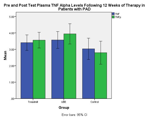
142 Figure*6a:* * 122#

143 Figure 6b 123#

144 Figure 6c 124#

145 Figure 6d Figure 6e 125#
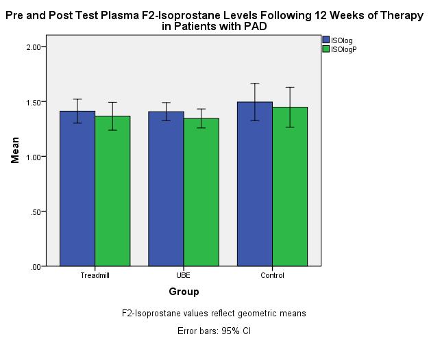
146 126#

147 Figure 7a 127#

148 Figure 7b 128#

149 Figure 7c 129#

150 Figure 8a Mean TNF change is expressed in pg/ml. 130#

151 Figure 8b 131#
152 Figure 8c 132#

153 Figure 9 TNF expressed in pg/ml: BMI is expressed in kg/m 2 133#
Biologic Oxidation BIOMEDICAL IMPORTAN
 Biologic Oxidation BIOMEDICAL IMPORTAN Chemically, oxidation is defined as the removal of electrons and reduction as the gain of electrons. Thus, oxidation is always accompanied by reduction of an electron
INFLAMMATION & REPAIR
 INFLAMMATION & REPAIR Lecture 7 Chemical Mediators of Inflammation Winter 2013 Chelsea Martin Special thanks to Drs. Hanna and Forzan Course Outline i. Inflammation: Introduction and generalities (lecture
Dyslipidemia Endothelial dysfunction Free radicals Immunologic
 ATHEROSCLEROSIS Hossein Mehrani Professor of Clinical Biochemistry Definition Atherosclerosis: Is a chronic inflammatory process characterized by plaque formation within the vessel wall of arteries and
III. 6. Test. Respiració cel lular
 III. 6. Test. Respiració cel lular Chapter Questions 1) What is the term for metabolic pathways that release stored energy by breaking down complex molecules? A) anabolic pathways B) catabolic pathways
Cell-Derived Inflammatory Mediators
 Cell-Derived Inflammatory Mediators Introduction about chemical mediators in inflammation Mediators may be Cellular mediators cell-produced or cell-secreted derived from circulating inactive precursors,
Energy Production In A Cell (Chapter 25 Metabolism)
") Energy Production In A Cell (Chapter 25 Metabolism) Large food molecules contain a lot of potential energy in the form of chemical bonds but it requires a lot of work to liberate the energy. Cells need
Energy Production In A Cell (Chapter 25 Metabolism) Large food molecules contain a lot of potential energy in the form of chemical bonds but it requires a lot of work to liberate the energy. Cells need
PHM142 Lecture 4: Platelets + Endothelial Cells
 PHM142 Lecture 4: Platelets + Endothelial Cells 1 Hematopoiesis 2 Platelets Critical in clotting - activated by subendothelial matrix proteins (e.g. collagen, fibronectin, von Willebrand factor) and thrombin
10. Which of the following immune cell is unable to phagocytose (a) neutrophils (b) eosinophils (c) macrophages (d) T-cells (e) monocytes
 neutrophils (b) eosinophils (c) macrophages (d) T-cells (e) monocytes") Chapter 2. Acute and chronic inflammation(6): 1. In acute inflammation, which events occur in the correct chronological order? (Remembered from 2000, 2004 exam.) p50 (a) transient vasoconstriction, stasis
Chapter 2. Acute and chronic inflammation(6): 1. In acute inflammation, which events occur in the correct chronological order? (Remembered from 2000, 2004 exam.) p50 (a) transient vasoconstriction, stasis
Enzymes and Metabolism
 PowerPoint Lecture Slides prepared by Vince Austin, University of Kentucky Enzymes and Metabolism Human Anatomy & Physiology, Sixth Edition Elaine N. Marieb 1 Protein Macromolecules composed of combinations
Chemical and Biochemical Mechanism Of Cell Injury.
 Chemical and Biochemical Mechanism Of Cell Injury. Professor Dr. M. Tariq Javed Dept. of Pathology Faculty of Vet. Science The University Of Agriculture Faisalabad Cell Injury When the cell is exposed
Pyruvate + NADH + H + ==== Lactate + NAD +
 1 UNIVERSITY OF PAPUA NEW GUINEA SCHOOL OF MEDICINE AND HEALTH SCIENCES DIVISION OF BASIC MEDICAL SCIENCES DISCIPLINE OF BIOCHEMISTRY AND MOLECULAR BIOLOGY PBL SEMINAR ANAEROBIC METABOLISM - An Overview
Chemical Energy. Valencia College
 9 Pathways that Harvest Chemical Energy Valencia College 9 Pathways that Harvest Chemical Energy Chapter objectives: How Does Glucose Oxidation Release Chemical Energy? What Are the Aerobic Pathways of
The Role of Massage in Blood Circulation, Pain Relief, and the Recovery Process: Implications of Existing Research
 The Role of Massage in Blood Circulation, Pain Relief, and the Recovery Process: Implications of Existing Research I. Basic Physiology of Circulation A. The Vascular Endothelium The endothelium is a complex
Arteriosclerosis & Atherosclerosis
 Arteriosclerosis & Atherosclerosis Arteriosclerosis = hardening of arteries = arterial wall thickening + loss of elasticity 3 types: -Arteriolosclerosis -Monckeberg medial sclerosis -Atherosclerosis Arteriosclerosis,
Electron Transport Chain and Oxidative phosphorylation
 Electron Transport Chain and Oxidative phosphorylation So far we have discussed the catabolism involving oxidation of 6 carbons of glucose to CO 2 via glycolysis and CAC without any oxygen molecule directly
Ch 9: Cellular Respiration
 Ch 9: Cellular Respiration Cellular Respiration An overview Exergonic reactions and catabolic pathway Energy stored in bonds of food molecules is transferred to ATP Cellular respiration provides the energy
Ch. 9 Cell Respiration. Title: Oct 15 3:24 PM (1 of 53)
") Ch. 9 Cell Respiration Title: Oct 15 3:24 PM (1 of 53) Essential question: How do cells use stored chemical energy in organic molecules and to generate ATP? Title: Oct 15 3:28 PM (2 of 53) Title: Oct 19
Ch. 9 Cell Respiration Title: Oct 15 3:24 PM (1 of 53) Essential question: How do cells use stored chemical energy in organic molecules and to generate ATP? Title: Oct 15 3:28 PM (2 of 53) Title: Oct 19
Metabolism. Metabolism. Energy. Metabolism. Energy. Energy 5/22/2016
 5//016 Metabolism Metabolism All the biochemical reactions occurring in the body Generating, storing and expending energy ATP Supports body activities Assists in constructing new tissue Metabolism Two
The Study of Endothelial Function in CKD and ESRD
 The Study of Endothelial Function in CKD and ESRD Endothelial Diversity in the Human Body Aird WC. Circ Res 2007 Endothelial Diversity in the Human Body The endothelium should be viewed for what it is:
How Cells Harvest Chemical Energy
 How Cells Harvest Chemical Energy Global Athlete Outreach Program US CytoThesis Systems Medicine Center www.cytothesis.us US OncoTherapy Systems BioMedicine Group CytoThesis Bioengineering Research Group
Stroke, the most common medical emergency, is a
 Report on Progress 2016 Advances in Our Knowledge of Stroke Mechanisms and Therapy Xuefang Ren, M.D., and James W. Simpkins Ph. D Department of Physiology and Pharmacology, Experimental Stroke Core, Center
Pathology of Coronary Artery Disease
 Pathology of Coronary Artery Disease Seth J. Kligerman, MD Pathology of Coronary Artery Disease Seth Kligerman, MD Assistant Professor Medical Director of MRI University of Maryland Department of Radiology
 Glossary For TheFatNurse s For All Ages Series Adipocytes, also known as lipocytes and fat cells, are the cells that primarily compose adipose tissue, specialized in storing energy as fat. Apolipoprotein
shehab Moh Tarek ... ManarHajeer
 3 shehab Moh Tarek... ManarHajeer In the previous lecture we discussed the accumulation of oxygen- derived free radicals as a mechanism of cell injury, we covered their production and their pathologic
La Trombosi Arteriosa
 La Trombosi Arteriosa Prof. Giovanni Davì Medicina Interna Chieti Platelet activation and thrombosis Harrison 19 edizione Platelets are essential for primary hemostasis and repair of the endothelium They
Biochemistry: A Short Course
 Tymoczko Berg Stryer Biochemistry: A Short Course Second Edition CHAPTER 20 The Electron-Transport Chain 2013 W. H. Freeman and Company Chapter 20 Outline Oxidative phosphorylation captures the energy
The 10 th International & 15 th National Congress on Quality Improvement in Clinical Laboratories
 The 10 th International & 15 th National Congress on Quality Improvement in Clinical Laboratories Cardiac biomarkers in atherosclerosis Najma Asadi MD-APCP Ross and Colleagues in 1973: Response to Injury
Oxidative phosphorylation & Photophosphorylation
 Oxidative phosphorylation & Photophosphorylation Oxidative phosphorylation is the last step in the formation of energy-yielding metabolism in aerobic organisms. All oxidative steps in the degradation of
4. Which step shows a split of one molecule into two smaller molecules? a. 2. d. 5
 1. Which of the following statements about NAD + is false? a. NAD + is reduced to NADH during both glycolysis and the citric acid cycle. b. NAD + has more chemical energy than NADH. c. NAD + is reduced
Part IV Antithrombotics, Anticoagulants and Fibrinolytics
 Part IV Antithrombotics, Anticoagulants and Fibrinolytics "The meaning of good and bad, of better and worse, is simply helping or hurting" Emerson Chapter 16: Blood Coagulation and Fibrinolytic System
Electron transport chain chapter 6 (page 73) BCH 340 lecture 6
 BCH 340 lecture 6") Electron transport chain chapter 6 (page 73) BCH 340 lecture 6 The Metabolic Pathway of Cellular Respiration All of the reactions involved in cellular respiration can be grouped into three main stages
Electron transport chain chapter 6 (page 73) BCH 340 lecture 6 The Metabolic Pathway of Cellular Respiration All of the reactions involved in cellular respiration can be grouped into three main stages
Pathophysiology of Catheter-Related Infection. All sources of infection are potential targets for prevention. Infusates/ drugs. hub/lines Dressing
 Pathophysiology of Catheter-Related Infection All sources of infection are potential targets for prevention catheter hematogeneous Infusates/ drugs hub/lines Dressing skin Critically ill patient: 2-4 vascular
BIOLOGY - CLUTCH CH.9 - RESPIRATION.
 !! www.clutchprep.com CONCEPT: REDOX REACTIONS Redox reaction a chemical reaction that involves the transfer of electrons from one atom to another Oxidation loss of electrons Reduction gain of electrons
Class XI Chapter 14 Respiration in Plants Biology. 1. It is a biochemical process. 1. It is a physiochemical process.
 Question 1: Differentiate between (a) Respiration and Combustion (b) Glycolysis and Krebs cycle (c) Aerobic respiration and Fermentation (a) Respiration and combustion Respiration Combustion 1. It is a
Mitochondria and ATP Synthesis
 Mitochondria and ATP Synthesis Mitochondria and ATP Synthesis 1. Mitochondria are sites of ATP synthesis in cells. 2. ATP is used to do work; i.e. ATP is an energy source. 3. ATP hydrolysis releases energy
 Question 1: Differentiate between (a) Respiration and Combustion (b) Glycolysis and Krebs cycle (c) Aerobic respiration and Fermentation (a) Respiration and combustion Respiration Combustion 1. It is a
Part 1 Risk Factors and Atherosclerosis. LO1. Define the Different Forms of CVD
 Week 3: Cardiovascular Disease Learning Outcomes: 1. Define the difference forms of CVD 2. Describe the various risk factors of CVD 3. Describe atherosclerosis and its stages 4. Describe the role of oxidation,
Marah Bitar. Faisal Nimri ... Nafeth Abu Tarboosh
 8 Marah Bitar Faisal Nimri... Nafeth Abu Tarboosh Summary of the 8 steps of citric acid cycle Step 1. Acetyl CoA joins with a four-carbon molecule, oxaloacetate, releasing the CoA group and forming a six-carbon
BY: RASAQ NURUDEEN OLAJIDE
 BY: RASAQ NURUDEEN OLAJIDE LECTURE CONTENT INTRODUCTION CITRIC ACID CYCLE (T.C.A) PRODUCTION OF ACETYL CoA REACTIONS OF THE CITIRC ACID CYCLE THE AMPHIBOLIC NATURE OF THE T.C.A CYCLE THE GLYOXYLATE CYCLE
19 Oxidative Phosphorylation and Photophosphorylation W. H. Freeman and Company
 19 Oxidative Phosphorylation and Photophosphorylation 2013 W. H. Freeman and Company CHAPTER 19 Oxidative Phosphorylation and Photophosphorylation Key topics: Electron transport chain in mitochondria Capture
How Cells Harvest Energy. Chapter 7. Respiration
 How Cells Harvest Energy Chapter 7 Respiration Organisms classified on how they obtain energy: autotrophs: produce their own organic molecules through photosynthesis heterotrophs: live on organic compounds
MULTIPLE CHOICE QUESTIONS
 MULTIPLE CHOICE QUESTIONS 1. Which of the following statements concerning anabolic reactions is FALSE? A. They are generally endergonic. B. They usually require ATP. C. They are part of metabolism. D.
PCTH 400. Endothelial dysfunction and cardiovascular diseases. Blood vessel LAST LECTURE. Endothelium. High blood pressure
 PCTH 400 LAST LECTURE Endothelial dysfunction and cardiovascular diseases. Classic Vascular pharmacology -chronic -systemic Local Vascular pharmacology -acute -targeted High blood pressure Blood pressure
Cellular Respiration. Cellular Respiration. C 6 H 12 O 6 + 6O > 6CO 2 + 6H energy. Heat + ATP. You need to know this!
 Cellular Respiration LISA Biology Cellular Respiration C 6 H 12 O 6 + 6O 2 - - - - - > 6CO 2 + 6H 2 0 + energy You need to know this! Heat + ATP 1 Did that equation look familiar? * The equation for cellular
Metabolism. Metabolic pathways. BIO 5099: Molecular Biology for Computer Scientists (et al) Lecture 11: Metabolic Pathways
 Lecture 11: Metabolic Pathways") BIO 5099: Molecular Biology for Computer Scientists (et al) Lecture 11: Metabolic Pathways http://compbio.uchsc.edu/hunter/bio5099 Larry.Hunter@uchsc.edu Metabolism Metabolism is the chemical change of
BIO 5099: Molecular Biology for Computer Scientists (et al) Lecture 11: Metabolic Pathways http://compbio.uchsc.edu/hunter/bio5099 Larry.Hunter@uchsc.edu Metabolism Metabolism is the chemical change of
number Done by Corrected by Doctor Nayef Karadsheh
 number 17 Done by Abdulrahman Alhanbali Corrected by Lara Abdallat Doctor Nayef Karadsheh 1 P a g e Pentose Phosphate Pathway (PPP) Or Hexose Monophosphate Shunt In this lecture We will talk about the
Plant Respiration. Exchange of Gases in Plants:
 Plant Respiration Exchange of Gases in Plants: Plants do not have great demands for gaseous exchange. The rate of respiration in plants is much lower than in animals. Large amounts of gases are exchanged
Introduction. Every organism must exchange materials and energy with its environment, and this exchange ultimately occurs at the cellular level.
 Introduction Every organism must exchange materials and energy with its environment, and this exchange ultimately occurs at the cellular level. Cells live in aqueous environments. The resources that they
) one consumes in breathing is converted to:, which of the following would be found in the oxidized state?
 one consumes in breathing is converted to:, which of the following would be found in the oxidized state?") MCB 102: Pantea s Sxn Chapter 19 Problem Set Answer Key 1) Page: 690 Ans: E Almost all of the oxygen (O 2 ) one consumes in breathing is converted to: A) acetyl-coa. B) carbon dioxide (CO 2 ). C) carbon
MCB 102: Pantea s Sxn Chapter 19 Problem Set Answer Key 1) Page: 690 Ans: E Almost all of the oxygen (O 2 ) one consumes in breathing is converted to: A) acetyl-coa. B) carbon dioxide (CO 2 ). C) carbon
Cellular Respiration. Biochemistry Part II 4/28/2014 1
 Cellular Respiration Biochemistry Part II 4/28/2014 1 4/28/2014 2 The Mitochondria The mitochondria is a double membrane organelle Two membranes Outer membrane Inter membrane space Inner membrane Location
Cellular Pathways That Harvest Chemical Energy. Cellular Pathways That Harvest Chemical Energy. Cellular Pathways In General
 Cellular Pathways That Harvest Chemical Energy A. Obtaining Energy and Electrons from Glucose Lecture Series 12 Cellular Pathways That Harvest Chemical Energy B. An Overview: Releasing Energy from Glucose
Cellular Respiration: Harvesting Chemical Energy Chapter 9
 Cellular Respiration: Harvesting Chemical Energy Chapter 9 Assemble polymers, pump substances across membranes, move and reproduce The giant panda Obtains energy for its cells by eating plants which get
10/25/2010 CHAPTER 9 CELLULAR RESPIRATION. Life is Work. Types of cellular respiration. Catabolic pathways = oxidizing fuels
 CHAPTER 9 CELLULAR RESPIRATION Life is Work Living cells require transfusions of energy from outside sources to perform their many tasks: Chemical work Transport work Mechanical work Energy stored in the
Chapter 8 Mitochondria and Cellular Respiration
 Chapter 8 Mitochondria and Cellular Respiration Cellular respiration is the process of oxidizing food molecules, like glucose, to carbon dioxide and water. The energy released is trapped in the form of
Chapter 6. How Cells Harvest Chemical Energy. Lecture by Richard L. Myers
 Chapter 6 How Cells Harvest Chemical Energy oweroint Lectures for Biology: Concepts & Connections, Sixth Edition Campbell, Reece, Taylor, Simon, and Dickey Copyright 2009 earson Education, Inc. Lecture
Structure of the Mitochondrion. Cell Respiration. Cellular Respiration. Catabolic Pathways. Photosynthesis vs. Cell Respiration ATP 10/14/2014
 Structure of the Mitochondrion Cellular Respiration Chapter 9 Pgs. 163 183 Enclosed by a double membrane Outer membrane is smooth Inner, or cristae, membrane is folded - this divides the mitochondrion
This slide belongs to iron lecture and it is to clarify the iron cycle in the body and the effect of hypoxia on erythropoitein secretion
 This slide belongs to iron lecture and it is to clarify the iron cycle in the body and the effect of hypoxia on erythropoitein secretion Topics of today lectures: Hemostasis Meaning of hemostasis Mechanisms
BIOLOGY 101. CHAPTER 9: Cellular Respiration - Fermentation: Life is Work
 BIOLOGY 101 CHAPTER 9: Cellular Respiration - Fermentation: Life is Work An Introduction to Metabolism: Energy of Life 8.3 ATP powers cellular work by coupling exergonic reactions to endergonic reactions
Cellular Respiration- -conversion of stored energy in glucose to usable energy for the cell -energy in cells is stored in the form of ATP
 Cellular Respiration Notes Chapter 7 How Cells Make ATP Energy Releasing Pathways Cellular Respiration- -conversion of stored energy in glucose to usable energy for the cell -energy in cells is stored
Cell Signaling (part 1)
") 15 Cell Signaling (part 1) Introduction Bacteria and unicellular eukaryotes respond to environmental signals and to signaling molecules secreted by other cells for mating and other communication. In multicellular
15 Cell Signaling (part 1) Introduction Bacteria and unicellular eukaryotes respond to environmental signals and to signaling molecules secreted by other cells for mating and other communication. In multicellular
Chapter 7 Cellular Respiration and Fermentation*
 Chapter 7 Cellular Respiration and Fermentation* *Lecture notes are to be used as a study guide only and do not represent the comprehensive information you will need to know for the exams. Life Is Work
WHY IS THIS IMPORTANT?
 CHAPTER 3 ESSENTIALS OF METABOLISM WHY IS THIS IMPORTANT? It is important to have a basic understanding of metabolism because it governs the survival and growth of microorganisms The growth of microorganisms
3.7.1 Define cell respiration [Cell respiration is the controlled release of energy from organic compounds in cells to form ATP]
![3.7.1 Define cell respiration [Cell respiration is the controlled release of energy from organic compounds in cells to form ATP]](/thumbs/72/66350985.jpg "3.7.1 Define cell respiration [Cell respiration is the controlled release of energy from organic compounds in cells to form ATP]") 3.7 Cell respiration ( Chapter 9 in Campbell's book) 3.7.1 Define cell respiration [Cell respiration is the controlled release of energy from organic compounds in cells to form ATP] Organic compounds store
3.7 Cell respiration ( Chapter 9 in Campbell's book) 3.7.1 Define cell respiration [Cell respiration is the controlled release of energy from organic compounds in cells to form ATP] Organic compounds store
Ch. 45 Blood Plasma proteins, Coagulation and Fibrinolysis Student Learning Outcomes: Describe basic components of plasma
 Chapt. 45 Ch. 45 Blood Plasma proteins, Coagulation and Fibrinolysis Student Learning Outcomes: Describe basic components of plasma Inheritance of X-linked gene for Factor VIII hemophilia A Explain the
Cellular Respiration
 Cellular Respiration 1. To perform cell work, cells require energy. a. A cell does three main kinds of work: i. Mechanical work, such as the beating of cilia, contraction of muscle cells, and movement
Metabolism Energy Pathways Biosynthesis. Catabolism Anabolism Enzymes
 Topics Microbial Metabolism Metabolism Energy Pathways Biosynthesis 2 Metabolism Catabolism Catabolism Anabolism Enzymes Breakdown of complex organic molecules in order to extract energy and dform simpler
Students will identify factors that affect blood flow and/or describe how these factors affect blood flow through the cardiovascular system.
 Students will identify factors that affect blood flow and/or describe how these factors affect blood flow through the cardiovascular system. Content Limits Items may address factors such as blood pressure,
Vocabulary. Chapter 20: Electron Transport and Oxidative Phosphorylation
 Vocabulary ATP Synthase: the enzyme responsible for production of ATP in mitochondria Chemiosmotic Coupling: the mechanism for coupling electron transport to oxidative phosphorylation; it requires a proton
Inflammation I. Dr. Nabila Hamdi MD, PhD
 Inflammation I Dr. Nabila Hamdi MD, PhD http://library.med.utah.edu/webpath/exam/m ULTGEN/examidx.htm 2 ILOs Distinguish between acute and chronic inflammation with respect to causes, nature of the inflammatory
Ch 07. Microbial Metabolism
 Ch 07 Microbial Metabolism SLOs Differentiate between metabolism, catabolism, and anabolism. Fully describe the structure and function of enzymes. Differentiate between constitutive and regulated enzymes.
INFLAMMATION. 5. Which are the main phases of inflammation in their "sequence": 1. Initiation, promotion, progression.
 INFLAMMATION 1. What is inflammation: 1. Selective anti-infective pathological reaction. 2. Pathological process, typical for vascularized tissues. 3. Self-sustained pathological condition. 4. Disease
CELL BIOLOGY - CLUTCH CH AEROBIC RESPIRATION.
 !! www.clutchprep.com CONCEPT: OVERVIEW OF AEROBIC RESPIRATION Cellular respiration is a series of reactions involving electron transfers to breakdown molecules for (ATP) 1. Glycolytic pathway: Glycolysis
2/4/17. Cellular Metabolism. Metabolism. Cellular Metabolism. Consists of all of the chemical reactions that take place in a cell.
 Metabolism Cellular Metabolism Consists of all of the chemical reactions that take place in a cell. Can be reactions that break things down. (Catabolism) Or reactions that build things up. (Anabolism)
Respiration. Respiration. Respiration. How Cells Harvest Energy. Chapter 7
 How Cells Harvest Energy Chapter 7 Organisms can be classified based on how they obtain energy: autotrophs: are able to produce their own organic molecules through photosynthesis heterotrophs: live on
Published on Second Faculty of Medicine, Charles University (http://www.lf2.cuni.cz )
") Published on Second Faculty of Medicine, Charles University (http://www.lf2.cuni.cz ) Biochemistry Submitted by Marie Havlová on 8. February 2012-0:00 Syllabus of Biochemistry Mechanisms of enzyme catalysis.
Published on Second Faculty of Medicine, Charles University (http://www.lf2.cuni.cz ) Biochemistry Submitted by Marie Havlová on 8. February 2012-0:00 Syllabus of Biochemistry Mechanisms of enzyme catalysis.
Moh Tarek + Faisal Massad. Tala Saleh ... Naif
 19 Moh Tarek + Faisal Massad Tala Saleh... Naif Last lecture we ve talked about the main antioxidant system which are the enzymes found in our body, mainly: 1. Glutathione peroxidase 2. Super oxide dismutase(sod)
MITOCHONDRIA LECTURES OVERVIEW
 1 MITOCHONDRIA LECTURES OVERVIEW A. MITOCHONDRIA LECTURES OVERVIEW Mitochondrial Structure The arrangement of membranes: distinct inner and outer membranes, The location of ATPase, DNA and ribosomes The
1- Which of the following statements is TRUE in regards to eukaryotic and prokaryotic cells?
 Name: NetID: Exam 3 - Version 1 October 23, 2017 Dr. A. Pimentel Each question has a value of 4 points and there are a total of 160 points in the exam. However, the maximum score of this exam will be capped
Relationship between serum glutathione peroxidase-1activity with endothelial dysfunction level in patients with coronary artery diseases
 Relationship between serum glutathione peroxidase-1activity with endothelial dysfunction level in patients with coronary artery diseases Introduction Reactive oxygen species (ROS),such as superoxide and
Metabolism. Chapter 8 Microbial Metabolism. Metabolic balancing act. Catabolism Anabolism Enzymes. Topics. Metabolism Energy Pathways Biosynthesis
 Chapter 8 Microbial Metabolism Topics Metabolism Energy Pathways Biosynthesis Catabolism Anabolism Enzymes Metabolism 1 2 Metabolic balancing act Catabolism and anabolism simple model Catabolism Enzymes
Reading Assignments. A. Energy and Energy Conversions. Lecture Series 9 Cellular Pathways That Harvest Chemical Energy. gasoline) or elevated mass.
 or elevated mass.") Lecture Series 9 Cellular Pathways That Harvest Chemical Energy Reading Assignments Review Chapter 3 Energy, Catalysis, & Biosynthesis Read Chapter 13 How Cells obtain Energy from Food Read Chapter 14
Lecture Series 9 Cellular Pathways That Harvest Chemical Energy Reading Assignments Review Chapter 3 Energy, Catalysis, & Biosynthesis Read Chapter 13 How Cells obtain Energy from Food Read Chapter 14
Oxidative Phosphorylation
 Oxidative Phosphorylation Energy from Reduced Fuels Is Used to Synthesize ATP in Animals Carbohydrates, lipids, and amino acids are the main reduced fuels for the cell. Electrons from reduced fuels are
Electron Transport and oxidative phosphorylation (ATP Synthesis) Dr. Howaida Nounou Biochemistry department Sciences college
 Dr. Howaida Nounou Biochemistry department Sciences college") Electron Transport and oxidative phosphorylation (ATP Synthesis) Dr. Howaida Nounou Biochemistry department Sciences college The Metabolic Pathway of Cellular Respiration All of the reactions involved
Electron Transport and oxidative phosphorylation (ATP Synthesis) Dr. Howaida Nounou Biochemistry department Sciences college The Metabolic Pathway of Cellular Respiration All of the reactions involved
ATHEROSCLEROSIS زيد ثامر جابر. Zaid. Th. Jaber
 ATHEROSCLEROSIS زيد ثامر جابر Zaid. Th. Jaber Objectives 1- Review the normal histological features of blood vessels walls. 2-define the atherosclerosis. 3- display the risk factors of atherosclerosis.
Cellular Respiration Harvesting Chemical Energy ATP
 Cellular Respiration Harvesting Chemical Energy ATP 2006-2007 What s the point? The point is to make ATP! ATP 2006-2007 Harvesting stored energy Energy is stored in organic molecules carbohydrates, fats,
2. What are the products of cellular respiration? Include all forms of energy that are products.
 Name Per Cellular Respiration An Overview Why Respire Anyhoo? Because bucko all cells need usable chemical energy to do work. The methods cells use to convert glucose into ATP vary depending on the availability
Cell Injury MECHANISMS OF CELL INJURY
 Cell Injury MECHANISMS OF CELL INJURY The cellular response to injurious stimuli depends on the following factors: Type of injury, Its duration, and Its severity. Thus, low doses of toxins or a brief duration
Integration Of Metabolism
 Integration Of Metabolism Metabolism Consist of Highly Interconnected Pathways The basic strategy of catabolic metabolism is to form ATP, NADPH, and building blocks for biosyntheses. 1. ATP is the universal
7 Pathways That Harvest Chemical Energy
 7 Pathways That Harvest Chemical Energy Pathways That Harvest Chemical Energy How Does Glucose Oxidation Release Chemical Energy? What Are the Aerobic Pathways of Glucose Metabolism? How Is Energy Harvested
Citric Acid Cycle and Oxidative Phosphorylation
 Citric Acid Cycle and Oxidative Phosphorylation Page by: OpenStax Summary The Citric Acid Cycle In eukaryotic cells, the pyruvate molecules produced at the end of glycolysis are transported into mitochondria,
Pathophysiology of Cardiovascular System. Dr. Hemn Hassan Othman, PhD
 Pathophysiology of Cardiovascular System Dr. Hemn Hassan Othman, PhD hemn.othman@univsul.edu.iq What is the circulatory system? The circulatory system carries blood and dissolved substances to and from
Chapter 5 Microbial Metabolism: The Chemical Crossroads of Life
 Chapter 5 Microbial Metabolism: The Chemical Crossroads of Life Copyright The McGraw-Hill Companies, Inc. Permission required for reproduction or display. The Metabolism of Microbes metabolism all chemical
Cellular Respiration Harvesting Chemical Energy ATP
 Cellular Respiration Harvesting Chemical Energy ATP 2006-2007 What s the point? The point is to make ATP! ATP 2006-2007 Harvesting stored energy Energy is stored in organic molecules carbohydrates, fats,
CH 7: Cell Respiration and Fermentation Overview. Concept 7.1: Catabolic pathways yield energy by oxidizing organic fuels
 CH 7: Cell Respiration and Fermentation Overview Living cells require energy from outside sources Some animals obtain energy by eating plants, and some animals feed on other organisms Energy flows into
Hemostasis and Thrombosis
 Hemostasis Hemostasis and Thrombosis Normal hemostasis is a consequence of tightly regulated processes that maintain blood in a fluid state in normal vessels, yet also permit the rapid formation of a hemostatic
Anatomy and Physiology
 Anatomy and Physiology For The First Class 2 nd Semester Thrombocytes = Platelets Thrombocytes = Platelets Blood platelets are non-nucleated disc like cell fragments 2-4 µm in diameter. Platelets are not
Eicosapentaenoic Acid and Docosahexaenoic Acid: Are They Different?
 Eicosapentaenoic Acid and Docosahexaenoic Acid: Are They Different? Trevor A Mori, Ph.D., Professor, School of Medicine and Pharmacology, Royal Perth Hospital Unit, University of Western Australia, Perth,
Review. Respiration. Glycolysis. Glycolysis is the decomposition (lysis) of glucose (glyco) to pyruvate (or pyruvic acid).
 of glucose (glyco) to pyruvate (or pyruvic acid).") Review Photosynthesis is the process of incorporating energy from light into energy-rich molecules like glucose. Respiration is the opposite process extracting that stored energy from glucose to form ATP
Review Photosynthesis is the process of incorporating energy from light into energy-rich molecules like glucose. Respiration is the opposite process extracting that stored energy from glucose to form ATP
number Done by Corrected by Doctor
 number 18 Done by Mahmoud Harbi Corrected by حسام أبو عوض Doctor Nayef Karadsheh Sources of Reactive Oxygen Species (ROS) 1 P a g e 1- Oxidases: there are some that produce hydrogen peroxide (H₂O₂) 2-
Chapter 5. Microbial Metabolism
 Chapter 5 Microbial Metabolism Metabolism Collection of controlled biochemical reactions that take place within a microbe Ultimate function of metabolism is to reproduce the organism Metabolic Processes