ULTRASONOGRAPHY RADIOLOGY Simple abdominal X-ray Intravenous urography Retrograde/anterograde pieloureterography Renal angiography CT
|
|
|
- Raymond Newman
- 5 years ago
- Views:
Transcription
1


2 ULTRASONOGRAPHY RADIOLOGY Simple abdominal X-ray Intravenous urography Retrograde/anterograde pieloureterography Renal angiography CT NUCLEAR MEDICINE Static studies: static renal scintigraphy Dynamic studies: renogram MRI
; - renal borders (normally smooth, regular); - renal arteries by color Doppler. Diseases: - Dilatation of pielocaliceal system. - Pielocaliceal stasis in different obstructive uropathies.")
3 Normal structures: - kidneys dimensions (normally = cm, with differences between the two kidneys < 2 cm); - parenchymal index (normally > 1.5 cm); - renal borders (normally smooth, regular); - renal arteries by color Doppler. Diseases: - Dilatation of pielocaliceal system. - Pielocaliceal stasis in different obstructive uropathies. - Kidney stones: hyperechoic images with posterior shadow cone. - Calcifications in the renal parenchyma. - Polycystic kidney disease. - Diagnosis of benign and malignant tumors. - Monitoring transplanted kidney. - Guiding percutaneous puncture for diagnostic / therapeutic reasons.


4 Liver SPLINA Normal view Right kidney Left kidney


5 B mode longitudinal transversal


6 color Doppler USG B Mode & Doppler Power Doppler

7 Doppler USG


8 RENAL ABSCESS
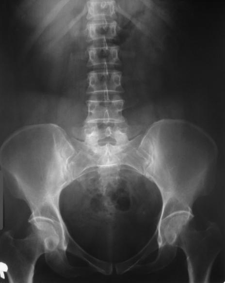
9 Simple abdominal X-ray Determine kidney area : Location Form (borders) Dimensions Radiopaque stones Calcifications Landmarks: m. psoas, 11-12th ribs, L1-L2 lumbar vertebral bodies


10

11 Preparation. Iodine contrast medium (ionic/non-ionic). Sequence of images.


12 Intravenous urography
13 Patient preparation for investigation: - young patients with regular chair don t requires preparation, light breakfast is recommended; - patients who are constipated will need purgative and laxative drugs or evacuator enema the day before the investigation + other evacuator enema 2-3 hours prior investigation; - patients with flatulence will take antiflatulents (Espumisan); - in special case can be prescribed calming remedies: valerian, chamomile infusion etc; - check urinalysis (urea, creatinine).


14 The first scan is performed on goal (RRVS). Intravenously administered iodine contrast media. Following administration of contrast solution is made to: min; min; last radiography is performed orthostatic at min. NB!!!! Sometimes it needed to perform radiography 2-3 min after the administrations of contrast media, to view nefrograma. On nefrograma can be detected early signs of pyelonephritis (parenchymal heterogeneous structure).
15 -shows all three components of calyx: cup, body, rod; -towards X-rays calyx can be located perpendicular (see themselves as the triangle) or orthogonal (see themselves as ring formation); -three main groups of calyx: upper, middle, lower; -the renal pelvis has clear border, is homogeneous and can be located intrarenal, extrarenal or mixed; -the urethers have 25-30cm in length and 4mm in width, shows areas of insignificant narrowing and widening (normal anatomical structure) and aren t homogeneous contrasted; - the urethers are open in upper part of Lie-to triangle of urinary bladder, posterior side; - urinary bladder at men is round, at female oval, need to be homogeneous contrasted.

16 IVU- normal anatomical structure Kidney longitudinal diameter L = 13 cm Kidney transverse diameter B = 6 cm. 1 - renal axis tilt 10, compared to the longitudinal axis; 2 - distance between renal pole: cm - upper pole cm - lower pole; 3 - uretheral diameter mm. (Average = ADULT)


17 Simple X-rays IVU 5-7 min IVU min
18 -Allergy to iodine solutions; -In case of tiriotoxicoses; -Asotemy (in this case we won t see the kidneys); -Decompensate cardiovascular malformations; -Malformations of liver; -Renal failure; -Glomerulonefritis; -Pregnancy.


19


20


21


22 AORTOGRAPHY: LEFT RENAL ARTERY THROMBOSIS AORTOGRAPHY & UIV: SUPLIMENTARY RENAL ARTERY AT THE INFERIOR POLE

23 Indicated in all renal pathology, including: - Inflammatory diseases; - Congenital malformations; - Trauma; - Positive diagnosis and staging of malignant tumors; - Post-nephrectomy control after cancer; - Guiding punctures / percutaneous drainage; - Detection of uretheral stones; - Study of the renal vessels.

24


25


26 CT
27 Indications: - Diagnosis and staging of malignant tumors; - The study of renal arteries (Angio-MRI); - Acute and chronic infections; -URO-MRI can replace IVU if there is contraindications to iodinated contrast agent injection; - Excellent assessment of urinary bladder, pelvic and retroperitoneal lymph node.


28 T1, T2 normal kidney


29 renal carcinoma


30 - Appreciate the percentage of each kidney that participate in the global renal function; - Detects areas of hyper- or afixation; Normal scintigraphy
 kidney transplant congenital anomalies")
31 The renal function and perfusion renal obstruction kidney infection ( chronic and acute pielonephritis ) kidney transplant congenital anomalies tumors
32 Extract. fraction Clearance Tc-99m DTPA 20% ml/min (to appreciate the function of renal glomeruls) Tc-99m MAG % ~ 300 ml/min (to appreciate the function of renal tubules)

33 Isotopic renogram The technique involves measuring the scintillation probes the time variation of renal radioactivity after administration of a radiotracer with predominantly renal elimination. The pulses are processed and recorded as the renogram. That is a dynamic study of kidney function. It is the renogram curves that show transit of tracer through the kidneys, so the curves are more important than the images. Upslope of curves demonstrate kidney uptake. Downslope of curves demonstrate elimination. The curve has three segments: - ascending segment steep, short duration = vascular time; - slow upward slope = parenchyma time (capture, secretion); - progressive downward slope = renal elimination.!!! The technique has the disadvantage of poor specificity results.
34 Concentration Concentration Concentration Concentration Extravascular Tissue Time Blood Time Kidney Tubules + pelvis Time Bladder
35 Concentration Concentration Concentration Concentration Extravascular Tissue Time Blood This is the curve that we want Kidney Tubules + pelvis Time Time Bladder


36 Normal radioactive substance capturing
37 Pyelonephritis Pyelonephritis are classified into acute and chronic. By acute pyelonephritis means suppurative- inflammatory process of kidneys with varying degrees of intensity, which extends from the renal tubules to cortical. Chronic pyelonephritis may follow an acute pyelonephritis or may have started as a quiet allure. Chronic pyelonephritis is inflammation of the renal parenchyma that develops slowly, with periodic worsening and with kidney sclerosis in final phase.
38 Clinical: -violent beginning - chills, fever, unilateral or bilateral back pain, -piuria, terminal hematuria. -polakiuria, pain in urination, oliguria, -headache, asthenia, nausea, vomiting. Ultrasonography: -enlarged kidney, hypoechogenyc parenchyma, -cortico-medullary differentiation is absent, -moderate enlargement of calyx and renal pelvis.!!! IN ACUTE PYELONEPHRITIS I/V UROGRAPHY IS NOT MADE, IT ALLOWS FEW DAYS AFTER ONSET. I/v urography: -syndrome of "white" kidney - is due to accumulation of contrast in glomeruls, without accumulate in calyx; -contrasting of calyx and pelvis is late and less intensive, with clear contours; -contrasting of pelvis without visualization of contrast in calyx (due to spasm).


39


40 Acute pyelonephritis IVU : enlarged left kidney, delayed nefrograma, narrowed calyx CT: bilateral enlarged kidneys, ribbed nefrograma, cortico-medullary layer is tickened, reduced contrasting, perinefral tissue opacity.


41 post L post R Cold" catchments - single or multiple, renal border clear, regular, low diffuse absorption. LPO RPO
42 Clinical picture depends on phase process: active, latent or remission. In general, patients have: - lumbar pains, exacerbated by efforts; - unclear urine, urinary frequency; -subfebrility, fatigue, asthenia, hypertension. Ultrasonography: - kidney border is irregular, -reduced parenchyma index, - hyper echoic parenchyma +/- transonic zone. Intravenous urography: -the kidney are increased in size because of inflammatory infiltration; -the calyx are elongated, dilated, deployed, spasms of calyx rods; - tubular-parenchyma low tide; - heterogeneous contrasted renal pelvis because of mucosal edema; sometimes it is hypotonic, widened; -hypotonic urethers and bladder; -in terminal stages kidney shrinks in volume.


43 IVU: CPN - Cortical thinned, calyx dilated, hypotonic, dislocated


44 MRI- shrinked kidney with irregular border, thickened cortical-medullar layer, calyx-pelvical system deformed. CT- shrinked kidney with irregular border, thickened cortical-medullar layer, calyxpelvical system deformed, perirenal adipose tissue endured.


45 Viewing minimal changes in I, II scan phases. The III scanner phase is disturbed, with delay evacuation phase, without obstruction evidence.
46 Glomerulonephritis is inflammation of the renal glomerules. This inflammation will cause damage to the glomerular membrane and renal capillary endothelium. One of the most feared complication is the evolution of glomerulonephritis towards chronic kidney disease! From an evolutionary standpoint, glomerulonephritis can be: acute, sub-acute (rapidly progressive), chronic. Clinical: - nephritic syndrome with edema, oliguria (decreased 24 hours urine at ml), hypertension, hematuria (blood in urine); - nephrotic syndrome with hematuria, frothy urine (due to elimination of increased urinary protein), soft edema which at the pressure will leave a trace.
 and parenchyma changes (sometimes appear small cysts).")
47 Because of the low density of urine and low kidney function, X-ray imaging investigations (RRVS, intravenous urography, CT with and without contrast) or magnetic field investigations (MRI) are less informative. USG can be used to determine the dimensions of kidneys (kidneys become enlarged in size) and parenchyma changes (sometimes appear small cysts). The most specific investigations are the laboratory one.
.")
48 Renal failure is defined by the rapid decline (acute renal failure) or slow decline (chronic renal failure) of renal function, resulting in inability to maintain electrolyte balance and excrete nitrogen products. Serum creatinine is a convenient marker for evaluation of renal function: creatinine value increase by mg/ dl / day (acute RF). Renal failure can be described also as a decrease in glomerular filtration rate.
49 ARF appear usually after an exacerbation of pre-existing renal disease, such as chronic glomerulonephritis, kidney diabetic or hypertensive damage, drug abuse (especially painkillers), or can be caused by an acute event (acute glomerulonephritis, autoimmune disease, infections, surgery, sepsis, etc). Acute renal failure is in most cases reversible. Symptoms that may occur in case of acute renal failure are: - anuria (ceasing production of urine); - oliguria (decreased production of urine below 400 ml/day); - hematuria (blood in urine); - lower extremities edema; - thirsty, lack of appetite, nausea and vomiting; - headache, abdominal pain; - drowsiness, confusion, anxiety; - heart rhythm disorder; - convulses, coma.
50 Chronic renal failure develops over several years. Symptoms usually appear at an advanced stage. The most common causes of chronic kidney disease are high blood pressure, type 2 diabetes and polycystic kidney disease. Ultrasonography: -reducing kidney size; -thinning cortical layer with increased echogenicity; -renal pyramids low differentiation; - papillary calcifications; -cysts. X-ray imaging investigations (RRVS, intravenous urography, CT with and without contrast) or magnetic field investigations (MRI) are less informative in the early stages, the use of contrast is a contraindication to the final stages.

51
Excretory urography (EU) or IVP US CT & radionuclide imaging
 or IVP US CT & radionuclide imaging") Excretory urography (EU) or IVP US CT & radionuclide imaging MRI arteriography studies requiring catherization or direct puncture of collecting system EU & to a lesser extent CT provide both functional
Excretory urography (EU) or IVP US CT & radionuclide imaging MRI arteriography studies requiring catherization or direct puncture of collecting system EU & to a lesser extent CT provide both functional
Functions of the kidney:
 Diseases of renal system : Normal anatomy of renal system : Each human adult kidney weighs about 150 gm, the ureter enters the kidney at the hilum, it dilates into a funnel-shaped cavity, the pelvis, from
PROFESSIONAL SKILLS 1 3RD YEAR SEMESTER 6 RADIOGRAPHY. THE URINARY SYSTEM Uz. Fatema shmus aldeen Tel
 PROFESSIONAL SKILLS 1 3RD YEAR SEMESTER 6 RADIOGRAPHY THE URINARY SYSTEM Uz. Fatema shmus aldeen Tel. 0925111552 Professional skills-2 THE URINARY SYSTEM The urinary system (review anatomy and physiology)
Radiology of hepatobiliary diseases
 GI cycle - Lecture 14 436 Teams Radiology of hepatobiliary diseases Objectives 1. To Interpret plan x-ray radiograph of abdomen with common pathologies. 2. To know the common pathologies presentation.
Outline. Introduction to imaging modalities of the urinary system. Case base learning of common diseases in urinary tract
 Outline Introduction to imaging modalities of the urinary system Case base learning of common diseases in urinary tract Outline Introduction to imaging modalities of the urinary system Case base learning
Outline. Introduction to imaging modalities of the urinary system. Case base learning of common diseases in urinary tract
 Outline Introduction to imaging modalities of the urinary system Case base learning of common diseases in urinary tract Diagnostic Investigations in Urinary System PLAIN KUB EXCRETORY UROGRAPHY RETROGRADE
Urinary System. Dr. Thorson
 Urinary System Dr. Thorson Lesson Objectives Upon completion of this lesson, students should be able to Define and spell the terms to learn for this chapter. Describe the purpose and function of the urinary
Nephrology - the study of the kidney. Urology - branch of medicine dealing with the male and female urinary systems and the male reproductive system
 Urinary System Nephrology - the study of the kidney Urology - branch of medicine dealing with the male and female urinary systems and the male reproductive system Functions of the Urinary System 1. Regulation
Guidelines, Policies and Statements D5 Statement on Abdominal Scanning
 Guidelines, Policies and Statements D5 Statement on Abdominal Scanning Disclaimer and Copyright The ASUM Standards of Practice Board have made every effort to ensure that this Guideline/Policy/Statement
Alterations of Renal and Urinary Tract Function
 Alterations of Renal and Urinary Tract Function Chapter 29 Urinary Tract Obstruction Urinary tract obstruction is an interference with the flow of urine at any site along the urinary tract The obstruction
URINARY SYSTEM I. Kidneys II. Nephron Unit and Urine Formation
 URINARY SYSTEM I. Kidneys A. Location and Structure 1. Retroperitoneal 2. Between T12 and L3 3. Rt. kidney slightly lower 4. Two bean shaped organs 5. Adrenal gland 6. Internal construction a. Renal cortex
Acute flank pain in children: Imaging considerations
 Acute flank pain in children: Imaging considerations Carlos J. Sivit MD Rainbow Babies and Children s Hospital Case Western Reserve School of Medicine Flank pain Results from distention of ureter or renal
Disorders of the kidney. Urine analysis. Nephrotic and nephritic syndrome.
 Disorders of the kidney. Urine analysis. Nephrotic and nephritic syndrome. Azotemia and Urinary Abnormalities Disturbances in urine volume oliguria, anuria, polyuria Abnormalities of urine sediment red
Proceedings of the 34th World Small Animal Veterinary Congress WSAVA 2009
 www.ivis.org Proceedings of the 34th World Small Animal Veterinary Congress WSAVA 2009 São Paulo, Brazil - 2009 Next WSAVA Congress : Reprinted in IVIS with the permission of the Congress Organizers IMAGING
URINARY TRACT IMAGING - BASIC PRINCIPLES
 URINARY TRACT IMAGING - BASIC PRINCIPLES Clinical Radiology Every physician needs a basic understanding of diagnostic imaging to understand how to order the appropriate studies and to understand the resulting
Contents. Review anatomy of the urinary tract Imaging modalities
 Contents Review anatomy of the urinary tract Imaging modalities The Urinary Tract Kidneys ตาแหน งไต (position) อย ใน retroperitoneum ระด บ T12-L3 โดยไต ขวาจะม ระด บตากว าไตซ ายเล กน อย ร ปร าง (shape)
RADIO-NUCLIDE STUDIES FOR THE EVALUATION OF KIDNEY FUNCTIONS. DR RIANA NEL NUCLEAR MEDICINE DEPT 27 Sept 2010
 RADIO-NUCLIDE STUDIES FOR THE EVALUATION OF KIDNEY FUNCTIONS DR RIANA NEL NUCLEAR MEDICINE DEPT 27 Sept 2010 Alive patient RA + tracers Tc-99m(86%) + MAG 3 DMSA (DTPA) T½ = 6,5h Gamma camera ect. Radio-active
R adio logical investigations of urinary system
 R adio logical investigations of urinary system There are 4 main radiological Ix: 1 IVU: Intravenous urography. 2- U/S 3-CT scan 4-Radioisotope scan. Others (not frequently used): MRI, arteriography, antegrade
Find Medical Solutions to Your Problems HYDRONEPHROSIS. (Distension of Renal Calyces & Pelvis)
") HYDRONEPHROSIS (Distension of Renal Calyces & Pelvis) Hydronephrosis is the distension of the renal calyces and pelvis due to accumulation of the urine as a result of the obstruction to the outflow of
HYDRONEPHROSIS (Distension of Renal Calyces & Pelvis) Hydronephrosis is the distension of the renal calyces and pelvis due to accumulation of the urine as a result of the obstruction to the outflow of
Controversies around antenatally detected PUJ syndrom. Amy Piepsz, CHU St Pierre, Brussels, Belgium
 Controversies around antenatally detected PUJ syndrom Amy Piepsz, CHU St Pierre, Brussels, Belgium Editors : Anthony Caldamone, USA Pierre Mouriquand, France Newborn boy History of prenatally diagnosed
Renal Disease. Please refer to the assignment page Three online modules TBLs
 Renal Disease Please refer to the assignment page Three online modules TBLs 1 Renal Embryology 2 Lab Tests UA CBC Enzymes Creatinine Creatinine clearance Ammonia Abs C Bx 3 BUN Creatinine Creatinine Clearance
RENAL SCINTIGRAPHY IN THE 21 st CENTURY
 RENAL SCINTIGRAPHY IN THE 21 st CENTURY 99m Tc- MAG 3 with zero time injection of Furosemide (MAG 3 -F 0 ) : A Fast and Easy Protocol, One for All Indications Clinical Experience Congenital Disorders PROTOCOL
Chapter 6: Genitourinary and Gastrointestinal Systems 93
 Chapter 6: Genitourinary and Gastrointestinal Systems 93 Chapter 6 Genitourinary and Gastrointestinal Systems Embryology Three sets of excretory organs or kidneys develop in human embryos: Pronephros:
Five Views of Transitional Cell Carcinoma: One Man s Journey
 September 2006 Five Views of Transitional Cell Carcinoma: One Man s Journey Amsalu Dabela, Harvard Medical School III Outline Overview: Renal Anatomy Our Patient s Story Diagnostic Imaging Studies Appearance
Sex: 女 Age: 51 Occupation: 無 Admission date:92/07/22
 Sex: 女 Age: 51 Occupation: 無 Admission date:92/07/22 Chief complaint Unknown fever for one month Hand tremor and left huge renal tumor was noted Present illness Suffered from fever for one month, hand
Index. Note: Page numbers of article titles are in boldface type.
 Magn Reson Imaging Clin N Am 12 (2004) 587 591 Index Note: Page numbers of article titles are in boldface type. A Adenoma(s), adrenal, gadolinium-enhanced MR imaging in, 533 534 hyperfunctioning versus
General Anatomy of Urinary System
 General Anatomy of Urinary System URINARY SYSTEM ORGANS Kidneys (2) Ureters (2) Urinary bladder Urethra KIDNEY FUNCTIONS Control blood volume and composition KIDNEY FUNCTIONS Filter blood plasma, eliminate
Urinary System. consists of the kidneys, ureters, urinary bladder and urethra
 Urinary System 1 Urinary System consists of the kidneys, ureters, urinary bladder and urethra 2 Location of Kidneys The kidneys which are positioned retroperitoneally lie on either side of the vertebral
Fetal Renal Malformations: The Role of Ultrasound in Diagnosis & Management
 Fetal Renal Malformations: The Role of Ultrasound in Diagnosis & Management 12 weeks Alfred Abuhamad, M.D. Eastern Virginia Medical School 13 weeks 2nd trimester Medullary pyramids Renal Sinus Cortex 2nd
Scintiphotography Transplants
 Scintiphotography Transplants In Evaluation of Renal HALCOTT T. HADEN, M.D. Associate Professor of Radiology and Medicine, Chief. Nuclear Medicine Section, McGuire VA Hospital, Richmond. Virginia Renal
Kidney & Urinary Tract Ultrasound. Fatina Fadel Hafez Bazaraa
 Kidney & Urinary Tract Ultrasound Fatina Fadel Hafez Bazaraa Ultrasonography Ultrasound Available Rapid Inexpensive Painless & no sedation needed No adverse effects/ complications Can be repeated Useful
Kidneys and Urinary Tract Content Outline. Anatomy Coverings. Location. (Effective February 2007) (16%-24%)
 (16%-24%)") Kidneys and Urinary Tract Content Outline (Effective February 2007) (16%-24%) Anatomy Coverings true capsule perirenal fat surrounds capsule Gerota s fascia separates perirenal from extraperitoneal fat
Kidneys and Urinary Tract Content Outline (Effective February 2007) (16%-24%) Anatomy Coverings true capsule perirenal fat surrounds capsule Gerota s fascia separates perirenal from extraperitoneal fat
Kristina M. Nowitzki, M.D., Ph.D. and Hao S. Lo, M.D. University of Massachusetts Medical School, Worcester, MA
 Kristina M. Nowitzki, M.D., Ph.D. and Hao S. Lo, M.D. University of Massachusetts Medical School, Worcester, MA Outline I. Introduction highlighting normal renal enhancement physiology including normal
BCH 450 Biochemistry of Specialized Tissues
 BCH 450 Biochemistry of Specialized Tissues VII. Renal Structure, Function & Regulation Kidney Function 1. Regulate Extracellular fluid (ECF) (plasma and interstitial fluid) through formation of urine.
Chapter 14 The Urinary System. Anatomy
 Chapter 14 The Urinary System Anatomy 14.1 Overview Role in removal of wastes to maintain homeostasis Acts as filtering system of the blood Produces urine Removes wastes, maintains ph, electrolyte composition,
RENAL SCINTIGRAPHY IN THE 21 st CENTURY
 RENAL SCINTIGRAPHY IN THE 21 st CENTURY 99m Tc- MAG 3 with zero time injection of Furosemide (MAG 3 -F 0 ) : A Fast and Easy Protocol, One for All Indications Introduction George N. Sfakianakis MD Professor
Urinary system Ultrasound (Renal & Urinary bladder)
") Urinary system Ultrasound (Renal & Urinary bladder) Edited & Presented by ; Hussien A.B ALI DINAR. Msc.Phd ISRRT Associate Member Lecturer (National university) Reporting Sonographer (PHC) Objective By
Urinary system Ultrasound (Renal & Urinary bladder) Edited & Presented by ; Hussien A.B ALI DINAR. Msc.Phd ISRRT Associate Member Lecturer (National university) Reporting Sonographer (PHC) Objective By
What s Your Diagnosis??? Renée Fahrenholz, Class of 2012
 Renée Fahrenholz, Class of 2012 What s Your Diagnosis??? Signalment Emma, a 9 year old, Female, Spayed, Domestic Short Haired Feline Presenting Complaint Weight loss, vomited the morning of her visit,
URINARY SYSTEM ANATOMY PART
 URINARY SYSTEM ANATOMY PART 1 DANIL HAMMOUDI.MD Urinary System Composed of kidneys, ureters, urinary bladder, and urethra Eliminates nitrogenous wastes from the body Regulates water, electrolyte, and ph
Nuclear medicine methods in the urogenital system
 Nuclear medicine methods in the urogenital system Anatomy of the kidneys I. Anatomy of the kidneys II. The types of examinations Static examinations (scintigraphy): 1) the radiopharmaceutical is administered
Obstructive Uropathy. PATHOPHYSIOLOGIC CHANGES UUO vs BUO. Arry Rodjani Urology Department Ciptomangunkusumo Hospital Jakarta
 Obstructive Uropathy PATHOPHYSIOLOGIC CHANGES UUO vs BUO Arry Rodjani Urology Department Ciptomangunkusumo Hospital Jakarta INTRODUCTION Obstructive uropathy refers to the functional or anatomic obstruction
Role of imaging in RCC. Ultrasonography. Solid lesion. Cystic RCC. Solid RCC 31/08/60. From Diagnosis to Treatment: the Radiologist Perspective
 Role of imaging in RCC From Diagnosis to Treatment: the Radiologist Perspective Diagnosis Staging Follow up Imaging modalities Limitations and pitfalls Duangkamon Prapruttam, MD Department of Therapeutic
US in non-traumatic acute abdomen. Lalita, M.D. Radiologist Department of radiology Faculty of Medicine ChiangMai university
 US in non-traumatic acute abdomen Lalita, M.D. Radiologist Department of radiology Faculty of Medicine ChiangMai university Sagittal Orientation Transverse (Axial) Orientation Coronal Orientation Intercostal
A&P of the Urinary System
 A&P of the Urinary System Week 44 1 Objectives Identify the organs of the urinary system, from a Identify the parts of the nephron (the functional unit List the characteristics of a normal urine specimen.
Urinary system. Urinary system
 INTRODUCTION. Several organs system Produce urine and excrete it from the body Maintenance of homeostasis. Components. two kidneys, produce urine; two ureters, carry urine to single urinary bladder for
Renal. Prof John Buscombe
 Renal Prof John Buscombe Renal nuclear Medicine Only consistent test of kidney func7on Many good tests for renal anatomy Ultrasound good looking at cysts and renal pelvis CT can look at perfusion, size
TOPICS FOR PRACTICAL LESSONS, DISCIPLINE RADIOLOGY For the IIIrd year students Faculty of Medicine, university year
 TOPICS FOR PRACTICAL LESSONS, DISCIPLINE RADIOLOGY For the IIIrd year students Faculty of Medicine, university year 2018-2019 I. Evolution of radiology. Notion of Radiophysics. 1. Medical imaging definition.
HMM 4401 Genito-urinary tract diseases
 HMM 4401 Genito-urinary tract diseases Urine production Core elements: Glomerulus, proximal and distal convoluted tube, loop of Henle, collecting tubules, ureters, bladder, sphincter, uretra, and out
Uroradiology For Medical Students
 Uroradiology For Medical Students Lesson 4: Cystography & Urethrography - Part 2 American Urological Association Review Cystography is useful in evaluating the bladder, the urethra and the competence of
Elevated Serum Creatinine, a simplified approach
 Elevated Serum Creatinine, a simplified approach Primary Care Update Creighton University School of Medicine. April 27 th, 2018 Disclosure Slide I have no disclosures and have no conflicts with this presentation.
HEALTHYSTART TRAINING MANUAL. Living well with Kidney Disease
 HEALTHYSTART TRAINING MANUAL Living well with Kidney Disease KIDNEY DISEASE CAN AFFECT ANYONE! 1 HEALTHYSTART PROGRAMME HEALTHYSTART is a lifestyle management programme to assist you to remain healthy
URINARY SYSTEM ANATOMY
 URINARY SYSTEM ANATOMY Adapted from Human Anatomy & Physiology Marieb and Hoehn (9 th ed.) OVERVIEW Metabolism of nutrients by the body produces wastes that must be removed from the body. Although excretory
Radiographic Procedures III (RAD 228)
") Radiographic Procedures III (RAD 228) Urinary System RADIOGRAPHIC EXAMINATIONS Urinary System Antegrade Exam IVU Functional test Hypertensive evaluation as per protocol Retrograde Exams Retrograde Urography
Radiographic Procedures III (RAD 228) Urinary System RADIOGRAPHIC EXAMINATIONS Urinary System Antegrade Exam IVU Functional test Hypertensive evaluation as per protocol Retrograde Exams Retrograde Urography
1. Urinary System, General
 S T U D Y G U I D E 16 1. Urinary System, General a. Label the figure by placing the numbers of the structures in the spaces by the correct labels. 7 Aorta 6 Kidney 8 Ureter 2 Inferior vena cava 4 Renal
THE URINARY SYSTEM. The cases we will cover are:
 THE URINARY SYSTEM The focus of this week s lab will be pathology of the urinary system. Diseases of the kidney can be broken down into diseases that affect the glomeruli, tubules, interstitium, and blood
Acute Pyelonephritis
 Acute Pyelonephritis Variant 1: Acute pyelonephritis. Uncomplicated patient (eg, no history of diabetes or immune compromise or history of stones or obstruction or prior renal surgery or lack of response
THE URINARY SYSTEM. The cases we will cover are:
 THE URINARY SYSTEM The focus of this week s lab will be pathology of the urinary system. Diseases of the kidney can be broken down into diseases that affect the glomeruli, tubules, interstitium, and blood
Acute Kidney Injury. I. David Weiner, M.D. Division of Nephrology, Hypertension and Transplantation University of Florida and NF/SGVHS
 Acute Kidney Injury I. David Weiner, M.D. Division of Nephrology, Hypertension and Transplantation University of Florida and NF/SGVHS 374-6102 David.Weiner@medicine.ufl.edu www.renallectures.com Concentration
Index. mri.theclinics.com. Note: Page numbers of article titles are in boldface type.
 Index Note: Page numbers of article titles are in boldface type. A Angiogenesis, and cancer of prostate, 689 690 Angiography, MR. See MR angiography. Apoptosis, MR imaging of, 637 Apparent diffusion coefficient,
Abdominal Ultrasound : Aorta, Kidneys, Bladder
 Abdominal Ultrasound : Aorta, Kidneys, Bladder Nilam J. Soni, MD, MSc Associate Professor of Medicine Divisions of Hospital Medicine and Pulmonary/Critical Care Medicine Department of Medicine University
Hydronephrosis. What is hydronephrosis?
 What is hydronephrosis? Hydronephrosis Hydronephrosis describes the situation where the urine collecting system of the kidney is dilated. This may be a normal variant or it may be due to an underlying
Human Urogenital System 26-1
 Human Urogenital System 26-1 Urogenital System Functions Filtering of blood, Removal of wastes and metabolites Regulation of blood volume and composition concentration of blood solutes ph of extracellular
Uroradiology Tutorial For Medical Students
 Uroradiology Tutorial For Medical Students Lesson 3: Cystography & Urethrography Part 1 American Urological Association Introduction Conventional radiography of the urinary tract includes several diagnostic
Mr PA. Clinical assessment of hydration. Poor urine output Sunken eyes Moistness of mucosa Cool peripheries Reduction in weight Postural hypotension
 X Anthony Warrens Mr PA 54 years old Previously well Went to Thailand Developed serious diarrhoea and vomiting two days before coming home 24 hours after return, still unwell GP found: urea 24 mmol/l creatinine
The Urinary System. Medical Assisting Third Edition. Booth, Whicker, Wyman, Pugh, Thompson The McGraw-Hill Companies, Inc. All rights reserved
 The Urinary System PowerPoint presentation to accompany: Medical Assisting Third Edition Booth, Whicker, Wyman, Pugh, Thompson 30-2 Learning Outcomes 30.1 Describe the structure, location, and functions
HIHIM 409 7/26/2009. Kidney and Nephron. Fermamdo Vega, M.D. 1
 Function of the Kidneys Nephrology Fernando Vega, M.D. Seattle Healing Arts Center Remove Wastes Regulate Blood Pressure Regulate Blood Volume Regulates Electrolytes Converts Vitamin D to active form Produces
Urinary System. Analyze the Anatomy and Physiology of the urinary system
 Urinary System Analyze the Anatomy and Physiology of the urinary system Kidney Bean-shaped Located between peritoneum and the back muscles (retroperitoneal) Renal pelvis funnelshaped structure at the beginning
Abdominal ultrasound:
 Abdominal ultrasound: Non-traumatic acute abdomen Wittanee Na-ChiangMai, MD Department of Radiology ChiangMai University 26/04/2017 Contents Technique of examination Normal anatomy Emergency conditions
My Patient Has Abdominal Pain PoCUS of the Biliary Tract and the Urinary Tract
 My Patient Has Abdominal Pain PoCUS of the Biliary Tract and the Urinary Tract Objectives PoCUS for Biliary Disease PoCUS for Renal Colic PoCUS for Urinary Retention Biliary Disease A patient presents
Congenital Pediatric Anomalies: A Collection of Abdominal Scintigraphy Findings: An Imaging Atlas
 ISPUB.COM The Internet Journal of Nuclear Medicine Volume 5 Number 1 Congenital Pediatric Anomalies: A Collection of Abdominal Scintigraphy Findings: An Imaging Atlas V Vijayakumar, T Nishino Citation
The urinary system consists of:
 Urinary system The urinary system consists of: - Two kidneys: this organ extracts wastes from the blood, balance body fluids and form urine. - Two ureters: this tube conducts urine from the kidneys to
ISUOG Basic Training. Distinguishing between Normal & Abnormal Appearances of the Urinary Tract. Seshadri Suresh, India
 ISUOG Basic Training Distinguishing between Normal & Abnormal Appearances of the Urinary Tract Seshadri Suresh, India Learning objectives 13 & 14 At the end of the lecture you will be able to: describe
URINARY SYSTEM. These organs lie posterior or inferior to the. (membrane).
.") URINARY SYSTEM I. INTRODUCTION Each kidney is made up of about a million tiny tubules called nephrons. Each nephron individually filters the blood and makes urine and it does the job completely, from start
URINARY SYSTEM I. INTRODUCTION Each kidney is made up of about a million tiny tubules called nephrons. Each nephron individually filters the blood and makes urine and it does the job completely, from start
Pediatric Retroperitoneal Masses Radiologic-Pathologic Correlation
 Acta Radiológica Portuguesa, Vol.XVIII, nº 70, pág. 61-70, Abr.-Jun., 2006 Pediatric Retroperitoneal Masses Radiologic-Pathologic Correlation Marilyn J. Siegel Mallinckrodt Institute of Radiology, Washington
weighing risks against benefits ALARA principle appropriate activities (radiopharmaceutical doses)
") weighing risks against benefits ALARA principle appropriate activities (radiopharmaceutical doses) based on EANM references adequate appointment method (patient booking system) Appropriate activities (doses)
weighing risks against benefits ALARA principle appropriate activities (radiopharmaceutical doses) based on EANM references adequate appointment method (patient booking system) Appropriate activities (doses)
RENAL FAILURE IN CHILDREN Dr. Mai Mohamed Elhassan Assistant Professor Jazan University
 RENAL FAILURE IN CHILDREN Dr. Mai Mohamed Elhassan Assistant Professor Jazan University OBJECTIVES By the end of this lecture each student should be able to: Define acute & chronic kidney disease(ckd)
Day 1 Bell Work We will be discussing one of FIVE excretory organs in the human body. We have already studied four of them. The kidneys are considered
 URINARY SYSTEM 1 Day 1 Bell Work We will be discussing one of FIVE excretory organs in the human body. We have already studied four of them. The kidneys are considered the main organ in the excretory system.
Renal size in healthy Malaysian adults by ultrasonography
 Med. J. Malaysia Vol. 44 No. 1 March 1989 Renal size in healthy Malaysian adults by ultrasonography F. Wang, FRCPEd, FRACP Professor and Head Department ofmedicine Faculty ofmedicine, University ofmalaya,
Obstetrics Content Outline Obstetrics - Fetal Abnormalities
 Obstetrics Content Outline Obstetrics - Fetal Abnormalities Effective February 2007 10 16% renal agenesis complete absence of the kidneys occurs when ureteric buds fail to develop Or degenerate before
Transducer Selection. Renal Artery Duplex Exam. Renal Scan. Renal Scan Echogenicity. How to Perform an Optimal Renal Artery Doppler Examination
 How to Perform an Optimal Renal Artery Doppler Examination Director of Ultrasound Education & Quality Assurance Baylor College of Medicine Division of Maternal-Fetal Medicine Maternal Fetal Center Imaging
Urinary System Laboratory
 Urinary System Laboratory 1 Adrenal gland Organs of The Urinary System Renal artery and vein Kidney Ureter Urinary bladder Figure 26.1 2 Urethra Functions of the urinary system organs: Urethra expels urine
ASSESSING THE PLAIN ABDOMINAL RADIOGRAPH M A A M E F O S U A A M P O F O
 ASSESSING THE PLAIN ABDOMINAL RADIOGRAPH M A A M E F O S U A A M P O F O Introduction The abdomen (less formally called the belly, stomach, is that part of the body between the thorax (chest) and pelvis,
Radiological and pathologic findings of fetal renal cystic diseases and associated fetal syndromes: A pictorial review
 Radiological and pathologic findings of fetal renal cystic diseases and associated fetal syndromes: A pictorial review Poster No.: C-2835 Congress: ECR 2010 Type: Educational Exhibit Topic: Pediatric Authors:
Request Card Task ANSWERS
 Request Card Task ANSWERS Medical Student Workbook Author: Dr Sam Leach, SpR Case 1 What differential diagnoses are most likely? Which investigation is most appropriate? Case 1 The most likely diagnosis
Lecture 7. The Urinary System
 Lecture 7 The Urinary System Copyright 2006 Thomson Delmar Learning The Urinary System The urinary system removes wastes from the body The urinary system also maintains homeostasis or a constant internal
Caveat sonologist Mistakes to avoid in Kidney Ultrasound
 Caveat sonologist Mistakes to avoid in Kidney Ultrasound Simon Freeman Derriford Hospital, Plymouth simonfreeman@nhs.net Bear trap 1 Report: There is a 4cm solid mass arising from the left kidney likely
Urinary tract infections, renal malformations and scarring
 Urinary tract infections, renal malformations and scarring Yaacov Frishberg, MD Division of Pediatric Nephrology Shaare Zedek Medical Center Jerusalem, ISRAEL UTI - definitions UTI = growth of bacteria
19. RENAL PHYSIOLOGY ROLE OF THE URINARY SYSTEM THE URINARY SYSTEM. Components and function. V BS 122 Physiology II 151 Class of 2011
 19. RENAL PHYSIOLOGY THE URINARY SYSTEM Components and function The urinary system is composed of two kidneys, the functionally filtering apparatus, which connect through two tubular structures called
CLINICAL PRESENTATION AND RADIOLOGY QUIZ QUESTION
 Donald L. Renfrew, MD Radiology Associates of the Fox Valley, 333 N. Commercial Street, Suite 100, Neenah, WI 54956 1/22/2011 Radiology Quiz of the Week # 4 Page 1 CLINICAL PRESENTATION AND RADIOLOGY QUIZ
Radiological Assessment of the Kidney in Patients with Hematuria
 March 2005 Radiological Assessment of the Kidney in Patients with Hematuria Jeremy L. McKay, Harvard Medical School Year III Hematuria Signs and Symptoms Microscopic or gross hematuria Abdominal pain Fever
Policies, Standards, and Guidelines. Guidelines for Abdominal Ultrasound Examination
 Policies, Standards, and Guidelines Guidelines for Abdominal Ultrasound Examination Approved by Council Feb 2018 Disclaimer and Copyright The ASUM Standards of Practice Board have made every effort to
Urinary System Multiple Choice Practice Test. c. Kidneys have three protective layers d. The adrenal gland is located deep within the kidney
 Urinary System Multiple Choice Practice Test 1. Which of the following is a function of the urinary system? a. Regulates water b. Regulates balance of acids, bases, and electrolytes c. Filters waste from
The Adnexal Mass. Handout NCUS 3/18/2017 Suzanne Dixon, MD
 The Adnexal Mass Handout NCUS 3/18/2017 Suzanne Dixon, MD Objectives: Pelvic mass differential Characteristics of the normal ovary Standard terminology for ovarian masses Benign vs. malignant features
KIDNEY FAILURE. What causes kidney failure People who are most at risk for kidney failure usually have one or more of the following causes:
 KIDNEY FAILURE Your kidneys are a pair of organs located toward your lower back. One kidney is on each side of your spine. They filter your blood and remove toxins from your body. Your kidneys send toxins
Ascites, a New Cause for Bilateral Hydronephrosis: Case Report
 Case Study TheScientificWorldJOURNAL (2009) 9, 1035 1039 TSW Urology ISSN 1537-744X; DOI 10.1100/tsw.2009.112 Ascites, a New Cause for Bilateral Hydronephrosis: Case Report D. Jain*, S. Dorairajan, and
Advanced Concept of Nursing- II UNIT-VI Advance Nursing Management of Genitourinary (GU) Diseases.
 Diseases.") In The Name of God (A PROJECT OF NEW LIFE COLLEGE OF NURSING KARACHI) Advanced Concept of Nursing- II UNIT-VI Advance Nursing Management of Genitourinary (GU) Diseases. Shahzad Bashir RN, BScN, DCHN,MScN
In The Name of God (A PROJECT OF NEW LIFE COLLEGE OF NURSING KARACHI) Advanced Concept of Nursing- II UNIT-VI Advance Nursing Management of Genitourinary (GU) Diseases. Shahzad Bashir RN, BScN, DCHN,MScN
URINARY SYSTEM. Urinary System
 URINARY SYSTEM Urinary System Kidney Functions Excretion Regulation of blood volume and pressure Regulation of electrolyte and ph levels Kidney Structure Gross Anatomy Fibrous Capsule Renal Cortex Renal
Chapter 23. Composition and Properties of Urine
 Chapter 23 Composition and Properties of Urine Composition and Properties of Urine (1 of 2) urinalysis the examination of the physical and chemical properties of urine appearance - clear, almost colorless
XANTHOGRANULOMATOUS PYELONEPHRITIS: radiologic review.
 XANTHOGRANULOMATOUS PYELONEPHRITIS: radiologic review. Poster No.: C-0557 Congress: ECR 2014 Type: Educational Exhibit Authors: M. Barral, J. M. Sánchez Crespo, J. C. Pérez Herrera, J. L. 1 2 3 1 1 1 Ortega
Bladder Schistosomes. Normally, urine is sterile. Presence of blood may indicate an infection.
 Bladder Schistosomes Normally, urine is sterile. Presence of blood may indicate an infection. 17.1 Introduction -Cells produce waste that can become toxic if they accumulate Functions the urinary system