Eun-Young Kang, M.D., Jae Wook Lee, M.D., Ji Yung Choo, M.D., Hwan Seok Yong, M.D., Ki Yeol Lee, M.D., Yu-Whan Oh, M.D.
|
|
|
- Aldous Daniels
- 5 years ago
- Views:
Transcription


1 Eun-Young Kang, M.D., Jae Wook Lee, M.D., Ji Yung Choo, M.D., Hwan Seok Yong, M.D., Ki Yeol Lee, M.D., Yu-Whan Oh, M.D. Department of Radiology, Korea University Guro Hospital, College of Medicine, Korea University, Seoul, Korea
2 No financial disclosures for all authors Presenting author contact information: Eun-Young Kang ) 2


3 Normal Age-Related Alterations on Chest Radiographs 31/F 82/F 3

4 31/F 82/F 4
5 Purpose Radiologic examinations of the elderly are rising continuously due to progressive increase in life expectancy. Structural and functional changes occur in the thorax involving lung parenchyma, airways, mediastinum, chest wall, and diaphragm with advancing age. For accurate interpretation of the chest radiographs and CT, distinguishing physiological and pathological changes is important. The purpose of this exhibit is to review age-related structural changes in chest radiographs and chest CT. 5
6 Contents Changes in the Lung with Age Changes in the Airways with Age Changes in the Mediastinum with Age Changes in the Chest Wall with Age Changes in the Diaphragm with Age By having an understanding of the typical imaging findings frequently seen in the elderly, radiologists can better distinguish normal aging from pathology and thus avoid common pitfalls and provide appropriate management. 6


7 Changes in the Lung with Age Age-related alveolar hyperinflation Ground-glass opacity, basal dependent Mosaic attenuation pattern Reticular densities, subpleural basal Incidental lung nodules Air cysts 7
8 Normal Age-Related Lung Alterations on Chest Radiography A longitudinal investigation, Age range: years Three chest radiographs: initial, 10 years, 18.5 years (40-49 years)(50-59 years) (60+ years) Persons demonstrating at least one of the 4 evaluated pulmonary parenchymal findings: 13% initially and 27% finally, increasing prevalence with age. None of these findings correlated with smoking status Acta Radiologica 1993;34:53 8

9 CT Feature of Lung Morphology in the Elderly (over 75 years) (under 55 years) All findings are independent of pack-year smoking history CT findings usually associated with interstitial lung disease may be part of the normal spectrum of senescent lung and should not be overinterpreted to represent clinically important disease. Radiology 2009;251:566 9
10 Age-related Alveolar Hyperinflation So-called Senile Emphysema Physiological alveolar hyperinflation without destruction During the aging process, the alveolar ducts increase in diameter and the alveoli become larger and shallower. Age-related density reduction of the lungs on CT: Approx. 50 HU between the ages of 20 and 70 Mean lung attenuation: statistically significantly decrease with increasing age Semin Nucl Med 2007;37:103 10


11 69/M Age-related alveolar hyperinflation in a non-smoker. Chest radiograph shows uniform hyperlucent lungs with flattened diaphragms which mimic findings of emphysema. However, chest CT shows no emphysema. Age-related alveolar hyperinflation: homogeneously distributed across the entire lung vs Actual emphysema: mainly affect the upper or lower lobe, depending on the phenotype 11

 in CT scans")
12 Ground Glass Opacity Homogeneous ground glass opacity in the basal dependent lungs. Due to shallow inspiration depth and dependent position of the parenchyma 67/M Normal >> Interstitial lung disease Homogeneous Reversible in prone position 69/M Homogeneous ground glass opacity in the basal dependent lungs (arrows) in CT scans 12
13 Mosaic Attenuation Pattern Common finding in aging patients Result of accidental images of expiration or respiration due to limited patient cooperation during the examination Air-trapping: Aging and smoking are possible predisposing factors. 76% in an asymptomatic cohort 61 years of age. Lower lobe predominance Radiology 2000;214:831 (1-5%) (5-25%) 13


14 78/F 76/F Mosaic attenuation pattern in lower lungs on CT scan. It is caused by expiration or limited respiration during CT examination. 14
15 Reticular Densities Subpleural and basal reticular pattern Frequently seen in asymptomatic elderly, 60% of the older non-smokers group (age 80.6 ± 4.2, N= 40) Radiology 2009;251:566 A normal spectrum of morphology of aging lung Possible causes: Increased collagen deposit and progressive fibrosis with aging, Alveolar collapse in relatively underventilated portions of the lung. No relationships between smoking history and reticular pattern No correlation between PFT and reticular pattern CT findings usually associated with IPF are frequently seen in asymptomatic elderly individuals (75 years or older). These findings may not necessarily represent clinically relevant interstitial lung disease. Therefore, CT diagnosis of IPF must be cautious in the elderly. 15


16 72/M 76/F Subtle reticular densities in both lower subpleural lungs on CT in asymptomatic nonsmokers. This limited subpleural basal reticular pattern is not associated with traction bronchiectasis or honeycombing. Incidental findings >> subclinical low-grade ILD Limited extent No honeycombing and traction bronchiectasis 16

 in right lower lobe, adjacent to a thoracic spine")
17 Localized reticular densities Incidental finding Focal interstitial fibrosis Associated with right-sided thoracic spine osteophytes AJR 2002;179:893 67/F 69/M Focal reticular densities (circle) in right lower lobe, adjacent to a thoracic spine osteophyte on CT 17
18 Incidental Pulmonary Nodules Chest Radiographs 1950s: 1/500 (0.2%) on chest radiographs 1999, ELCAP: 7% on chest radiographs Low dose CT 8~74% on screening LDCT, in asymptomatic volunteers, mainly smokers or former smokers between the ages of 55 and 75 Non-calcified nodule detected CT: 25~70 % Malignant nodule: 1 % of detected nodules Early diagnosis of lung cancer vs Common false-positive findings 18

")


19 Intrapulmonary lymph nodes A common cause of incidental small pulmonary nodule on CT Well-circumscribed, angular in shape, solid nodule Subpleural region, below the level of the carina Clinical imaging 2013;37:487 Small nodules on right lower lobe subpleural lung (arrows) on CT in a 55-year-old woman with history of uterine cervical cancer. These nodules are confirmed histologically as intrapulmonary lymph nodes. Subpleural nodules located in the lower lungs should be kept in mind that they may be intrapulmonary lymph nodes even though the patient has malignancy.


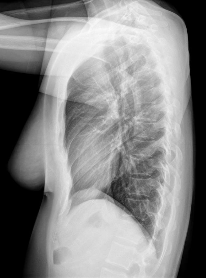
20 Perifissural nodules on CT Well-circumscribed, smooth, solid nodules Triangular or oval shape, <10mm in diameter, below the level of the carina Intrapulmonary lymph node A low likelihood for malignancy No CT follow-up required Radiology 2010;154:949 66/F 76/M Small ovoid nodules in interlobar fissures (arrows) on CT. These perifissural nodules are consistent with intrapulmonary lymph node and are not needed further follow-up.
21 Air cysts A part of the normal aging process of the lungs 7.6% on chest CT (2633 participants, mean age 59.2 years, range years; 50% female), prevalence increased with age. Asymptomatic individuals older than 40 years No association with cigarette smoking or emphysema Associated decreases in DLCO and BMI Solitary, the peripheral area of the lower lobes, remain unchanged or slightly increase in size over time Thorax 2015;70:


22 67/F 54/M, Birt-Hogg-Dube syndrome 73/F Small thin-walled air cysts in RML and LLL (arrows) on CT. But, Multiple air cysts with its count of five or more may need to be evaluated for the possibility of cystic lung diseases. 22


23 Changes in the Airways Tracheobroncheal wall cartilage calcification Common appearance in the elderly female No clinical significance Diffuse cartilage calcification along the tracheobronchial wall in 83-year-old-woman 23


24 Saber-sheath trachea Parietal malacia increases the tracheal AP diameter and collapse of the lateral walls A sign of hyperinflation, but normal aging findings Increased AP diameter of intrathoracic trachea with diffuse tracheal wall calcification in a 74- year-old man. He is a past smoker with no emphysema or chronic obstructive lung disease. 24

 in lower")
25 Increased bronchoarterial ratio and bronchial wall thickness Increased CT bronchoarterial ratio and bronchial wall thickness with age Relative hypoxemia existing in an aged cohort vasoconstriction and relative bronchial dilation AJR 2003;180:513 Caution for the diagnosis of bronchiectasis in the elderly : bronchoarterial ratio greater than 1.5, bronchi visible into the lung periphery, and lack of distal tapering of the bronchial diameter Increased bronchoarterial ratio (arrows) in lower lobes on thin-section CT in an asymptomatic 76-year-old woman. She is a nonsmoker and has no known lung disease. 25


26 Changes in the Mediastinum Heart Cardiac enlargement Increased myocardial muscle mass and thickness due to myocyte hypertrophy and increased connective tissue matrix Coronary artery calcification Extracoronary calcifications Aortic valve calcifications, mitral valve/annulus calcifications Due to fat, collagen, and calcium salt deposition Mild heart valve regurgitation in 90% of healthy patients over 80 years of age Chest radiograph shows mild cardiomegaly, mitral annular calcification (arrow), and diffuse aortic wall calcification in a 92-year-old woman. Coronary artery calcification and extracoronary calcifications in the elderly: No clinical significance in healthy patients, but cardiovascular disease risk factors 26


27 Aorta and Great Vessels Lengthening and dilation with advancing age enlargement of mediastinal contours Atheroma calcifications: most commonly seen in the aortic arch and in the descending thoracic aorta Chest radiograph in a 71-year-old man shows a bulging soft tissue opacity in right paratracheal area (arrow) corresponding to a tortuous right 77/F brachiocephalic to subclavian artery on CT. 27


28 Excessive Fat Deposition Superior mediastinum, Paravertebral regions, Cardiophrenic angles, Extrapleural space Chest radiographs in 72-year-old woman show mass opacity in left cardiophrenic angle area which is classic appearance of prominent paracardaic fat pad. This is confirmed on CT. Prominent paracardiac fat pads are usually mistaken for mass, atelectasis, or pneumonia on chest radiographs. 28
29 Changes in the Chest Wall Decreased chest wall compliance with increasing age Muscle Aging-related muscle mass loss one of the major causes of increased pulmonary transparency on chest X-rays in the elderly Rib Costal cartilage calcification: mistaken for pulmonary nodules Thoracic spines Osteoporosis Degenerative changes: reduced intervertebral space, bone sclerosis adjacent to the intervertebral discs, marginal vertebral osteophytes Barrel chest 29


30 Chest lateral radiograph shows increased thoracic AP diameter in an asymptomatic 74-year-old man. He has no emphysema and COPD. Barrel chest: increased thoracic AP diameter relating to osteoporosis and partial or complete vertebral fractures. A manifestation of COPD/ Emphysema, but typically seen in the elderly. 30


31 Chest PA radiograph in a 75-year-old woman shows a nodular opacity in left paraspinal area (arrow), which is corresponding to bridging osteophytes on CT. 31

32 Changes in the Diaphragm Diaphragmatic Bulging Diaphragmatic atrophy, weakness and dyskinesia Elevation and bulging of the hemidiaphragm Esophageal Hiatal Hernia Increases with age, women>men Enlargement of the anterior-posterior distance of the thorax stretching of the diaphragm in the anterior direction widen the esophageal hiatus increased hiatal hernia. J Thorac Imaging ;27:372 32

.")


33 Chest PA radiograph in an 80-year-old woman shows a large mass opacity with internal air density in left retrocardiac area (arrows). Chest CT confirms esophageal hiatal hernia. Increased anterior-posterior distance of the thorax causes stretching of the diaphragm, and results in widening of the esophageal hiatus. 33
34 AGING CHEST Lung Airway Mediastinum Chest Wall Diaphragm Normal Structural Changes in Radiologic Images Age-related alveolar hyperinflation Ground-glass opacity, basal dependent Mosaic attenuation pattern Reticular densities, subpleural and basal Incidental lung nodules Air cysts Cartilage calcifications Increased AP diameter of trachea Increased CT bronchoarterial ratio Increased bronchial wall thickness Cardiac enlargement Coronary and cardiac valve/annulus calcification Vessel lengthening, dilation, and calcification Excessive fat deposition Osteoporosis Decreased muscle mass Rib cartilage calcifications Increased thoracic AP diameter Bulging contour Hiatal hernia 34
35
Chest X-ray Interpretation
 Chest X-ray Interpretation Introduction Routinely obtained Pulmonary specialist consultation Inherent physical exam limitations Chest x-ray limitations Physical exam and chest x-ray provide compliment
Do you want to be an excellent Radiologist? - Focus on the thoracic aorta on lateral chest image!!!
 The lateral chest radiograph: Challenging area around the thoracic aorta!!! Do you want to be an excellent Radiologist? - Focus on the thoracic aorta on lateral chest image!!! Dong Yoon Han 1, So Youn
An Image Repository for Chest CT
 An Image Repository for Chest CT Francesco Frajoli for the Chest CT in Antibody Deficiency Group An Image Repository for Chest CT he Chest CT in Antibody Deficiency Group is an international and interdisciplinary
10/17/2016. Nuts and Bolts of Thoracic Radiology. Objectives. Techniques
 Nuts and Bolts of Thoracic Radiology October 20, 2016 Carleen Risaliti Objectives Understand the basics of chest radiograph Develop a system for interpreting chest radiographs Correctly identify thoracic
Radiological Anatomy of Thorax. Dr. Jamila Elmedany & Prof. Saeed Abuel Makarem
 Radiological Anatomy of Thorax Dr. Jamila Elmedany & Prof. Saeed Abuel Makarem Indications for Chest x - A chest x-ray may be used to diagnose and plan treatment for various conditions, including: Diseases/Fractures
Imaging of Thoracic Trauma: Tips and Traps. Arun C. Nachiappan, MD Associate Professor of Clinical Radiology University of Pennsylvania
 Imaging of Thoracic Trauma: Tips and Traps Arun C. Nachiappan, MD Associate Professor of Clinical Radiology University of Pennsylvania None Disclosures Objectives Describe blunt and penetrating traumatic
Chest XRay interpretation INTERPRETATIONS Identifications: Name & Date Technical evaluation Basic Interpretations
 Chest XRay interpretation INTERPRETATIONS Identifications: Name & Date Technical evaluation Basic Interpretations TECHNICAL EVALUATION 1. Projection: AP/PA view To differentiate between AP & PA films,
TB Radiology for Nurses Garold O. Minns, MD
 TB Nurse Case Management Salina, Kansas March 31-April 1, 2010 TB Radiology for Nurses Garold O. Minns, MD April 1, 2010 TB Radiology for Nurses Highway Patrol Training Center Salina, KS April 1, 2010
4/16/2017. Learning Objectives. Interpretation of the Chest Radiograph. Components. Production of the Radiograph. Density & Appearance
 Interpretation of the Arthur Jones, EdD, RRT Learning Objectives Identify technical defects in chest radiographs Identify common radiographic abnormalities This Presentation is Approved for 1 CRCE Credit
Chest Radiology Interpretation: Findings of Tuberculosis
 Chest Radiology Interpretation: Findings of Tuberculosis Get out your laptops, smart phones or other devices pollev.com/chestradiology Case #1 1 Plombage Pneumonia Cancer 2 Reading the TB CXR Be systematic!
UERMMMC Department of Radiology. Basic Chest Radiology
 UERMMMC Department of Radiology Basic Chest Radiology PHYSICS DENSITIES BONE SOFT TISSUES WATER FAT AIR TELEROENTGENOGRAM Criteria for an Ideal Chest Radiograph 1. Upright 2. Posteroanterior View 3. Full
Undergraduate Teaching
 Prof. James F Meaney Undergraduate Teaching Chest X-Ray Understanding the normal anatomical by reference to cross sectional imaging Radiology? It s FUN! Cryptic puzzle Sudoku (Minecraft?) It s completely
Interpreting thoracic x-ray of the supine immobile patient: Syllabus
 Interpreting thoracic x-ray of the supine immobile patient: Syllabus Johannes Godt Dep. of Radiology and Nuclear Medicine Oslo University Hospital Ullevål NORDTER 2017, Helsinki Content - Why bedside chest
Financial disclosure COMMON DIAGNOSES IN HRCT. High Res Chest HRCT. HRCT Pre test. I have no financial relationships to disclose. Anatomy Nomenclature
 Financial disclosure I have no financial relationships to disclose. Douglas Johnson D.O. Cardiothoracic Imaging Gaston Radiology COMMON DIAGNOSES IN HRCT High Res Chest Anatomy Nomenclature HRCT Sampling
Congenital Lung Malformations: Radiologic-Pathologic Correlation
 Acta Radiológica Portuguesa, Vol.XVIII, nº 70, pág. 51-60, Abr.-Jun., 2006 Congenital Lung Malformations: Radiologic-Pathologic Correlation Marilyn J. Siegel Mallinckrodt Institute of Radiology, Washington
Avoid the Traps! Tips for Identifying and Distinguishing Normal Thoracic CT Findings from Pathology.
 Avoid the Traps! Tips for Identifying and Distinguishing Normal Thoracic CT Findings from Pathology. Aman Jivraj, Joy Borgaonkar, Daria Manos, Robert Miller Dalhousie University, Halifax NS, Canada -None
B-I-2 CARDIAC AND VASCULAR RADIOLOGY
 (YEARS 1 3) CURRICULUM FOR RADIOLOGY 13 B-I-2 CARDIAC AND VASCULAR RADIOLOGY KNOWLEDGE To describe the normal anatomy of the heart and vessels including the lymphatic system as demonstrated by radiographs,
Eun-Young Kang, M.D., Jae Wook Lee, M.D., Ji Yung Choo, M.D., Hwan Seok Yong, M.D., Ki Yeol Lee, M.D., Yu-Whan Oh, M.D.
 Eun-Young Kang, M.D., Jae Wook Lee, M.D., Ji Yung Choo, M.D., Hwan Seok Yong, M.D., Ki Yeol Lee, M.D., Yu-Whan Oh, M.D. Department of Radiology, Korea University Guro Hospital, College of Medicine, Korea
Web Chapter 3. Image Gallery: Lesion detection on low dose chest CT
 Web Chapter 3 Image Gallery: Lesion detection on low dose chest CT Sarabjeet Singh, MD Mannudeep K. Kalra, MD *Eugene J. Mark, MD *James Stone, MD James H. Thrall, MD Department of Radiology and *Department
An Introduction to Radiology for TB Nurses
 An Introduction to Radiology for TB Nurses Garold O. Minns, MD September 14, 2017 TB Nurse Case Management September 12 14, 2017 EXCELLENCE EXPERTISE INNOVATION Garold O. Minns, MD has the following disclosures
Bronchiectasis: An Imaging Approach
 Bronchiectasis: An Imaging Approach Travis S Henry, MD Associate Professor of Clinical Radiology Cardiac and Pulmonary Imaging Section University of California, San Francisco Large Middle Small 1 Bronchiectasis
Non-neoplastic Conditions in the Mediastinum
 2017 WCTI Non-neoplastic Conditions in the Mediastinum Mi Young Kim, MD, PhD Hyun Jung Koo, MD Jae Woo Song, MD, PhD Department of Radiology and Research Institute of Radiology Asan Medical Center, University
Interesting Cases. Pulmonary
 Interesting Cases Pulmonary 54M with prior history of COPD, hep B/C, and possible history of TB presented with acute on chronic dyspnea, and productive cough Hazy opacity overlying the left hemithorax
Approach to CXR. Terminology. 1.Identification. Greg Blecher SCH Respir Fellow. Correct patient Correct date and time Correct examination
 Approach to CXR Greg Blecher SCH Respir Fellow From Rob Posteraro http://home.earthlink.net/~rhpos/cxr_interpret.txt.html ; http://home.earthlink.net/~rhpos/cxr_main.txt.html) Approach to viewing Chest
PULMONARY TUBERCULOSIS RADIOLOGY
 PULMONARY TUBERCULOSIS RADIOLOGY RADIOLOGICAL MODALITIES Medical radiophotography Radiography Fluoroscopy Linear (conventional) tomography Computed tomography Pulmonary angiography, bronchography Ultrasonography,
Workshop Cyst & Lucency. How to Approach
 Workshop Cyst & Lucency How to Approach To Approach Cystic Lung Disease True cysts? Cavitary disease Cystic bronchiectasis Mosaic attenuation Subpleural cysts Bullae Paraseptal emphysema Honeycombing Birt
I9 COMPLETION INSTRUCTIONS
 The I9 Form is completed for each screening exam at T0, T1, and T2. At T0 (baseline), the I9 documents comparison review of the baseline screen (C2 Form) with any historical images available. At T1 and
Low-dose CT Lung Cancer Screening Guidelines for Pulmonary Nodules Management Version 2
 Low-dose CT Lung Cancer Screening Guidelines for Pulmonary Nodules Management Version 2 The Committee for Management of CT-screening-detected Pulmonary Nodules 2009-2011 The Japanese Society of CT Screening
X-Rays. Prepared by Prof.Dr. Magda Hassab Allah Assist.lecturer Marwa Al Hady
 X-Rays Prepared by Prof.Dr. Magda Hassab Allah Assist.lecturer Marwa Al Hady CHEST X-RAYS Normal Chest X-ray Comments on chest X ray includes examination of 1- Bony cage (ribs,clavicles &vertebral column
Chest and cardiovascular
 Module 1 Chest and cardiovascular A. Doss and M. J. Bull 1. Regarding the imaging modalities of the chest: High resolution computed tomography (HRCT) uses a slice thickness of 4 6 mm to identify mass lesions
Sectional Anatomy Quiz - III
 Sectional Anatomy - III Rashid Hashmi * Rural Clinical School, University of New South Wales (UNSW), Wagga Wagga, NSW, Australia A R T I C L E I N F O Article type: Article history: Received: 30 Jun 2018
Interactive Lecture. Lecture 7 - Interactive. Radiology of cardiorespiratory disease. Editing File. Done By. Color Coding Important Notes Extra
 Lecture 7 - Interactive 436 Teams Interactive Lecture Radiology of cardiorespiratory disease Done By Team Leaders: Khalid Alshehri Hanin Bashaikh Team Members: Ghaida Alsaeed Maha Alissa Nawwaf AlHarbi
X-Rays. Kunal D Patel Research Fellow IMM
 X-Rays Kunal D Patel Research Fellow IMM The 12-Steps } 1: Name 2: Date 3: Old films 4: What type of view(s) 5: Penetration } Pre-read 6: Inspiration 7: Rotation Quality Control 8: Angulation 9: Soft tissues
Radiology of the respiratory disease
 Radiology of the respiratory disease [ Color index: Important Notes Extra ] [ Editing file Feedback Share your notes Shared notes ] Resources: - 435 Slides - 434 Team - 435 Notes Done by: - Mai Alageel
Chapter 5: Other mediastinal structures. The Large Arteries. The Aorta. Ascending aorta
 Chapter 5: Other mediastinal structures The Large Arteries The Aorta The aorta is the main arterial trunk of the systemic circulation and in the healthy state its wall contain a large amount of yellow
Combined pulmonary fibrosis and emphysema; prevalence and follow up among health-care personnel
 Combined pulmonary fibrosis and emphysema; prevalence and follow up among health-care personnel Poster No.: C-0698 Congress: ECR 2013 Type: Scientific Exhibit Authors: K. Chae, G. Jin, S. Chon, Y. Lee;
11/10/2014. Multi-disciplinary Approach to Diffuse Lung Disease: The Imager s Perspective. Radiology
 Multi-disciplinary Approach to Diffuse Lung Disease: The Imager s Perspective Radiology Pathology Clinical 1 Role of HRCT Diagnosis Fibrosis vs. inflammation Next step in management Response to treatment
Large veins of the thorax Brachiocephalic veins
 Large veins of the thorax Brachiocephalic veins Right brachiocephalic vein: formed at the root of the neck by the union of the right subclavian & the right internal jugular veins. Left brachiocephalic
Radiological conference. Left upper lobe collapse. Citation Hong Kong Practitioner, 1998, v. 20 n. 9, p
 Title Radiological conference. Left upper lobe collapse Author(s) Wong, LLS; Peh, WCG Citation Hong Kong Practitioner, 1998, v. 20 n. 9, p. 513-517 Issued Date 1998 URL http://hdl.handle.net/10722/44672
October 2012 Imaging Case of the Month. Michael B. Gotway, MD Associate Editor Imaging. Department of Radiology Mayo Clinic Arizona Scottsdale, AZ
 October 2012 Imaging Case of the Month Michael B. Gotway, MD Associate Editor Imaging Department of Radiology Mayo Clinic Arizona Scottsdale, AZ Clinical History: A 65-year-old non-smoking woman presented
Learning Objectives. 1. Identify which patients meet criteria for annual lung cancer screening
 Disclosure I, Taylor Rowlett, DO NOT have a financial interest /arrangement or affiliation with one or more organizations that could be perceived as a real or apparent conflict of interest in the context
Lecturer: Ms DS Pillay ROOM 2P24 25 February 2013
 Lecturer: Ms DS Pillay ROOM 2P24 25 February 2013 Thoracic Wall Consists of thoracic cage Muscle Fascia Thoracic Cavity 3 Compartments of the Thorax (Great Vessels) (Heart) Superior thoracic aperture
Chest X-ray (CXR) Interpretation Brent Burbridge, MD, FRCPC
 Interpretation Brent Burbridge, MD, FRCPC") Chest X-ray (CXR) Interpretation Brent Burbridge, MD, FRCPC An approach to reviewing a chest x-ray will create a foundation that will facilitate the detection of abnormalities. You should create your own
Chest X-ray (CXR) Interpretation Brent Burbridge, MD, FRCPC An approach to reviewing a chest x-ray will create a foundation that will facilitate the detection of abnormalities. You should create your own
Pediatric TB Intensive Houston, Texas October 14, 2013
 Pediatric TB Intensive Houston, Texas October 14, 2013 Radiologic Presentation of Childhood TB Susan D. John, MD, FACR October 14, 2013 Disclosures I have no disclosures or conflicts of interest to report
Sectional Anatomy Quiz II
 Sectional Anatomy II Rashid Hashmi Rural Clinical School, University of New South Wales, Wagga Wagga, New South Wales, Australia A R T I C L E I N F O Article type: Article history: Received: 3 Aug 2017
Chest X rays and Case Studies. No disclosures. Outline 5/31/2018. Carlo Manalo, M.D. Department of Radiology Loma Linda University Children s Hospital
 Chest X rays and Case Studies Carlo Manalo, M.D. Department of Radiology Loma Linda University Children s Hospital No disclosures. Outline Importance of history Densities delineated on radiography An approach
Signs in Chest Radiology
 Signs in Chest Radiology Jonathan H. Chung, MD Disclosures No pertinent disclosures Jonathan H. Chung, MD Assistant Professor Institute t of fadvanced d Biomedical Imaging National Jewish Health Denver,
Shedding Light on Neonatal X-rays. Objectives. Indications for X-Rays 5/14/2018
 Shedding Light on Neonatal X-rays Barbara C. Mordue, MSN, NNP-BC Neonatal Nurse Practitioner LLUH Children s Hospital, NICU Objectives Utilize a systematic approach to neonatal x-ray interpretation Identify
Systemic lupus erythematosus (SLE): Pleuropulmonary Manifestations
: Pleuropulmonary Manifestations") 08/30/10 09/26/10 Systemic lupus erythematosus (SLE): Pleuropulmonary Manifestations Camila Downey S. Universidad de Chile, School of Medicine, Year VII Harvard University, School of Medicine Sept 17,
08/30/10 09/26/10 Systemic lupus erythematosus (SLE): Pleuropulmonary Manifestations Camila Downey S. Universidad de Chile, School of Medicine, Year VII Harvard University, School of Medicine Sept 17,
5/9/2015. Multi-disciplinary Approach to Diffuse Lung Disease: The Imager s Perspective. No, I am not a pulmonologist! Radiology
 Multi-disciplinary Approach to Diffuse Lung Disease: The Imager s Perspective No, I am not a pulmonologist! Radiology Pathology Clinical 1 Everyone needs a CT Confidence in diagnosis Definitive HRCT +
Chest imaging (Case-based teaching) 胸腔病例教學 謝叔強 財團法人恩主公醫院主治醫師萬芳醫院放射科兼任主治醫師.
 胸腔病例教學 謝叔強 財團法人恩主公醫院主治醫師萬芳醫院放射科兼任主治醫師.") Chest imaging (Case-based teaching) 胸腔病例教學 謝叔強 財團法人恩主公醫院主治醫師萬芳醫院放射科兼任主治醫師 e510019@gmail.com Check List(1) 1. Check patient data, position, technical quality and normal anatomy. 2. Review systematically
Chest imaging (Case-based teaching) 胸腔病例教學 謝叔強 財團法人恩主公醫院主治醫師萬芳醫院放射科兼任主治醫師 e510019@gmail.com Check List(1) 1. Check patient data, position, technical quality and normal anatomy. 2. Review systematically
Is it really honeycombing? Limitations and pitfalls in radiological diagnosis of honeycombing.
 Is it really honeycombing? Limitations and pitfalls in radiological diagnosis of honeycombing. J. Arenas-Jiménez, E. García-Garrigós, M. Sirera-Matilla; M.C. Planells-Alduvin, F.I. Aranda* Hospital General
Syllabus: 6 pages (Page 6 lists corresponding figures for Grant's Atlas 11 th & 12 th Eds.)
") PLEURAL CAVITY AND LUNGS Dr. Milton M. Sholley SELF STUDY RESOURCES Essential Clinical Anatomy 3 rd ed. (ECA): pp. 70 81 Syllabus: 6 pages (Page 6 lists corresponding figures for Grant's Atlas 11 th &
PLEURAL CAVITY AND LUNGS Dr. Milton M. Sholley SELF STUDY RESOURCES Essential Clinical Anatomy 3 rd ed. (ECA): pp. 70 81 Syllabus: 6 pages (Page 6 lists corresponding figures for Grant's Atlas 11 th &
The External Anatomy of the Lungs. Prof Oluwadiya KS
 The External Anatomy of the Lungs Prof Oluwadiya KS www.oluwadiya.com Introduction The lungs are the vital organs of respiration Their main function is to oxygenate the blood by bringing inspired air into
Neonatal Chest X-Ray Interpretation
 CHAPTER 7 Neonatal Chest X-Ray Interpretation Prof. Praveen Kumar Neonatal unit, Department of Pediatrics, PGIMER, Chandigarh Learning Objectives At the end of this session, you should be able to: 1. Schematically
Differential diagnosis
 Differential diagnosis Idiopathic pulmonary fibrosis (IPF) is part of a large family of idiopathic interstitial pneumonias (IIP), one of four subgroups of interstitial lung disease (ILD). Differential
How to Analyse Difficult Chest CT
 How to Analyse Difficult Chest CT Complex diseases are:- - Large lesion - Unusual or atypical pattern - Multiple discordant findings Diffuse diseases are:- - Numerous findings in both sides 3 basic steps
Identify the lines used in anatomical surface descriptions of the thorax. median line mid-axillary line mid-clavicular line
 L 14 A B O R A T O R Y Thorax THORACIC WALL Identify the lines used in anatomical surface descriptions of the thorax. median line mid-axillary line mid-clavicular line Identify the surface landmarks of
Learning Radiology: Recognizing the Basics. Text with Student Consult Online Access Code
 Learning Radiology: Recognizing the Basics. Text with Student Consult Online Access Code Herring, W ISBN-13: 9780323074445 Table of Contents 1. Recognizing Anything The "colorful" world of radiology A
Pulmonary Nodules & Masses
 Pulmonary Nodules & Masses A Diagnostic Approach Heber MacMahon The University of Chicago Department of Radiology Disclosure Information Consultant for Riverain Technology Minor equity in Hologic Royalties
Visual Assessment of CT Findings in Smokers With Nonobstructed Spirometric Abnormalities in The COPDGene Study
 88 Chronic Obstructive Pulmonary Diseases: Journal of the COPD Foundation Original Research. Visual Assessment of CT Findings in Smokers With Nonobstructed Spirometric Abnormalities in The COPDGene Study
Spectrum of Cystic Lung Disease and its Mimics. Kathleen Jacobs MD and Elizabeth Weihe MD UC San Diego Medical Center, Department of Radiology
 Spectrum of Cystic Lung Disease and its Mimics Kathleen Jacobs MD and Elizabeth Weihe MD UC San Diego Medical Center, Department of Radiology No Financial Disclosures Learning Objectives 1. Review the
Intrathoracic extrapleural lesions: MDCT
 Intrathoracic extrapleural lesions: MDCT Poster No.: C-0994 Congress: ECR 2010 Type: Educational Exhibit Topic: Chest Authors: S. S. Kim, Y. T. Kim, S. S. Jou, J. K. Han; Cheonan/KR Keywords: Thoracic
Pulmonary Manifestations of Systemic Lupus Erythematosus 1
 Pulmonary Manifestations of Systemic Lupus Erythematosus 1 Kee Hyuk Yang, M.D., Yo Won Choi, M.D., Seok Chol Jeon, M.D., Choong Ki Park, M.D., Kyung in Joo, M.D., Chang Kok Hahm, M.D., Seung Ro Lee, M.D.
Bronchioloalveolar Carcinoma Mimicking DILD:
 Bronchioloalveolar Carcinoma Mimicking DILD: A Case Report 1 Ju Young Lee, M.D., In Jae Lee, M.D., Dong Gyu Kim, M.D. 2, Soo Kee Min, M.D. 3, Min-Jeong Kim, M.D., Sung Il Hwang, M.D., Yul Lee, M.D., Sang
Cardiac Radiography. Jared D. Christensen, M.D.
 Cardiac Radiography Jared D. Christensen, M.D. Cardiac radiography Jared D. Christensen, M.D. Overview Basic Concepts Technique Normal anatomy Cases Technique 3 Standard Views Posterior-Anterior (PA) Anterior-Posterior
8/14/2017. Objective: correlate radiographic findings of common lung diseases to actual lung pathologic features
 What is that lung disease? Pulmonary Patterns & Correlated Pathology Dr. Russell Tucker, DACVR Objective: correlate radiographic findings of common lung diseases to actual lung pathologic features Improved
GUIDELINES FOR CANCER IMAGING Lung Cancer
 GUIDELINES FOR CANCER IMAGING Lung Cancer Greater Manchester and Cheshire Cancer Network Cancer Imaging Cross-Cutting Group April 2010 1 INTRODUCTION This document is intended as a ready reference for
Pulmonary Sarcoidosis - Radiological Evaluation
 Original Research Article Pulmonary Sarcoidosis - Radiological Evaluation Jayesh Shah 1, Darshan Shah 2*, C. Raychaudhuri 3 1 Associate Professor, 2 1 st Year Resident, 3 Professor and HOD Radiology Department,
CT of saber-sheath trachea
 Acta Radiologica ISSN: 0284-1851 (Print) 1600-0455 (Online) Journal homepage: https://www.tandfonline.com/loi/iard20 CT of saber-sheath trachea Jean Paul Trigaux, G. Hermes, P. Dubois, B. Van Beers, L.
Pediatric High-Resolution Chest CT
 Pediatric High-Resolution Chest CT Alan S. Brody, MD Professor of Radiology and Pediatrics Chief, Thoracic Imaging Cincinnati Children s s Hospital Cincinnati, Ohio, USA Pediatric High-Resolution CT Short
American College of Radiology ACR Appropriateness Criteria
 American College of Radiology ACR Criteria Radiologic Management of Thoracic Nodules and Masses Variant 1: Middle-aged patient (35 60 years old) with an incidental 1.5-cm lung nodule. The lesion was smooth.
Approach to Pulmonary Nodules
 Approach to Pulmonary Nodules Edwin Jackson, Jr., DO Assistant Professor-Clinical Director, James Early Detection Clinic Department of Internal Medicine Division of Pulmonary, Allergy, Critical Care and
Acute and Chronic Lung Disease
 KATHOLIEKE UNIVERSITEIT LEUVEN Faculty of Medicine Acute and Chronic Lung Disease W De Wever, JA Verschakelen Department of Radiology, University Hospitals Leuven, Belgium Clinical utility of HRCT To detect
The Imaging Analysis of Pulmonary Sarcodiosis
 www.cancercellresearch.org ISSN: 2161-2609 Article The Imaging Analysis of Pulmonary Sarcodiosis Xin He, Chuanyu Zhang* Department of Radiology, Affiliated Hospital of Qingdao University, Qingdao, China
Objectives. What is a Chest X Ray? CXR Workshop. Definition (diagnostic tool/internal PE) Types. Cost
 Types. Cost") Objectives CAPA 2011 Christy Wilson, PA C Georgia Lung Associates Identify the radiographic landmarks on a chest radiograph Recognize identifiers of poor quality on the chest radiograph Outline an approach
Objectives CAPA 2011 Christy Wilson, PA C Georgia Lung Associates Identify the radiographic landmarks on a chest radiograph Recognize identifiers of poor quality on the chest radiograph Outline an approach
HRCT in Diffuse Interstitial Lung Disease Steps in High Resolution CT Diagnosis. Where are the lymphatics? Anatomic distribution
 Steps in High Resolution CT Diagnosis Pattern of abnormality Distribution of disease Associated findings Clinical history Tomás Franquet MD What is the diagnosis? Hospital de Sant Pau. Barcelona Secondary
CT Manifestations of Late Sequelae in Patients with Tuberculous Pleuritis
 CT Manifestations of Late Sequelae in Patients with Tuberculous Pleuritis T uberculous pleuritis remains one of the major causes of pleural effusion with an incidence ranging from as high as 86% in a population
OBJECTIVES. Solitary Solid Spiculated Nodule. What would you do next? Case Based Discussion: State of the Art Management of Lung Nodules.
 Organ Imaging : September 25 2015 OBJECTIVES Case Based Discussion: State of the Art Management of Lung Nodules Dr. Elsie T. Nguyen Dr. Kazuhiro Yasufuku 1. To review guidelines for follow up and management
The Spectrum of Management of Pulmonary Ground Glass Nodules
 The Spectrum of Management of Pulmonary Ground Glass Nodules Stanley S Siegelman CT Society 10/26/2011 No financial disclosures. Noguchi M et al. Cancer 75: 2844-2852, 1995. 236 surgically resected peripheral
Liebow and Carrington's original classification of IIP
 Liebow and Carrington's original classification of IIP-- 1969 Eric J. Stern MD University of Washington UIP Usual interstitial pneumonia DIP Desquamative interstitial pneumonia BIP Bronchiolitis obliterans
ARTICLE IN PRESS. Ahuva Grubstein a, Daniele Bendayan b, Ithak Schactman c, Maya Cohen a, David Shitrit b, Mordechai R. Kramer b,
 Respiratory Medicine (2005) 99, 948 954 Concomitant upper-lobe bullous emphysema, lower-lobe interstitial fibrosis and pulmonary hypertension in heavy smokers: report of eight cases and review of the literature
C2 COMPLETION INSTRUCTIONS
 The C2 Form is completed for each screening exam at T0, T1, and T2. The C2 Form is to be completed by each of the following ACRIN-NLST study staff: the research associate (study coordinator), CT technologist,
ARDS - a must know. Page 1 of 14
 ARDS - a must know Poster No.: C-1683 Congress: ECR 2016 Type: Authors: Keywords: DOI: Educational Exhibit M. Cristian; Turda/RO Education and training, Edema, Acute, Localisation, Education, Digital radiography,
Case N 1. Anterior thoracic paint with increasing dyspnea for few days. No cough. Decrease of cardiac sounds. Courtesy Dr Van den Homberg-Tanzania
 Case N 1 Anterior thoracic paint with increasing dyspnea for few days. No cough. Decrease of cardiac sounds Courtesy Dr Van den Homberg-Tanzania Case N 1 Enlargment of cardiac silhouette. Notice the symetry
New Horizons in the Imaging of the Lung
 New Horizons in the Imaging of the Lung Postprocessing. How to do it and when do we need it? Peter M.A. van Ooijen, MSc, PhD Principal Investigator, Radiology, UMCG Discipline Leader Medical Imaging Informatics
Pulmonary Patterns & Correlated Pathology
 Pulmonary Patterns & Correlated Pathology Russell Tucker, DVM, DACVR Washington State University College of Veterinary Medicine Objective: correlate radiographic findings of common lung diseases to actual
Right lung. -fissures:
 -Right lung is shorter and wider because it is compressed by the right copula of the diaphragm by the live.. 2 fissure, 3 lobes.. hilum : 2 bronchi ( ep-arterial, hyp-arterial ), one artery mediastinal
Interpretation of the chest radiograph Elizabeth Puddy MB ChB FCARCSI Catherine Hill MB ChB MRCP FRCR
 Interpretation of the chest radiograph Elizabeth Puddy MB ChB FCARCSI Catherine Hill MB ChB MRCP FRCR The traditional technique used in the acquisition and development of a chest radiograph uses methods
The radiological differential diagnosis of the UIP pattern
 5th International Conference on Idiopathic Pulmonary Fibrosis, Modena, 2015, June 12th The radiological differential diagnosis of the UIP pattern Simon Walsh King s College Hospital Foundation Trust London,
Pediatric TB Intensive Houston, Texas
 Pediatric TB Intensive Houston, Texas November 13, 2009 Radiographic Manifestations of Pediatric TB Susan D. John, MD, FACR November 13, 2009 Radiologic Presentation of Childhood TB Susan D. John, MD,
Advances in MDCT of Thoracic Trauma
 Baltic Congress of Radiology, Riga 2010 Advances in MDCT of Thoracic Trauma Robert A. Novelline, MD Professor of Radiology, Harvard Medical School Director of Emergency Radiology, Massachusetts General
Cystic Lung Disease: a Comparison of Cystic Size, as Seen on Expiratory and Inspiratory HRCT Scans
 Cystic Lung Disease: a Comparison of Cystic Size, as Seen on Expiratory and Inspiratory HRCT Scans Ki-Nam Lee, MD 1 Seong-Kuk Yoon, MD 1 Seok Jin Choi, MD 2 Jin Mo Goo, MD 3 Kyung-Jin Nam, MD 1 Index words:
CT Chest. Verification of an opacity seen on the straight chest X ray
 CT Chest Indications: To assess equivocal plain x-ray findings Staging of lung neoplasm Merastatic workup of extra thoraces malignancies Diagnosis of diffuse lung diseases with HRCT Assessment of bronchietasis
I8 COMPLETION INSTRUCTIONS
 The I8 Form is completed for each screening exam at T0, T1, and T2. At T0 (baseline), the I8 Form documents comparison review of the baseline screen (DR Form) with any historical images available. At T1
Dr. Weyrich G07: Superior and Posterior Mediastina. Reading: 1. Gray s Anatomy for Students, chapter 3
 Dr. Weyrich G07: Superior and Posterior Mediastina Reading: 1. Gray s Anatomy for Students, chapter 3 Objectives: 1. Subdivisions of mediastinum 2. Structures in Superior mediastinum 3. Structures in Posterior
Anterior Mediastinal Masses: The 4 T s
 May 2001 Anterior Mediastinal Masses: The 4 T s Rachel Van Sambeek, Harvard Medical School, Year III 1 Mediastinal Compartments 3 arbitrary divisions that do not correlate with anatomic planes: Anterior
Boy 8 months TPRC. 21 Sep 06 CXR. Flat and. CLE findings. BPD findings a. Left opacity
 CLE in BPD lung Boy 8 months 17 Sep 06 21 Sep 06 CXR Flat and low position of the diaphragm ICD insertion, right; ET tube slightly shift to the left RUL atelectasis RML hyperinflation, herniating across
Manage TB Dr. A. Chitrakumar Madras Medical College and RGGGH Institute of Thoracic Medicine, Chennai
 Manage TB Dr. A. Chitrakumar Madras Medical College and RGGGH Institute of Thoracic Medicine, Chennai Lecture 16 Radiology in diagnosis of Tuberculosis Session 01 So, welcome to the session Radiology in