Non-neoplastic Conditions in the Mediastinum
|
|
|
- Lucy Holland
- 5 years ago
- Views:
Transcription


1 2017 WCTI Non-neoplastic Conditions in the Mediastinum Mi Young Kim, MD, PhD Hyun Jung Koo, MD Jae Woo Song, MD, PhD Department of Radiology and Research Institute of Radiology Asan Medical Center, University of Ulsan College of Medicine, South Korea
2 Disclosures Contents 1. Acute mediastinitis None. 2. Mesenchymal lesion 3. Vascular lesion 4. LN disease, other than tumor 5. Mediastinal cysts 6. Others Perforation or rupture of esophagus Descending necrotizing mediastinitis Direct extension by adjacent infections Thymolipoma Fibrosing mediastinitis Hemangioma Perforation or rupture of airways Lymphangioma Post surgical complication-mc

, UI 24cm")


3 Acute Mediastinitis 68/F Perforation of esophagus, fish bone (red arrow), UI 24cm Abscess pocket Gas bubbles Esophageal wall thickening Increased attenuation of mediastinal fat


4 Acute Mediastinitis 65/M Iatrogenic rupture of esophagus during S-B tube insertion Extraluminal gas, pneumomediastinum Single or multiple mediastinal abscesses with fluid collection

5 Acute Mediastinitis 39/M C.C. vomiting Distal esophageal rupture with acute mediastinitis Pleural effusion Pericardial effusion pneumomediastinum
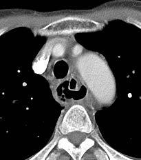
6 Acute Mediastinitis 55/M Esophageal dissection and perforation Mucosal flap with submucosal distribution of gas or contrast, the classic double-barreled appearance Occur posterior to the true lumen of the esophagus


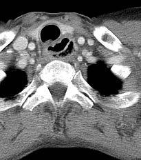
7 Acute Mediastinitis 56/M C.C. fever Descending necrotizing mediastinitis and retropharyngeal abscess caused by K. pneumoniae Multiple retropharyngeal and mediastinal abscesses Increased soft tissue density and obliteration of normal fat planes, neck and mediastinum


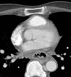
8 Acute Mediastinitis 56/M S/P MV repair Mediastinitis after cardiac surgery, S. aureus on pus culture Retrosternal complicated fluid collection Air bubbles Fistula Focal osteomyelitis, nonunion of bone


9 Mediastinitis 20/M Trauma Post traumatic bronchial rupture Pneumomediastinum Contour deformity of airway or airway narrowing Atelectasis or aspiration pneumonia


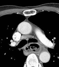
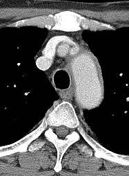
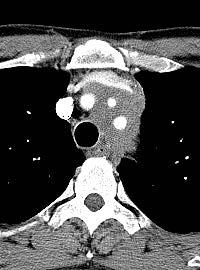
10 Mediastinitis 20/M C.C. dyspnea Fibrosing mediastinitis Diffuse mediastinal soft tissue attenuation Hilar or mediastinal mass Obstruction or narrowing of a pulmonary artery Obstruction or narrowing of SVC Tracheobronchial narrowing or irregularities Calcification Multiple collateral veins Enlarged left superior intercostals vein ( )


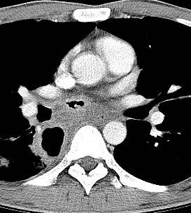
11 Mediastinitis 51/M C.C. dyspnea Fibrosing mediastinitis, companion case Steroid Tx Soft tissue, ( mediastinum ), biopsy: Sclerosing inflammation


12 Thymolipoma Contents 17/M 17/M 1. Acute mediastinitis 2. Mesenchymal lesion 3. Vascular lesion Thymolipoma Hemangioma Lymphangioma 4. LN disease, other than tumor 5. Mediastinal cysts 6. Others Predominant fat attenuation and intermingled soft tissue attenuation Connection between tumor and the thymus Well circumscribed consisting of mature adipose tissue with interspersed islands of thymic tissue Incidentally found in young adults Fat suppression or chemical shift MR imaging technique may be helpful
 Arise in the")

13 Hemangioma 35/M Occur in the first four decades of life (75 %) Arise in the anterior mediastinum (68 %). Smoothly outlined Contain punctate calcification; phleboliths


,")
14 Lymphangioma 47/F Well-circumscribed lesion of low (or water) attenuation molding to the mediastinal contours and enveloping the great vessels 3 types, unilocular (most common), cavernous, and intermediate types


15 Contents Iatrogenic Left Innominate Vein Injury 47/F 1. Acute mediastinitis 2. Mesenchymal lesion 3. Vascular lesion 4. LN disease, other than tumor 5. Mediastinal cysts 6. Others Dilatation of mediastinal veins, pulmonary artery, and bronchial artery Vascular injury Acute aortic disease Hematoma in anterior mediastinum


16 Iatrogenic aortic injury 73/F Focal contrast extravasation, active bleeding. Pericardial effusion Diffuse high attenuation at mediastinum, hematoma


17 Aneurysms of bronchial arteries 60/M Tortuous and engorged left bronchial arteries Right Thrombosed bronchial aneurysm artery: Hypertrophic of right bronchial and tortuous artery ( appearance. ) Left Collateral bronchial vessels artery: Hypertrophic, tortuous appearance and aneurysm at the orifice of left Ill-defined bronchial mediastinitis artery. embolization


18 Contents Sarcoidosis 47/F 47/F 1. Acute mediastinitis 2. Mesenchymal lesion 3. Vascular lesion 4. LN disease, other than tumor 5. Mediastinal cysts Sarcoidosis Tuberculosis Castleman s disease 6. Others Bilateral hilar lymph node enlargement is the most common finding, followed by interstitial lung disease. Sarcoidosis is a multisystem disorder that is characterized by noncaseous epithelioid cell granulomas. RadioGraphics 2010


19 Tuberculous lymphadenopathy 47/F Young adult Preponderance of involvement of the right paratracheal and subcarinal lymph nodes. Nodes larger than 2 cm in diameter invariably show central areas of relative low density and peripheral rim enhancement after injection of contrast medium Radiology 1987


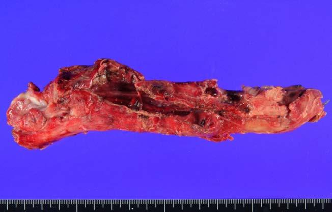
20 Nodo-Esophago-bronchial fistula caused by TB lymphadenitis 36/M


21 Castleman's disease, Hyaline vascular type 22/F A solitary, well-circumscribed mediastinal mass Strong enhancement The hyaline-vascular type (90%, unicentric form) AJR 2004


22 Castleman's disease, Plasma cell type 59/F Disseminated Castleman s disease manifests with diffuse mediastinal lymphadenopathy Plasma cell type (10%, multicentric form, systemic)
")


23 Contents Mediastinal cysts, Common findings 1. Acute mediastinitis Smooth and sharply marginated masses Water density (about 50%) or homogeneous soft tissue density (50%) 2. Mesenchymal on CT lesion Do not enhance after IV administration of contrast 3. Vascular lesion Variable pattern on T1-weighted sequence 4. LN disease, other than tumor Hyperintense on T2-weighted sequence regardless of the nature of the cyst content 5. Mediastinal cysts 60/F Thymic cyst, ant. mediastinum 39/M Thymic Pericardial cyst cyst, Rt cardiophrenic angle Pericardial cyst 6. Others Bronchogenic cyst Forgut duplication cyst Thoracic duct cyst Mediastinal pancreatic pseudocyst


24 57/M Lung 54/F posterior mediastinum Bronchogenic cyst 59/M Mediastinal, subcarinal, complicated 38/M Mediastinal, paratracheal


25 Esophageal duplication cyst 21/F 21/F Esophageal cyst: intramural or adjacent to the wall of esophagus


26 Thoracic duct cyst 58/F Thoracic duct cyst and dilatation 47/M Rare cystic lesion in the mediastinum. Weakness in the thoracic duct wall allows formation of a cyst. Chyle in the fluid of the cyst Symptomless or compression of surrounding structures


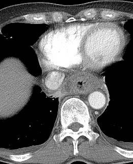
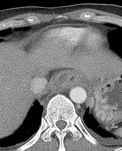
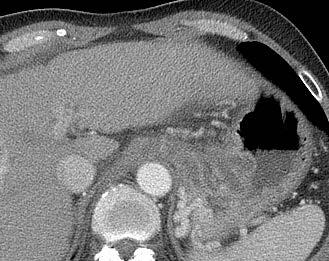
27 Mediastinal pancreatic pseudocyst 44/M, chronic pancreatitis Mediastinal extension of pseudocyst can occur through anatomical openings of diaphragm. The posterior mediastinum is the most common location of the mediastinal pseudocysts through esophageal and aortic hiatus.


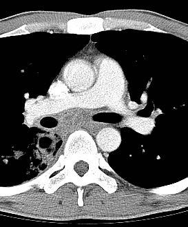
28 Intrathoracic Contents goiter 47/F 1. Acute mediastinitis 2. Mesenchymal lesion 3. Vascular lesion 4. LN disease, other than tumor 5. Mediastinal cysts 6. Others Intrathoracic goiter Diverticulum Continuity with the cervical gland Mediastinal lipomatosis Focal calcification Extramedullary hematopoiesis Arise in the posterolateral portion of the thyroid gland and descend inferiorly to the posterior mediastinum


")

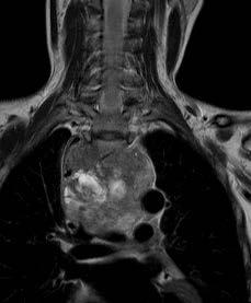
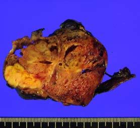
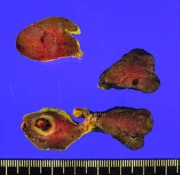
29 Intrathoracic goiter T1WI 45/F Pre enhanced Post enhanced Enhanced T1WI T2WI Sharp borders (>90%) High attenuation on unenhanced CT (>100 HU) After IV contrast administration, thyroid tissue exhibits early and prolonged enhancement Continuity with the cervical gland


30 Zenker's diverticulum at upper esophagus 56/M Protrusion of esophageal mucosa through the posterior muscle layers of the pharynx through a defect in the hypopharyngeal wall. Found on the posterior wall of the hypopharynx Fluid-filled, characteristic Infected Zenker's diverticulum The American Journal of Medicine 2003


31 Esophageal diverticulum 68/M, UI 30cm middle esophageal diverticulum 72/M, UI 37cm distal esophageal diverticulum
 hyperplasia Increased number of lymphoid follicles, seen in")

32 Thymic lymphoid hyperplasia 26/F Grave s disease f/u Thymic lymphoid (follicular) hyperplasia Increased number of lymphoid follicles, seen in immune disorders including systemic lupus erythematosus, rheumatoid arthritis, scleroderma, thyrotoxicosis, and Grave s disease Commonly associated with myasthenia gravis


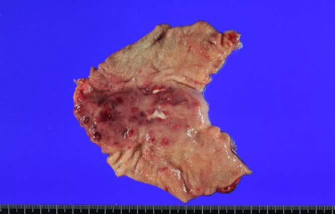
33 Mediastinal lipomatosis 60/M S/P left nephrectomy d/t RCC Soft tissue, ( mediastinum ), excision : Low grade lipogenic tumor, favor lipoma


34 Extramedulary hematopoiesis 61/M AML, Anemia Well-circumscribed, smooth, soft tissue attenuation masses, usually at multiple levels in a paraspinal location without erosion or pressure changes on the adjacent ribs or vertebral bodies Hypertrophy of the medullary cavity of the ribs Rare cause of posterior mediastinal masses and is usually seen in patients with severe, long-standing anemia
35 Summary CT is most often used in the assessment of mediastinal non-neoplastic diseases, with MR imaging usually being used as an adjunct to CT An awareness of the CT findings associated with the spectrum of mediastinal non-neoplastic diseases facilitates the accurate and prompt diagnosis The compartments of the mediastinum may help narrow the differential diagnosis. Thank you for your attention. Contact:
Mediastinal Tumors: Imaging
 Mediastinal Tumors: Imaging References Imaging in Oncology, Husband and Reznek Computed Tomography and Magnetic Resonance of the thorax, Naidich, Zerhouni, Siegelman, Mediastinal compartments Anterior:
Sectional Anatomy Quiz - III
 Sectional Anatomy - III Rashid Hashmi * Rural Clinical School, University of New South Wales (UNSW), Wagga Wagga, NSW, Australia A R T I C L E I N F O Article type: Article history: Received: 30 Jun 2018
Dr. Weyrich G07: Superior and Posterior Mediastina. Reading: 1. Gray s Anatomy for Students, chapter 3
 Dr. Weyrich G07: Superior and Posterior Mediastina Reading: 1. Gray s Anatomy for Students, chapter 3 Objectives: 1. Subdivisions of mediastinum 2. Structures in Superior mediastinum 3. Structures in Posterior
TB Intensive Houston, Texas
 TB Intensive Houston, Texas October 15-17, 17 2013 Diagnosis of TB: Radiology Rosa M Estrada-Y-Martin, MD MSc FCCP October 16, 2013 Rosa M Estrada-Y-Martin, MD MSc FCCP, has the following disclosures to
Esophageal Perforation
 Esophageal Perforation Dr. Carmine Simone Thoracic Surgeon, Division of General Surgery Head, Division of Critical Care May 15, 2006 Overview Case presentation Radiology Pre-operative management Operative
Sectional Anatomy Quiz II
 Sectional Anatomy II Rashid Hashmi Rural Clinical School, University of New South Wales, Wagga Wagga, New South Wales, Australia A R T I C L E I N F O Article type: Article history: Received: 3 Aug 2017
Case of the Day Chest
 Case of the Day Chest Darin White MDCM FRCPC Department of Radiology, Mayo Clinic 76 th Annual Scientific Meeting Canadian Association of Radiologists Montreal, QC April 26, 2013 2013 MFMER slide-1 Disclosures
Pediatric TB Intensive Houston, Texas October 14, 2013
 Pediatric TB Intensive Houston, Texas October 14, 2013 Radiologic Presentation of Childhood TB Susan D. John, MD, FACR October 14, 2013 Disclosures I have no disclosures or conflicts of interest to report
Acute Aortic Syndromes
 Acute Aortic Syndromes Carole J. Dennie, MD Acute Thoracic Aortic Syndromes Background Non-Traumatic Acute Thoracic Aortic Syndromes Carole Dennie MD FRCPC Associate Professor of Radiology and Cardiology
Eun-Young Kang, M.D., Jae Wook Lee, M.D., Ji Yung Choo, M.D., Hwan Seok Yong, M.D., Ki Yeol Lee, M.D., Yu-Whan Oh, M.D.
 Eun-Young Kang, M.D., Jae Wook Lee, M.D., Ji Yung Choo, M.D., Hwan Seok Yong, M.D., Ki Yeol Lee, M.D., Yu-Whan Oh, M.D. Department of Radiology, Korea University Guro Hospital, College of Medicine, Korea
Pathology of Mediastinal Tumors
 SAMO Meeting Lucerne 2009 Pathology of Mediastinal Tumors Alex Soltermann Most common lesions (adults) Clinical presentation 50% of the patients are asymptomatic, lesion discovered incidentally Symptoms
Pediatric TB Intensive Houston, Texas
 Pediatric TB Intensive Houston, Texas November 13, 2009 Radiographic Manifestations of Pediatric TB Susan D. John, MD, FACR November 13, 2009 Radiologic Presentation of Childhood TB Susan D. John, MD,
Imaging of Thoracic Trauma: Tips and Traps. Arun C. Nachiappan, MD Associate Professor of Clinical Radiology University of Pennsylvania
 Imaging of Thoracic Trauma: Tips and Traps Arun C. Nachiappan, MD Associate Professor of Clinical Radiology University of Pennsylvania None Disclosures Objectives Describe blunt and penetrating traumatic
Basic Data. Sex:Male 31 years old Occupation: 搬家工人
 Basic Data Sex:Male 31 years old Occupation: 搬家工人 Chief Complaint Intermittent chest pain with shortness of breath for 2-3 months. Present Illness 4 months ago, he started having occasional chest pain
Incidental Esophageal Findings on Chest CT. Amira Hussien, MD, Elliot Fishman, MD, Bouchra Younes, MD, Ahmed Hatw. Johns Hopkins Medical Institution
 Incidental Esophageal Findings on Chest CT Amira Hussien, MD, Elliot Fishman, MD, ouchra Younes, MD, Ahmed Hatw. Johns Hopkins Medical Institution I have nothing to disclose. DISCLOSURE INTRODUCTION Although
Imaging Of Cystic Paravertebral Masses:
 Imaging Of Cystic Paravertebral Masses: Differential Diagnosis and Key Discriminators John P. Lichtenberger III, MD, Maj, USAF, MC Brent McCarragher, MD, CPT, USA John R. Dryden, MD, LT, USN P. Gabriel
Chest Radiology Interpretation: Findings of Tuberculosis
 Chest Radiology Interpretation: Findings of Tuberculosis Get out your laptops, smart phones or other devices pollev.com/chestradiology Case #1 1 Plombage Pneumonia Cancer 2 Reading the TB CXR Be systematic!
Dr. Pratik Mukherjee, MMed, FRCR Dr. Ashish Chawla, MD, ABR (USA) Khoo Teck Puat Hospital, Singapore
 Khoo Teck Puat Hospital, Singapore") Dr. Pratik Mukherjee, MMed, FRCR Dr. Ashish Chawla, MD, ABR (USA) Khoo Teck Puat Hospital, Singapore The authors declare no financial disclosures. To revisit the basics of approach to mediastinal masses
Dr. Pratik Mukherjee, MMed, FRCR Dr. Ashish Chawla, MD, ABR (USA) Khoo Teck Puat Hospital, Singapore The authors declare no financial disclosures. To revisit the basics of approach to mediastinal masses
10/14/2018 Dr. Shatarat
 2018 Objectives To discuss mediastina and its boundaries To discuss and explain the contents of the superior mediastinum To describe the great veins of the superior mediastinum To describe the Arch of
Do you want to be an excellent Radiologist? - Focus on the thoracic aorta on lateral chest image!!!
 The lateral chest radiograph: Challenging area around the thoracic aorta!!! Do you want to be an excellent Radiologist? - Focus on the thoracic aorta on lateral chest image!!! Dong Yoon Han 1, So Youn
Large veins of the thorax Brachiocephalic veins
 Large veins of the thorax Brachiocephalic veins Right brachiocephalic vein: formed at the root of the neck by the union of the right subclavian & the right internal jugular veins. Left brachiocephalic
Chest XRay interpretation INTERPRETATIONS Identifications: Name & Date Technical evaluation Basic Interpretations
 Chest XRay interpretation INTERPRETATIONS Identifications: Name & Date Technical evaluation Basic Interpretations TECHNICAL EVALUATION 1. Projection: AP/PA view To differentiate between AP & PA films,
UERMMMC Department of Radiology. Basic Chest Radiology
 UERMMMC Department of Radiology Basic Chest Radiology PHYSICS DENSITIES BONE SOFT TISSUES WATER FAT AIR TELEROENTGENOGRAM Criteria for an Ideal Chest Radiograph 1. Upright 2. Posteroanterior View 3. Full
Children are not small adults Children are Not Small Adults Anatomic considerations Pliable bony & cartilagenous structures - Significant thoracic inj
 PEDIATRIC CHEST TRAUMA Children are not small adults Role of imaging Spectrum of injury Children are not small adults Children are Not Small Adults Anatomic considerations Pliable bony & cartilagenous
Diagnostic Value of EBUS-TBNA in Various Lung Diseases (Lymphoma, Tuberculosis, Sarcoidosis)
") Diagnostic Value of EBUS-TBNA in Various Lung Diseases (Lymphoma, Tuberculosis, Sarcoidosis) Sevda Sener Cömert, MD, FCCP. SBU, Kartal Dr.Lütfi Kırdar Training and Research Hospital Department of Pulmonary
Diagnostic Value of EBUS-TBNA in Various Lung Diseases (Lymphoma, Tuberculosis, Sarcoidosis) Sevda Sener Cömert, MD, FCCP. SBU, Kartal Dr.Lütfi Kırdar Training and Research Hospital Department of Pulmonary
Anterior Mediastinal Masses: The 4 T s
 May 2001 Anterior Mediastinal Masses: The 4 T s Rachel Van Sambeek, Harvard Medical School, Year III 1 Mediastinal Compartments 3 arbitrary divisions that do not correlate with anatomic planes: Anterior
An Introduction to Radiology for TB Nurses
 An Introduction to Radiology for TB Nurses Garold O. Minns, MD September 14, 2017 TB Nurse Case Management September 12 14, 2017 EXCELLENCE EXPERTISE INNOVATION Garold O. Minns, MD has the following disclosures
Chest X-ray Interpretation
 Chest X-ray Interpretation Introduction Routinely obtained Pulmonary specialist consultation Inherent physical exam limitations Chest x-ray limitations Physical exam and chest x-ray provide compliment
Learning Radiology: Recognizing the Basics. Text with Student Consult Online Access Code
 Learning Radiology: Recognizing the Basics. Text with Student Consult Online Access Code Herring, W ISBN-13: 9780323074445 Table of Contents 1. Recognizing Anything The "colorful" world of radiology A
100 Chest X Rays for Study Group. by Dr. Suneet Khurana
 100 Chest X Rays for Study Group by Dr. Suneet Khurana Approach to - Chest X Ray (shadow of the viscera on a photographic plate) Gas appears Black Fat appears Dark Grey Water Appears as Light Grey Bone
ACUTE PANCREATITIS: NEW CLASSIFICATION OF AN OLD FOE. T Barrow, A Nasrullah, S Liong, V Rudralingam, S A Sukumar
 ACUTE PANCREATITIS: NEW CLASSIFICATION OF AN OLD FOE T Barrow, A Nasrullah, S Liong, V Rudralingam, S A Sukumar LEARNING OBJECTIVES q Through a series of cases illustrate the updated Atlanta symposium
Mediastinum and pericardium
 Mediastinum and pericardium Prof. Abdulameer Al-Nuaimi E-mail: a.al-nuaimi@sheffield.ac.uk E. mail: abdulameerh@yahoo.com The mediastinum: is the central compartment of the thoracic cavity surrounded by
CT of Acute Thoracic Aortic Syndromes Stuart S. Sagel, M.D.
 CT of Acute Thoracic Aortic Syndromes Stuart S. Sagel, M.D. Thoracic Aortic Aneurysms Atherosclerotic Dissection Penetrating ulcer Mycotic Inflammatory (vasculitis) Traumatic Aortic Imaging Options Catheter
PULMONARY TUBERCULOSIS RADIOLOGY
 PULMONARY TUBERCULOSIS RADIOLOGY RADIOLOGICAL MODALITIES Medical radiophotography Radiography Fluoroscopy Linear (conventional) tomography Computed tomography Pulmonary angiography, bronchography Ultrasonography,
Diffuse Mediastinal Widening on the Chest Radiograph: How to Narrow the Differential Diagnosis
 Diffuse Mediastinal Widening on the Chest Radiograph: How to Narrow the Differential Diagnosis Ian Russell, Pierre Maldjian Rutgers New Jersey Medical School Department of Radiology Category: Other Mediastinal
Anterior Mediastinal Masses
 Residents Section Pattern of the Month Shahrzad et al. nterior Mediastinal Masses Residents Section Pattern of the Month Maryam Shahrzad 1 Thi Som Mai Le Mario Silva lexander. ankier Ronald L. Eisenberg
CT 101 :Pancreas and Spleen
 CT 101 :Pancreas and Spleen Shikha Khullar,, MD, MPH Division of Radiology University of South Alabama The Pancreas Normal Pancreas 3 Phase Pancreatic CT Non contrast Arterial phase : 30-35 35 second
Mediastinum It is a thick movable partition between the two pleural sacs & lungs. It contains all the structures which lie
 Dr Jamila EL medany OBJECTIVES At the end of the lecture, students should be able to: Define the Mediastinum. Differentiate between the divisions of the mediastinum. List the boundaries and contents of
May 2017 Imaging Case of the Month. Prasad M. Panse, MD and Michael B. Gotway, MD. Department of Radiology Mayo Clinic Arizona Scottsdale, Arizona USA
 May 2017 Imaging Case of the Month Prasad M. Panse, MD and Michael B. Gotway, MD Department of Radiology Mayo Clinic Arizona Scottsdale, Arizona USA Clinical History: A 32-year-old man presented for routine
PLEURAE and PLEURAL RECESSES
 PLEURAE and PLEURAL RECESSES By Dr Farooq Aman Ullah Khan PMC 26 th April 2018 Introduction When sectioned transversely, it is apparent that the thoracic cavity is kidney shaped: a transversely ovoid space
TB Radiology for Nurses Garold O. Minns, MD
 TB Nurse Case Management Salina, Kansas March 31-April 1, 2010 TB Radiology for Nurses Garold O. Minns, MD April 1, 2010 TB Radiology for Nurses Highway Patrol Training Center Salina, KS April 1, 2010
GIT RADIOLOGY. Water-soluble contrast media (e.g. gastrograffin) are the other available agents.which doesn t cause inflammatory peritonitis..
 are the other available agents.which doesn t cause inflammatory peritonitis..") GIT RADIOLOGY Imaging techniques-general principles: Contrast examinations: Barium sulphate is the best contrast for GIT (with good mucosal coating & excellent opacification & being inert); but is contraindicated
GIT RADIOLOGY Imaging techniques-general principles: Contrast examinations: Barium sulphate is the best contrast for GIT (with good mucosal coating & excellent opacification & being inert); but is contraindicated
4/16/2017. Learning Objectives. Interpretation of the Chest Radiograph. Components. Production of the Radiograph. Density & Appearance
 Interpretation of the Arthur Jones, EdD, RRT Learning Objectives Identify technical defects in chest radiographs Identify common radiographic abnormalities This Presentation is Approved for 1 CRCE Credit
Signs in Chest Radiology
 Signs in Chest Radiology Jonathan H. Chung, MD Disclosures No pertinent disclosures Jonathan H. Chung, MD Assistant Professor Institute t of fadvanced d Biomedical Imaging National Jewish Health Denver,
ACUTE AORTIC SYNDROMES
 ACUTE AORTIC SYNDROMES AGNETA FLINCK MD, PhD Dept. of Thoracic Radiology Sahlgrenska University Hospital ACUTE AORTIC SYNDROMES Aortic dissection Intramural hematoma (IMH) 5-20% Penetrating atherosclerotic
Chest X rays and Case Studies. No disclosures. Outline 5/31/2018. Carlo Manalo, M.D. Department of Radiology Loma Linda University Children s Hospital
 Chest X rays and Case Studies Carlo Manalo, M.D. Department of Radiology Loma Linda University Children s Hospital No disclosures. Outline Importance of history Densities delineated on radiography An approach
CT Chest. Verification of an opacity seen on the straight chest X ray
 CT Chest Indications: To assess equivocal plain x-ray findings Staging of lung neoplasm Merastatic workup of extra thoraces malignancies Diagnosis of diffuse lung diseases with HRCT Assessment of bronchietasis
Undergraduate Teaching
 Prof. James F Meaney Undergraduate Teaching Chest X-Ray Understanding the normal anatomical by reference to cross sectional imaging Radiology? It s FUN! Cryptic puzzle Sudoku (Minecraft?) It s completely
Neckmasses in infancy and childhood: Clinical and radiological classification and imaging approaches M. Mearadji
 Neckmasses in infancy and childhood: Clinical and radiological classification and imaging approaches M. Mearadji International Foundation for Pediatric Imaging Aid Introduction Neck masses are a frequent
Case Acute ascending thoracic aortic rupture due to penetrating atherosclerotic ulcer
 Case 12305 Acute ascending thoracic aortic rupture due to penetrating atherosclerotic ulcer Lopes Dias J, Costa NV, Leal C, Alves P, Bilhim T Section: Chest Imaging Published: 2014, Dec. 19 Patient: 68
Chief Complain. For chemotherapy
 Chief Complain For chemotherapy Present Illness 93.12 Progressive weakness of R t arm for 1 year X-ray: peneative lesion over right proximal humorous Bone scan: multiple increased intake Biopsy of distal
Approach to CXR. Terminology. 1.Identification. Greg Blecher SCH Respir Fellow. Correct patient Correct date and time Correct examination
 Approach to CXR Greg Blecher SCH Respir Fellow From Rob Posteraro http://home.earthlink.net/~rhpos/cxr_interpret.txt.html ; http://home.earthlink.net/~rhpos/cxr_main.txt.html) Approach to viewing Chest
!"#$%&'%()'*+,-%&&.'+('*/%)+%,#+0' 12/.,'3%)+"4#%52.
'*+,-%&&.'+('*/%)+%,#+0' 12/.,'3%)+4#%52.") !"#$%&'%()'*+,-%&&.'+('*/%)+%,#+0' 12/.,'3%)+"4#%52.!"#$%&'()$*+&,--#&$.//,0'1232'!-#0'45 *6 '7849!!"#$%&'"(&)*+),$-.*/*01) 2$34/&1)*+)5"-.3.(") 6%.(3")*+)7*(08/$)9(.:"%;.&1)) )?
!"#$%&'%()'*+,-%&&.'+('*/%)+%,#+0' 12/.,'3%)+"4#%52.!"#$%&'()$*+&,--#&$.//,0'1232'!-#0'45 *6 '7849!!"#$%&'"(&)*+),$-.*/*01) 2$34/&1)*+)5"-.3.(") 6%.(3")*+)7*(08/$)9(.:"%;.&1)) )?
Introduction to Chest CT Interpretation. Objectives 8/28/2017
 Introduction to Chest CT Interpretation Deborah Stein ACNP BC, CCRN NP Education Specialist Department of Anesthesia and Critical Care Medicine August 28, 2017 Objectives Basic Principles Thoracic Anatomy
CT of Fatty Thoracic Masses
 V ii8i CT of Fatty Thoracic Masses -.-. Fig. 1.-Mediastinal lipomatosis. A, CT scan shows abundant fat throughout superior mediastinum. Fat is homogeneous and similar in attenuation to subcutaneous fat.
Shedding Light on Neonatal X-rays. Objectives. Indications for X-Rays 5/14/2018
 Shedding Light on Neonatal X-rays Barbara C. Mordue, MSN, NNP-BC Neonatal Nurse Practitioner LLUH Children s Hospital, NICU Objectives Utilize a systematic approach to neonatal x-ray interpretation Identify
Lecture 2: Clinical anatomy of thoracic cage and cavity II
 Lecture 2: Clinical anatomy of thoracic cage and cavity II Dr. Rehan Asad At the end of this session, the student should be able to: Identify and discuss clinical anatomy of mediastinum such as its deflection,
PULMONARY COLLAPSE Whole lung collapse: There is complete opacity of the L hemi-thorax. The mediastinum is invisible and displaced into
 1 PULMONARY COLLAPSE: A cause of opacity on chest X-ray but there are several features to distinguish it from alveolar shadowing or interstitial lung disease. The loss of lung volume may affect the position
Avoid the Traps! Tips for Identifying and Distinguishing Normal Thoracic CT Findings from Pathology.
 Avoid the Traps! Tips for Identifying and Distinguishing Normal Thoracic CT Findings from Pathology. Aman Jivraj, Joy Borgaonkar, Daria Manos, Robert Miller Dalhousie University, Halifax NS, Canada -None
Case Discussion Splenic Abscess
 Case Discussion Splenic Abscess Personal Data Gender: male Birth Date: 1928/Mar/06th Allergy: Mefenamic Smoking: 0.5 PPD for 55 years Alcohol: negative (?) 4 Months Ago Abdominal pain: epigastric area
Sex: 女 Age: 51 Occupation: 無 Admission date:92/07/22
 Sex: 女 Age: 51 Occupation: 無 Admission date:92/07/22 Chief complaint Unknown fever for one month Hand tremor and left huge renal tumor was noted Present illness Suffered from fever for one month, hand
How to Analyse Difficult Chest CT
 How to Analyse Difficult Chest CT Complex diseases are:- - Large lesion - Unusual or atypical pattern - Multiple discordant findings Diffuse diseases are:- - Numerous findings in both sides 3 basic steps
Radiological Anatomy of Thorax. Dr. Jamila Elmedany & Prof. Saeed Abuel Makarem
 Radiological Anatomy of Thorax Dr. Jamila Elmedany & Prof. Saeed Abuel Makarem Indications for Chest x - A chest x-ray may be used to diagnose and plan treatment for various conditions, including: Diseases/Fractures
B-I-2 CARDIAC AND VASCULAR RADIOLOGY
 (YEARS 1 3) CURRICULUM FOR RADIOLOGY 13 B-I-2 CARDIAC AND VASCULAR RADIOLOGY KNOWLEDGE To describe the normal anatomy of the heart and vessels including the lymphatic system as demonstrated by radiographs,
Objectives. Thoracic Inlet. Thoracic Inlet Boundaries. Thoracic Inlet Sagittal View ANTERIOR SCALENE ANTERIOR SCALENE
 Objectives Thoracic Inlet Deborah L. Reede M.D. SUNY Downstate Medical Center Learn the anatomy of the thoracic inlet (TI) Review the clinical and radiographic findings of common lesions encountered in
Chapter 2 Cardiac Interpretation of Pediatric Chest X-Ray
 Chapter 2 Cardiac Interpretation of Pediatric Chest X-Ray Ra-id Abdulla and Douglas M. Luxenberg Key Facts The cardiac silhouette occupies 50 55% of the chest width on an anterior posterior chest X-ray
Congenital Lung Malformations: Radiologic-Pathologic Correlation
 Acta Radiológica Portuguesa, Vol.XVIII, nº 70, pág. 51-60, Abr.-Jun., 2006 Congenital Lung Malformations: Radiologic-Pathologic Correlation Marilyn J. Siegel Mallinckrodt Institute of Radiology, Washington
Advances in MDCT of Thoracic Trauma
 Baltic Congress of Radiology, Riga 2010 Advances in MDCT of Thoracic Trauma Robert A. Novelline, MD Professor of Radiology, Harvard Medical School Director of Emergency Radiology, Massachusetts General
ANATOMY OF THE PLEURA. Dr Oluwadiya KS
 ANATOMY OF THE PLEURA Dr Oluwadiya KS www.oluwadiya.sitesled.com Introduction The thoracic cavity is divided mainly into: Right pleural cavity Mediastinum Left Pleural cavity Pleural cavity The pleural
Right lung. -fissures:
 -Right lung is shorter and wider because it is compressed by the right copula of the diaphragm by the live.. 2 fissure, 3 lobes.. hilum : 2 bronchi ( ep-arterial, hyp-arterial ), one artery mediastinal
Contents. Basic Ultrasound Principles and Terminology. Ultrasound Nodule Characteristics
 Contents Basic Ultrasound Principles and Terminology Basic Ultrasound Principles... 1 Ultrasound System... 2 Linear Transducer for Superficial Images and Ultrasound-Guided FNA... 3 Scanning Planes... 4
Case 9087 Retropharyngeal nodular fasciitis
 Case 9087 Retropharyngeal nodular fasciitis Santiago I 1; Cavalheiro F 2; Noruégas MJ 3; Sanches MC3 1 Hospital Infante D. Pedro, Aveiro, Portugal 2 Hospitais da Universidade de Coimbra, Portugal 3 Hospital
Lung Cancer - Suspected
 Lung Cancer - Suspected Shared Decision Making Lung Cancer: http://www.enhertsccg.nhs.uk/ Patient presents with abnormal CXR Lung cancer - clinical presentation History and Examination Incidental finding
EDUCATIONAL CASES E1 & E2. Natasha Inglis 20/03/15
 EDUCATIONAL CASES E1 & E2 Natasha Inglis 20/03/15 CASE E1 79 year old female Rectum. Altemeier operation Histology Superficial erosions and mucosal congestion volcano lesion and pseudomembrane formation
Post-Op Aorta: Differentiating Normal Post-Op vs. Complications. Linda C. Chu, MD Assistant Professor of Radiology Johns Hopkins University
 Post-Op Aorta: Differentiating Normal Post-Op vs. Complications Linda C. Chu, MD Assistant Professor of Radiology Johns Hopkins University No disclosures Disclosures Goals and Objectives To review CT technique
The External Anatomy of the Lungs. Prof Oluwadiya KS
 The External Anatomy of the Lungs Prof Oluwadiya KS www.oluwadiya.com Introduction The lungs are the vital organs of respiration Their main function is to oxygenate the blood by bringing inspired air into
Cardiac Radiography. Jared D. Christensen, M.D.
 Cardiac Radiography Jared D. Christensen, M.D. Cardiac radiography Jared D. Christensen, M.D. Overview Basic Concepts Technique Normal anatomy Cases Technique 3 Standard Views Posterior-Anterior (PA) Anterior-Posterior
Radiology Pathology Conference
 Radiology Pathology Conference Sharlin Johnykutty,, MD, Cytopathology Fellow Sara Majewski, MD, Radiology Resident Friday, August 28, 2009 Presentation material is for education purposes only. All rights
Radiological staging of lung cancer. Shukri Loutfi,MD,FRCR Consultant Thoracic Radiologist KAMC-Riyadh
 Radiological staging of lung cancer Shukri Loutfi,MD,FRCR Consultant Thoracic Radiologist KAMC-Riyadh Bronchogenic Carcinoma Accounts for 14% of new cancer diagnoses in 2012. Estimated to kill ~150,000
Lecture 3. Inflammatory Processes
 Lecture 3 Inflammatory Processes Process: Increased vascular permeability Water and cellular infiltrations Results: Abscess, ulceration, cavitation Penetration, perforation and fistula formation Scarring,
Typical and atypical imaging of thoracic and abdominal aortic rupture
 Typical and atypical imaging of thoracic and abdominal aortic rupture Poster No.: C-0453 Congress: ECR 2014 Type: Educational Exhibit Authors: J. Isogai, T. Ichihara, T. Inoue, T. Kanamori ; Asahi/JP,
The use of advanced imaging in the diagnosis of TB. Pierre Goussard, Robert Gie Tygerberg Children`s Hospital and University of Stellenbosch
 The use of advanced imaging in the diagnosis of TB Pierre Goussard, Robert Gie Tygerberg Children`s Hospital and University of Stellenbosch Imaging Bronchoscopy Tracheo-bronchograms Chest CT-scan Ultrasound
Cervical Lymph Nodes
 Cervical Lymph Nodes Diana Gaitini, MD Unit of Ultrasound, Department of Medical Imaging Rambam Medical Center and Faculty of Medicine Technion, Israel Institute of Technology Haifa, Israel Learning Targets
Chest and cardiovascular
 Module 1 Chest and cardiovascular A. Doss and M. J. Bull 1. Regarding the imaging modalities of the chest: High resolution computed tomography (HRCT) uses a slice thickness of 4 6 mm to identify mass lesions
objectives Pitfalls and Pearls in PET/CT imaging Kevin Robinson, DO Assistant Professor Department of Radiology Michigan State University
 objectives Pitfalls and Pearls in PET/CT imaging Kevin Robinson, DO Assistant Professor Department of Radiology Michigan State University To determine the regions of physiologic activity To understand
Complicated echinococcal cyst to Biopsy or not to biopsy. V. Rusanov MR Kramer Pulmonary Institute, Rabin medical center
 Complicated echinococcal cyst to Biopsy or not to biopsy V. Rusanov MR Kramer Pulmonary Institute, Rabin medical center Case 1 84 y.o. Male, Iraq descend, past smoker 40 PY Medical History- HTN, Rheumatoid
Imaging abdominal vascular emergencies. V.Stoynova
 Imaging abdominal vascular emergencies V.Stoynova Abdominal vessels V. Stoynova 2 Acute liver bleeding trauma anticoagulant therapy liver disease : HCC, adenoma, meta, FNH, Hemangioma Diagnosis :CT angiography
The thymus: A pictorial review of thymic pathology
 The thymus: A pictorial review of thymic pathology Poster No.: C-1692 Congress: ECR 2013 Type: Educational Exhibit Authors: D. J. Conces, S. Rissing, P. J. Loehrer; Indianapolis, IN/US Keywords: Cancer,
Radiological conference. Left upper lobe collapse. Citation Hong Kong Practitioner, 1998, v. 20 n. 9, p
 Title Radiological conference. Left upper lobe collapse Author(s) Wong, LLS; Peh, WCG Citation Hong Kong Practitioner, 1998, v. 20 n. 9, p. 513-517 Issued Date 1998 URL http://hdl.handle.net/10722/44672
BOGOMOLETS NATIONAL MEDICAL UNIVERSITY DEPARTMENT OF HUMAN ANATOMY. Guidelines. Module 2 Topic of the lesson Aorta. Thoracic aorta.
 BOGOMOLETS NATIONAL MEDICAL UNIVERSITY DEPARTMENT OF HUMAN ANATOMY Guidelines Academic discipline HUMAN ANATOMY Module 2 Topic of the lesson Aorta. Thoracic aorta. Course 1 The number of hours 3 1. The
Shadow because the air
 Thyroid Ultrasound Thyroid US examination needs: 1. high frequency transducer 2. extended patient's neck 3. check all the neck area because the swelling could be in areas other than the thyroid such as
THE DESCENDING THORACIC AORTA
 Intercostal Arteries and Veins Each intercostal space contains a large single posterior intercostal artery and two small anterior intercostal arteries. The anterior intercostal arteries of the lower spaces
Guidelines, Policies and Statements D5 Statement on Abdominal Scanning
 Guidelines, Policies and Statements D5 Statement on Abdominal Scanning Disclaimer and Copyright The ASUM Standards of Practice Board have made every effort to ensure that this Guideline/Policy/Statement
Role of imaging in RCC. Ultrasonography. Solid lesion. Cystic RCC. Solid RCC 31/08/60. From Diagnosis to Treatment: the Radiologist Perspective
 Role of imaging in RCC From Diagnosis to Treatment: the Radiologist Perspective Diagnosis Staging Follow up Imaging modalities Limitations and pitfalls Duangkamon Prapruttam, MD Department of Therapeutic
Chest X-ray (CXR) Interpretation Brent Burbridge, MD, FRCPC
 Interpretation Brent Burbridge, MD, FRCPC") Chest X-ray (CXR) Interpretation Brent Burbridge, MD, FRCPC An approach to reviewing a chest x-ray will create a foundation that will facilitate the detection of abnormalities. You should create your own
Chest X-ray (CXR) Interpretation Brent Burbridge, MD, FRCPC An approach to reviewing a chest x-ray will create a foundation that will facilitate the detection of abnormalities. You should create your own
DESCRIPTION: This is the part of the trunk, which is located between the root of the neck and the superior border of the abdominal region.
 1 THE THORACIC REGION DESCRIPTION: This is the part of the trunk, which is located between the root of the neck and the superior border of the abdominal region. SHAPE : T It has the shape of a truncated
SETTING Fudan University Shanghai Cancer Center. RESPONSIBLE PARTY Haiquan Chen MD.
 OFFICIAL TITLE A Phase Ⅲ Study of Left Side Thoracotomy Approach (SweetProcedure) Versus Right Side Thoracotomy Plus Midline Laparotomy Approach (Ivor-Lewis Procedure) Esophagectomy in Middle or Lower
Lecturer: Ms DS Pillay ROOM 2P24 25 February 2013
 Lecturer: Ms DS Pillay ROOM 2P24 25 February 2013 Thoracic Wall Consists of thoracic cage Muscle Fascia Thoracic Cavity 3 Compartments of the Thorax (Great Vessels) (Heart) Superior thoracic aperture