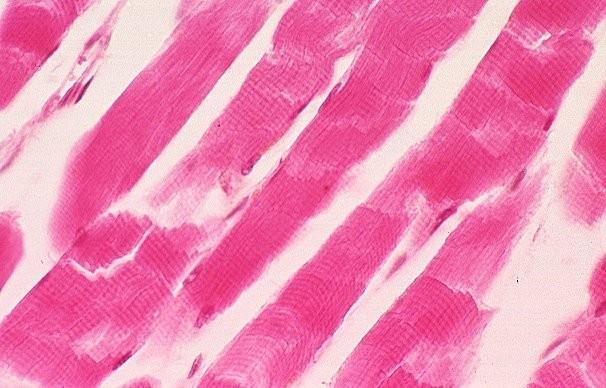
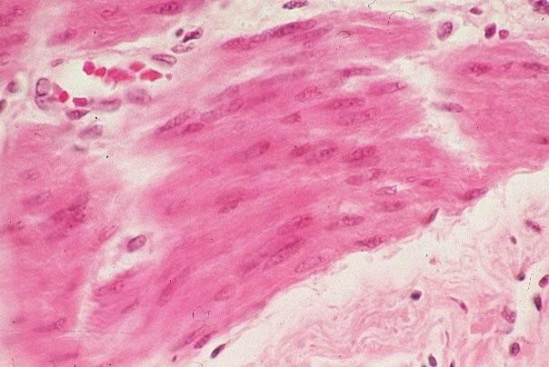
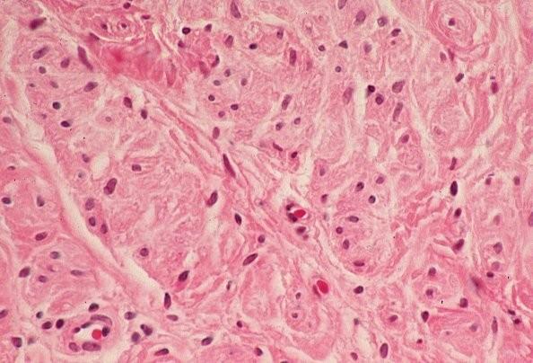
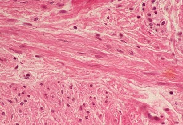
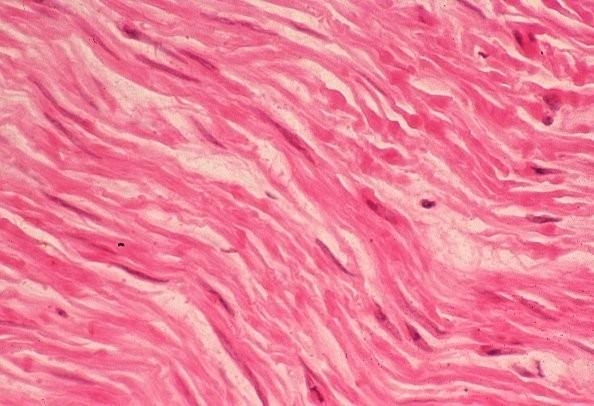
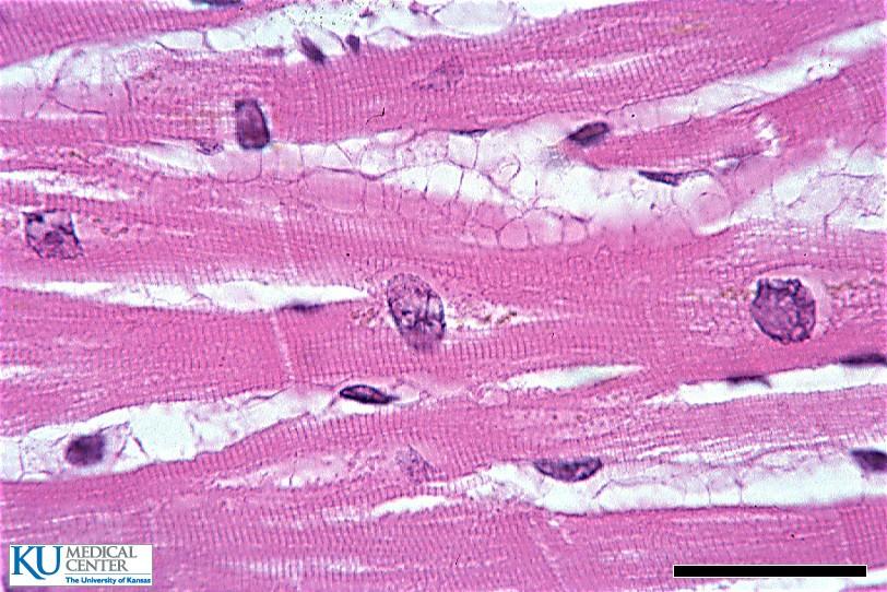
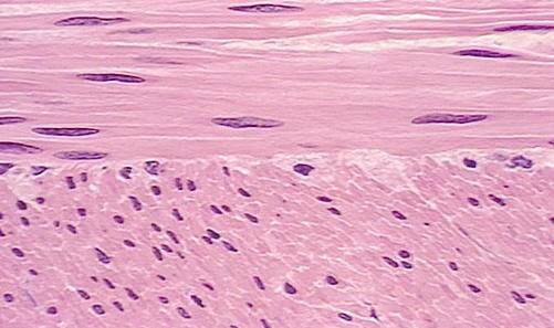
Development and teratology of cardiovascular and lymphatic systems. Repetition: Muscle tissue
|
|
|
- Louisa Jackson
- 5 years ago
- Views:
Transcription

1 Development and teratology of cardiovascular and lymphatic systems Repetition: Muscle tissue
2
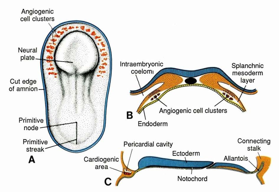
3 Beginning of the cardiovascular system development the 3rd week: Hemangiogenesis (day 15 16) blood islets (insulae sanguinae) in extraembryonic mesoderm and splanchnic mesenchyme of embryo
4 Clusters of mesenchyme cells (angiogenic cells) differentiate into: - angioblasts endothelium (at the periphery of blood islets) - hemoblasts primitive erythrocytes (in the center of blood islets)
5 Clusters of angiogenic cells form a "horseshoe-shaped" space between somatic and splanchnic layer of mesoderm = pericardial cavity. Two endothelial tubes arrise in splanchnic mesoderm. The ventral portion of these tubes forms the cardiogenic area with two heart tubes, while the lateral portions form the dorsal aortae.
6

7 Germ disc: prosencephalon mesencephalon eye rhombencephalon heart lateral mesoderm somites 8,9 Spine small blood vessels blood islands primitive streak
8


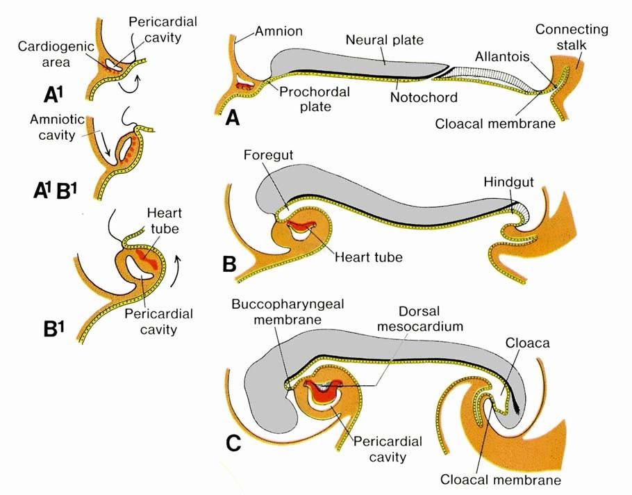
9 Initially, the cardiogenic area is located anterior to the prechordal plate and the neural plate. The growth of the central nervous system pulls the cardiogenic area and prechordal plate (buccopharyngeal membrane ventrally and caudally ( ).
10


11 Cardiogenic region just cranial to the prechordal plate.
12

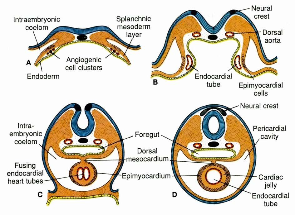
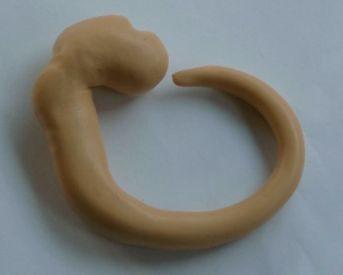
13 The canalization of cardiogenic clusters in the splanchnic mesoderm results in the formation of the paired heart tubes. Folding of embryo and primitive gut separation from yolk sac. Fusion of the heart tubes
14 a single heart tube is, temporarily attached to the dorsal side of the pericardial cavity by the dorsal mesocardium (4). The splanchnic mesoderm (5) around the heart tube (3 - future endocardium) thickens and forms the heart jelly = myoepicardial mantle (future myocardium and epicardium). 3
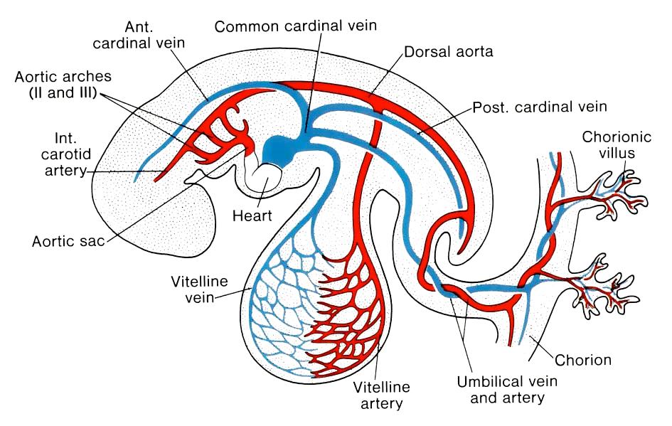
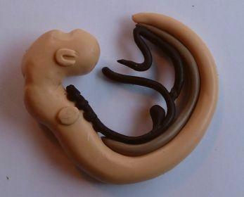
15 Cor tubulare Cor sigmoideum Cor quadricameratum Heart tube Truncus arteriosus + aortic roots Bulbus cordis Ventricle Atrium Sinus venosus Common cardinal veins Umbilical veins Vitelline veins


16 Looping of the heart tube - because growth of the heart tube is faster then growth of the pericardial cavity
17 Cor quadricameratum

18 Atrioventricular septum Endocardial cushions develop in the dorsal and ventral walls of the heart in the region of the atrioventricular canal. They grow towards each other and fuse, thus dividing the atrioventricular canal into right and left orifices. The lateral interventricular cushions appear on the sides of the canal. These proliferations of the mesenchymal tissue subsequently differentiate into bicuspid and tricuspid atrioventricular valves.
 6 - membranaceous part of septum 7 - IV")
19 Ventricle Septum interventriculare Grows from the heart apex cranily to the endocardial cushions (AV septum) 6 - membranaceous part of septum 7 - IV septum
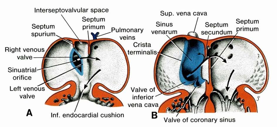
20 Atrium Septum atriorum -septum primum with ostium primum (obliterates); ostium secundum -septum secundum with foramen ovale
21 Atrium At the end of the 4th week, septum primum grows from the dorsocranial wall of the primitive atrium towards the endocardial cushions. The temporary opening (ostium primum - op) between the lower rim of the septum primum and the endocardial cushions gradually closes. Perforations in the upper part of the septum primum form the ostium secundum - os. Subsequently, another crest (septum secundum) grows from the right side of the septum primum and gradually covers the foramen secundum. The oval opening left by the septum secundum is called foramen ovale. Septum primum Septum secundum os op foramen ovale
22
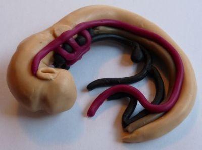
23 Sinus venosus Common cardinal veins Umbilical veins Vitelline veins left veins obliterate and - left portion of sinus venosus is transformed into sinus coronarius - right portion becomes to be part of right atrium
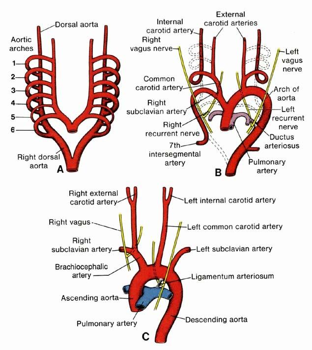
24 Truncus arteriosus + aortic roots -cranial part of bulbus cordis divides into: -aortic roots, branched into 6 pairs of aortic arches
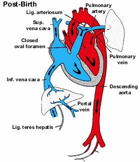
25 Bulbus cordis - cranial part give rise to truncus arteriosus - middle part give rice to conus arteriosus distal part participate in formation of ventricle wall
26 Bulbus cordis and truncus arteriosus a pair of opposing ridges appear in the walls of the bulbus cordis and truncus arteriosus. These ridges twist around each other, forming a spiral course of the aorticopulmonary septum. This septum divides the bulbus cordis and truncus arteriosus into two channels, the aorta and the pulmonary artery. It also participates in the closure of the interventricular foramen

27 Formation of aorticopulmonary septum 1. Aorta 2. Left pulmonary artery 3. Pulmonary trunk 4. Muscular interventricular septum 5. Right ventricle 6. Membranous interventricular septum


28 1. Aorticopulmonary septum 2. Pulmonary valve 3. Pulmonary artery 4. Aortic valve 5. Aorta
29

30 1. Internal carotid artery 2. External carotid artery 3. Common carotid artery 4. Right subclavian artery 5. Arch of aorta 6. Brachiocephalic artery 7. Ductus arteriosus 8. 7th intersegmental artery 9. Pulmonary artery 10. Carotid duct 11. Obliterated right dorsal aorta
")
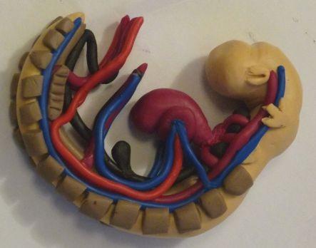
31 Primitive blood circulation (2)

32 the primitive blood circulation in an embryo.
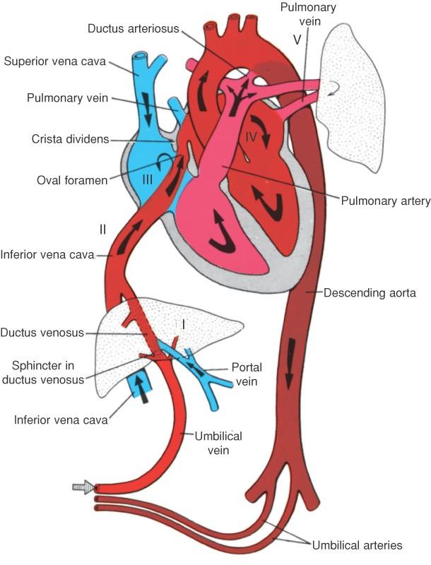
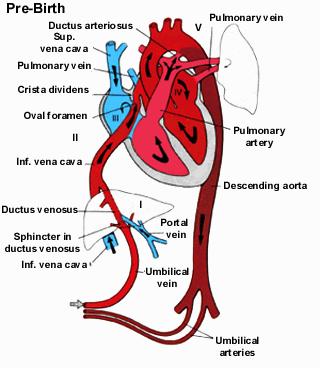
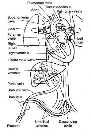
33 The fetal circulation


34 At birth, the circulation of the fetal blood through the placenta is stopped and the lungs begin to function. The foramen ovale, ductus arteriosus, ductus venosus and umbilical vessels subsequently obliterate and transform into corresponding ligaments.
35
36 Congenital malformations in CVS (the most frequent) With left right shunt (without cyanosis) atrial septum defect ventricular septum defect ductus arteriosus apertus (patens, persistens) With right left shunt (with cyanosis) Fallot tetralogy transposition of great vessels truncus arteriosus (common aorticopulmonal canal) tricuspid valve atresia Without shunt coarctation of aorta aortic stenosis pulmonary stenosis dextrocardia (+situs inversus) ectopia cordis
37 Atrial Septal Defects a group of common congenital anomalies defects occuring in a number of different forms and more often in females. patent foramen ovale left-right shunting
38 Ventricular Septal Defect occurs in the interventricular septum, and is more frequent in males that females. left-right shunting
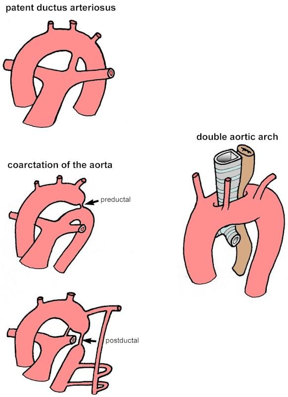
39 Patent Ductus Arteriosus occurs commonly in preterm infants, can close spontaneously (by day three in 60% of normal term neonates) the remainder are ligated simply and with little risk. left-right shunting
40 Tetralogy of Fallot named after Etienne-Louis Arthur Fallot (1888) who described it as "la maladie blue" and is a common developmental cardiac defect. The syndrome consists of a number of cardiac defects possibly stemming from abnormal neural crest migration. consists of: 1. ventricular septal defect 2. pulmonary stenosis (valvular or infundibular) 3. results in an overriding aorta 4. right ventricular hypertrophy right-left shunting
41 Transposition of Great Vessels Characterized by aorta arising from right ventricle and pulmonary artery from the left ventricle and often associated with other cardiac abnormalities (e.g. ventricular septal defect). right-left shunting
42 Tricuspid Atresia Blood is shunted through an atrial septal defect to the left atrium and through the ventricular septal defect to the pulmonary artery. The shaded arrows indicate mixing of the blood. right-left shunting
43 Coarctation of Aorta (preductal or postductal) before or behind ductus arteriosus
44 Aortic Stenosis Pulmonary Stenosis

45 Hypoplastic Left Heart Characterized by hypoplasia (underdevelopment or absence) of the left ventricle obstructive valvular and vascular lesion of the left side of the heart. hypoplastic left hear functional hypoplastic left heart
46 Double Outlet Right Ventricle De-oxygenated blood enters the aorta from the right ventricle and is returned to the body.
47 Pulmonary Atresia Abnormal blood flow (as indicated by the shaded blue arrow) is from the right atrium and right ventricle through an atrial septal defect to the left side of the heart. Blood can reach the pulmonary arteries only through a patent ductus arteriosus.
48
49 Heart Sinus venosus (R horn): smooth part of right atrium (sinus venarum) and the "valve" of the superior vena cava. The sino-atrial node. Sinus venosus (L horn): coronary sinus, valve of coronary sinus Valve of sinus venosus (R): border of smooth part of right atrium (crista terminalis) Valve of sinus venosus (L): part of atrial septum Common atrium: rough part of right and left atria and the auricles Embryonic pulmonary vein: large part of the left atrial wall Septum primum: left side of the atrial septum Septum secundum: right side of the atrial septum Foramen secundum: dimple in the atrial septum seen from left side (fossa ovalis, ok) Foramen ovale: fossa ovalis Endocardial cushions: parts of the atrioventricular wall, mitral, and tricuspid valves Endocardial cushions: membraneous part of ventricular septum, part of arterial trunk Bulbus cordis: trabeculated part of R ventricle and aortic vestibule of L ventricle Truncus arteriosus: ascending (ventral) aorta and pulmonary artery Common ventricle: parts of the right and left ventricle
50 Arterial System Aortic sac (R and L sides of sac): brachiocephalic artery (right) and part of the ascending aorta (left), large parts of common carotid arteries 1st aortic arch (R and L): part of the maxillary artery and external carotid arteries 2nd aortic arch (R and L): part of the hyoid and stapedial arteries, part of external carotids 3rd aortic arch (R and L): part of common carotids and first part of the internal carotids 4th aortic arch (R): small proximal part of the right subclavian artery 4th aortic arch (L): small part of arch of the aorta just proximal to the left subclavian artery 6th aortic arch (R): proximal part of right pulmonary artery 6th aortic arch (L=ductus): proximal left pulmonary artery and and ligamentum arteriosum Ductus arteriosus: ligamentum arteriosum Dorsal aorta (R and L): part of right subclavian, descending aorta below left subclavian Unpaired ventral (or vitelline) arteries: celiac, superior mesenteric, and inferior mesenteric arteries Paired dorsal segmental arteries: intercostal arteries and vertebral arteries Umbilical arteries: internal iliac, superior vescial arteries, medial umbilical ligaments
51 Venous System Ductus venosus: ligamentum venosum Umbilical vein (L): round ligament (ligamentum teres) Vitelline vein (R): superior mesenteric vein and the inferior vena cava Vitelline vein (L): most of the portal vein Anterior cardinal veins (R and L): internal jugular veins (left brachiocephalic vein is an anastomosis) Anterior cardinal vein (R): part of superior vena cava and right brachiocephalic vein Common cardinal vein (R): proximal part of superior vena cava Common cardinal vein (L): lateral part of coronary sinus and oblique vein of left atrium Posterior cardinal vein (R): part of azygos vein and common iliac veins Supracardinal veins: hemiazygos vein (L) and caudal part of azygos vein (R) Subcardinal vein (R): renal segment of inferior vena cava
52

53 Cardiogenic precursors form the linear heart tube. Mesenchymal cells form the cardiac valves from the conotruncal (CT) and atrioventricular valve (AVV) segments. A atrium; Ao aorta; DA ductus arteriosus; LA left atrium; LCC left common carotid; LSCA left subclavian artery; LV left ventricle; PA pulmonary artery; RA right atrium; RCC right common carotid; RSCA right subclavian artery; RV right ventricle; V ventricle.

54

55 transposition of the great vessels - the aorta exits from the right ventricle, and the pulmonary trunk exits from the left ventricle. This is only compatible with life postnatally if the ductus arteriosus remains patent, allowing communication between the otherwise closed systemic and pulmonary circulations driven by the left and right sides of the heart.
56
57

58
59
60
61
62
63
64
65
66
67
68
DEVELOPMENT OF THE CIRCULATORY SYSTEM L E C T U R E 5
 DEVELOPMENT OF THE CIRCULATORY SYSTEM L E C T U R E 5 REVIEW OF CARDIAC ANATOMY Heart 4 chambers Base and apex Valves Pericardial sac 3 layers: epi, myo, endo cardium Major blood vessels Aorta and its
Embryology of the Heart
 *Page 1A: Embryology of the Heart Human embryonic disc is divided into three layers: ectoderm, intraembryonic mesoderm, and endoderm. The embryonic disc lies between the amniotic cavity and the primary
6. Development of circulatory system II. Cardiac looping. Septation of atria and ventricles. Common heart malformations.
 6. Development of circulatory system II. Cardiac looping. Septation of atria and ventricles. Common heart malformations. Formation of heart tube paired endothelial-lined heart tube is formed from blood
Development of the heart
 Development of the heart Prof. Abdulameer Al-Nuaimi E-mail: a.al-nuaimi@sheffield.ac.uk abdulameerh@yahoo.com Early Development of the Circulatory System Appears in the middle of the third week, when the
When you see this diagram, remember that you are looking at the embryo from above, through the amniotic cavity, where the epiblast appears as an oval
 When you see this diagram, remember that you are looking at the embryo from above, through the amniotic cavity, where the epiblast appears as an oval disc 2 Why the embryo needs the vascular system? When
The Cardiovascular System (Part I) 黃敏銓 解剖學暨細胞生物學研究所
 黃敏銓 解剖學暨細胞生物學研究所") The Cardiovascular System (Part I) 黃敏銓 解剖學暨細胞生物學研究所 1 Congenital heart defects (CHDs) 台灣兒童心臟學會 Sinus venarum Membranous septum Conus arteiosus (infundibulum) Aortic vestibule The Cardiovascular System
The Cardiovascular System (Part I) 黃敏銓 解剖學暨細胞生物學研究所 1 Congenital heart defects (CHDs) 台灣兒童心臟學會 Sinus venarum Membranous septum Conus arteiosus (infundibulum) Aortic vestibule The Cardiovascular System
The sinus venosus represent the venous end of the heart It receives 3 veins: 1- Common cardinal vein body wall 2- Umbilical vein from placenta 3-
 1 2 The sinus venosus represent the venous end of the heart It receives 3 veins: 1- Common cardinal vein body wall 2- Umbilical vein from placenta 3- Vitelline vein from yolk sac 3 However!!!!! The left
Development of the Heart
 Development of the Heart Thomas A. Marino, Ph.D. Temple University School of Medicine Stages of Development of the Heart 1. The horseshoe-shaped pericardial cavity. 2. The formation of the single heart
The Cardiovascular System (Part II)
") The Cardiovascular System (Part II) 黃敏銓 mchuang@ntu.edu.tw 解剖學暨細胞生物學研究所 1 Development of veins Three paired veins drain into the tubular heart of a 4-week embryo Vitelline veins: poorly oxygenated blood
The Cardiovascular System (Part II) 黃敏銓 mchuang@ntu.edu.tw 解剖學暨細胞生物學研究所 1 Development of veins Three paired veins drain into the tubular heart of a 4-week embryo Vitelline veins: poorly oxygenated blood
IN THE NAME OF GOD. Development of the Heart and Vasculature
 IN THE NAME OF GOD Development of the Heart and Vasculature Overview vascular system appears (middle of 3 rd week) when the embryo is not able to satisfy its nutrition by diffusion Heart is the first functional
Development of the Great Vessels and Conduc6on Tissue
 Development of the Great Vessels and Conduc6on Tissue Development of the heart fields h:p://php.med.unsw.edu.au/embryology/ index.php?6tle=advanced_- _Heart_Fields! 2 Septa6on of the Bulbus Cordis Bulbus
W.S. O The University of Hong Kong
 W.S. O The University of Hong Kong Objectives: Describe early angiogenesis. Describe the heart tube formation. Describe the partitioning into a 4- chambered heart. List the formation of heart valves and
Heart & vascular system I. Dawei Dong
 Heart & vascular system I Dawei Dong Lecture goal Learn the basics of heart and vascular development. Development of Heart, Blood, and Blood Vessels LEARNING GOALS: 1. explain the early development of
Circulatory system. Lecture #2
 Circulatory system Lecture #2 The essential components of the human cardiovascular system: Heart Blood Blood vessels Arteries - blood vessels that conduct arterial blood from heart ventricle to organs
Notes: 1)Membranous part contribute in the formation of small portion in the septal cusp.
Membranous part contribute in the formation of small portion in the septal cusp.") Embryology 9 : Slide 16 : There is a sulcus between primitive ventricular and bulbis cordis that will disappear gradually and lead to the formation of one chamber which is called bulboventricular chamber.
Embryology 9 : Slide 16 : There is a sulcus between primitive ventricular and bulbis cordis that will disappear gradually and lead to the formation of one chamber which is called bulboventricular chamber.
W.S. O. School of Biomedical Sciences, University of Hong Kong
 W.S. O School of Biomedical Sciences, University of Hong Kong Objectives: Describe early angiogenesis. Describe the heart tube formation. Describe the partitioning into a 4- chambered heart. List the formation
6. HEART AND CIRCULATORY SYSTEM I
 6. HEART AND CIRCULATORY SYSTEM I Dr. Taube P. Rothman P&S 12-520 Tpr2@columbia.edu 212-305-7930 RECOMMENDED READING: Larsen Human Embryology, 3rd Edition, pp. 195-199; 157-169 top left; 172-174; bottom
Congenital Heart Defects
 Normal Heart Congenital Heart Defects 1. Patent Ductus Arteriosus The ductus arteriosus connects the main pulmonary artery to the aorta. In utero, it allows the blood leaving the right ventricle to bypass
Heart Development and Congenital Heart Disease
 Heart Development and Congenital Heart Disease Sally Dunwoodie s.dunwoodie@victorchang.edu.au Developmental and Stem Cell Biology Division Victor Chang Cardiac Research Institute for the heart of Australia...
Transcription for Narration of Embryology of the Great Arteries
 Transcription for Narration of Embryology of the Great Arteries Slide 1: In this presentation I am going to describe for you the development of what are known as the great arteries. The great arteries
Chapter 4: The thoracic cavity and heart. The Heart
 Chapter 4: The thoracic cavity and heart The thoracic cavity is divided into right and left pleural cavities by a central partition, the mediastinum. The mediastinum is bounded behind by the vertebral
Heart and Lungs. LUNG Coronal section demonstrates relationship of pulmonary parenchyma to heart and chest wall.
 Heart and Lungs Normal Sonographic Anatomy THORAX Axial and coronal sections demonstrate integrity of thorax, fetal breathing movements, and overall size and shape. LUNG Coronal section demonstrates relationship
A. Incorrect! Think of a therapy that reduces prostaglandin synthesis. B. Incorrect! Think of a therapy that reduces prostaglandin synthesis.
 USMLE Step 1 - Problem Drill 02: Embryology Question No. 1 of 10 1. A premature infant is born with a patent ductus arteriosis. Which of the following treatments may be used as part of the treatment regimen?
human anatomy 2016 lecture thirteen Dr meethak ali ahmed neurosurgeon
 Heart The heart is a hollow muscular organ that is somewhat pyramid shaped and lies within the pericardium in the mediastinum. It is connected at its base to the great blood vessels but otherwise lies
Large Ventricular Septal Defect with Irreversible Pulmonary Hypertension
 Week 20 Blue Is For Boys Tetralogy of Fallot Presenting Symptoms Child, 4 Cyanosis Exercise Tolerance Failure to thrive Clubbing Lower Sternal Heave Right ventricular hypertrophy Loud ejection systolic
"Lecture Index. 1) Heart Progenitors. 2) Cardiac Tube Formation. 3) Valvulogenesis and Chamber Formation. 4) Epicardium Development.
 Heart Progenitors. 2) Cardiac Tube Formation. 3) Valvulogenesis and Chamber Formation. 4) Epicardium Development.") "Lecture Index 1) Heart Progenitors. 2) Cardiac Tube Formation. 3) Valvulogenesis and Chamber Formation. 4) Epicardium Development. 5) Septation and Maturation. 6) Changes in Blood Flow during Development.
"Lecture Index 1) Heart Progenitors. 2) Cardiac Tube Formation. 3) Valvulogenesis and Chamber Formation. 4) Epicardium Development. 5) Septation and Maturation. 6) Changes in Blood Flow during Development.
Adult Congenital Heart Disease: What All Echocardiographers Should Know Sharon L. Roble, MD, FACC Echo Hawaii 2016
 1 Adult Congenital Heart Disease: What All Echocardiographers Should Know Sharon L. Roble, MD, FACC Echo Hawaii 2016 DISCLOSURES I have no disclosures relevant to today s talk 2 Why should all echocardiographers
CARDIAC DEVELOPMENT CARDIAC DEVELOPMENT
 CARDIAC DEVELOPMENT CARDIAC DEVELOPMENT Diane E. Spicer, BS, PA(ASCP) University of Florida Dept. of Pediatric Cardiology Curator Van Mierop Cardiac Archive This lecture is given with special thanks to
Development of the Heart *
 OpenStax-CNX module: m46673 1 Development of the Heart * OpenStax This work is produced by OpenStax-CNX and licensed under the Creative Commons Attribution License 3.0 By the end of this section, you will
the Cardiovascular System I
 the Cardiovascular System I By: Dr. Nabil A Khouri MD, MsC, Ph.D MEDIASTINUM 1. Superior Mediastinum 2. inferior Mediastinum Anterior mediastinum. Middle mediastinum. Posterior mediastinum Anatomy of
CONGENITAL HEART DISEASE (CHD)
") CONGENITAL HEART DISEASE (CHD) DEFINITION It is the result of a structural or functional abnormality of the cardiovascular system at birth GENERAL FEATURES OF CHD Structural defects due to specific disturbance
CONGENITAL HEART DISEASE (CHD) DEFINITION It is the result of a structural or functional abnormality of the cardiovascular system at birth GENERAL FEATURES OF CHD Structural defects due to specific disturbance
Figure 10.1A Transparency Master 79
 Brain Carotid arteries Jugular vein Right front leg Lungs (inflated) Cranial Right atrium To left front leg Left subclavian Bronchus capillaries Brachiocephalic vein Left atrium Dorsal aorta Right ventricle
Slide 1. Slide 2. Slide 3 CONGENITAL HEART DISEASE. Papworth Hospital NHS Trust INTRODUCTION. Jakub Kadlec/Catherine Sudarshan INTRODUCTION
 Slide 1 CONGENITAL HEART DISEASE Jakub Kadlec/Catherine Sudarshan NHS Trust Slide 2 INTRODUCTION Most common congenital illness in the newborn Affects about 4 9 / 1000 full-term live births in the UK 1.5
Lab Activity 23. Cardiac Anatomy. Portland Community College BI 232
 Lab Activity 23 Cardiac Anatomy Portland Community College BI 232 Cardiac Muscle Histology Branching cells Intercalated disc: contains many gap junctions connecting the adjacent cell cytoplasm, creates
HUMAN HEART. Learn the following structures on the heart models.
 HUMAN HEART Learn the following structures on the heart models. The human heart has four chambers that consist of the right atrium, left atrium, right ventricle, and left ventricle. The atria are smaller
Anomalous Systemic Venous Connection Systemic venous anomaly
 World Database for Pediatric and Congenital Heart Surgery Appendix B: Diagnosis (International Paediatric and Congenital Cardiac Codes (IPCCC) and definitions) Anomalous Systemic Venous Connection Systemic
MODULE 2: CARDIOVASCULAR SYSTEM ANTOMY An Introduction to the Anatomy of the Heart and Blood vessels
 MODULE 2: CARDIOVASCULAR SYSTEM ANTOMY An Introduction to the Anatomy of the Heart and Blood vessels The cardiovascular system includes a pump (the heart) and the vessels that carry blood from the heart
ULTRASOUND OF THE FETAL HEART
 ULTRASOUND OF THE FETAL HEART Cameron A. Manbeian, MD Disclosure Statement Today s faculty: Cameron Manbeian, MD does not have any relevant financial relationships with commercial interests or affiliations
THE CARDIOVASCULAR SYSTEM. Part 1
 THE CARDIOVASCULAR SYSTEM Part 1 CARDIOVASCULAR SYSTEM Blood Heart Blood vessels What is the function of this system? What other systems does it affect? CARDIOVASCULAR SYSTEM Functions Transport gases,
CV Anatomy Quiz. Dr Ella Kim Dr Pip Green
 CV Anatomy Quiz Dr Ella Kim Dr Pip Green Q1 The location of the heart is correctly described as A) lateral to the lungs. B) medial to the sternum. C) superior to the diaphragm. D) posterior to the spinal
List of Videos. Video 1.1
 Video 1.1 Video 1.2 Video 1.3 Video 1.4 Video 1.5 Video 1.6 Video 1.7 Video 1.8 The parasternal long-axis view of the left ventricle shows the left ventricular inflow and outflow tract. The left atrium
Cardiovascular Respiratory Renal/ Urinary. VOLUME 111 Nervous System. Organ ~ Systems ~
 Cardiovascular Respiratory Renal/ Urinary VOLUME 111 Nervous System Organ ~ Systems ~ Contents Section I: Cardiovascular System Chapter 1. Embryology... 3 Chapter 2. Histology... 13 Chapter 3. Anatomy...
Blood Vessels. Types of Blood Vessels Arteries carry blood away from the heart Capillaries smallest blood vessels. Veins carry blood toward the heart
 C H A P T E R Blood Vessels 20 Types of Blood Vessels Arteries carry blood away from the heart Capillaries smallest blood vessels The site of exchange of molecules between blood and tissue fluid Veins
Development of Blood Vessels and Fetal Circulation *
 OpenStax-CNX module: m46610 1 Development of Blood Vessels and Fetal Circulation * OpenStax This work is produced by OpenStax-CNX and licensed under the Creative Commons Attribution License 3.0 By the
Middle mediastinum---- heart & pericardium. Dep. of Human Anatomy Zhou Hongying
 Middle mediastinum---- heart & pericardium Dep. of Human Anatomy Zhou Hongying eaglezhyxzy@163.com Subdivisions of the mediastinum Contents of Middle mediastinum Heart Pericardium: a serous sac enclosing
The Physiology of the Fetal Cardiovascular System
 The Physiology of the Fetal Cardiovascular System Jeff Vergales, MD, MS Department of Pediatrics Division of Pediatric Cardiology jvergales@virginia.edu Disclosures I serve as the medical director for
LECTURE 5. Anatomy of the heart
 LECTURE 5. Anatomy of the heart Main components of the CVS: Heart Blood circulatory system arterial compartment haemomicrocirculatory (=microvascular) compartment venous compartment Lymphatic circulatory
Absent Pulmonary Valve Syndrome
 Absent Pulmonary Valve Syndrome Fact sheet on Absent Pulmonary Valve Syndrome In this condition, which has some similarities to Fallot's Tetralogy, there is a VSD with narrowing at the pulmonary valve.
Large veins of the thorax Brachiocephalic veins
 Large veins of the thorax Brachiocephalic veins Right brachiocephalic vein: formed at the root of the neck by the union of the right subclavian & the right internal jugular veins. Left brachiocephalic
Spleen. Vertebrate hearts Pericardial cavity division in coelum. Vessel walls. Endocardium = endothelium of blood vessels. Artery elastic tissue
 Spleen White pulp macrophages, monocyte storage Red pulp - (RBC) storage, and prod n (in nonmammals) Vertebrate hearts Pericardial cavity division in coelum Endocardium = endothelium of blood vessels Fig.
Chapter 14. The Cardiovascular System
 Chapter 14 The Cardiovascular System Introduction Cardiovascular system - heart, blood and blood vessels Cardiac muscle makes up bulk of heart provides force to pump blood Function - transports blood 2
Part 1. Copyright 2011 Pearson Education, Inc. Figure Copyright 2011 Pearson Education, Inc.
 PowerPoint Lecture Slides prepared by Leslie Hendon University of Alabama, Birmingham C H A P T E R The Heart 19 Part 1 The Heart A muscular double pump circuit vessels transport blood to and from the
THE VESSELS OF THE HEART
 1 THE VESSELS OF THE HEART The vessels of the heart include the coronary arteries, which supply the heart and the veins and lymph vessels, which drain the heart. THE CORONARY ARTERIES These are the blood
ECHOCARDIOGRAPHIC APPROACH TO CONGENITAL HEART DISEASE: THE UNOPERATED ADULT
 ECHOCARDIOGRAPHIC APPROACH TO CONGENITAL HEART DISEASE: THE UNOPERATED ADULT Karen Stout, MD, FACC Divisions of Cardiology University of Washington Medical Center Seattle Children s Hospital NO DISCLOSURES
Lab Photo Review Sheet
 9 8 0. Posterior Median Sulcus. Central Canal. Dorsal (Posterior) Horn. Ventral (Anterior) Horn. Grey Matter. White Matter. Anterior Median Fissure 8. Ventral (Anterior) Root (ramus) 9. Dorsal (Posterior)
By Dickens ATURWANAHO & ORIBA DAN LANGOYA MAKchs, MBchB CONGENTAL HEART DISEASE
 By Dickens ATURWANAHO & ORIBA DAN LANGOYA MAKchs, MBchB CONGENTAL HEART DISEASE Introduction CHDs are abnormalities of the heart or great vessels that are present at birth. Common type of heart disease
Heart Anatomy. 7/5/02 Stephen G Davenport 1
 Heart Anatomy Copyright 1999, Stephen G. Davenport, No part of this publication may be reproduced, stored in a retrieval system, or transmitted, in any form without prior written permission. 7/5/02 Stephen
Dr. Weyrich G07: Superior and Posterior Mediastina. Reading: 1. Gray s Anatomy for Students, chapter 3
 Dr. Weyrich G07: Superior and Posterior Mediastina Reading: 1. Gray s Anatomy for Students, chapter 3 Objectives: 1. Subdivisions of mediastinum 2. Structures in Superior mediastinum 3. Structures in Posterior
Chapter 20 (1) The Heart
 The Heart") Chapter 20 (1) The Heart Learning Objectives Describe the location and structure of the heart Describe the path of a drop of blood from the superior vena cava or inferior vena cava through the heart out
Chapter 20 (1) The Heart Learning Objectives Describe the location and structure of the heart Describe the path of a drop of blood from the superior vena cava or inferior vena cava through the heart out
Chapter 2 Cardiac Interpretation of Pediatric Chest X-Ray
 Chapter 2 Cardiac Interpretation of Pediatric Chest X-Ray Ra-id Abdulla and Douglas M. Luxenberg Key Facts The cardiac silhouette occupies 50 55% of the chest width on an anterior posterior chest X-ray
Cardiac Catheterization Cases Primary Cardiac Diagnoses Facility 12 month period from to PRIMARY DIAGNOSES (one per patient)
") PRIMARY DIAGNOSES (one per patient) Septal Defects ASD (Atrial Septal Defect) PFO (Patent Foramen Ovale) ASD, Secundum ASD, Sinus venosus ASD, Coronary sinus ASD, Common atrium (single atrium) VSD (Ventricular
PRIMARY DIAGNOSES (one per patient) Septal Defects ASD (Atrial Septal Defect) PFO (Patent Foramen Ovale) ASD, Secundum ASD, Sinus venosus ASD, Coronary sinus ASD, Common atrium (single atrium) VSD (Ventricular
2. right heart = pulmonary pump takes blood to lungs to pick up oxygen and get rid of carbon dioxide
 A. location in thorax, in inferior mediastinum posterior to sternum medial to lungs superior to diaphragm anterior to vertebrae orientation - oblique apex points down and to the left 2/3 of mass on left
CJ Shuster A&P2 Lab Addenum Beef Heart Dissection 1. Heart Dissection. (taken from Johnson, Weipz and Savage Lab Book)
") CJ Shuster A&P2 Lab Addenum Beef Heart Dissection 1 Heart Dissection. (taken from Johnson, Weipz and Savage Lab Book) Introduction When you have finished examining the model, you are ready to begin your
CJ Shuster A&P2 Lab Addenum Beef Heart Dissection 1 Heart Dissection. (taken from Johnson, Weipz and Savage Lab Book) Introduction When you have finished examining the model, you are ready to begin your
Human Anatomy and Physiology Chapter 19 Worksheet 1- The Heart
 Human Anatomy and Physiology Chapter 19 Worksheet 1- The Heart Name Date Period 1. The "double pump" function of the heart includes the right side, which serves as the circuit pump, while the left side
Assessing Cardiac Anatomy With Digital Subtraction Angiography
 485 JACC Vol. 5, No. I Assessing Cardiac Anatomy With Digital Subtraction Angiography DOUGLAS S., MD, FACC Cleveland, Ohio The use of intravenous digital subtraction angiography in the assessment of patients
The Heart. The Heart A muscular double pump. The Pulmonary and Systemic Circuits
 C H A P T E R 19 The Heart The Heart A muscular double pump circuit takes blood to and from the lungs Systemic circuit vessels transport blood to and from body tissues Atria receive blood from the pulmonary
Anatomy & Physiology
 1 Anatomy & Physiology Heart is divided into four chambers, two atrias & two ventricles. Atrioventricular valves (tricuspid & mitral) separate the atria from ventricles. they open & close to control flow
Anatomy lab -1- Imp note: papillary muscle Trabeculae Carneae chordae tendineae
 Anatomy lab -1- Imp note: the arrangement of this sheet is different than the lab recording, it has been arranged in a certain way to make it easier to study. When you open the left ventricle you can see
Overview of Anatomy and Physioloy II Second Year Students
 University of Baghdad College of Nursing Department of Basic Medical Sciences Overview of Anatomy and Physioloy II Second Year Students Asaad Ismail Ahmad, Ph.D. Asaad Ismail Ahmad, Ph.D. Electrolyte and
Anatomy of the Heart. Figure 20 2c
 Anatomy of the Heart Figure 20 2c Pericardium & Myocardium Remember, the heart sits in it s own cavity, known as the mediastinum. The heart is surrounded by the Pericardium, a double lining of the pericardial
C3, 4, 5, 6, & 7 Worksheet. C3 Describe the inter-relationships of the structures of the heart
 Name: Date: C3, 4, 5, 6, & 7 Worksheet C3 Describe the inter-relationships of the structures of the heart 1. Label and give the functions of the following: a. left and right atrium: b. left and right ventricle:
The Blood Vessels of the Thorax
 5 The Blood Vessels of the Thorax Chapter Outline Large Arteries of the Thorax 78 The Aorta 78 Ascending Aorta and the Pericardium 78 Aneurysm of the Arch of the Aorta 78 Coarctation of the Aorta 78 Trauma
Segmental Analysis. Gautam K. Singh, M.D. Washington University School of Medicine St. Louis
 Segmental Analysis Gautam K. Singh, M.D. Washington University School of Medicine St. Louis Segmental Analysis Segmental Analysis: From Veins to Ventricles Segmental Approach to Evaluation of Congenital
Congenital Heart Disease: Physiology and Common Defects
 Congenital Heart Disease: Physiology and Common Defects Jamie S. Sutherell, M.D, M.Ed. Associate Professor, Pediatrics Division of Cardiology Director, Medical Student Education in Pediatrics Director,
EMBRYOLOGY NOTES. I. Summary of First Three Weeks a. Definitions b. Germ layers. Central Nervous System. III. Gastrointestinal System
 EMBRYOLOGY NOTES I. Summary of First Three Weeks a. Definitions b. Germ layers II. Central Nervous System III. Gastrointestinal System IV. Urogenital System and Homologues V. Cardiovascular System and
Heart and Soul Evaluation of the Fetal Heart
 Heart and Soul Evaluation of the Fetal Heart Ivana M. Vettraino, M.D., M.B.A. Clinical Associate Professor, Michigan State University College of Human Medicine Objectives Review the embryology of the formation
This lab activity is aligned with Visible Body s A&P app. Learn more at visiblebody.com/professors
 1 This lab activity is aligned with Visible Body s A&P app. Learn more at visiblebody.com/professors 2 PRE-LAB EXERCISES: A. Watch the video 29.1 Heart Overview and make the following observations: 1.
BOGOMOLETS NATIONAL MEDICAL UNIVERSITY DEPARTMENT OF HUMAN ANATOMY. Guidelines. Module 2 Topic of the lesson Aorta. Thoracic aorta.
 BOGOMOLETS NATIONAL MEDICAL UNIVERSITY DEPARTMENT OF HUMAN ANATOMY Guidelines Academic discipline HUMAN ANATOMY Module 2 Topic of the lesson Aorta. Thoracic aorta. Course 1 The number of hours 3 1. The
The Cardiovascular System (Part II)
") The Cardiovascular System (Part II) 1 Aortic arch derivatives Aortic arches 2 Pharyngeal and aortic arches 4 th week 3 (1st, 2nd pairs disappeared) 6 th week (37 days) 4 8th week: transformed into the
The Cardiovascular System (Part II) 1 Aortic arch derivatives Aortic arches 2 Pharyngeal and aortic arches 4 th week 3 (1st, 2nd pairs disappeared) 6 th week (37 days) 4 8th week: transformed into the
Figure ) The specific chamber of the heart that is indicated by letter A is called the. Diff: 1 Page Ref: 364
 The specific chamber of the heart that is indicated by letter A is called the. Diff: 1 Page Ref: 364") Essentials of Anatomy and Physiology, 9e (Marieb) Chapter 11 The Cardiovascular System Short Answer Figure 11.1 Using Figure 11.1, identify the following: 1) The Purkinje fibers are indicated by label.
Essentials of Anatomy and Physiology, 9e (Marieb) Chapter 11 The Cardiovascular System Short Answer Figure 11.1 Using Figure 11.1, identify the following: 1) The Purkinje fibers are indicated by label.
Large Arteries of Heart
 Cardiovascular System (Part A-2) Module 5 -Chapter 8 Overview Arteries Capillaries Veins Heart Anatomy Conduction System Blood pressure Fetal circulation Susie Turner, M.D. 1/5/13 Large Arteries of Heart
was judged subjectively. The left ventricle was considered to be slightly hypoplastic when the cardiac
 British Heart J7ournal, 1976, 38, 1124-1132. Double outlet right ventricle Study of 27 cases A. H. Cameron, F. Acerete, M. Quero, and M. C. Castro From the Department of Patlology, Children's Hospital,
Mediastinum and pericardium
 Mediastinum and pericardium Prof. Abdulameer Al-Nuaimi E-mail: a.al-nuaimi@sheffield.ac.uk E. mail: abdulameerh@yahoo.com The mediastinum: is the central compartment of the thoracic cavity surrounded by
Chapter 14. Circulatory System Images. VT-122 Anatomy & Physiology II
 Chapter 14 Circulatory System Images VT-122 Anatomy & Physiology II The mediastinum Dog heart Dog heart Cat heart Dog heart ultrasound Can see pericardium as distinct bright line Pericardial effusion Fluid
THE HEART OBJECTIVES: LOCATION OF THE HEART IN THE THORACIC CAVITY CARDIOVASCULAR SYSTEM
 BIOLOGY II CARDIOVASCULAR SYSTEM ACTIVITY #3 NAME DATE HOUR THE HEART OBJECTIVES: Describe the anatomy of the heart and identify and give the functions of all parts. (pp. 356 363) Trace the flow of blood
The Heart & Pericardium Dr. Rakesh Kumar Verma Assistant Professor Department of Anatomy KGMU UP Lucknow
 The Heart & Pericardium Dr. Rakesh Kumar Verma Assistant Professor Department of Anatomy KGMU UP Lucknow Fibrous skeleton Dense fibrous connective tissue forms a structural foundation around AV & arterial
Cardiovascular system:
 Cardiovascular system: Mediastinum: The mediastinum: lies between the right and left pleura and lungs. It extends from the sternum in front to the vertebral column behind, and from the root of the neck
A SURGEONS' GUIDE TO CARDIAC DIAGNOSIS
 A SURGEONS' GUIDE TO CARDIAC DIAGNOSIS PART II THE CLINICAL PICTURE DONALD N. ROSS B. Sc., M. B., CH. B., F. R. C. S. CONSULTANT THORACIC SURGEON GUY'S HOSPITAL, LONDON WITH 53 FIGURES Springer-Verlag
Foetal Cardiology: How to predict perinatal problems. Prof. I.Witters Prof.M.Gewillig UZ Leuven
 Foetal Cardiology: How to predict perinatal problems Prof. I.Witters Prof.M.Gewillig UZ Leuven Cardiopathies Incidence : 8-12 / 1000 births ( 1% ) Most frequent - Ventricle Septum Defect 20% - Atrium Septum
The Cardiovascular System
 C H A P T E R 1 4 The Cardiovascular System OBJECTIVES After studying this chapter, you should be able to: 1. Describe how the heart is positioned in the thoracic cavity. 2. List and describe the layers
Pathological physiology of cardiovascular system Congenital heart diseases
 Pathological physiology of cardiovascular system Congenital heart diseases Rácz Oliver, Sedláková Eva Institute of Pathological Physiology, Medical School, P.J. Šafárik University Oliver Rácz, Eva Sedláková
Common Defects With Expected Adult Survival:
 Common Defects With Expected Adult Survival: Bicuspid aortic valve :Acyanotic Mitral valve prolapse Coarctation of aorta Pulmonary valve stenosis Atrial septal defect Patent ductus arteriosus (V.S.D.)
Hypoplastic Left Heart Syndrome: Echocardiographic Assessment
 Hypoplastic Left Heart Syndrome: Echocardiographic Assessment Craig E Fleishman, MD, FACC, FASE Director, Non-invasive Cardiac Imaging The Hear Center at Arnold Palmer Hospital for Children, Orlando SCAI
Paediatric Cardiology. Acyanotic CHD. Prof F F Takawira
 Paediatric Cardiology Acyanotic CHD Prof F F Takawira Aetiology Chromosomal Down syndrome, T13, T18 Genetic syndromes (gene defects) Velo-Cardio-facial (22 del) Genetic syndromes (undefined aetiology)
Organogenesis Part 2. V. Lateral Plate Mesoderm VI. Endoderm VII. Development of the Tetrapod Limb VIII. Sex Determination. V. Lateral Plate Mesoderm
 Organogenesis Part 2 V. Lateral Plate Mesoderm VI. Endoderm VII. Development of the Tetrapod Limb VIII. Sex Determination V. Lateral Plate Mesoderm chordamesoderm paraxial mesoderm intermediate mesoderm
THE NORMAL AND ABNORMAL INTER-ATRIAL SEPTUM
 THE NORMAL AND ABNORMAL INTER-ATRIAL SEPTUM BY REGINALD HUDSON From the Institute of Cardiology and National Heart Hospital Received April 5, 1954 This paper is an elementary study of the normal and abnormal
ANATDMY. lecture # : Date : Lecturer : Maher Hadidi
 ANATDMY 27 lecture # : Date : Lecturer : Maher Hadidi Pericardium A double-walled fibroserous conical-shaped sac, within middle mediastinum. Enclose the heart and roots of its large vessels. Vagus nerves
Read Me. covering the Heart Anatomy. Labs. textbook. use. car: you
 Heart Anatomy Lab Pre-Lab Exercises Read Me These exercises should be done before coming to lab, after watching the videos covering the Heart Anatomy Labs. Answer the questions in this guide using the
Lab 6: Blood. BIO104 Laboratory Handouts 147. Unit 12: Blood and Lymphatics. 1. Blood Characteristics Volume Functions Composition -
 147 Lab 6: Blood Unit 12: Blood and Lymphatics Ex. 12-1: Formed Elements (Cells) of Blood, p. 313-316 1. Blood Characteristics Volume Functions Composition - 2. Leukocytes (WBCs) a. WBC count normal b.
MEDICAL MANAGEMENT WITH CAVEATS 1. In one study of 50 CHARGE patients with CHD, 75% required surgery. 2. Children with CHARGE may be resistant to chlo
 CARDIOLOGY IN CHARGE SYNDROME: FOR THE PHYSICIAN Angela E. Lin, M.D. Teratology Program/Active Malformation Surveillance, Brigham and Women's Hospital, Old PBBH-B501, 75 Francis St., Boston, MA 02115 alin@partners.org