Echo is Still Strong in Myocardial Viability Wook-Jin Chung, MD, PhD
|
|
|
- Chrystal Sherman
- 5 years ago
- Views:
Transcription
1 Echo is Still Strong in Myocardial Viability Wook-Jin Chung, MD, PhD Noninvasive CV Imaging Lab, HF & PAH Clinic Gachon University Gil Hospital Incheon, Korea


2 LV dysfunction Necrosis Ischemia Stunned Hibernating Viable?
3 Remodeling Necrotic tissue- replaced by fibrotic and scar tissue Stage I. For hours to a few days Stage 2. Three weeks Stage 3. Three or four weeks later
4 Techniques to Detect Viable Myocardium Characteristics of viable myocardium Contractile reserve Technique Dobutame Echo Dobutamine MRI Signs of viability Improved contraction during infusion of low-dose dobutamine Intact cell membrane Thallium-201 SPECT Tracer activity >50% Redistribution (>10%) Intact mitochondria Technetium-99m SPECT Tracer activity >50% Improved tracer uptake after nitrates Glucose metabolism 18F-FDG imaging Tracer activity >50% Preserved perfusion/18f- FDG uptake Schinkel AFL, et al. Heart 2005;91: Perfusion-metabolism mismatch

5 Spectrum of myocardial dysfunction in ICM Normal Repetitive stunning Hibernation Scar Peak Flow Flow reserve Resting Flow Systolic function CFR CFR CFR CFR X CR CR CR CR X PERF PERF PERF PERF X MET MET MET MET X Courtesy of DH Kim, AMC.


6 Myocardial Viability Myocardial Contrast Echo Otto. Practice of Clinical Echocardiography, 4 th ed, p54

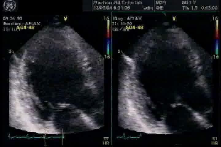
7 Myocardial Viability Dobutamine Stress Echo Otto. Practice of Clinical Echocardiography, 4 th ed, p313
8 Comparison of the various imaging techniques for detecting Hibernating Myocardium Technique No. of studies No. of patients Mean EF (%) Sensitivity (%) Specificity (%) Dobutamine echocardiography.total Low-dose DbE High-dose DbE Myocardial contrast echocardiography. total Thallium scintigraphy. total Tl-201 rest-redistribution Tl-201 re-injection Technetium scintigraphy.total Without nitrates protocol With nitrates protocol Positron emission tomography.total Cardiovascular magnetic resonance. total Low-dose dobutamine protocol Late gadolinium-enhancement protocol Shah et al. EHJ 2013 review
9 Detection of Myocardial Viability Functional Recovery After Revascularization European Heart Journal (2010) 31,
10 Dobutamine Stress Echocardiography Detection of Mycardial Viability Stress Rest Low High Hyperkinetic Normal Worsening Ischemic Sustained Improvement Viable, patent IRA Biphasic Viable, stenosed IRA No Change Infarction


11 Abnormal segments revascularized (%) Dobutamine Stress Echocardiography Detection of Mycardial Viability Recovery No recovery Qureshi U et al, Circ 95:626, Biphasic worsening Worsening Sustained sustained No change n=43 n=8 improvement n=70 n=27


12 `
13 Regional Wall Motion in 2D Echo Wall thickening Endocardial motion Synchronicity

14 Regional Wall Motion in 2D Echo Problems of `Eye Ball` Assessment 1. Limited temporal resolution 2. Only radial motion 3. Interobserver variability 4. Long learning curve Need Quantitative Measurements!

15 Importance of Quantification If you can t measure it, you can t manage it. Peter F. Drucker ( )
16 Quantification of Wall Motion Tissue Doppler Imaging Deformation Imaging


17 Longitudinal Myocardial Velocity Apical Mid Basal Annulus Palmes et al., J Am Soc Echocardiogr, 2001

18 The Left Ventricle is Isaac K, et al., J Am Soc Echocardiogr, % 70% of the muscle fibers are longitudinal!

19 Segmental TDI (S ) for IHD during Dobutamine Stress Echo Marwick T. Am J Cardiol 2001;87:
:657-64.")
20 Change of S during LD-Dobutamine Stress Echocardiography c/w PET, n=36 Yuan D,,Hoffmann R. Echocardiogr 2001;18(8):

21 Change of S during LD-Dobutamine Stress Echocardiography c/w 3M F/U Echo, n=56 Schneider C,et al. Int J cardiol 2006;110: By ROC analysis, a cut-off value of 1.0 cm/s was the best parameter to differ viable from nonviable myocardium (area under the curve 0.85; p <0.01; 95% CI 0.79 to 0.90).
22 TDI Strain Differentiates Transmurality in AMI After Reperfusion Derumeaux G et al. Circulation 2001;103:589-96
23 Limitations of TDI Angle dependency Frame rate dependency Tethering ( 묶임현상 )
24 Angle Dependency of TDI 임상심초음파학제 2 판 p187

25 Low Frame Rate TDI Data Extrapolated information 30 FPS Each square represents a frame of information More data points, More accurate waveforms!
26 Tethering( 묶임현상 ) of TDI
27 Limitations of TDI Stress Echo Angle dependent -> Need of Narrow angle Not simultaneous assessment -> Color spectral TDI (frame rate dependent) Tethering
28 Strain Rate : Rate of Deformation (Spatial velocity gradient) stress v v Strain Rate 1 2 d v 1 v 2 d No influence of translation Really local information


29 Advantage of Strain Rate Imaging vs TDI Basal septal akinesia TVI SRI cm / s s -1 cm / s s -1
30
31 Peak Systolic Strain Rate during Dobutamine Stress Echocardiography Hoffmann et al. JACC 2002;39:443-9.

32 Incremental Value of SRI Assessment of Myocardial Viability after Revascularization Hanecom et al. Circulation 2005;20;112(25):

 E/E =9.")

33 Baseline 10ug DSE F/60, NSTEMI s/p tirofiban EF=53% (LVEDV/ESV=80.5/37.8ml) E/E =9.38 NT-proBNP=476ng/L Lt main and 2VD, LVEDP=9mmHg, PCWP=5mmHg # (MD#97) 15 Months F/U

34 Altered Myocardial Stiffness Stunned Vs. Transmural Infarct Pislaru et al. Circulation 2004;109:


35 S E =0.7 s -1 S E =1.5 s -1 S A =0.2 s -1 S A =2.2 s -1 Baseline Dobu 20 Normal Viable by PET Nonviable by PET Viable & nonviab Peak SR E at rest 1.13± ± ± Peak SR E with dob 1.30± ± ±0.48 <.001 Peak SR E with dob 0.17± ± ± Peak SR A at rest 0.98± ± ± Peak SR A with dob 1.24± ± ±0.58 <.001 n=37 Hoffmann R, et al. Am Soc Echocardiogr 2005;18(4):330-5.


36 Strain Rate for Assessing Viability Acute myocardial infarction in man (LAD occlusion)


37 Strain Rate for Assessing Viability Recovery acute MI 1 day 2 weeks

38 Hatle L, et al, Eur Heart J, 2000 Apex Normal Base Apex Acute Inferior MI Base Apex Chronic Inferior MI Base


39 Strain Rate for Assessing Viability Acute myocardial infarction in man (LAD occlusion) [s -1 ] [s -1 ] AVC


40 Tennant R and Wiggers CJ. Am J Physiol. 1935;112:
41 Post Systolic Thickening First described by Tennant and Wiggers. Common finding in myocardial infarction. Small area of contraction interspersed by akinetic and hypokinetic segment after MI. Good predictor of viable myocardium. Underlying mechanisms are unclear. (Active fiber shortening or passive thickening)
 0 0-10")

42 Post Systolic Shortening Defined as predominantly negative Strain between AVC and E-wave (MVO) PSS < 20% of tot. Strain PSS > 20% of tot. Strain AVC MVO AVC MVO systole diastole systole diastole

43 Post Systolic Strain Index


44 Weidemann et al. Circulation 2003;107(6): Strain rate, Baseline Strain rate, Dobu 20
45 PSI

46 Automated Functional Imaging


47 Automated Functional Imaging

48 Automated Functional Imaging


49 Gobal Longitudinal Strain predicts LV remodeling in recent NSTEMI D Andrea A, et al. Int J Cardiol. 2011;153:


50 Automated Functional Imaging Exercise Echo baseline LAD os 70% LN peak


51 3D Strain
 and with dobutamine (0.170.08 versus 0.370.10; P<0.001). Ecc was significantly decreased in nonviable regions compared with viable segments at rest (0.080.06 versus 0.170.10; P<0.001) and with dobutamine (0.070.")
52 3D LV Strain in LD Dobutamine MR Circumferential strain in mid-ventricle Compared with normal volunteers, global left ventricular Ecc was significantly decreased in patients with ICM at rest ( versus ; P<0.001) and with dobutamine ( versus ; P<0.001). Ecc was significantly decreased in nonviable regions compared with viable segments at rest ( versus ; P<0.001) and with dobutamine ( versus ; P<0.001). Bree et al. Circulation 2006;114(suppl I):I-33-6.

53 3D Speckle Tracking Longtudinal Strain Strain 20.1% 14.9% global 3-dimensional longitudinal strains Abate E, et al. Am J Cardiol. 2012;110:


54 Take Home Message 1. Low dose dobutamine stress echocardiography showed high specificity and comparable sensitivity with other imaging modalities 2. Measurement of both systolic and post-systolic deformation both at rest and during a graded dobutamine infusion may provide prognostic information about myocardial viability. 3. Implement of these new indices in the routine clinical practice will need additional clinical and large-scale studies.

55 To be, or not to be, that is the question! Shakespeare, Hamlet, 1601
, Eun Ok Shim, Kyeong Soon Kim, Ok Ryun Kim, Eun Ju You, Jung Geun Moon, Mi-Seung Shin (Gachon")
56 To be, or not to be, that is on your hands! Special thanks to Hye-Jin Kim (GE Health Korea), Eun Ok Shim, Kyeong Soon Kim, Ok Ryun Kim, Eun Ju You, Jung Geun Moon, Mi-Seung Shin (Gachon Gil Echo Lab), Dae Hee Kim, Soo-Jin Kang (Ulsan Univ.) and Goo- Yeong Cho (Hallym Univ.) for this slide.

57 Thank you for your attention!!! Wook-Jin Chung, MD, PhD Noninvasive CV Imaging Lab., HF & PAH Clinic Cardiology Division, Heart Center Gachon University Gil Hospital Incheon, Korea
Assessment of Ischemia and Viability
 EAE Teaching Course Bucharest, 2010 Assessment of Ischemia and Viability Jens-Uwe Voigt Dpt. of Cardiology University Leuven Belgium Assessment of Ischemia & Viability resting wall motion Stress Testing
Insights into Viability- Function and Contractile Reserve
 Insights into Viability- Function and Contractile Reserve Tom Marwick Cleveland Clinic Conflicts research grants and consulting with GE, Philips, Siemens Off-label use of Definity for assessment of myocardial
Rational use of imaging for viability evaluation
 EUROECHO and other imaging modalities 2011 Rational use of imaging for viability evaluation Luc A. Pierard, MD, PhD, FESC, FACC Professor of Medicine Head, Department of Cardiology, CHU Liège, Belgium
Myocardial viability testing. What we knew and what is new
 Myocardial viability testing. What we knew and what is new Dr B K S Sastry, MD, DM. CARE Hospitals, Hyderabad What is Viability Viability Dysfunctional myocardium subtended by diseased coronary arteries
Velocity, strain and strain rate: Doppler and Non-Doppler methods. Thoraxcentre, Erasmus MC,Rotterdam
 Velocity, strain and strain rate: Doppler and Non-Doppler methods J Roelandt J. Roelandt Thoraxcentre, Erasmus MC,Rotterdam Basics of tissue Doppler imaging Instantaneous annular velocity profiles IVCT
DISCLOSURE. Myocardial Mechanics. Relevant Financial Relationship(s) Off Label Usage
 Off Label Usage") 7th Annual Team Echocardiography: The Heart of Cardiovascular Medicine Tissue Doppler, Strain, Speckle: What? How? Christopher J Kramer RDCS Aurora Medical Group Advanced Cardiovascular Services, Aurora
7th Annual Team Echocardiography: The Heart of Cardiovascular Medicine Tissue Doppler, Strain, Speckle: What? How? Christopher J Kramer RDCS Aurora Medical Group Advanced Cardiovascular Services, Aurora
MR Assessment of Myocardial Viability
 MR Assessment of Myocardial Viability Definition of Viability Clinical Metabolism: Presence of glucose uptake Perfusion / Perfusion reserve Morphology: Wall thickness, wall thickening Contractility: Recovery
Tissue Doppler Imaging in Congenital Heart Disease
 Tissue Doppler Imaging in Congenital Heart Disease L. Youngmin Eun, M.D. Department of Pediatrics, Division of Pediatric Cardiology, Kwandong University College of Medicine The potential advantage of ultrasound
Echo in CAD: Wall Motion Assessment
 Echo in CAD: Wall Motion Assessment Joe M. Moody, Jr, MD UTHSCSA and STVHCS October 2007 Relevant References ACC/AHA/ASE 2003 Guideline Update for the Clinical Application of Echocardiography Bayes de
Radiologic Assessment of Myocardial Viability
 November 2001 Radiologic Assessment of Myocardial Viability Joshua Moss, Harvard Medical School Year III Patient EF 66yo female with a 3-year history of intermittent chest pain previously relieved by sublingual
1. LV function and remodeling. 2. Contribution of myocardial ischemia due to CAD, and
 1 The clinical syndrome of heart failure in adults is commonly associated with the etiologies of ischemic and non-ischemic dilated cardiomyopathy, hypertrophic cardiomyopathy, hypertensive heart disease,
Evaluation of Myocardial Viability: What Have We Learned from STICH? Professor of Medicine David Geffen School of Medicine at UCLA. Heart Failure (HF)
") Evaluation of Myocardial Viability: What Have We Learned from STICH? Daniel S. Berman, MD Director, Cardiac Imaging Cedars-Sinai Heart Institute CSMC 2013 Professor of Medicine David Geffen School of Medicine
Evaluation of Myocardial Viability: What Have We Learned from STICH? Daniel S. Berman, MD Director, Cardiac Imaging Cedars-Sinai Heart Institute CSMC 2013 Professor of Medicine David Geffen School of Medicine
DECLARATION OF CONFLICT OF INTEREST. None
 DECLARATION OF CONFLICT OF INTEREST None Hot Topics in Echocardiography: The position of the EAE EAE / ASE recommendation about Echo Assessment of Cardiac Mechanics Jens-Uwe Voigt Dpt. of Cardiovascular
Journal of the American College of Cardiology Vol. 39, No. 3, by the American College of Cardiology ISSN /02/$22.
 Journal of the American College of Cardiology Vol. 39, No. 3, 2002 2002 by the American College of Cardiology ISSN 0735-1097/02/$22.00 Published by Elsevier Science Inc. PII S0735-1097(01)01763-6 Strain
Strain and Strain Rate Imaging How, Why and When?
 Strain and Strain Rate Imaging How, Why and When? João L. Cavalcante, MD Advanced Cardiac Imaging Fellow Cleveland Clinic Foundation Disclosures: No conflicts of interest Movement vs Deformation Movement
Cardial MRI; Approaching the Level of Gold Standard for Viability Assessment
 Cardial MRI; Approaching the Level of Gold Standard for Viability Assessment 용환석 고려대학교구로병원영상의학과 Viability Hibernating myocardium a state of myocardial hypocontractility during chronic hypoperfusion, in
Role of echocardiography in the assessment of ischemic heart disease 분당서울대학교병원윤연이
 Role of echocardiography in the assessment of ischemic heart disease 분당서울대학교병원윤연이 Outline Evaluation of Chest pain Evaluation of MI complications Prediction of Outcomes Evaluation of Chest pain Evaluation
VECTORS OF CONTRACTION
 1/3/216 Strain, Strain Rate, and Torsion: Myocardial Mechanics Simplified and Applied VECTORS OF CONTRACTION John Gorcsan, MD University of Pittsburgh, Pittsburgh, PA Shortening Thickening Twisting No
The best from Euro-Echo Ischemic heart disease. Fausto Rigo,FESC Department of Cardiology Mestre-Venezia Hospital,Italy
 The best from Euro-Echo 2011 Ischemic heart disease Fausto Rigo,FESC Department of Cardiology Mestre-Venezia Hospital,Italy faustorigo@alice.it DECLARATION OF CONFLICT OF INTEREST No conflict of interest
Cardiac Viability Testing A Clinical Perspective Annual Cardiac Imaging Symposium. Lisa M Mielniczuk MD FRCPC University of Ottawa Heart Institute
 Cardiac Viability Testing A Clinical Perspective Annual Cardiac Imaging Symposium Lisa M Mielniczuk MD FRCPC University of Ottawa Heart Institute 62 year old male Anterior STEMI late presentation, occluded
Imaging and heart failure
 Imaging and heart failure Jeroen J Bax Dept of Cardiology Leiden Univ Medical Center The Netherlands Davos, feb 2013 Research grants: Medtronic, Biotronik, Boston, St Jude, BMS imaging, GE Healthcare,
LV FUNCTION ASSESSMENT: WHAT IS BEYOND EJECTION FRACTION
 LV FUNCTION ASSESSMENT: WHAT IS BEYOND EJECTION FRACTION Jamilah S AlRahimi Assistant Professor, KSU-HS Consultant Noninvasive Cardiology KFCC, MNGHA-WR Introduction LV function assessment in Heart Failure:
How does the heart pump? From sarcomere to ejection volume
 How does the heart pump? From sarcomere to ejection volume Piet Claus Cardiovascular Imaging and Dynamics Department of Cardiovascular Diseases University Leuven, Leuven, Belgium Course on deformation
When is strain assessment mandatory?
 When is strain assessment mandatory? Geneviève Derumeaux University of Lyon France Presenter Disclosure Information Geneviève Derumeaux When is strain assessment mandatory? DISCLOSURE INFORMATION: None
3D-stress echocardiography Bernard Cosyns, MD, PhD
 3D-stress echocardiography Bernard Cosyns, MD, PhD No Disclosure The Pro-Technology bias Sicari et al. Cardiovascular Ultrasound 2006, 4:11 Overview 2D stress echocardiography: main limitations 3D echocardiography:
Gated blood pool ventriculography: Is there still a role in myocardial viability?
 Gated blood pool ventriculography: Is there still a role in myocardial viability? Oliver C. Alix, MD Adult Clinical and Nuclear Cardiology St. Luke s Medical Centre - Global City Case Presentation A 62-year-old
Novel echocardiographic modalities: 3D echo, speckle tracking and strain rate imaging. Potential roles in sports cardiology. Stefano Caselli, MD, PhD
 Novel echocardiographic modalities: 3D echo, speckle tracking and strain rate imaging. Potential roles in sports cardiology. Stefano Caselli, MD, PhD Ospedale San Pietro Fatebenefratelli Rome, Italy Differential
Old and new insights into viability:perfusion and Perfusion Reserve
 Old and new insights into viability:perfusion and Perfusion Reserve R.Senior Professor of Clinical Cardiology Consultant Cardiologist and Director of Echo,Royal Brompton Hospital,London and Northwick Park
22 nd Annual Conference of the Saudi Heart Association Riyadh, Saudi Arabia
 22 nd Annual Conference of the Saudi Heart Association Riyadh, Saudi Arabia New Echocardiographic Modalities to Evaluate Ventricular Function in Congenital Heart Disease: Tissue Doppler & Strain Rate Imaging
Strain Imaging: Myocardial Mechanics Simplified and Applied
 9/28/217 Strain Imaging: Myocardial Mechanics Simplified and Applied John Gorcsan III, MD Professor of Medicine Director of Clinical Research Division of Cardiology VECTORS OF CONTRACTION Shortening Thickening
Tissue Doppler and Strain Imaging. Steven J. Lester MD, FRCP(C), FACC, FASE
, FACC, FASE") Tissue Doppler and Strain Imaging Steven J. Lester MD, FRCP(C), FACC, FASE Relevant Financial Relationship(s) None Off Label Usage None a. Turn the wall filters on and turn down the receiver gain. b. Turn
Tissue Doppler and Strain Imaging Steven J. Lester MD, FRCP(C), FACC, FASE Relevant Financial Relationship(s) None Off Label Usage None a. Turn the wall filters on and turn down the receiver gain. b. Turn
10/7/2013. Systolic Function How to Measure, How Accurate is Echo, Role of Contrast. Thanks to our Course Director: Neil J.
 Systolic Function How to Measure, How Accurate is Echo, Role of Contrast Neil J. Weissman, MD MedStar Health Research Institute & Professor of Medicine Georgetown University Washington, D.C. No Disclosures
Current Guidelines for Diagnosis of AMI Chest pain ST change on EKG Cardiac Enzymes
 Noninvasive Cardiac Imaging in Myocardial Infarction Sangchol Lee Sungkyunkwan University Samsung Medical Center Current Guidelines for Diagnosis of AMI Chest pain ST change on EKG Cardiac Enzymes Do We
Coronary Revascularization in Patients witj Severe LV Dysfunction.: Is the concept of viability still viable?
 Coronary Revascularization in Patients witj Severe LV Dysfunction.: Implications of the STICH trial Is the concept of viability still viable? Banff 2016 3041435-1 Prognosis of Patients With LV Dysfunction
2/2/2011. Strain and Strain Rate Imaging How, Why and When? Movement vs Deformation. Doppler Myocardial Velocities. Movement. Deformation.
 Strain and Strain Rate Imaging How, Why and When? João L. Cavalcante, MD Advanced Cardiac Imaging Fellow Cleveland Clinic Foundation Disclosures: No conflicts of interest Movement vs Deformation Movement
Tissue Doppler and Strain Imaging
 Tissue Doppler and Strain Imaging Steven J. Lester MD, FRCP(C), FACC, FASE Relevant Financial Relationship(s) None Off Label Usage None 1 Objective way with which to quantify the minor amplitude and temporal
Strain/Untwisting/Diastolic Suction
 What Is Diastole and How to Assess It? Strain/Untwisting/Diastolic Suction James D. Thomas, M.D., F.A.C.C. Cardiovascular Imaging Center Department of Cardiology Cleveland Clinic Foundation Cleveland,
3/27/2014. Introduction.
 Introduction. Myocardial perfusion & contractility becomes abnormal immediately after the onset of ischaemia, even before the development of the symptoms & ST segment changes. 1 Myocardial Wall Motion
Detection and Assessment of MI: Use of Imaging Methods. Robert O. Bonow, M.D.
 Detection and Assessment of MI: Use of Imaging Methods Robert O. Bonow, M.D. Detection and Assessment of MI: Use of Imaging Methods Robert O. Bonow, M.D. No Relationships to Disclose Expert Consensus Document
Qualitative and Quantitative Assessment of Perfusion
 APCDE 2011 Qualitative and Quantitative Assessment of Perfusion Hyun Ju Yoon Chonnam National University Hospital Gwangju, Korea ISCHEMIC CASCADE Blood flow mismatch Perfusion defects on nuclear imaging,
Validation of echocardiographic 2-dimensional. speckle tracking longitudinal strain imaging for
 Chapter 10 Validation of echocardiographic 2-dimensional speckle tracking longitudinal strain imaging for viability assessment in patients with chronic ischemic left ventricular dysfunction and comparison
Perfusion, Viability, Edema and Hemorrhage: How it Can (and Should) Change Clinical Practice. Rohan Dharmakumar, Ph.D.
 Change Clinical Practice. Rohan Dharmakumar, Ph.D.") Perfusion, Viability, Edema and Hemorrhage: How it Can (and Should) Change Clinical Practice Rohan Dharmakumar, Ph.D. Director, Translational Cardiac Imaging Research Associate Director, Biomedical Imaging
Perfusion, Viability, Edema and Hemorrhage: How it Can (and Should) Change Clinical Practice Rohan Dharmakumar, Ph.D. Director, Translational Cardiac Imaging Research Associate Director, Biomedical Imaging
How To Perform Strain Imaging; Step By Step Approach. Maryam Bo Khamseen Echotechnoligist II EACVI, ARDMS, RCS King Abdulaziz Cardiac Center- Riyadh
 How To Perform Strain Imaging; Step By Step Approach Maryam Bo Khamseen Echotechnoligist II EACVI, ARDMS, RCS King Abdulaziz Cardiac Center- Riyadh Outlines: Introduction Describe the basic of myocardium
Advanced Multi-Layer Speckle Strain Permits Transmural Myocardial Function Analysis in Health and Disease:
 Advanced Multi-Layer Speckle Strain Permits Transmural Myocardial Function Analysis in Health and Disease: Clinical Case Examples Jeffrey C. Hill, BS, RDCS Echocardiography Laboratory, University of Massachusetts
The importance of left atrium in LV diastolic function
 II Baltic Heart Failure Meeting and Congress of Latvian Society of Cardiology The importance of left atrium in LV diastolic function Dr. Artem Kalinin Eastern Clinical University Hospital Riga 30.09.2010.
Evaluation of Left Ventricular Diastolic Dysfunction by Doppler and 2D Speckle-tracking Imaging in Patients with Primary Pulmonary Hypertension
 ESC Congress 2011.No 85975 Evaluation of Left Ventricular Diastolic Dysfunction by Doppler and 2D Speckle-tracking Imaging in Patients with Primary Pulmonary Hypertension Second Department of Internal
Global left ventricular circumferential strain is a marker for both systolic and diastolic myocardial function
 Global left ventricular circumferential strain is a marker for both systolic and diastolic myocardial function Toshinari Onishi 1, Samir K. Saha 2, Daniel Ludwig 1, Erik B. Schelbert 1, David Schwartzman
Strain-Rate Imaging During Dobutamine Stress Echocardiography Provides Objective Evidence of Inducible Ischemia
 Strain-Rate Imaging During Dobutamine Stress Echocardiography Provides Objective Evidence of Inducible Ischemia Jens-Uwe Voigt, MD; Bert Exner; Kristin Schmiedehausen, MD; Cord Huchzermeyer; Udo Reulbach,
Tissue Doppler and Strain Imaging
 Tissue Doppler and Strain Imaging Steven J. Lester MD, FRCP(C), FACC, FASE Relevant Financial Relationship(s) None Off Label Usage None 1 Objective way with which to quantify the minor amplitude and temporal
Aortic valve Stenosis: Insights in the evaluation of LV function. Erwan DONAL Cardiologie CHU Rennes
 Aortic valve Stenosis: Insights in the evaluation of LV function Erwan DONAL Cardiologie CHU Rennes erwan.donal@chu-rennes.fr Preload Afterload Myocardial Fiber Shortening Circumferential Longitudinal
MRI ACS-ben. Tamás Simor MD, PhD, Med Hab. University of Pécs, Heart Institute
 MRI ACS-ben Tamás Simor MD, PhD, Med Hab Time Course of Changes in Infarct Size, Viable Myocardium, and LV Mass After Reperfused and Nonreperfused MI Blue lines denote reperfused myocardial infarction
Cardiac Resynchronization Therapy Selection therapy Echocardiography
 Cardiac Resynchronization Therapy Selection therapy Echocardiography Prof. Patrizio LANCELLOTTI CHU Sart Tilman, Liège April, 2010 Candidates for CRT : class IA NYHA Functional Class III or IV (Subjective)
Revascularization of ischemic LV dysfunction: past, present and future
 Revascularization of ischemic LV dysfunction: past, present and future Paolo G Camici, MD, FESC, FACC, FAHA, FRCP Vita-Salute University and San Raffaele Scientific Institute Milan Advances in Cardiovascular
The Value of 2D Strain Imaging during Stress Testing
 The Value of 2D Strain Imaging during Stress Testing Marie Moonen, M.D., Patrizio Lancellotti, M.D., Ph.D., Dimitrios Zacharakis, M.D., and Luc Pierard, M.D., Ph.D., F.E.S.C., F.A.C.C. CHU Sart Tilman,
Left Ventricular Dyssynchrony in Patients Showing Diastolic Dysfunction without Overt Symptoms of Heart Failure
 ORIGINAL ARTICLE DOI: 10.3904/kjim.2010.25.3.246 Left Ventricular Dyssynchrony in Patients Showing Diastolic Dysfunction without Overt Symptoms of Heart Failure Jae Hoon Kim, Hee Sang Jang, Byung Seok
Sung A Chang Department of Internal Medicine, Division of Cardiology, Sungkyunkwan University School of Medicine, Samsung Medical Center
 CMR Perfusion and Viability A STICH Out of Time? Sung A Chang Department of Internal Medicine, Division of Cardiology, Sungkyunkwan University School of Medicine, Samsung Medical Center Can Imaging Improve
LA Function analysis Marcia Barbosa Vice Presidente - Brazilian Soc of Cardiology President-elect - Interamerican Soc of Cardiology
 LA Function analysis Marcia Barbosa Vice Presidente - Brazilian Soc of Cardiology President-elect - Interamerican Soc of Cardiology Belo Horizonte Brazil DECLARATION OF CONFLICT OF INTEREST Nothing to
Cardiovascular Imaging Stress Echo
 Cardiovascular Imaging Stress Echo Theodora A Zaglavara, MD, PhD Cardiac Imaging Department INTERBALKAN MEDICAL CENTER Thessaloniki GREECE Evolution of Stress Echo: From Innovation to a Widely Established
Cardiology for the Practitioner Advanced Cardiac Imaging: Worth the pretty pictures?
 Keenan Research Centre Li Ka Shing Knowledge Institute Cardiology for the Practitioner Advanced Cardiac Imaging: Worth the pretty pictures? Howard Leong-Poi, MD, FRCPC Associate Professor of Medicine St.
The Value of Stress MRI in Evaluation of Myocardial Ischemia
 The Value of Stress MRI in Evaluation of Myocardial Ischemia Dr. Saeed Al Sayari, MBBS, EBCR, MBA Department of Radiology and Nuclear Medicine Mafraq Hospital, Abu Dhabi United Arab Emirates Introduction
Viability Testing Using Dynamic Echocardiography
 Viability Testing Using Dynamic Echocardiography Theodora A Zaglavara, MD, PhD Director of Echocardiography EUROMEDICA KYANOUS STAVROS HOSPITAL Thessaloniki GREECE Goals of Cardiac Imaging in Coronary
Νεότερα ςτην Υπερηχοκαρδιογραφία. Βαςίλειοσ Καμπερίδησ Clinical research fellow in Cardiology
 Νεότερα ςτην Υπερηχοκαρδιογραφία Βαςίλειοσ Καμπερίδησ Clinical research fellow in Cardiology Disclosures ESC training grant EACVI research grant HCS training grant ELIKAR research grant Evolution of Echocardiography
Advanced imaging of the left atrium - strain, CT, 3D, MRI -
 Advanced imaging of the left atrium - strain, CT, 3D, MRI - Monica Rosca, MD Carol Davila University of Medicine and Pharmacy, Bucharest, Romania Declaration of interest: I have nothing to declare Case
Imaging of Coronary Artery Disease: II
 Acta Radiológica Portuguesa, Vol.XIX, nº 74, pág. 45-51, Abr.-Jun., 2007 Imaging of Coronary Artery Disease: II Jean Jeudy University of Maryland School of Medicine Department of Diagnostic Radiology Armed
HYPERTROPHY: Behind the curtain. V. Yotova St. Radboud Medical University Center, Nijmegen
 HYPERTROPHY: Behind the curtain V. Yotova St. Radboud Medical University Center, Nijmegen Disclosure of interest: none Relative wall thickness (cm) M 0.22 0.42 0.43 0.47 0.48 0.52 0.53 F 0.24 0.42 0.43
CT for Myocardial Characterization of Cardiomyopathy. Byoung Wook Choi, Yonsei University Severance Hospital, Seoul, Korea
 CT for Myocardial Characterization of Cardiomyopathy Byoung Wook Choi, Yonsei University Severance Hospital, Seoul, Korea Cardiomyopathy Elliott P et al. Eur Heart J 2008;29:270-276 The European Society
The road to successful CRT implantation: The role of echo
 The road to successful CRT implantation: The role of echo Tae-Ho Park Dong-A University Hospital, Busan, Korea Terminology Cardiac Resynchronization Therapy (CRT) = Biventricular pacing (BiV) = Left ventricular
Alicia Armour, MA, BS, RDCS
 Alicia Armour, MA, BS, RDCS No disclosures Review 2D Speckle Strain (briefly) Discuss some various patient populations & disease pathways where Strain can be helpful Discuss how to acquire images for Strain
Magnetic Resonance Imaging (MRI) for the Assessment of Myocardial Viability
 for the Assessment of Myocardial Viability") Ontario Health Technology Assessment Series 2010; Vol. 10, No. 15 Magnetic Resonance Imaging (MRI) for the Assessment of Myocardial Viability An Evidence-Based Analysis Presented to the Ontario Health
Ontario Health Technology Assessment Series 2010; Vol. 10, No. 15 Magnetic Resonance Imaging (MRI) for the Assessment of Myocardial Viability An Evidence-Based Analysis Presented to the Ontario Health
Carlos Eduardo Suaide Silva, Luiz Darcy Cortez Ferreira, Luciana Braz Peixoto, Claudia Gianini Monaco, Manuel Adán Gil, Juarez Ortiz
 Silva et al Original Article Arq Bras Cardiol Study of the Myocardial Contraction and Relaxation Velocities through Doppler Tissue Imaging Echocardiography. A New Alternative in the Assessment of the Segmental
THE first objective of this thesis was to explore possible shape parameterizations
 8 SUMMARY Columbus is not the only person who has discovered a new continent. So too have I. Anak Semua Bangsa (Child of All Nations) PRAMOEDYA ANANTA TOER 8.1 Myocardial wall motion modeling THE first
Pearls & Pitfalls in nuclear cardiology
 Pearls & Pitfalls in nuclear cardiology Maythinee Chantadisai, MD., NM physician Division of Nuclear Medicine, Department of radiology, KCMH Principle of myocardial perfusion imaging (MPI) Radiotracer
Cardiac Imaging. Kimberly Delcour, DO, FACC. Mahi Ashwath, MD, FACC, FASE. Director, Cardiac CT. Director, Cardiac MRI
 Cardiac Imaging Kimberly Delcour, DO, FACC Director, Cardiac CT Mahi Ashwath, MD, FACC, FASE Director, Cardiac MRI Cardiac Imaging Discuss the clinical applications of and indications for: Cardiac CT Nuclear
Imaging in Ischemic Heart Disease: Role of Cardiac MRI
 Imaging in Ischemic Heart Disease: Role of Cardiac MRI Chiara Bucciarelli Ducci MD, PhD, FESC, FRCP Consultant Senior Lecturer Cardiologist Bristol Heart Institute, University of Bristol, UK Chair elect,
Three-dimensional Wall Motion Tracking:
 Three-dimensional Wall Motion Tracking: A Novel Echocardiographic Method for the Assessment of Ventricular Volumes, Strain and Dyssynchrony Jeffrey C. Hill, BS, RDCS, FASE Jennifer L. Kane, RCS Gerard
Little is known about the degree and time course of
 Differential Changes in Regional Right Ventricular Function Before and After a Bilateral Lung Transplantation: An Ultrasonic Strain and Strain Rate Study Virginija Dambrauskaite, MD, Lieven Herbots, MD,
How to assess ischaemic MR?
 ESC 2012 How to assess ischaemic MR? Luc A. Pierard, MD, PhD, FESC, FACC Professor of Medicine Head, Department of Cardiology University Hospital Sart Tilman, Liège ESC 2012 No conflict of interest Luc
Strain imaging in children: from Tissue Doppler to 3 D
 Strain imaging in children: from Tissue Doppler to 3 D Mark kk. Friedberg Fi Outline Deformation in the fetus and neonate Deformation in pediatric cardiomyopathy y (briefly!) Deformation in Congenital
Coronary Revascularization for Severe LV Dysfunction Is s. Is the concept of viability testing still viable?
 Coronary Revascularization for Severe LV Dysfunction Is s Is the concept of viability testing still viable? Banff 2017 2015 MFMER 3492638-7 Prognosis of Patients With LV Dysfunction and CAD Major determinants
Mechanisms and role of contrast echocardiography
 Mechanisms and role of contrast echocardiography Seol Sang-Hoon Inje University College of Medicine, Haeundae Paik Hospital, Busan, Korea Physical Principles of Contrast Ultrasound Contrast echocardiography
Nancy Goldman Cutler, MD Beaumont Children s Hospital Royal Oak, Mi
 Nancy Goldman Cutler, MD Beaumont Children s Hospital Royal Oak, Mi Identify increased LV wall thickness (WT) Understand increased WT in athletes Understand hypertrophic cardiomyopathy (HCM) Enhance understanding
Grading of Myocardial Dysfunction by Tissue Doppler Echocardiography A Comparison Between Velocity, Displacement, and Strain Imaging in Acute Ischemia
 Journal of the American College of Cardiology Vol. 47, No. 8, 2006 2006 by the American College of Cardiology Foundation ISSN 0735-1097/06/$32.00 Published by Elsevier Inc. doi:10.1016/j.jacc.2006.01.051
Value of Assessment of Viable and Ischemic Myocardium and Techniques Such as MRI, Radionuclide Imaging
 Chapter 2 Imaging for Viable and Ischemic Myocardium Value of Assessment of Viable and Ischemic Myocardium and Techniques Such as MRI, Radionuclide Imaging Catalin Loghin and K. Lance Gould Introduction
Prognostic Value of Left Atrial Size and Function
 Prognostic Value of Left Atrial Size and Function James D. Thomas, M.D., F.A.C.C. Cardiovascular Imaging Center Department of Cardiology Cleveland Clinic Foundation Cleveland, Ohio, USA Conflicts: None
@02-126_Coronary_calcification.ppt. Professor Molecular and Medical Pharmacology
 Assessment of Myocardial Viability Jamshid Maddahi, M.D., FACC, FASNC Professor Molecular and Medical Pharmacology (Nuclear Medicine) and Medicine (Cardiology) David Geffen School of Medicine at UCLA Director,
Echo assessment of the failing heart
 Echo assessment of the failing heart Mark K. Friedberg, MD The Labatt Family Heart Center The Hospital for Sick Children Toronto, Ontario, Canada Cardiac function- definitions Cardiovascular function:
Le FDG dans l évaluation de la viabilité : Quelle place par rapport aux autres traceurs?
 Le FDG dans l évaluation de la viabilité : Quelle place par rapport aux autres traceurs? Pierre-Yves MARIE, Médecine Nucléaire, CHU de Nancy PET Gated-SPECT MRI FDG imaging: effect of dietary status fasting
DOWNLOAD PDF MYOCARDIAL CONTRAST TWO DIMENSIONAL ECHOCARDIOGRAPHY (DEVELOPMENTS IN CARDIOVASCULAR MEDICINE)
") Chapter 1 : Imaging Cardiovascular Medicine Stanford Medicine contrast two-dimensional echocardiography (MC-2DE), a new and exciting diagnostic methodology for assessment of myocardial perfusion, which
Chapter 1 : Imaging Cardiovascular Medicine Stanford Medicine contrast two-dimensional echocardiography (MC-2DE), a new and exciting diagnostic methodology for assessment of myocardial perfusion, which
Quantitation of right ventricular dimensions and function
 SCCS Basics of cardiac assessment Quantitation of right ventricular dimensions and function Tomasz Kukulski, MD PhD Dept of Cardiology, Congenital Heart Disease and Electrotherapy Silesian Medical University
Correlation Between Regional Wall Motion Abnormalities via 2-Dimensional Echocardiography, and Coronary Angiographic Findings
 THE ECHOCARDIOGRAPHY, IRAQI POSTGRADUATE MEDICAL AND CORONARY JOURNAL ANGIOGRAPHIC FINDINGS VOL.11, SUPPLEMENT,2012 Correlation Between Regional Wall Motion Abnormalities via 2-Dimensional Echocardiography,
Cardiac Imaging Tests
 Cardiac Imaging Tests http://www.medpagetoday.com/upload/2010/11/15/23347.jpg Standard imaging tests include echocardiography, chest x-ray, CT, MRI, and various radionuclide techniques. Standard CT and
Quantifying LV function how good are we?
 Quantifying LV function how good are we? Professor Alan G Fraser Wales Heart Research Institute Cardiff University, U.K. Support for research from Hitachi Aloka, & GE Ultrasound Visual assessment of synchronicity
Cardiovascular deformation imaging Part II: cardiac
 Electrical Engineering, Cardiology and Physics Cardiac Imaging Research Cardiovascular deformation imaging Part II: cardiac Jan D hooge 1-3, Chris L. de Korte 4 1 Medical Image Computing Dept. of Electrical
Mechanisms of False Positive Exercise Electrocardiography: Is False Positive Test Truly False?
 Mechanisms of False Positive Exercise Electrocardiography: Is False Positive Test Truly False? Masaki Izumo a, Kengo Suzuki b, Hidekazu Kikuchi b, Seisyo Kou b, Keisuke Kida b, Yu Eguchi b, Nobuyuki Azuma
Impaired Regional Myocardial Function Detection Using the Standard Inter-Segmental Integration SINE Wave Curve On Magnetic Resonance Imaging
 Original Article Impaired Regional Myocardial Function Detection Using the Standard Inter-Segmental Integration Ngam-Maung B, RT email : chaothawee@yahoo.com Busakol Ngam-Maung, RT 1 Lertlak Chaothawee,
Myocardial Deformation Imaging Based on Ultrasonic Pixel Tracking to Identify Reversible Myocardial Dysfunction
 Journal of the American College of Cardiology Vol. 51, No. 15, 2008 2008 by the American College of Cardiology Foundation ISSN 0735-1097/08/$34.00 Published by Elsevier Inc. doi:10.1016/j.jacc.2007.10.066
Cardiac Chamber Quantification by Echocardiography
 Cardiac Chamber Quantification by Echocardiography Maryam Bokhamseen, RCS, RCDS, EACVI Echotechnologist ǁ, Non invasive Cardiac Laboratory King Abdulaziz Cardiac Center. Outline: Introduction. Background
Χ.Τρίκκα Καρδιολογική Κλινική Νοζοκομείο Ερρίκος Νησνάν
 Χ.Τρίκκα Καρδιολογική Κλινική Νοζοκομείο Ερρίκος Νησνάν Efficacy of Myocardial Contrast Echocardiography in the Diagnosis and Risk Stratification of Acute Coronary Syndrome Am J Cardiol 2005;96:1498 1502
Outline. EuroScore II. Society of Thoracic Surgeons Score. EuroScore II
 SURGICAL RISK IN VALVULAR HEART DISEASE: WHAT 2D AND 3D ECHO CAN TELL YOU AND WHAT THEY CAN'T Ernesto E Salcedo, MD Professor of Medicine University of Colorado School of Medicine Director of Echocardiography
Stephen Glen ISCHAEMIC HEART DISEASE AND LEFT VENTRICULAR FUNCTION
 Stephen Glen ISCHAEMIC HEART DISEASE AND LEFT VENTRICULAR FUNCTION Overview Coronary arteries Terminology to describe contractility Measuring ventricular function Systolic dysfunction Practice cases- LV