Diseases of Stomach & Abomasum. Physical influences Gastric Ulcers Gastritis Parasitic Diseases
|
|
|
- Erik Ward
- 5 years ago
- Views:
Transcription
1 Diseases of Stomach & Abomasum Physical influences Gastric Ulcers Gastritis Parasitic Diseases Neoplasia
2 Physical Influences Acute gastric dilation & volvulus (GDV) Displaced abomasum Chronic gastric dilation Abomasal dilation & emptying defect
3 GDV - Pathogenesis Failure of normal eructation & pyloric outflow Mostly large dog breeds, rarely horses & pigs Follows large meal (dry or highly fermentable) Xs gas production functional obstruction of cardia & pylorus dilation torsion volvulus Compression of lung & posterior vena cava Circulatory collapse (shock) Death from respiratory & circulatory failure
4 GDV - Gross lesions Severe abdominal distension Clock-wise rotation of stomach Hemorrhagic infarction Rupture of stomach (equine) V-shaped bending of enlarged spleen Congestion of intestines


5 Schematic and actual illustration of Gastric dilation and volvulus, dog. The stomach is filled with fluid and gas and the serosa is congested. The spleen is engorged, displaced to the right, and V-shaped. Death is usually from hypovolemic shock following compression of lungs and posterior vena cava.


6 GDV, V-shaped bending of enlarged spleen GDV, Clock-wise rotation of stomach GDV, compression of the caudal vena cava
7 Abomasal Displacement LDA mostly in dairy cows Common GI disorder requiring surgery Associated with parturient stress Affects mostly high producers Feeding high grain diet Rarely fatal RDA mostly in calves - fatal


8 Ab l l l d dil i Abomasal volvulus and dilation, calf. The abomasum is normally on the ventral side but here, it has been displaced to the right and dorsally. Abomasal volvulus and dilation, calf.
9 Stomach/Abomasum (contd) Gastric Dilation Acute dilation & rupture Horses ingesting fermentable feed Could be secondary to intestinal obstruction Distinguish from post mortem rupture Chronic Secondary to other conditions Abomasal Dilation and Emptying Defect


10 Stomach rupture, horse. Stomach rupture occurs following ingestion of highly fermentable feed. Omasal impaction, cow. Abomasal impaction, cow.
11 Gastric Ulcers Important but less so than in humans Imbalance of aggressive & protective factors in mucosa Epithelial necrosis erosion ulceration perforation peritonitis Main signs: Hematemesis Melena Anemia Abdominal pain
12 Schematic illustration of gastric ulcer formation in humans (Kumar, Abbas & Fausto, 2005)


13 Gastric Ulcers - Etiologic Factors Local mucosal injury Normal or high h gastric acidity Mastocytoma - via xs histamine Zollinger-Ellison syndrome - via xs gastrin Local ischemia (stress-induced) Steroids & NSAIDs (aspirin) Others Interference with PG synthesis Direct epithelial necrosis Diet, foreign bodies, infections, uremia Role of Helicobacter spp undetermined in animals, unlike in humans.
14 Gastric Ulcers - Gross lesions Cattle Punched out areas in pylorus, may perforate Pigs Round/oval/stellate craters in pars esophagea Craters covered with fibrin i & blood clots Most are incidental but may cause melena Few lead dto acute fatal hemorrhage h into GIT Horses: often subclinical Dogs: body, pylorus & duodenum


 and")


")

15 Gastric ulcers in animals. In calves, gastric ulcers may perforate (top left) and cause fatal peritonitis. In pigs, they occur as a oval or rectangular crater (top right), often as incidental finding, but could be fatal. In dogs, they often involve the pylorus (bottom left) and the dog could bleed to death. Bleeding peptic ulcer, duodenum, dog


16 Gastritis Dogs & cats Cause often undetermined Uremia gastric vasculopathy Helicobacter spp?? Cattle, sheep & goats Braxy (bradshot) due to Cl. septicum Mycotic infections Uremic gastritis, dog Helicobacter sp


,")
.")

17 Acute hemorrhagic gastritis involving glandular stomach, pig with acute septicemia. Multifocal hemorrhagic infarcts in mycotic gastritis (abomasitis), calf Braxy, lambs (bottom). Acute, hemorrhagic and emphysematous abomasitis due to exotoxin of Clostridium septicum
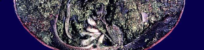
18 Parasitic diseases Ruminants Haemonchosis Ostertagiosis (types I & II) Trichostrongylosis Equine Gastric bots Draschia megastoma Trichostrongylosis Swine Hyostrongylosis


19 Abomasal parasitism & hemorrhage, sheep with hemonchosis. Blood within the gut of the parasite gives it the characteristic barber s pole appearance (below) Hyperplastic abomasitis, bovine ostertagiosis, (top). Confluence of multiple nodules gives it cobblestone appearance. Stomach bots, horse (below). Incidental finding Draschia megastoma, horse
20 Neoplasia Primary gastric tumors are uncommon in animals Lymphoma (cats, cattle etc) Squamous cell carcinoma (equine) Adenocarcinoma (dogs) Polyps Thick, ulcerated plaque Marked scirrhous reaction (desmoplasia) Metastases common

 and")


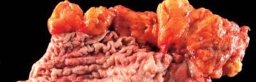


21 Gastric lymphoma, cat stomach (left) and bovine abomasum (right). Expansile, lobulated & ulcerated masses Gastric squamous cell carcinoma, equine. Proliferative & ulcerated mass Gastric adenocarcinoma, ulcerated (left), and leather bottle appearance in linitis plastica right)
22 Small & Large Intestines: Structure & Function Long coiled tube with large surface area for digestion & absorption Folded mucosa Villi (7-14 fold increase) Microvilli (15-40 fold increase) Cecum Good defense mechanism


23 Small intestine Colon Structure & function of intestines. Digestive and absorptive surface area is markedly increased by mucosal folds (left), villi in small intestine (middle) and microvilli on enterocytes (right)
24 Diseases of Small & Large Intestines Congenital anomalies Obstruction & Functional disorders Inflammation Specific enteric diseases (mostly infectious) Neoplastic diseases


25 Congenital anomalies Segmental defects Stenosis Atresia Persistent Meckel s diverticulum Derived from omphalomesenteric duct Megacolon Types of stenosis & atresia Blind end atresia Abdominal distension, atresia coli, feeder pig. Results from obstruction of distal part of gut


26 Meckel diverticulum. The blind pouch is located on the antimesenteric side of the small bowel. Atresia ilei in a lamb. Marked distension and hyperemia of the blind end is due to stasis, of ingesta, bacterial overgrowth and gas produccion. Megacolon, cat. Fecal-filled, enlarged colon could be congenital (congenital lack of intestinal innervation), or acquired (following nerve injury).


")
")


27 Obstruction & functional disorders Obturation (from within or intraluminal) Compression (external) Stenosis (strictures or narrowing) Enterolith, horse Foreign body (sock), intestine, dog Ascarid impaction, jejunum, horse
28 Consequences of obstruction Vary with type and location of obstruction Death from Toxemia (bacterial overgrowth) Shock (dehydration, etc) Starvation or toxicosis Gross lesions Distended abdomen Dilated bowel proximal to obstruction Collapsed and empty distal part Congested/infarcted area of obstruction


29 Distended d abdomen, pig Stricture, intestine horse. Healing by fibrosis narrowing of the lumen partial or complete obstruction dilation (D) of proximal intestine. Initial injury could be penetrating or nonpenetrating wounds, or vascular injury

 ileus Absence of normal tone &")


30 Visceral displacements & functional disorders that lead to intestinal obstruction Intussusception Telescoping (intussusceptum/intussusci piens) Paralytic (adynamic) ileus Absence of normal tone & peristalsis i Herniation Displacement through a foramen Volvulus and torsion Twisting on mesenteric axis Rotation along long axis Major causes of intestinal obstruction (Kumar, Abbas & Fausto, 2005) Intussusception, dog


31 Intussusception, intestine pig (above), dog (below). Affected segments are folded like accordion as a result of telescoping of one segment into another. Congestion is due to impaired venous return strangulation and infarction. The portion of the intestine distal to the intussusception is small, relative to the proximal portion (below)


")
32 Displacement through foramen Internal External May cause incarceration, infarction, ileus or perforation Herniation Herniation through the epiploic foramen (horse, above) and through the diaphragm (cat, below) Umbilical hernia


")


33 Volvulus of small intestine (horse, above and bottom left) and of spiral colon (pig, above right) results in vascular compromise and infarction. Pedunculated lipomas in horses (arrows) can also cause strangulation and death.


34 Thank you! Have a good weekend Ateloprosopia: Incomplete development of the face
Pathology of the Alimentary System Lecture 6 Diseases of intestine
 Systemic Pathology I - VPM 221 Pathology of the Alimentary System Lecture 6 Diseases of intestine Enrique Aburto Fall 2014 VII. Small & Large Intestines Structure & Function Long coiled tube, large surface
Pathology of Intestinal Obstruction. Dr. M. Madhavan, MBBS., MD., MIAC, Professor of Pathology Saveetha Medical College
 Pathology of Intestinal Obstruction Dr. M. Madhavan, MBBS., MD., MIAC, Professor of Pathology Saveetha Medical College Pathology of Intestinal Obstruction Objectives list the causes of intestinal obstruction
Gastrointestinal Disorders. Disorders of the Esophagus 3/7/2013. Congenital Abnormalities. Achalasia. Not an easy repair. Types
 Gastrointestinal Disorders Congenital Abnormalities Disorders of the Esophagus Types Stenosis Atresia Fistula Newborn aspirates while feeding. Pneumonia Not an easy repair Achalasia Lack of relaxation
Pathology of the Alimentary Tract
 Pathology of the Alimentary Tract Lab 2: Lower alimentary tract SI, LI, cecum, and peritoneum GIST in the cecum of a dog Shannon Martinson: http://people.upei.ca/smartinson VPM 221: November, 2011 3 year
Gastrointestinal Tract Imaging. Objectives. Reference. VMB 960 April 6, Stomach Small Intestine Colon. Radiography & Ultrasound
 Gastrointestinal Tract Imaging VMB 960 April 6, 2009 Stomach Small Intestine Colon Objectives Radiography & Ultrasound Contrast Examination of the Small Intestine Reference Chapters 45 47 Pages 750 805
Gastrointestinal Obstruction
 Customer Name, Street Address, City, State, Zip code Phone number, Alt. phone number, Fax number, e-mail address, web site Gastrointestinal Obstruction (Blockage of the Gastrointestinal Tract) Basics OVERVIEW
Intestinal Obstruction Clinical Presentation & Causes
 Intestinal Obstruction Clinical Presentation & Causes V Chidambaram-Nathan Consultant Transplant and General Surgeon Sheffield Kidney Institute Northern General Hospital Intestinal Obstruction One of the
Bowel obstruction and tumors
 Bowel obstruction and tumors Intestinal Obstruction Obstruction of the GI tract may occur at any level, but the small intestine is most often involved because of its relatively narrow lumen. Causes: Hernias
Polyps in general: is a descriptive term of forming a mass that is exophytic & polypoid.
 ميحرلا نمحرلا هللا مسب Gastric Tumors: Benign tumours & tumor-like conditions: -Mucosal: Gastric polyps (they are uncommon) -Mesenchymal tumours: Leiomyoma & Lipoma (can occur anywhere in the body) Malignant:
Development of pancreas and Small Intestine. ANATOMY DEPARTMENT DR.SANAA AL-AlSHAARAWY DR.ESSAM Eldin Salama
 Development of pancreas and Small Intestine ANATOMY DEPARTMENT DR.SANAA AL-AlSHAARAWY DR.ESSAM Eldin Salama OBJECTIVES At the end of the lecture, the students should be able to : Describe the development
Introduction and Definitions
 Bowel obstruction Introduction and Definitions Accounts for 5% of all acute surgical admissions Patients are often extremely ill requiring prompt assessment, resuscitation and intensive monitoring Obstruction
GIT RADIOLOGY. Water-soluble contrast media (e.g. gastrograffin) are the other available agents.which doesn t cause inflammatory peritonitis..
 are the other available agents.which doesn t cause inflammatory peritonitis..") GIT RADIOLOGY Imaging techniques-general principles: Contrast examinations: Barium sulphate is the best contrast for GIT (with good mucosal coating & excellent opacification & being inert); but is contraindicated
GIT RADIOLOGY Imaging techniques-general principles: Contrast examinations: Barium sulphate is the best contrast for GIT (with good mucosal coating & excellent opacification & being inert); but is contraindicated
The term inflammatory bowel disease is used to designate two related inflammatory intestinal disorders:
 3. Disorders of the Small and Large Intestines a. Irritable Bowel Syndrome: The term irritable bowel syndrome is used to describe a functional gastrointestinal disorder characterized by a variable combination
Gastric ulcer Duodenal ulcer Pancreatitis Ileus. Barbora Konečná
 Gastric ulcer Duodenal ulcer Pancreatitis Ileus Barbora Konečná basa.konecna@gmail.com Peptic ulcers of stomach and duodenum (PUD) Ulcers are chronic, often solitary lesions, that occur in any part of
Pathology of the Liver and Biliary Tract 5 Diseases of the Biliary Tract. Shannon Martinson, March 2017
 Pathology of the Liver and Biliary Tract 5 Diseases of the Biliary Tract Shannon Martinson, March 2017 http://people.upei.ca/smartinson/ OUTLINE Normal anatomy & function Hepatobiliary injury and responses
3. Gastro-Intestinal Tract, updated pages 27 to 31. The Equine GI Tract and Physiology Lips. Teeth. Esophagus
 3. Gastro-Intestinal Tract, updated 02-12-2018 - pages 27 to 31 The Equine GI Tract and Physiology Lips Teeth Esophagus Stomach Simple stomach divided into two regions Cardiac region is lined with a non-glandular,
Good morning! July 24, 2014
 Good morning! July 24, 2014 Prep #1 A 2-year-old boy presents to your office with a 2-day history of swelling of the right eye. He has been otherwise well. There are scattered insect bites on his body,
A rare case of intestinal obstruction due to internal hernia. Dr. Jayanth 3 rd year PG Dept. Of General Surgery
 A rare case of intestinal obstruction due to internal hernia Dr. Jayanth 3 rd year PG Dept. Of General Surgery One of the common cause of acute abdomen May lead to high morbidity and mortality if not treated
Bowel obstruction and tumors
 Bowel obstruction and tumors Intestinal Obstruction Obstruction of the GI tract may occur at any level, but the small intestine is most often involved because of its relatively narrow lumen. Causes: Hernias
Pathology of the Liver and Biliary Tract 5 Diseases of the Biliary Tract. Shannon Martinson, April 2016
 Pathology of the Liver and Biliary Tract 5 Diseases of the Biliary Tract Shannon Martinson, April 2016 http://people.upei.ca/smartinson/ OUTLINE Normal anatomy & function Hepatobiliary Injury and responses
2nd week. preexam. GIT system. Atyaf group. Qs team
 2nd week preexam GIT system Qs team 2nd week 2009 Atyaf group بسم االله الرحمن الرحيم 1) a patient with autoimmune gastritis. He is not likely to develop: A. H.pylori colonization. B. pernicious anemia.
Nordic Forum - Trauma & Emergency Radiology. Bowel Obstruction: Imaging Update
 Nordic Forum - Trauma & Emergency Radiology Bowel Obstruction: Imaging Update Borut Marincek Institute of Diagnostic Radiology University Hospital Zurich, Switzerland Acute Abdomen Bowel Obstruction Bowel
Fareed Khdair, MD Assistant Professor Chief, Section of Pediatric Gastroenterology, Hepatology, and Nutrition University of Jordan School of Medicine
 Fareed Khdair, MD Assistant Professor Chief, Section of Pediatric Gastroenterology, Hepatology, and Nutrition University of Jordan School of Medicine Outline Lecture one : Gut formation Foregut: esophagus,
Proceedings of the American Association of Equine Practitioners - Focus Meeting. Focus on Colic. Indianapolis, IN, USA 2011
 www.ivis.org Proceedings of the American Association of Equine Practitioners - Focus Meeting Focus on Colic Indianapolis, IN, USA 2011 Next Focus Meetings: July 22-24, 2012 - Focus on Hind Limb Lameness
- Tamara Wahbeh. - Fareed Khdair. 0 P a g e
 -1 - Tamara Wahbeh - - Fareed Khdair 0 P a g e GI Embryology Note: I included everything in the records and slides; anything in the slide not included in this sheet was not mentioned by the doctor during
UNDERSTANDING X-RAYS: ABDOMINAL IMAGING THE ABDOMEN
 UNDERSTANDING X-RAYS: ABDOMINAL IMAGING THE ABDOMEN Radiology Enterprises radiologyenterprises@gmail.com www.radiologyenterprises.com STOMACH AND SMALL BOWEL STOMACH AND SMALL BOWEL Swallowed air is a
1. Esophageal diverticulum located above the upper esophageal sphincter is called
 Test Bank for Robbins Basic Pathology 9th Edition by Kumar Link full download: http://testbankair.com/download/test-bank-for-robbins-basic-pathology-9thedition-by-kumar/ Chapter 14: Oral Cavity and Gastrointestinal
Gastroenterology Tutorial
 Gastroenterology Tutorial Gastritis Poorly defined term that refers to inflammation of the stomach. Infection with H. pylori is the most common cause of gastritis. Most patients remain asymptomatic Some
Topics for discussion. Pediatric General Surgery. Physiology. Surgical Newborns. Neonatal Intestinal Obstruction
 Topics for discussion Pediatric General Surgery Professor General & Thoracic Surgery What makes Pediatric Surgery unique? Neonatal intestinal obstruction Abdominal wall defects Inguinal hernias Appendicitis
The surface mucous cells and the cardiac and pyloric glands secrete mucus which protects the stomach from self-digestion.
 PATHOLOGY OF THE STOMACH Stomach mucosa Gastric mucosa is covered by a layer of mucus. The mucosal glands comprise the cardiac glands, the fundic glands in the fundus and body of the stomach, and the pyloric
Patient. Male 76 year old C.C: abdominal pain
 Patient Male 76 year old C.C: abdominal pain Bowel stool retention Suspected pulmonary TB at right upper lung Infiltration in right lower lung Pleural thickening at the Right chest Localized dilated small
What is Your Diagnosis?
 What is Your Diagnosis? Izabela Ragan, Class of 2014 Signalment Species: Canine Breed: English Bulldog Sex: Male castrated Date of birth: 04/14/11 Presenting Complaint Dog was presented for vomiting and
Intestinal Obstruction
 By the Name of ALLAH the Most Gracious the Most Merciful Intestinal Obstruction د. أحمد اسامة حسن Specialist in General Surgery and Laparoscopic Surgery To be read in Bailey & Love s Short Practice of
U Lecture Objectives. U Nordic Forum Trauma & Emergency Radiology. Bowel obstruction. U Bowel Obstruction: Etiologies
 Nordic Forum Trauma & Emergency Radiology Lecture Objectives Bowel Obstruction To illustrate the spectrum of acute obstruction of the small and the large bowel To explain how these bowel obstructions may
Gastrointestinal Tract. Anatomy of GI Tract. Anatomy of GI Tract. (Effective February 2007) (1%-5%)
 (1%-5%)") Gastrointestinal Tract (Effective February 2007) (1%-5%) Anatomy of GI Tract Esophagus bulls-eye or target EG junction seen on sagittal scan posterior to left lobe of liver and anterior to aorta Anatomy
Gastrointestinal Tract (Effective February 2007) (1%-5%) Anatomy of GI Tract Esophagus bulls-eye or target EG junction seen on sagittal scan posterior to left lobe of liver and anterior to aorta Anatomy
KK College of Nursing Peptic Ulcer Badil D ass Dass, Lecturer 25th July, 2011
 KK College of Nursing Peptic Ulcer Badil Dass, Lecturer 25 th July, 2011 Objectives: By the end of this lecture, the students t will be able to: Define peptic pp ulcer Describe the etiology and pathology
LOOKING FOR AIR IN ALL THE WRONG PLACES Richard M. Gore, MD North Shore University Health System University of Chicago Evanston, IL
 SIGNIFICANCE OF EXTRALUMINAL ABDOMINAL GAS: LOOKING FOR AIR IN ALL THE WRONG PLACES Richard M. Gore, MD North Shore University Health System University of Chicago Evanston, IL SCBT/MR 2012 October 26,
GASTROINTESTINAL SYSTEM
 GASTROINTESTINAL SYSTEM Topographic Anatomy of the Abdomen Surface Landmarks Xiphoid process T9/T10 Inferior costal margin L2/L3 Iliac Crest L4 level ASIS L5/S1 level Pubic symphysis level of greater trochanter
Development of the Digestive System. W.S. O The University of Hong Kong
 Development of the Digestive System W.S. O The University of Hong Kong Plan for the GI system Then GI system in the abdomen first develops as a tube suspended by dorsal and ventral mesenteries. Blood
5. Which component of the duodenal contents entering the stomach causes the most severe changes to gastric mucosa:
 Gastro-intestinal disorders 1. Which are the most common causes of chronic gastritis? 1. Toxic substances 2. Chronic stress 3. Alimentary factors 4. Endogenous noxious stimuli 5. Genetic factors 2. Chronic
Plain abdomen The standard films are supine & erect AP views (alternative to erect, lateral decubitus film is used in ill patients).
.") Plain abdomen The standard films are supine & erect AP views (alternative to erect, lateral decubitus film is used in ill patients). The stomach can be readily identified by its location, gastric rugae
Plain abdomen The standard films are supine & erect AP views (alternative to erect, lateral decubitus film is used in ill patients). The stomach can be readily identified by its location, gastric rugae
GASTRIC HETEROTOPIA IN THE ILEUM WITH ULCERATION AND CHRONIC BLEEDING
 GASTROENTEROLOGY 66: 113-117, 1974 Copyright 1974 by The Williams & Wilkins Co. Vol. 66, No.1 Printed in U.S.A. CASE REPORTS GASTRIC HETEROTOPIA IN THE ILEUM WITH ULCERATION AND CHRONIC BLEEDING KARIM
National Museum of Health and Medicine
 National Museum of Health and Medicine Otis Historical Archives Bower Photograph Collection Date of Records: 1910s-1920s Size: 1 box Finding Aid: by Eric W. Boyle (2012) Biographical Note: Col. Morris
ABDOMINAL RADIOLOGY UNDERSTANDING
 ABDOMINAL RADIOLOGY UNDERSTANDING CACVT 2017 SPRING CONFERENCE - GREENWOOD VILLAGE, CO Amy Newfield, CVT, VTS (ECC) BluePearl Massachusetts - Waltham, MA INTRODUCTION As a technician you will likely be
Systemic Pathology I VPM 221 PATHOLOGY OF THE ALIMENTARY SYSTEM LAB 2
 Systemic Pathology I VPM 221 PATHOLOGY OF THE ALIMENTARY SYSTEM LAB 2 Enrique Aburto Fall 2014 Some advice for your final exam: Focus on the info contained in the pp presentations (on Moodle) Handouts
Postgastrectomy Syndromes
 Postgastrectomy Syndromes Postgastrectomy syndromes are iatrogenic conditions that may arise from partial gastrectomies, independent of whether the gastric surgery was initially performed for peptic ulcer
RECTAL PROLAPSE objectives
 RECTAL PROLAPSE objectives 1.Classify rectal prolapse 2. Enumerate the causes of rectal prolapse 3. Differentiate between complete rectal prolapse and intussusception 4. List the modalities of treatment
The Physician as Medical Illustrator
 The Physician as Medical Illustrator Francois Luks Arlet Kurkchubasche Division of Pediatric Surgery Wednesday, December 9, 2015 Week 5 A good picture is worth a 1,000 bad ones How to illustrate an operation
GI tract pathology MCQs
 GI tract pathology MCQs 1):The most common cause of intestinal obstruction is A. volvulus B. neoplasm C. intussusception D. hernia E. adhesions 2):A filling defect on a barium examination of the gastrointestinal
58 year old male complaining of 3-week history of increasing epigastric pain
 Peptic Ulcer Disease 58 year old male complaining of 3-week history of increasing epigastric pain Has had dyspepsia in the past for which he took Tums, but this is much worse and only partially relieved
McHenry Western Lake County EMS System Paramedic, EMT-B and PHRN Optional Continuing Education 2018 #10 Acute GI Bleeds
 McHenry Western Lake County EMS System Paramedic, EMT-B and PHRN Optional Continuing Education 2018 #10 Acute GI Bleeds Gastrointestinal bleeding is a very common problem in emergency medicine. Between
The Nature of Disease Pathology for the Health Professions. Chapter 11. Disorders of the GI Tract. Lecture 11
 The Nature of Disease Pathology for the Health Professions Thomas H. McConnell Chapter 11 Disorders of the GI Tract Lecture 11 Review of the GI Tract Anatomy & Function Figures from: McConnell, The Nature
Abdominal Assessment
 Abdominal Assessment Mary Marian, MS,RD,CSO University of AZ, Tucson, AZ Neha Parekh, MS,RD,LD,CNSC Cleveland Clinic, Cleveland, OH Objectives: 1. Outline the steps in performing an abdominal examination.
Digestive System. In one end and out the other.
 Digestive System In one end and out the other. Overview Every cell in the body needs nourishment, yet most cells cannot leave their position in the body and travel to a food source, so the food must be
Preview from Notesale.co.uk Page 1 of 34
 Abdominal viscera and digestive tract Digestive tract Abdominal viscera comprise majority of the alimentary system o Terminal oesophagus, stomach, pancreas, spleen, liver, gallbladder, kidneys, suprarenal
Home FAQ Archives ABP Topics NeoReviews.org My Bookmarks CME Information Help. Print this Page Add to my Bookmarks Page 3 of 10
 Welcome Kristin Ingstrup [ Logout ] SEARCH Home FAQ Archives ABP Topics NeoReviews.org My Bookmarks CME Information Help Overview Editorial Board My Learning Plan January February March May June July August
Clinical & Sonographic Approach To GI Disease, Obstructions & Foreign Bodies Parts 1 & 2
 Clinical & Sonographic Approach To GI Disease, Obstructions & Foreign Bodies Parts 1 & 2 Eric Lindquist DMV (Italy), Cert./President IVUSS Further information regarding interventional procedures and case
GASTROINTESTINAL IMAGING STUDY GUIDE
 GASTROINTESTINAL IMAGING STUDY GUIDE Pharynx Diverticula Foreign bodies Trauma o Motility Disorders Esophagus Diverticula Trauma Esophagitis Barrett esophagus Rings, webs, and strictures Varices Benign
Development of the Digestive System. W.S. O School of Biomedical Sciences, University of Hong Kong.
 Development of the Digestive System W.S. O School of Biomedical Sciences, University of Hong Kong. Organization of the GI tract: Foregut (abdominal part) supplied by coeliac trunk; derivatives include
Midgut. Over its entire length the midgut is supplied by the superior mesenteric artery
 Gi Embryology 3 Midgut the midgut is suspended from the dorsal abdominal wall by a short mesentery and communicates with the yolk sac by way of the vitelline duct or yolk stalk Over its entire length the
Gastroduodenal Ulcer Disease
 Gastroduodenal Ulcer Disease (Ulcers in the Stomach and Upper Small Intestine [Duodenum]) Basics OVERVIEW Gastro- refers to the stomach; duodenal refers to the upper small intestine or duodenum Ulcers
ABDOMEN - GI. Duodenum
 TALA SALEH ABDOMEN - GI Duodenum - Notice the shape of the duodenum, it looks like capital G shape tube which extends from the pyloroduodenal junction to the duodenojejunal junction. - It is 10 inches
Spleen indications of splenectomy complications OPSI
 Intestinal obstruction Differences between adynamic ileus and mechanical obstruction Aetiology Pathophysiology (Cluster contractions- bowel proximal to the obstruction dilate- wall of obstructed gut is
Gastrointestinal Pathology of Pigs. Jerome C. Nietfeld, DVM, MS, PhD Kansas State Veterinary Diagnostic Lab Department DMP Kansas State University
 Gastrointestinal Pathology of Pigs Jerome C. Nietfeld, DVM, MS, PhD Kansas State Veterinary Diagnostic Lab Department DMP Kansas State University Neonatal Diarrhea Likely the number 1 killer of neonatal
Pathophysiology of the
 Pathophysiology of the digestive e systemstem Blagoi Marinov, MD, PhD Pathophysiology Department Medical University of Plovdiv Digestive system overview 1 Most frequent GI disorders Gastritis Peptic ulcer
Exploring Anatomy: the Human Abdomen
 Exploring Anatomy: the Human Abdomen PERITONEUM AND PERITONEAL CAVITY PERITONEUM The peritoneum is a thin serous membrane that lines the abdominal cavity and covers, in variable amounts, the viscera within
Role of imaging in the evaluation of the acute abdomen
 Prof. András Palkó MD, PhD Role of imaging in the evaluation of the acute abdomen Faculty of General Medicine University of Szeged Hungary 1 Definition Sudden onset of severe symptoms requiring emergency
Radiography of the Acute Abdomen
 The 3 rd Annual Vet Education Online Veterinary Conference - July 2012 Radiography of the Acute Abdomen Dr Angela Hartman DVM Dipl. ACVR Massy University, New Zealand Vet Education Pty Ltd Vet Education
Digestive Systems of Livestock. A basic Look. To understand the basic anatomy of livestock digestive systems.
 Digestive Systems of Livestock A basic Look Goal: To understand the basic anatomy of livestock digestive systems. Objectives: To understand methods of prehension. To understand the different types of digestive
Chapter Outline. Structural defects. Obstructive disorders. Preview from Notesale.co.uk Page 3 of 98. Cleft lip and cleft palate
 Structural defects Chapter Outline Cleft lip and cleft palate Page 3 of 98 Esophageal atresia and tracheoesophageal fistula Hernias Obstructive disorders Hypertrophic pyloric stenosis Intussusception Anorectal
Post Mortal Approach to the Respiratory System Part 1
 Post Mortal Approach to the Respiratory System Part 1 System examination Before the carcass is opened examination of the nasal openings is carried out. Observe for any evidence of nasal discharge or nasal
PATHOLOGY OF LIVER & BILIARY TRACT. Lecture 5. Idiopathic & proliferative conditions; diseases of the biliary tract
 PATHOLOGY OF LIVER & BILIARY TRACT Lecture 5 Idiopathic & proliferative conditions; diseases of the biliary tract Enrique Aburto Winter 2015 IX. Diseases of uncertain origin Equine serum hepatitis Idiopathic
Pathology of the Liver and Biliary Tract 2 Developmental, Circulatory and Metabolic Disorders
 Pathology of the Liver and Biliary Tract 2 Developmental, Circulatory and Metabolic Disorders Shannon Martinson, August 2017 http://people.upei.ca/smartinson/ DEVELOPMENTAL ANOMOLIES Congenital Cysts Congenital
GASTROINTESTINAL TRACT
 GASTROINTESTINAL TRACT ESOPHAGUS Clinical manifestations: 1-Dysphagia (difficulty in swallowing), which is attributed either to deranged esophageal motor function or to narrowing or obstruction of the
Gastrointestinal tract
 Chapter 7 Gastrointestinal tract NORMAL SONOGRAPHIC ANATOMY Sonographically, the fetal stomach is visible from 9 weeks of gestation as a sonolucent cystic structure in the upper left quadrant of the abdomen.
OPEN ACCESS TEXTBOOK OF GENERAL SURGERY
 OPEN ACCESS TEXTBOOK OF GENERAL SURGERY PEPTIC ULCER DISEASE PC Bornman RS Du Toit EPIDEMIOLOGY AND PATHOGENESIS The prevalence of duodenal ulcer disease has a variable geographical distribution and differs
Health Center Krusevac, Department of Surgery, Krusevac, Serbia
 Health Center Krusevac, Department of Surgery, Krusevac, Serbia SURGICAL TREATMENT OF MECKEL S DIVERTICULUM Milan Jovanovic, R. Zdravkovic, S. Zajic, M. Smiljkovic, V. Kulic, A. Kitanovic, G. Filipovic,
2015 SENIOR DIGESTIVE
 2015 SENIOR DIGESTIVE EASY 2495 (easy) TH190/HIH705-1 Q: In reference to digestion, describe 2 main functions of the horse's mouth. A: Intake food, chew food, wet food with saliva 2496 (easy) TH93/HIH405-1,2
Chapter 32 Gastroenterology General Pathophysiology General Risk Factors for GI emergencies: Excessive Consumption Excessive Smoking Increased
 1 2 3 4 5 6 7 Chapter 32 Gastroenterology General Pathophysiology General Risk Factors for GI emergencies: Excessive Consumption Excessive Smoking Increased Ingestion of Caustic Substances Poor Bowel Habits
The use of percutaneous abdominal ultrasound examination in diagnosing equine small intestinal disorders
 Polish Journal of Veterinary Sciences Vol. 15, No. 4 (212), 759-766 DOI 1.2478/v1181-12-115-2 Original article The use of percutaneous abdominal ultrasound examination in diagnosing equine small intestinal
Vomiting in children: The good coordination between radiologists and pediatricians is the key to success
 Vomiting in children: The good coordination between radiologists and pediatricians is the key to success C. Santos Montón 1, M. T. Garzon Guiteria 2, A. Hortal Benito-Sendín 1, K. El Karzazi 1, P. Sanchez
Close window to return to IVIS. in collaborazione con RICHIESTO ACCREDITAMENTO. organizzato da certificata ISO 9001:2000
 in collaborazione con Close window to return to IVIS RICHIESTO ACCREDITAMENTO SOCIETÀ CULTURALE ITALIANA VETERINARI PER ANIMALI DA COMPAGNIA SOCIETÀ FEDERATA ANMVI organizzato da certificata ISO 9001:2000
Radiographic Positioning. Small Animal Abdominal Radiography. Lecture Outline. Matthew Paek, VMD, MS, DACVR
 Small Animal Abdominal Radiography Matthew Paek, VMD, MS, DACVR Email: Matthew.Paek@SynergyVIP.com 7/30/2018 1 Lecture Outline Radiographic technique Introduction to systematic review and principles of
Small Intestine, Large Intestine and anal cannel
 Small Intestine, Large Intestine and anal cannel 32409 Small intestine Large intestine Small intestine General Structure of the Digestive Tract rat 32409 Epithelium with goblet cells and absorptive cells
INVESTIGATIONS OF GASTROINTESTINAL DISEAS
 INVESTIGATIONS OF GASTROINTESTINAL DISEAS Lecture 1 and 2 دز اسماعيل داود فرع الطب كلية طب الموصل Radiological tests of structure (imaging) Plain X-ray: May shows soft tissue outlines like liver, spleen,
Inflammation Laboratory 1
 Inflammation Laboratory 1 Lab1 Emphasis: The exudates of acute inflammation Descriptions Morphologic Diagnoses Shannon Martinson: http://people.upei.ca/smartinson VPM 152: February 2012 Describing Lesions
Case: C : Six year old, male castrated, malamute with a history of large bowel diarrhea, tenesmus, mucous and frank blood in the stool.
 Case: E25436-98: 21 year old horse. Describe the lesion: A pedunculated, firm, globoid mass approximately 2cm in greatest diameter that is attached to the serosal surface of the colon. Morphologic diagnosis:
Gastrointestinal pathology 2018 lecture 4. Dr Heyam Awad FRCPath
 Gastrointestinal pathology 2018 lecture 4 Dr Heyam Awad FRCPath Topics to be covered Peptic ulcer disease Hiatal hernia Gastric neoplasms Peptic ulcer disease (PUD)= chronic gastric ulcer Causes H pylori
A novel plain abdominal radiograph sign to diagnose malrotation with volvulus
 A novel plain abdominal radiograph sign to diagnose malrotation with volvulus Nataraja RM 1, Mahomed AA 1* 1. Department of Paediatric Surgery, Royal Alexandra Hospital for Sick Children, Brighton,UK *
Neonatal intestinal obstruction: how to make etiological diagnosis?
 Neonatal intestinal obstruction: how to make etiological diagnosis? Poster No.: C-1414 Congress: ECR 2013 Type: Educational Exhibit Authors: W. Mnari, M. Zguidi, A. Zrig, M. Maatouk, B. Hmida, R. Salem,
Unit Title: Digestive Systems. Instructor: Ms. Hutchinson. Objectives: After completion of this unit of instruction, students will:
 Course: Livestock Production TEKS: 130.3(c)(5)(a) Unit Title: Digestive Systems Instructor: Ms. Hutchinson Objectives: After completion of this unit of instruction, students will: A. Define what a digestive
ACUTE ABDOMEN. Dr. M Asadi. Surgical Oncology Research Center MUMS. Assistant Professor of General Surgery
 ACUTE ABDOMEN Dr. M Asadi Assistant Professor of General Surgery Surgical Oncology Research Center MUMS Definition I. The term Acute Abdomen refers to signs & symptoms of abdominal pain and tenderness,
Neonatal intestinal obstruction: how to make etiological diagnosis?
 Neonatal intestinal obstruction: how to make etiological diagnosis? Poster No.: C-1414 Congress: ECR 2013 Type: Educational Exhibit Authors: W. MNARI, M. Zguidi, A. Zrig, M. MAATOUK, B. Hmida, R. Salem,
Abdominal radiology 腹部放射線學
 Abdominal radiology 腹部放射線學 台北醫學大學 - 市立萬芳醫院 留偉順 laowilson@hotmail.com The Normal Abdominal Series Chest Supine abdomen Erect abdomen Left lateral decubitus abdomen Learning objectives Understanding normal
Pathogenesis Most individuals with the infection also have the associated gastritis but are asymptomatic
 STOMACH Chronic Gastritis The presence of chronic inflammatory changes in the mucosa leading eventually to mucosal atrophy and epithelial metaplasia. In the Western world the prevalence of chronic gastritis
HCPCS Codes (Alphanumeric, CPT AMA) ICD-9-CM Codes Covered by Medicare Program
 ICD-9-CM Codes Covered by Medicare Program") HCPCS s (Alphanumeric, CPT AMA) 82272 Blood, occult, by peroxidase activity (e.g., guaiac), qualitative, feces, 1-3 simultaneous determinations, performed for other than colorectal neoplasm screening ICD-9-CM
HCPCS s (Alphanumeric, CPT AMA) 82272 Blood, occult, by peroxidase activity (e.g., guaiac), qualitative, feces, 1-3 simultaneous determinations, performed for other than colorectal neoplasm screening ICD-9-CM
Pediatric Surgical Emergencies Veronica Victorian, PA-C
 Pediatric Surgical Emergencies Veronica Victorian, PA-C Texas Children s Hospital Division of Pediatric General Surgery Assistant Professor, Baylor College of Medicine Objectives 1. Define Pediatric Surgical
Lectures of Human Embryology
 Lectures of Human Embryology "Body Cavities & GIT" By DR. ABDEL-MONEM AWAD HEGAZY M.B. with honor 1983, Dipl."Gynecology and Obstetrics "1989, Master "Anatomy and Embryology" 1994, M.D. "Anatomy and Embryology"
The embryonic endoderm initially is widely connected with the yolk sac. As a consequence of cephalocaudal and lateral folding, a portion of the
 DIGESTIVE SYSTEM The embryonic endoderm initially is widely connected with the yolk sac. As a consequence of cephalocaudal and lateral folding, a portion of the endoderm-lined yolk sac cavity is incorporated
Mesenteric jejunal diverticulitis causing peritonitis
 Case Report: Mesenteric jejunal diverticulitis causing peritonitis Chinmay Gandhi, Moses Ingty, Sadanand Prasadi Department of surgery, Bharati Medical College, Sangali, Maharashtra, India Corresponding