REPRODUCTIVE SYSTEM By Dr.Ahmed Salman
|
|
|
- Amy Dayna Houston
- 5 years ago
- Views:
Transcription

1 The University Of Jordan Faculty Of Medicine Anatomy Department REPRODUCTIVE SYSTEM By Dr.Ahmed Salman Assistant Professor of Anatomy &embryology
2 Male genital system
3 Learning Objectives 1. Identify External and Internal male organs 2. Discuses different scrotal layers 3. Know different content of the scrotum 4. Learn anatomy of the penis 5. Identify structure of the prostate 6. Know the course and relation of vas deferens 7. Enumerate blood, nerve supply and lymphatic drainage of External male genitalia
4 1. Scrotum 2. Testis 3. Epididymis 4. Spermatic cord 5. Penis Male External Genital Organs

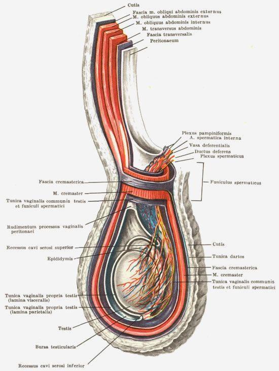
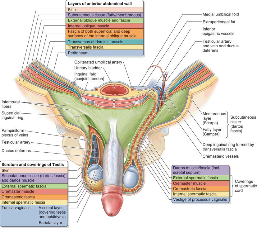
5 The scrotum The scrotum is a cutaneous pouch, containing testis, epididymis and lower part of the spermatic cord (of both sides). Layers of scrotum Skin :- The skin of the scrotum is pigmented, rugose and is marked by a longitudinal median raphe. Superficial fascia of the scrotum:- The fatty layer is absent (to assist heat loss) and is replaced by the subcutaneous dartos muscle formed of involuntary muscle fibers. The muscle is supplied by sympathetic nerve fibers reaching it through the genital branch of the genitofemoral nerve. The muscle aids heat regulation of testis and scrotum. The deep membranous layer of the scrotum is called Colles' fascia. It is continuous superiorly with Scarpa's fascia of the anterior abdominal wall
6 A comparison between layers of scrotum and that of anterior abdominal wall Layers of the anterior abdominal wall Skin Superficial fascia Superficial fatty layer Deep membranous layer (Scarpa's fascia) External oblique muscle Internal oblique muscle Transversus abdominis Transversus fascia Extraperitoneal tissue Peritoneum Layers of the scrotum Skin Superficial fascia Replaced by Dartos muscle Deep membranous layer (Fascia of Colles) External spermatic fascia Cremastric muscle and fascia No corresponding layer Internal spermatic fascia Loose connective tissue Tunica vaginalis around the testis
7
8 Cremaster muscle, Is formed by the lowermost fascicles of the internal oblique muscle arising from the inguinal ligament The cremaster muscle reflexively draws the testis superiorly in the scrotum, particularly in response to cold. It is supplied by genital branch of the genitofemoral nerve Dartos muscle, Smooth muscle of the fat-free subcutaneous tissue of the scrotum (dartos fascia), which inserts into the skin, assisting testicular elevation as it produces contraction of the skin of the scrotum. It is supplied by sympathetic nerve fibres reaching it through the genital branch of the genitofemoral nerve Cremasteric Reflex : Contraction of the cremaster muscle is caused by lightly stroking the skin on the medial aspect of the superior part of the thigh leads to rapid elevation of the testis on the same side Afferent: ilioinguinal nerve Efferent : Genital branch of the genitofemoral nerve
9
10 Blood supply :- Cremastric branch of the inferior epigastric artery Superficial and deep external pudendal branches of femoral artery Scrotal branches of internal pudendal artery. Nerve supply :- Anterior 1/3: Ilioinguinal nerve (L1 dermatome) + genital branch of genitofemoral N. Posterior 2/3: Scrotal branches of pudendal nerve and posterior cutaneous nerve of the thigh (S3 dermatome). Lymphatic drainage :- Superficial inguinal lymph nodes With infraumbilical and perineal skin
11 Testis Testis is the male primary sex organ, suspended in the scrotum by the spermatic cord. The testis develops at the upper part of posterior abdominal wall, then descends into scrotum
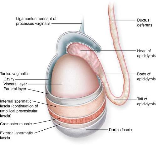
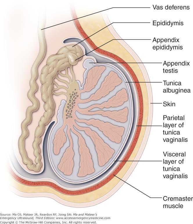
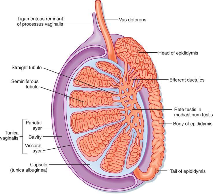
12 Testis has 2 poles, (the upper and lower), 2 borders, (anterior and posterior), and 2 surfaces, (medial and lateral). The epididymis (which is a long coiled duct) forms a cap at the upper pole of the testis, descending down lateral to the posterior border towards its lower pole. Coverings (tunics) of the testis : From outside inwards 1. Tunica vaginalis: It is the lower part of the processus vaginalis of the peritoneum. It is invaginated by the testis from behind, It has parietal and visceral layers with a cavity in between. The tunica vaginalis covers the whole testis except its posterior border. Sinus of epididymis is that part of the cavity of tunica vaginalis which extends between lateral side of testis and the epididymis. 2. Tunica albuginea: It is the tough white fibrous coat which covers the testis all around. 3. Tunica vasculosa: It is formed of vascularized connective tissue, deep to the tunica albuginea and extends between the lobules of the testis
13
14 Structure of the testis The postero-superior part of tunica albuginea is thickened to form the mediastinum testis. Numerous septa pass from the mediastinum to the inner surface of the rest of tunica albuginea dividing the testis into lobules. Each lobule contains 2-3 seminiferous tubules with interstitial cells of Leydig in between the tubules. Near the mediastinum testis, the seminiferous tubules join together to form straight tubules, which enter the mediastinum anastomosing with each other to form a network of tubules called "rete testis" The rete testis gives rise to efferent ductules which emerge from the upper pole of the testis to form head of epididymis.
15

16 Upper end Ant border Lateral surface Lower end Tunica vaginalis Vas deferens Scrotal septum Epididymis Lateral view of right testis Posterior view of right testis
17 The epididymis It is the highly coiled comma shaped tube which is attached to the postero-iateral aspect of the testis. It may act as a reservoir for sperms. Length: In the comma shaped coiled form it is about 1.5 inches long. When it is uncoiled, it measures about 6 meters in length. It has 3 parts Head; forms a cap at upper pole of the testis, to which is connected by efferent ductules. These ductules form head of the epididymis Body; is the intermediate part and is made up of the single coiled tube Tail; is the lower end of the tube and it continues as vas deferens which ascends medial to the epididymis.

18 Arterial blood supply of the testis and epididymis By testicuiar artery, a branch of abdominal aorta at L2 vertebra. It descends on the posterior abdominal wall to reach the deep inguinal ring where it runs in the spermatic cord in the inguinal canal. It supplies epididymis and enters the testis It anastomoses with cremastric artery and artery of the vas. Venous drainage Venous blood from testis and epididymis drain into the pampiniform plexus. It surrounds and accompanies the testicuiar artery up to the superficial inguinal ring. In the inguinal canal, it gives rise to a single testicuiar vein. The right vein ends in the inferior vena cava and the left one ends in left renal vein.
 The")
19 Varicocele A varicocele is a condition in which the veins of the pampiniform plexus are elongated and dilated. It is a common disorder in adolescents and young adults It is more common occurring on the left side because : The right testicular vein joins the low-pressure inferior vena cava (by acute angle) The left vein joins the left renal vein, in which the venous pressure is higher (by right angle) Nerve supply Both testis and epididymis are supplied by sympathetic fibers derived from aortic plexus. It contains afferent sensory (for testicuiar sensation) and efferent (vasomotor). Lymphatic drainage : Into lateral aortic lymph nodes.

20 Thermoregulation of the testis The process of spermatogenesis needs a temperature 2-3 C below the body temperature. This is achieved through the following 3 mechanisms; 1. Cutaneous mechanism; The scrotal skin is very vascular and rich in sweat glands. Fat is absent in its subcutaneous tissue, all aid heat loss. 2.Muscular mechanism: Includes 2 muscles, dartos and cremastric. In cold cremastric muscle elevates the testis near the body, so preventing heat loss. In warm weather, the opposite effects are obtained. 3. Vascular mechanism: The pampiniform plexus aids heat loss by radiation, so helps to maintain low temperature around the testis.

21 The spermatic cord It is a group of structures which meet at deep inguinal ring and traverse the inguinal canal down to posterior border of the testis. Coverings: The spermatic cord is invested by 3 coverings; internal spermatic fascia, cremastric muscle and fascia, and external spermatic fascia. Constituents of the spermatic cord 1. Testicuiar artery (from aorta) 2. Cremastric artery (from inferior epigastric artery) 3. Artery of the vas (from the inferior vesical artery) 4. Vas deferens 5. Pampiniform venous plexus 6. Vestige of processus vaginalis. 7. Genital branch of genitofemoral nerve, 8. Sympathetic plexus around the testicuiar artery and artery of the vas 9. Lymphatics of testis and epididymis ascending to lateral aortic lymph nodes and Loose areolar tissue.
22


23 Torsion of the Testis Torsion of the testis is a rotation of the testis around the spermatic cord. It is often associated with an excessively large tunica vaginalis. The patient complains from severe pain. It is an emergency case, the testicular artery may be occluded, followed by necrosis of the testis.


24 Hydrocele It is an accumulation of fluid within the tunica vaginalis. Haematocele It is an accumulation of blood within the tunica vaginalis.
 and bulb of penis, all are present in the superficial perineal pouch of")

25 Penis It has a root (or attached portion) and a shaft (or free potion). The root is formed of 3 parts; two curura (right and left) and bulb of penis, all are present in the superficial perineal pouch of perineum. The bulb is covered on its outer surface by the bulbospongiosus muscles The shaft is formed of 3 columns of erectile tissue; two corpora cavernosa (right and left) and a median corpus spongiosum. Each crus is attached to the side of the pubic arch and is covered on its outer surface by the ischiocavernosus muscle.
26 The shaft of the penis The body of the penis is essentially composed of three cylinders of erectile tissue enclosed in a tubular sheath of fascia (Buck s fascia). A- The two corpora cavernosa:- They lie dorsally side by side in the shaft of penis. Each is firmly surrounded by fibrous tissue called tunica albuginea which also sends a median septum between the two Followed distally, the corpora cavernosa end in pointed projections within the cap-like glans penis of corpus spongiosum Followed proximally (towards the root of the penis), the two corpora cavernosa diverge from each other and each passes postero laterally to continue as the crus penis, which becomes firmly attached to the everted lip of the ischio pubic ramus The corpora cavernosa contain many irregular cavernous spaces which become filled by blood during erection.

, it remains in the attached to")
27 B- The corpus spongiosum It is median and lies in the ventral surface of the two corpora cavernosa. It is also surrounded by a separate sheath of tunica albuginea Followed distally, it forms glans penis which fits over the distal ends of the corpora cavernosa. The base of the glans penis is called the corona gtandis Followed proximally (towards the root of the penis), it remains in the attached to the inferior surface of the perineal membrane. The corpus spongiosum is traversed by the penile part of the urethra. It also contains cavernous tissue capable of erection.

28 The skin of the penis Followed distally, the skin forms a loose fold called the prepuce or foreskin which covers the glans. The deep layer of this fold is attached to the coronary sulcus of the glans by frenulum Followed proximally, the penile skin is continuous with skin of the scrotum. The fascia of the penis The superficial penile fascia is devoid of fat (like the scrotum) but rich in loose connective tissue to allow free movement of skin over the shaft of penis. Urethral opening frenulum


29 Circumcision is the operation of removing the greater part of the prepuce, or foreskin.

, extends from the symphysis")
30 The ligaments of the penis Fundiform ligament: arise from the lower part of the linea alba, its fibers split to surround the proximal part of the penile shaft to insert into the midline raphe of the scrotum. Suspensory ligament: (deep to the fundiform ligament), extends from the symphysis pubis and blends below with fascia penis.
.")

31 Fundiform ligament Suspensory ligament Arteries of the penis All are branches of internal pudendal artery and all are paired (right and left). Dorsal artery of the penis supplies the skin, fascia, and glans. Deep artery of the penis supplies the corpus cavernosum.with convoluted helicine arteries Artery of the bulb supplies the corpus spongiosum and glans penis
; divides into right and left-")
, passes below symphysis pubis to")
32 Venous drainage By 2 dorsal veins which are superficial and deep; 1. Superficial dorsal vein (superficial to the fascia penis); divides into right and left- Each ends in the corresponding superficial external pudendal vein. 2. Deep dorsal vein of the penis (deep to fascia penis), passes below symphysis pubis to terminate in prostatic venous plexus.
33 Lymph drainage From the penis into superficial inguinal lymph nodes (with the scrotum). From glans penis, lymphatics drain directly to gland of Cloquet in the femoral canal. Nerves of the penis 1. Dorsal nerve of the penis (sensory), is a branch of pudendal nerve, runs lateral to the dorsal artery of the penis 2. Cavernous nerves (autonomic) arise from the inferior hypogastric plexus,parasympathetic fibers (S2,3,4) produce vasodilatation & erection of penis

34 Ductus Deferens (Vas Deferens) It is thick walled muscular tube which springs from the lower end of the epididymis It ends behind the base of the bladder, by joining the duct of the seminal vesicle to form the ejaculatory duct

35 Length : about 45 cm, the same length of the following : a.thoracic duct b.spinal cord c.adult femur d.the distance from the incisor teeth to the cardiac end of the stomach. Function : transmit the spermatozoa from the epididymis to the ejaculatory duct, which in turn opens in the prostatic urethra.
36 Course and Relations of the Vas : Its distal part lies in the scrotum and inguinal canal, its proximal part runs in the pelvis. A. Of its distal part (in the scrotum and inguinal canal: From the tail of the epididymis, it ascends along the posterior aspect of the testis, medial to the epididymis. Then, it ascends in the posterior part of the spermatic cord to traverse the inguinal canal. At the deep inguinal ring, it leaves the cord and curves around the lateral side of the inferior epigastric artery to enter the pelvis. B. Of its proximal part (in the pelvis) : The vas descends posteriorly external to the peritoneum, crossing these structures in the side wall of the pelvis :- external iliac vessels, obliterated umbilical artery, obturator nerve and obturator vessels. Then, it turns medially and crosses the ureter near the superolateral angle of the base of the bladder. Behind the bladder base, it runs superior to, then medial to the seminal vesicle and expands to form ampulla of the vas, below which it narrows to join the duct of the seminal vesicle to form the ejaculatory duct.

37 Vessels of the Vas: Artery of the vas is derived either from the superior or inferior vesical artery. It runs in the spermatic cord and anastomoses with the testicular artery. Veins join the vesical venous plexus. Nerves : are derived from prostatic nerve plexus which comes from the inferior hypogastric plexus. Fibers are mainly sympathetic for the process of ejaculation. Applied Anatomy: bilateral vasectomy is a common operation for male sterilization


38 Seminal vesicles, ejaculatory ducts and bulbourethral glands
39 Seminal Vesicles It is a sacculated tube, about 5 cm long. Site and relations : it lies behind base of the bladder and has the following relations; Anteriorly : it contacts base of the bladder. Posteriorly : it contacts the rectum and rectovesical fascia. Superiorly : its upper end is covered by peritoneum of rectovesical pouch and related to the vas deferens. Medially : ampulla of the vas. Termination : inferiorly, it narrows into a small duct which joins the vas to form ejaculatory duct. Arterial supply : from inferior vesical and middle rectal arteries. Veins : to vesical venous plexus. Nerves: from prostatic nerve plexus (mainly sympathetic). Functions : the seminal vesicle produces an alkaline secretion rich in fructose and mucus. The secretion is added to the spermatozoa in ejaculation. Applied Anatomy : The seminal vesicles when enlarged, could be felt on rectal examination. Abscess in he seminal vesicle may rupture into the peritoneal cavity.

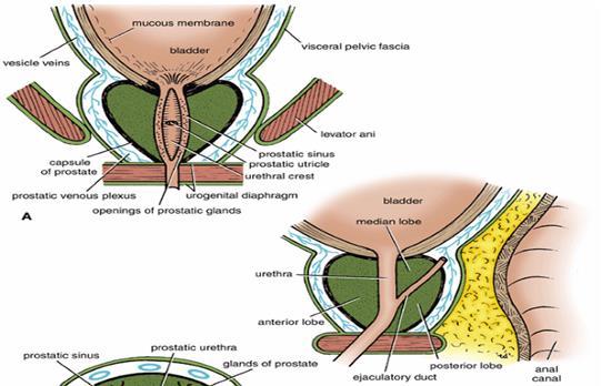
40 Ejaculatory Ducts : Each is about 2 cm long, formed by union of the ductus deferens and the duct of the seminal vesicle. The two ducts run antero-inferiorly between median and posterior lobes of the prostate along the sides of the prostatic utricle to open on the seminal colliculus of the prostatic urethra. Bulbourethral Glands : These small glands lie lateral to the membranous urethra in the deep perineal pouch Each gives rise to a long duct (3 cm) which pierces the perineal membrane to open on the floor of the spongy part of the urethra. Blood supply: by artery of the bulb of the penis. It is innervated by prostatic nerve plexus Function : It secretes a number of substances added to the spermatozoa during ejaculation.

41 The Prostate It is an accessory gland of male reproductive system, which surrounds the prostatic urethra
42 Site : it lies in the lower part of the lesser pelvis behind the inferior border of the pubic symphysis in front of the rectum, below neck of the bladder. Shape and Description: It simulates an inverted cone which has a base (directed superiorly); an apex (directed inferiorly), four surfaces: anterior, posterior, and two inferolateral surfaces. 1- Base of the prostate : It is directed upwards, separated from the bladder by a groove contains part of the prostatic venous plexus. It is pierced by the urethra. 2- Apex of the prostate: Is directed downwards It rests on the perineal membrane (roof of the deep perineal pouch). The urethra emerges from the prostate anterosuperior to the apex.
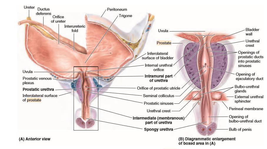
43 3-Anterior surface: It is convex and lies behind the lower part of the symphysis pubis. Its upper part is connected to the pubic bodies by puboprostatic ligaments. 4- Posterior surface: It is nearly fiat and is related to ampulla of the rectum separated from it by rectovesical fascia (fascia of Denonvilliers) The prostate is easily palpated by a finger in the rectum (the rectovesical fascia is attached above to the floor of rectovesical pouch and below to the perineal body). Near its upper border, this surface is pierced by the two ejaculatory ducts. 5- Right and left inferolateral surfaces: Are convex and related to levator prostatae parts of levator ani muscle. Structures that traverse the prostate : Prostatic urethra. The two ejaculatory ducts descend anteroinferiorly to open in the prostatic urethra The gland contains the utricle.


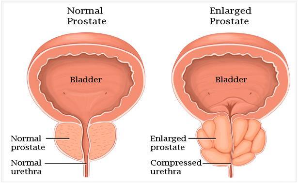
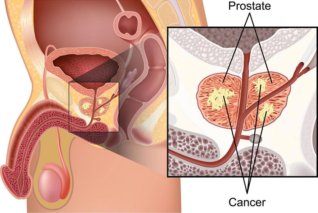
44 Lobes of the prostate: By means of the prostatic urethra and the two ejaculatory ducts, the prostate is divided into five lobes; Anterior lobe (isthmus): lies in front of the prostatic urethra. It consists a fibromuscular tissue with little glandular tissue. Right and left lateral lobes : one on each of the prostatic urethra. They are the usual sites for the senile enlargement of the prostate. Posterior lobe : lies behind the prostatic urethra, but below the two ejaculatory ducts. It is the usual site for cancer prostate. Median lobe: Lies between the upper part of prostatic urethra and the two ejaculatory ducts. After middle age, it produces uvula vesicae, in the lower part of the bladder trigone. It is also a common site for senile enlargement of the prostate. (BPH) The uvula vesicae may obstruct the flow of urine at the internal urethral meatus.
45
46
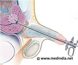
47 Transurethral resection of the prostate (TURP)
48 The prostatic capsules: 1. Inner true capsule : it is fibromuscular in structure. 2. Outer false capsule (prostatic sheath): it is a condensed visceral pelvic fascia. Between the two capsules, lies the prostatic venous plexus. Blood Supply of the Prostate: Arteries are derived from inferior vesical and middle rectal arteries. Venous drainage : the veins form the prostatic venous plexus which has the following features : It is embedded between the two capsules of the prostate. It is present only in front and sides of the gland Superiorly, it is continuous with the vesical venous plexus. Anteriorly : it receives the deep dorsal vein of penis. Posterolaterally : the plexus is drained to the internal iliac veins which in turn communicates with the internal vertebral venous plexuses by the lateral sacral veins. These veins are valveless and responsible for spread of cancer prostate to lumbar vertebrae Acid phosphatase and Prostate-Specific Antigen (PSA) are markedly elevated in prostatic diseases especially carcinoma Lymphatic Drainage: to internal, external iliac lymph nodes. Nerve Supply: by prostatic nerve plexus derived from the inferior hypogastric plexus.


49 BPH. The enlarged prostate indents and elevates the bladder floor. CT :Median lobe. This enlarged portion of the prostate produces a polypoid midline mass in the bladder lumen.

50
أحمد رواجبة- محمود الحربي- أحمد السالمان-
 -6 أحمد رواجبة- محمود الحربي- أحمد السالمان- 1 P a g e The Male Reproductive System The male genital system structures are divided into: Internal structures: 1- Prostate 3-Ejaculatory ducts External structures:
Inguinal Canal. It is an oblique passage through the lower part of the anterior abdominal wall. Present in both sexes
 Inguinal canal Inguinal Canal It is an oblique passage through the lower part of the anterior abdominal wall Present in both sexes It allows structures to pass to and from the testis to the abdomen in
M. Al-Mohtaseb. Tala Saleh. Faisal Nimri
 4 5 M. Al-Mohtaseb Tala Saleh Faisal Nimri Inguinal Hernia - An abdominal hernia is the protrusion of part of the abdominal content beyond the normal confines of the abdominal wall through weak points
Male Reproductive System. Dr Maan Al-Abbasi PhD, MSc, MBChB, MD
 Male Reproductive System Dr Maan Al-Abbasi PhD, MSc, MBChB, MD Learning Objectives 1. Describe the General Anatomy of the Male Reproductive System 2. Identify the structures that are related to the prostate.
REPRODUCTIVE SYSTEM By Dr.Ahmed Salman
 The University Of Jordan Faculty Of Medicine Anatomy Department REPRODUCTIVE SYSTEM By Dr.Ahmed Salman Assistant Professor of Anatomy &embryology Perineum It is the diamond-shaped lower end of the trunk
Male Reproductive System
 Male Reproductive System Please view our Editing File before studying this lecture to check for any changes. Color Code Important Doctors Notes Notes/Extra explanation Objectives At the end of the lecture,
ESUR SCROTAL AND PENILE IMAGING WORKING GROUP MULTIMODALITY IMAGING APPROACH TO SCROTAL AND PENILE PATHOLOGIES 2ND ESUR TEACHING COURSE
 ESUR SCROTAL AND PENILE IMAGING WORKING GROUP MULTIMODALITY IMAGING APPROACH TO SCROTAL AND PENILE PATHOLOGIES 2ND ESUR TEACHING COURSE NORMAL ANATOMY OF THE SCROTUM MICHAEL NOMIKOS M.D. F.E.B.U. UROLOGICAL
The Female and Male External Genitalia. Prof Oluwadiya KS
 The Female and Male External Genitalia Prof Oluwadiya KS www.oluwadiya.com Anatomy of the female external genitalia This consists of : The vulva which is made up of: o The clitoris o Vestibular apparatus
Rama Nada. - Ensherah Mokheemer. - Ahmed salman. 1 P a g e
 - 5 - Rama Nada - Ensherah Mokheemer - Ahmed salman 1 P a g e We will continue talking about the urinary bladder The ligaments of the bladder: 1-Median umbilical ligament: Continuous with apex of the bladder
Human Anatomy Unit 3 REPRODUCTIVE SYSTEM
 Human Anatomy Unit 3 REPRODUCTIVE SYSTEM In Anatomy Today Male Reproductive System Gonads = testes primary organ responsible for sperm production development/maintenan ce of secondary sex characteristics
حسام أبو عوض. -Dr. Mohammad Muhtasib. 1 P a g e
 5 حسام أبو عوض - -Dr. Mohammad Muhtasib 1 P a g e There are two types of inguinal hernia: direct and indirect. Hernia: protrusion of the small intestine or the greater omentum of the intra-abdominal organs
Anatomy & Physiology: Testis Penis. Hosam Serag ST7 Urology University Hospital of Wales
 Anatomy & Physiology: Testis Penis Hosam Serag ST7 Urology University Hospital of Wales Testis Dimensions: 4 to 5 cm long 3 cm wide 2.5 cm deep volume of 30 ml Prader Orchidometer Layers Visceral tunica
Yes, cranially with ovarian, caudally with vaginal. Yes, with uterine artery (collateral circulation between abdominal +pelvic source)
") Blood supply to internal female genitalia: uterine Internal iliac Sup. large branch: uterus, inf. Small branch: cervix+ sup. Vagina Yes, cranially with ovarian, caudally with vaginal Medially in base of
Blood supply to internal female genitalia: uterine Internal iliac Sup. large branch: uterus, inf. Small branch: cervix+ sup. Vagina Yes, cranially with ovarian, caudally with vaginal Medially in base of
DISSECTION 8: URINARY AND REPRODUCTIVE SYSTEMS
 8546d_c01_1-42 6/25/02 4:32 PM Page 38 mac48 Mac 48: 420_kec: 38 Cat Dissection DISSECTION 8: URINARY AND REPRODUCTIVE SYSTEMS Typically, the urinary and reproductive systems are studied together, because
The Repr duct ve System. Function: producing offspring
 The Repr duct ve System Function: producing offspring Anatomy of male reproductive system Location: The reproductive organs are classified as external and internal genitalia. The external genitalia are
Gross Anatomy of the Urinary System
 Gross Anatomy of the Urinary System Lecture Objectives Overview of the urinary system. Describe the external and internal anatomical structure of the kidney. Describe the anatomical structure of the ureter
Urinary Bladder. Prof. Imran Qureshi
 Urinary Bladder Prof. Imran Qureshi Urinary Bladder It develops from the upper end of the urogenital sinus, which is continuous with the allantois. The allantois degenerates and forms a fibrous cord in
ABDOMINAL WALL & RECTUS SHEATH
 ABDOMINAL WALL & RECTUS SHEATH Learning Objectives Describe the anatomy, innervation and functions of the muscles of the anterior, lateral and posterior abdominal walls. Discuss their functional relations
Primary sex organs (gonads): testes and ovaries. Accessory reproductive organs: ducts, glands, and external genitalia
: testes and ovaries. Accessory reproductive organs: ducts, glands, and external genitalia") Male Reproductive System Primary sex organs (gonads): testes and ovaries Produce sex cells (gametes) Secrete steroid sex hormones Androgens (males) Estrogens and progesterone (females) Accessory reproductive
Male Reproductive System Primary sex organs (gonads): testes and ovaries Produce sex cells (gametes) Secrete steroid sex hormones Androgens (males) Estrogens and progesterone (females) Accessory reproductive
NORMAL ANATOMY OF THE PENIS
 NORMAL ANATOMY OF THE PENIS IOANNIS VARKARAKIS ASOSCIATE PROFESSOR OF UROLOGY 2 ND DEPT OF UROLOGY NATIONAL & KAPODISTRIAN UNIVERSITY OF ATHENS PENILE GROSS ANATOMY 3 ERECTILE COLUMNS TWO CORPORA CAVERNOSA
The Scrotum & Testes Prof. Dr. Imran Qureshi
 The Scrotum & Testes Prof. Dr. Imran Qureshi The Scrotum It is a cutaneous pouch of the anterior abdominal wall. Most layers of the abdominal wall are represented in its structure. It contains the testes
The Male and Female Internal Genitalia. Dr Oluwadiya Kehinde
 The Male and Female Internal Genitalia Dr Oluwadiya Kehinde www.oluwadiya.com Overview The reproductive role of the male is to produce sperm, deliver them to the female Primary sex organs are the gonads
Inferior Pelvic Border
 Pelvis + Perineum Pelvic Cavity Enclosed by bony, ligamentous and muscular wall Contains the urinary bladder, ureters, pelvic genital organs, rectum, blood vessels, lymphatics and nerves Pelvic inlet (superior
Perineum. done by : zaid al-ghnaneem
 Perineum done by : zaid al-ghnaneem Hello everyone, this sheet will talk about 2 nd Lecture which is Perineum but there are some slides and info from 1 st Lecture. Everything included Slides + Pics Let
Male Reproductive System Dr. Gary Mumaugh
 Male Reproductive System Dr. Gary Mumaugh Reproductive System Basics Primary sex organs (gonads) testes in males, ovaries in females Gonads produce sex cells called gametes (gametes means spouses) and
THE ABDOMEN SUPRARENAL GLANDS KIDNEY URETERS URINARY BLADDER
 THE ABDOMEN SUPRARENAL GLANDS KIDNEY URETERS URINARY BLADDER THE SUPRARENAL GLANDS The suprarenal (adrenal) glands lie immediately superior and slightly anterior to the upper pole of either kidney. Golden
Dana Alrafaiah. - Amani Nofal. - Ahmad Alsalman. 1 P a g e
 - 2 - Dana Alrafaiah - Amani Nofal - Ahmad Alsalman 1 P a g e This lecture will discuss five topics as follows: 1- Arrangement of pelvic viscera. 2- Muscles of Pelvis. 3- Blood Supply of pelvis. 4- Nerve
Reproductive System. Where it all begins
 Reproductive System Where it all begins When it comes the reproductive anatomy of my gender, I would rate my knowledge (1 very poor, 10 excellent) When it comes the reproductive anatomy of the opposite
Pelvis MCQs. Block 1. B. Reproductive organs. C. The liver. D. Urinary bladder. 1. The pelvic diaphragm includes the following muscles: E.
 Pelvis MCQs Block 1 1. The pelvic diaphragm includes the following muscles: A. The obturator internus B. The levator ani C. The coccygeus D. The external urethral sphincter E. The internal urethral sphincter
GI anatomy Lecture: 2 د. عصام طارق
 GI anatomy Lecture: 2 د. عصام طارق Objectives: To define rectus sheath. To describe anatomy of inguinal canal. To relates types of inguinal hernia to the region. To explore spermatic cord. Rectus Abdominis
The posterior abdominal wall. Prof. Oluwadiya KS
 The posterior abdominal wall Prof. Oluwadiya KS www.oluwadiya.sitesled.com Posterior Abdominal Wall Lumbar vertebrae and discs. Muscles opsoas, quadratus lumborum, iliacus, transverse, abdominal wall
The Reproductive System
 PowerPoint Lecture Slide Presentation by Patty Bostwick-Taylor, Florence-Darlington Technical College The Reproductive System 16PART A The Reproductive System Gonads primary sex organs Testes in males
The Reproductive System
 Essentials of Human Anatomy & Physiology Elaine N. Marieb Seventh Edition Chapter 16 The Reproductive System Slides 16.1 16.20 Lecture Slides in PowerPoint by Jerry L. Cook The Reproductive System Gonads
The Reproductive System
 16 PART A The Reproductive System PowerPoint Lecture Slide Presentation by Jerry L. Cook, Sam Houston University ESSENTIALS OF HUMAN ANATOMY & PHYSIOLOGY EIGHTH EDITION ELAINE N. MARIEB The Reproductive
ANATOMY OF PELVICAYCEAL SYSTEM -DR. RAHUL BEVARA
 1 ANATOMY OF PELVICAYCEAL SYSTEM -DR. RAHUL BEVARA 2 KIDNEY:ANATOMY OVERVIEW Kidneys are retroperitoneal, in posterior abdominal region, extending from T12 L3 Bean-shaped Right kidney is lower than left
Femoral Triangle and Adductor Canal. Dr. Heba Kalbouneh Associate Professor of Anatomy and Histology
 Femoral Triangle and Adductor Canal Dr. Heba Kalbouneh Associate Professor of Anatomy and Histology Femoral Triangle and Adductor Canal Femoral triangle Is a triangular depressed area located in the upper
PELVIS II: FUNCTION TABOOS (THE VISCERA) Defecation Urination Ejaculation Conception
 Defecation Urination Ejaculation Conception") PELVIS II: FUNCTION TABOOS (THE VISCERA) Defecation Urination Ejaculation Conception REVIEW OF PELVIS I Pelvic brim, inlet Pelvic outlet True pelvis-- --viscera Tilt forward Mid-sagital views-- --how the
PELVIS II: FUNCTION TABOOS (THE VISCERA) Defecation Urination Ejaculation Conception REVIEW OF PELVIS I Pelvic brim, inlet Pelvic outlet True pelvis-- --viscera Tilt forward Mid-sagital views-- --how the
2. List the 8 pelvic spaces: list one procedure or dissection which involves entering that space.
 Name: Anatomy Quiz: Pre / Post 1. In making a pfannensteil incision you would traverse through the following layers: a) Skin, Camper s fascia, Scarpa s fascia, external oblique aponeurosis, internal oblique
Ureters, Urinary Bladder & Urethra
 Ureters, Urinary Bladder & Urethra Please check our Editing File هذا العمل ال يغني عن المصدر األساسي للمذاكرة Lecture 2 } و م ن ي ت و ك ع ل ا لل ه ف ه و ح س ب ه { Objectives o Describe the course of ureter
MALE REPRODUCTIVE SYSTEM
 MALE REPRODUCTIVE SYSTEM 1. The male reproductive system is made up of the following structures, EXCEPT: a. prostate; b. testicle; c. spermatic ducts; d. vestibular bulbs; e. seminal vesicles. 2.The testicle:
MALE REPRODUCTIVE SYSTEM
 MALE REPRODUCTIVE SYSTEM The male reproductive system consists of primary sex organs (testes) and secondary or accessory sex organs. The secondary organs consist of a series of genital ducts (ductules
STRUCTURAL BASIS OF MEDICAL PRACTICE EXAMINATION 3. October 17, 2014
 STRUCTURAL BASIS OF MEDICAL PRACTICE EXAMINATION 3 October 17, 2014 PART l. Answer in the space provided. (12 pts) 1. Identify the structures. (2 pts) A. B. A B C. D. C D 2. Identify the structures. (2
Benha University. Faculty of Medicine. Anatomy Department Course code (MED 0701) Model answer of Anatomy examination. (Abdomen,Pelvis and Thorax)
 Model answer of Anatomy examination. (Abdomen,Pelvis and Thorax)") 1 Benha University Faculty of Medicine Anatomy Department Course code (MED 0701) Model answer of Anatomy examination (Abdomen,Pelvis and Thorax) 1 st year 2 nd term Date :18 /5 /2013 2 I-Short account
1 Benha University Faculty of Medicine Anatomy Department Course code (MED 0701) Model answer of Anatomy examination (Abdomen,Pelvis and Thorax) 1 st year 2 nd term Date :18 /5 /2013 2 I-Short account
The front of the thigh. Dr.Amjad shatarat
 The front of the thigh Femoral triangle (Scarpa s triangle) Is a triangular depressed area located in the upper part of the medial aspect of the thigh immediately below the inguinal ligament. Superiorly:
Systematic Anatomy. Shanghai Medical College,Fudan University. Dr.Hongqi Zhang ( 张红旗 )
") Systematic Anatomy Department of Anatomy,Histology & Embryology Shanghai Medical College,Fudan University Dr.Hongqi Zhang ( 张红旗 ) Email: zhanghq58@yahoo.com.cn Office: Building 9,Room308, 54237151-9308
Systematic Anatomy Department of Anatomy,Histology & Embryology Shanghai Medical College,Fudan University Dr.Hongqi Zhang ( 张红旗 ) Email: zhanghq58@yahoo.com.cn Office: Building 9,Room308, 54237151-9308
STRUCTURAL BASIS OF MEDICAL PRACTICE EXAMINATION 3. October 16, 2015
 STRUCTURAL BASIS OF MEDICAL PRACTICE EXAMINATION 3 October 16, 2015 PART l. Answer in the space provided. (12 pts) 1. Identify the structures. (2 pts) A. B. A B C. D. C D 2. Identify the structures. (2
Table 2. First Generated List of Expert Responses. Likert-Type Scale. Category or Criterion. Rationale or Comments (1) (2) (3) (4)
 (2) (3) (4)") Table 2. First Generated List of Expert Responses. Likert-Type Scale Category or Criterion Anatomical Structures and Features Skeletal Structures and Features (1) (2) (3) (4) Rationale or Comments 1. Bones
Table 2. First Generated List of Expert Responses. Likert-Type Scale Category or Criterion Anatomical Structures and Features Skeletal Structures and Features (1) (2) (3) (4) Rationale or Comments 1. Bones
ANATYOMY OF The thigh
 ANATYOMY OF The thigh 1- Lateral cutaneous nerve of the thigh Ι) Skin of the thigh Anterior view 2- Femoral branch of the genitofemoral nerve 5- Intermediate cutaneous nerve of the thigh 1, 2 and 3 are
Scrotum Kacey Morrison Amanda Baxter Sabrina Tucker July 18, 2006 SCROTUM
 Scrotum Kacey Morrison Amanda Baxter Sabrina Tucker July 18, 2006 SCROTUM 1) Other Names: Scrotum None Testicles Testes (Curry Tempkin, p. 236, 2/3/2) Ductus deferens spermatic cord (Tempkin, p. 279, Anatomy
The Anterolateral Abdominal Wall By Prof. Dr. Muhammad Imran Qureshi
 1 P age The Anterolateral Abdominal Wall By Prof. Dr. Muhammad Imran Qureshi Introduction The abdomen is the region of the trunk located between the thorax and the pelvis. It includes the anterolateral
Chapter 22 Reproductive Systems. Male Reproductive Organs. Male Reproductive Organs. Specialized to produce, maintain the male sex cells (sperm)
") Chapter 22 Reproductive Systems Male reproductive organs 1 Male Reproductive Organs posterior view 2 Male Reproductive Organs Specialized to produce, maintain the male sex cells (sperm) Transport these
Chapter 22 Reproductive Systems Male reproductive organs 1 Male Reproductive Organs posterior view 2 Male Reproductive Organs Specialized to produce, maintain the male sex cells (sperm) Transport these
Biology 224 Human Anatomy and Physiology II Week 9; Lecture 2; Wednesday Stuart Sumida. Development and Structure, of the Reproductive System
 Biology 224 Human Anatomy and Physiology II Week 9; Lecture 2; Wednesday Stuart Sumida Development and Structure, of the Reproductive System Don t forget the relationships of the structures of the layers
ANATYOMY OF The thigh
 ANATYOMY OF The thigh 1- Lateral cutaneous nerve of the thigh Ι) Skin of the thigh Anterior view 2- Femoral branch of the genitofemoral nerve 5- Intermediate cutaneous nerve of the thigh 1, 2 and 3 are
GI module Lecture: 9 د. عصام طارق. Objectives:
 GI module Lecture: 9 د. عصام طارق Objectives: To list structures forming posterior abdominal wall. To follow aorta & its main branches. To describe IVC & its main tributaries. To list nerves of posterior
Lab #9: Kidney: Gross Anatomy & Histology
 Name Date Lab #9: Kidney: Gross Anatomy & Histology Lab #10: Male Reproductive System: Human Models & Histology Lab #11: Female Reproductive System: Human Models & Histology Stuff to Know Dr. L. Bacha
BLOCK IV: OFFICIAL BODY PARTS LIST FOR ANTERIOR ABDOMINAL WALL AND ABDOMINAL CONTENTS
 BLOCK IV: OFFICIAL BODY PARTS LIST FOR ANTERIOR ABDOMINAL WALL AND ABDOMINAL CONTENTS External oblique muscle Muscular portion Aponeurotic portion Superficial inguinal ring Lateral (inferior) crus Medial
The Thoracic wall including the diaphragm. Prof Oluwadiya KS
 The Thoracic wall including the diaphragm Prof Oluwadiya KS www.oluwadiya.com Components of the thoracic wall Skin Superficial fascia Chest wall muscles (see upper limb slides) Skeletal framework Intercostal
Perineum. Dept. of Human Anatomy Zhou Hong Ying
 Perineum Dept. of Human Anatomy Zhou Hong Ying OUTLINE Subdivision The Layers Urogenital Diaphragm Main Structures inside Superficial & Deep Perineal Spaces Ischioanal Fossa Perineum A narrow region Urogenital
Abdomen: Introduction. Prof. Oluwadiya KS
 Abdomen: Introduction Prof. Oluwadiya KS www.oluwadiya.com Abdominopelvic Cavity Abdominal Cavity Pelvic Cavity Extends from the inferior margin of the thorax to the superior margin of the pelvis and the
Aniko Szabo Hill 1 of 12
 Common Function: produce offsprings endocrine glands: sex hormone production Primary organs or gonads: sex cell or gametes production Secondary or accessory organs: glands nourish gametes ducts transport
The Urinary System Pearson Education, Inc.
 26 The Urinary System Introduction The urinary system does more than just get rid of liquid waste. It also: Regulates plasma ion concentrations Regulates blood volume and blood pressure Stabilizes blood
Group of students. - Rawan almujabili د. محمد المحتسب - 1 P a g e
 - 14 - Group of students - Rawan almujabili د. محمد المحتسب - 1 P a g e Nerves of the posterior abdominal wall The spinal cord gives off spinal nerves between the vertebrae. In the abdomen, through the
Pelvis Perineum MCQs. Block 1.1. A. Urinary bladder. B. Rectum. C. Reproductive organs. D. The thigh
 Pelvis Perineum MCQs Block 1.1 1. The pelvic diaphragm includes the following muscles: A. The coccygeus B. The levator ani C. The external urethral sphincter D. The internal urethral sphincter E. The obturator
Nerves on the Posterior Abdominal Wall
 Nerves on the Posterior Abdominal Wall Lumbar Plexus The lumbar plexus, which is one of the main nervous pathways supplying the lower limb, is formed in the psoasmuscle from the anterior ramiof the upper
The Male Reproductive System
 The Male Reproductive System YONG-MEI CHEN ( 陈咏梅 ) Dept. of Anatomy, Histology & Embryology Peking Union Medical College Tel:69156461 E-mail address: pumc_he@126.com Content Spermatogenesis Spermiogenesis
musculoskeletal system anatomy nerves of the lower limb 1 done by: dina sawadha & mohammad abukabeer
 musculoskeletal system anatomy nerves of the lower limb 1 done by: dina sawadha & mohammad abukabeer What is the importance of plexuses? plexuses provides us the advantage of a phenomenon called convergence
Borders of the Abdomen
 Abdominal wall Borders of the Abdomen Abdomen is the region of the trunk that lies between the diaphragm above and the inlet of the pelvis below Borders Superior: Costal cartilages 7-12. Xiphoid process:
Bony ypelvis. Composition: formed by coccyx, and their articulations Two portions
 Pelvis Bony ypelvis Composition: formed by paired hip bones, sacrum, coccyx, and their articulations Two portions Greater pelvis Lesser pelvis Terminal line ( pelvic inlet): formed by promontory of sacrum,
Penis and Prostate. Holly White Jennifer Zang September 7, Penis and Prostate. 1) Other Names None
 Other Names None") Penis and Prostate Penis and Prostate Holly White Jennifer Zang September 7, 2006 1) Other Names None 2) Definition/ Location The prostate is a doughnut-like gland that lies inferior to the urinary bladder
Penis and Prostate Penis and Prostate Holly White Jennifer Zang September 7, 2006 1) Other Names None 2) Definition/ Location The prostate is a doughnut-like gland that lies inferior to the urinary bladder
Abdomen. Retroperitoneal space
 Abdomen. Retroperitoneal space Abdominal cavity The space bounded by: Anterolateral abdominal wall Posterior abdominal wall Diaphragm Pelvic walls and pelvic floor. Subdivided into: True abdominal cavity
The functional anatomy of the urinary system. Human Anatomy Department Dr. Anastasia Bendelic
 The functional anatomy of the urinary system Human Anatomy Department Dr. Anastasia Bendelic Plan Development of the kidneys and their abnormalities Development of the urinary ways and their abnormalities
SUBJECTS 2nd year, 1st semester I. 1. Primitive gut - limits, derivatives 2. Foregut -limits, evolution, derivatives 3. Midgut -limits, evolution,
 SUBJECTS 2nd year, 1st semester I. 1. Primitive gut - limits, derivatives 2. Foregut -limits, evolution, derivatives 3. Midgut -limits, evolution, derivatives 4. Hindgut- limits, evolution, derivatives
ANATYOMY OF The thigh
 ANATYOMY OF The thigh 1- Lateral cutaneous nerve of the thigh Ι) Skin of the thigh Anterior view 2- Femoral branch of the genitofemoral nerve 1, 2 and 3 are From the lumber plexus 5- Intermediate cutaneous
Anatomy of the Large Intestine
 Large intestine Anatomy of the Large Intestine 2 Large Intestine Extends from ileocecal valve to anus Length = 1.5-2.5m = 5 feet Regions Cecum = 2.5-3 inch Appendix= 3-5 inch Colon Ascending= 5 inch Transverse=
Lab Schedule for Rest of Semester
 Laboratory 9 Cat Dissection II Respiratory Urinary/Reproductive Systems Lab Schedule for Rest of Semester Cat dissection labs Dissection II (today) Respiratory (Ex. 57 in Hole) Human Reproductive Systems
Histology of Male Reproductive System
 Histology of Male Reproductive System Lecture Objectives Describe the histological features of the male reproductive system Male Reproductive System The male structures of reproduction include the: testes,
The Male Reproductive System
 The Male Reproductive System The male reproductive system Testes Genital ducts Accessory sex glands: seminal vesicles prostate bulbourethral glands External genitalia: penis Structure of the Testis Tunica
Jordan University Faculty Of Medicine. Breast. Dr. Ahmed Salman. Assistant professor of anatomy & embryology
 Jordan University Faculty Of Medicine Breast Dr. Ahmed Salman Assistant professor of anatomy & embryology The breasts are specialized accessory glands of the skin that secretes milk. They are situated
Urinary System. Chapter 17 7/19/11. Introduction
 7/19/11 Chapter 17 Urinary System Introduction A. The urinary system consists of two kidneys that filter the blood, two ureters, a urinary bladder, and a urethra to convey waste substances to the outside.
Anatomy Lecture Notes Chapter 24
 primary sex organs = gonads produce gametes secrete hormones that control reproduction secondary sex organs = accessory structures Development and Differentiation A. gonads develop from mesoderm starting
Male Reproductive System
 Male Reproductive System organs that function in: gamete and hormone production not all in abdominal cavity paired testicles = controlled by LH & FSH duct systems accessory glands Testis: Gross Histology
Gross anatomy of the urinary system. Done by : razan krishan. slide in bold and book in green
 Gross anatomy of the urinary system Done by : razan krishan slide in bold and book in green Kidneys, ureters, urinary bladder & urethra Urine flows from each kidney, down its ureter to the bladder and
Pelvic Angiogram - Male
 Pelvic Angiogram - Male Common iliac artery Internal iliac artery Lateral sacral artery Iliolumbar artery Posterior trunk of internal iliac artery Superior gluteal artery Internal pudendal artery External
[ANATOMY #12] April 28, 2013
![[ANATOMY #12] April 28, 2013](/thumbs/86/93473883.jpg "[ANATOMY #12] April 28, 2013") Sympathetic chain : Sympathetic chain is each of the pair of ganglionated longitudinal cords of the sympathetic nervous system; extend from level of atlas (base of skull) till coccyx. It is paravertebral
Sympathetic chain : Sympathetic chain is each of the pair of ganglionated longitudinal cords of the sympathetic nervous system; extend from level of atlas (base of skull) till coccyx. It is paravertebral
Pancreas & Biliary System. Dr. Vohra & Dr. Jamila
 Pancreas & Biliary System Dr. Vohra & Dr. Jamila 1 Objectives At the end of the lecture, the student should be able to describe the: Location, surface anatomy, parts, relations & peritoneal reflection
Lumbar Plexus. Ventral rami L1 L4 Supplies: Major nerves.. Abdominal wall External genitalia Anteromedial thigh
 Lower Limb Nerves Lectures Objectives Describe the structure and relationships of the plexuses of the lower limb. Describe the course, relationships and structures supplied for the major nerves of the
Anatomy of the Body for Piercers
 Nipples are devoid of Raised structures on the areolae are Montgomery glands or tubercles, or areolar glands Normal variation Provide lubrication during breastfeeding Best to avoid piercing them Hair follicles
The abdominal Esophagus, Stomach and the Duodenum. Prof. Oluwadiya KS
 The abdominal Esophagus, Stomach and the Duodenum Prof. Oluwadiya KS www.oluwadiya.com Viscera of the abdomen Abdominal esophagus: Terminal part of the esophagus The stomach Intestines: Small and Large
4.05 Remember the structures of the reproductive system
 4.05 Remember the structures of the reproductive system Scrub In The external area between the vulva and the anus is the : a. Cervix b. Endometrium c. Perineum d. Vagina What structure connects the testes
The Reproductive System PowerPoint Lecture Presentations prepared by Steven Bassett Southeast Community College Lincoln, Nebraska
 27 The Reproductive System PowerPoint Lecture Presentations prepared by Steven Bassett Southeast Community College Lincoln, Nebraska Introduction The reproductive system is designed to perpetuate the species
Biology Human Anatomy Abdominal and Pelvic Cavities
 Biology 351 - Human Anatomy Abdominal and Pelvic Cavities Please place your name and I.D. number on the back of the last page of this exam. You must answer all questions on this exam. Because statistics
THYROID & PARATHYROID. By Prof. Saeed Abuel Makarem & Dr. Sanaa Al-Sharawy
 THYROID & PARATHYROID By Prof. Saeed Abuel Makarem & Dr. Sanaa Al-Sharawy 1 OBJECTIVES By the end of the lecture, the student should be able to: Describe the shape, position, relations and structure of
Anatomy of the Thorax
 Anatomy of the Thorax A) THE THORACIC WALL Boundaries Posteriorly by the thoracic part of the vertebral column Anteriorly by the sternum and costal cartilages Laterally by the ribs and intercostal spaces
Abdominal muscles. Subinguinal hiatus and ingiunal canal. Femoral and adductor canals. Neurovascular system of the lower limb. Sándor Katz M.D.,Ph.D.
 Abdominal muscles. Subinguinal hiatus and ingiunal canal. Femoral and adductor canals. Neurovascular system of the lower limb. Sándor Katz M.D.,Ph.D. External oblique muscle Origin: outer surface of the
4.05 Remember the structures of the reproductive system
 4.05 Remember the structures of the reproductive system 4.05 Remember the structures of the reproductive system Essential question What are the structures of the reproductive system? 2 Structures of the
MALE REPRODUCTIVE SYSTEM
 1 MALE REPRODUCTIVE SYSTEM SCPA 602 Anatomical Basis for Pathological Study Updated: 20.09.2018 Lect. Nisamanee Charoenchon, PhD nisamanee.cha@mahidol.ac.th Department of Pathobiology, Mahidol University
Anatomy of the renal system. Professor Nawfal K. Al-Hadithi
 Anatomy of the renal system Professor Nawfal K. Al-Hadithi Objectives To describe the posterior abdominal wall To identify the main anatomical landmarks of the kidneys & ureters To describe the suprarenal
Male Reproductive Structures I. Overview A. Main functions: 1. Produce a haploid male gamete (sperm) 2. Deposit sperm in the female so fertilization
 2. Deposit sperm in the female so fertilization") Male Reproductive Structures I. Overview A. Main functions: 1. Produce a haploid male gamete (sperm) 2. Deposit sperm in the female so fertilization may occur! A. Scrotum 1. Muscular pouch that holds the
Male Reproductive Structures I. Overview A. Main functions: 1. Produce a haploid male gamete (sperm) 2. Deposit sperm in the female so fertilization may occur! A. Scrotum 1. Muscular pouch that holds the
Individually, humans don t need to reproduce to survive. But to survive as a species,
 Chapter 13 Why Ask Y?: The Male Reproductive System In This Chapter Explaining the parts of male reproduction Understanding meiosis and what happens to chromosomes Individually, humans don t need to reproduce
Biomechanics. and Functional Anatomy. of Human Male Genitalia. For designers and creators of biomimetic androids, dolls and robots
 Biomechanics and Functional Anatomy of Human Male Genitalia For designers and creators of biomimetic androids, dolls and robots The Penis The shaft or body of the penis is formed principally by a fused