Pelvic Pain in the Pediatric Patient Susan D. John, M.D.
|
|
|
- Magnus Strickland
- 5 years ago
- Views:
Transcription

1 Pelvic Pain in the Pediatric Patient Susan D. John, M.D. RSNA 2012 Patients First
2 Objectives After attending this presentation, participants will be able to: Understand the common congenital and acquired conditions that may cause acute pelvic pain in children Recognize unusual or confusing findings in these conditions on US, CT, or MRI Design imaging protocols for children with pelvic pain that minimizes radiation exposure and provides prompt and accurate diagnoses
3 Question #1 What percentage of your practice involves children < 16 years of age? % % % 4. Greater than 75%
4 Challenges Pelvic Pain in Children Common pathologies differ from causes of pelvic pain in adults Localization of pain is less reliable in younger children CT is less desirable in children Little abdominal fat Radiation exposure Bladder filling for transabdominal US can be problematic
5 Pelvic Pain in Children But it s not all bad. Transabdominal ultrasound is easier in children (small body size) Certain conditions cluster in specific age groups
6 Imaging Approach Ultrasound can be used as a screening tool for the majority of causes of abdomen or pelvis pain Recommendation Use the highest frequency transducer that penetrates to the necessary depth Infants linear or curved 7-12 mhz Children 5-10 mhz Adolescents lower if needed
7 CT Great if Dose is Image Gently Managed Child size the kvp and ma (use body diameter) Single phase is often enough Scan only the indicated area IV contrast often needed Oral, rectal contrast only in select cases
8 MRI Excellent study for children who can hold still (usually over 6-7 years) GI applications are increasing Better than CT if spine, bones potentially involved
9 Pathology Groups Congenital/developmental abnormalities Inflammatory conditions Cysts Masses

10 Congenital Vaginal Obstruction Isolated anomaly Imperforate hymen Transverse septum Complex anomalies Uterine duplication Cloacal malformations Presents in infancy or near puberty

11 Hydrocolpos in the Newborn May have palpable mass, may go undetected Imperforate hymen most common Vagina filled with mucus or serous fluid


12 Hematocolpos in Adolescents Pelvic or back pain Primary amenorrhea 13 yo with pelvic pain


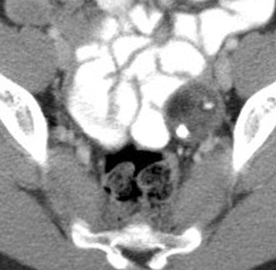
13 Hematometrocolpos May present with urinary retention or constipation 13 year old with pain and difficulty urinating


14 11 year old with hematometrocolpos T1 MRI MRI allows accurate determination of level of obstruction
15 Question #2 Unilateral obstruction of uterine duplications can be accompanied by what other anomaly? 1. Meckel s diverticulum 2. Ovarian agenesis 3. Renal agenesis 4. Ectopic pancreas
16 Question #2 Unilateral obstruction of uterine duplications can be accompanied by what other anomaly? 1. Meckel s diverticulum 2. Ovarian agenesis 3. Renal agenesis (ipsilateral) 4. Ectopic pancreas


17 12 year old with pelvic pain for 1 week


18 Abscess Perforated Appendicitis

19 Ovarian Cysts Follicles seen at all ages Seen on prenatal US as early as 28 wks Fewer after 1 year age Cysts > 2.5 cm > 5 cm may be treated surgically


")
20 Simple Ovarian Cyst Most are functional cysts (follicular, corpus luteum) Resolve in 4 12 wks


21 Hemmorhage Hemorrhagic Cysts Fine fibrinous strands to diffuse heterogenous echogenitiy


22 Ovarian cysts can extend to the liver in young infants


23 Ruptured Hemorrhagic Cyst Surgical emergency, if hemorrhage large Cyst may be difficult to see Crenated appearance


24 Ovarian Cyst with Torsion More likely with cysts > 5 cm May show internal hemorrhage Look for enlargement of ovary

 Small cysts lining")
25 Torsion of the Normal Ovary More common in adolescents Right ovary more common Abrupt sharp pain with vomiting, fever Enlarged ovary is most consistent finding (> 4 cm) Small cysts lining periphery Central edema


26 Ovarian Torsion Incomplete torsion can lead to massive edema Exquisite tenderness with compression


27 Follow-up after Detorsion Ovary may be fully or partially salvageable with laparoscopic detorsion Pre Post
28 Question #3 What is the earliest Doppler ultrasound finding of ovarian torsion? 1. Decreased arterial blood flow 2. Decreased venous blood flow 3. Complete absence of Doppler flow 4. Normal-appearing flow
29 Question #3 What is the earliest Doppler ultrasound finding of ovarian torsion? 1. Decreased arterial blood flow 2. Decreased venous blood flow 3. Complete absence of Doppler flow 4. Normal-appearing flow

30 Doppler US of Ovarian Torsion Venous and lymphatic flow initially compromised Arterial walls muscular Arterial flow compromise variable Normal arterial flow has been reported in 27-60% of cases. Twisted vascular pedicle


31 Torsion with Normal Doppler 47 cases of torsion 13% normal flow 13% no enlargement 60% no cyst or mass Overall accuracy 76.8% Maschiach, JUIM 2011 Multiple features are best predictor
32 Question # 4 Sonographer finds a large urinary bladder What should you do next? 1. Report as bladder outlet obstruction 2. Call pediatrician and suggest catheterization 3. Try to get patient to void and look again

33 Question # 4 Sonographer finds a large urinary bladder What should you do next? 1. Report as bladder outlet obstruction 2. Call pediatrician and suggest catheterization 3. Try to get patient to void and look again


34 Ovarian Teratoma Germ cell tumors most common ovarian neoplasms in children Classic mature teratoma Derived from at least two germ cell layers Primarily cystic (unilocular) Rokitansky nodule (dermoid plug) Hair follicles, fragments of bone, teeth Dermoid mesh Hair strands (linear interfaces)


35 CT or MRI appearance virtually pathognomonic Adipose tissue within cyst wall or dermoid plug Calcifications, amorphous or formed


36 Variable Appearance at US


37

38 Teratomas Bilateral in 10%


39 Mesenteric Cyst Lymphatic malformation May be unilocular or multiseptated Prone to hemorrhage or infection Increased complexity


40 Infected Lymphangioma

41 Enteric Duplication Cysts Can arise from any part of GI tract; ileum common. Bowel wall signature Echogenic mucosa Hypoechoic muscularis Echogenic serosa


42 Ovarian Cyst with Hemorrhage Hemorrhage lining cyst can mimic double layer wall of enteric duplication cyst
43 Urinary Tract Abnormalities Urinary tract infections most common Imaging used to identify VUR, renal abnormalities Bladder wall thickening may be seen, but of limited utility

44 Urachal Anomalies Incomplete obliteration of tract between allantois and bladder Patent urachus Sinus tract/fistula Cyst Diverticulum


45 Urachal Anomalies Asymptomatic unless infected Hemorrhage, rupture uncommon Carcinoma rare in children


46 Infected urachal cyst

47 16 year old male with pelvic pain and hematuria
48 Question # 5 Most likely cause of these findings is: 1. Bladder wall tumor 2. Hirschprung disease 3. Prostatic hypertrophy 4. Prostate/seminal vesicle anomaly


49 Seminal vesicle dilatation/cysts Congenital obstruction of ejaculatory duct Associated with ipsilateral renal agenesis in 66% Treated if symptomatic


50 Lower GU Tract Tumors Pelvic pain related to bladder outlet obstruction or constipation Rhabdomyosarcoma most common GU tumor Best prognosis (77% 5-yr survival) of all sites Majority with bladder or prostate involvement are <5 yrs of age Ddx: hemangioma, polyps, cystitis cystica


51 Presacral Lesions May be visible with US in young children, but MRI is best for evaluation


52 Neuroblastoma 2-3% occur in pelvis Detection of intraspinal extension valuable I-123 MIBG scans


53 Mature Teratoma Calcifications are common in both neuroblastoma and germ cell tumors
54 Other Presacral Masses/Cysts in Children Lymphoma Anterior meningocele Neurofibroma Tumors/lesions of sacrum Rectal duplication cyst Abscess


55 Question # 6 Where is this foreign body located? 1. Cecal lumen 2. Wall of cecum 3. Appendix 4. Patient s pocket 5. Can t tell

56 Nail in Appendix


57 Acute Appendicitis

58 US for Appendicitis Still accepted as best first screening exam Staged approach using CT for equivocal cases highly accurate Krishnamoorthi, Radiol Jan. 2011

59 Appendix Size in Appendicitis 6 mm or > in diameter PPV 63% NPV 100% More useful for excluding appendicitis Rettenbacher, Radiology 2011; 218: mm or > Similar accuracy Goldin, Pediatr Radiol 2011; 41: 993.


60 Lymphoid Hyperplasia of the Appendix Enlarged lymphoid tissue in the wall of appendix Response to viral infection Can mimic a fluid-filled appendix Look for central mucosal stripe May result in increased size


61 Compressibility difficult to demonstrate


62 Signs of Active or Impending Perforation Loss of mucosal lining Edematous fat Adjacent fluid collections Secondary findings can be strong indicators of appendicitis Wiersma, Eur Radiol 2009; 19: 455.

63 CT less desirable in young children, but useful in equivocal cases or suspected perforation


64 Crohn s Disease with Abscess
65 Take Home Points Ultrasound best first imaging study for most causes of pelvic pain in children Consider scanning beyond the pelvis CT for delineation of abscesses or troubleshooting MRI for presacral masses, congenital anomalies Use for inflammatory lesions is growing
Abdominal Pain in Pediatric Patients Image Gently
 Abdominal Pain in Pediatric Patients Image Gently Susan D. John, M.D. Baptist Health Emergency Radiology 2017 Disclosure I have no financial relationships with a commercial entity producing healthcarerelated
Intraperitoneal cysts in infancy and childhood An overview and sonographic differentiation
 Intraperitoneal cysts in infancy and childhood An overview and sonographic differentiation M. Mearadji International Foundation for Pediatric Imaging Aid Rotterdam, The Netherlands Intraperitoneal cysts
Patient Information. Age: 8 y/o Sex: Female. Date of Admission: Date of Discharge:
 Patient Information Age: 8 y/o Sex: Female Date of Admission: 92-10-08 Date of Discharge: 92-10-18 Chief Complaint Severe admominal pain and vomiting with dysuria since last afternoon Present Illness Lower
Obstetrics Content Outline Obstetrics - Fetal Abnormalities
 Obstetrics Content Outline Obstetrics - Fetal Abnormalities Effective February 2007 10 16% renal agenesis complete absence of the kidneys occurs when ureteric buds fail to develop Or degenerate before
ACUTE ABDOMEN IN OLDER CHILDREN. Carlos J. Sivit M.D.
 ACUTE ABDOMEN IN OLDER CHILDREN Carlos J. Sivit M.D. ACUTE ABDOMEN Clinical condition characterized by severe abdominal pain developing over several hours ACUTE ABDOMINAL PAIN Common childhood complaint
FHS Appendicitis US Protocol
 FHS Appendicitis US Protocol Reviewed By: Shireen Khan, MD; Sarah Farley, MD; Anna Ellermeier, MD Last Reviewed: May 2018 Contact: (866) 761-4200 **NOTE for all examinations: 1. If documenting possible
ACUTE PELVIC PAIN 강릉아산병원영상의학과 이은혜
 ACUTE PELVIC PAIN 강릉아산병원영상의학과 이은혜 Gynecologic PID Ruptured ovarian cyst Adnexal torsion Acute pelvic pain Pregnancy-related Ectopic pregnancy Placental abruption Nongynecologic Acute appendicitis Diverticulitis
A Practical Approach to Adnexal Masses
 A Practical Approach to Adnexal Masses Darcy J. Wolfman, MD Section Chief of Genitourinary Imaging American Institute for Radiologic Pathology Clinical Associate Johns Hopkins Community Radiology Division
IN THE NAME OF GOD POV: CYSTIC OVARIAN LESION
 IN THE NAME OF GOD POV: CYSTIC OVARIAN LESION CASE 1 20 years old girl with AUB and pelvic pain from 2 weeks ago Impression :Simple unilocular 6 cm ovarian cyst Next step? Almost certainly benign so FU
Always keep it in the differential
 Acute Appendicitis Lissa C. Sakata and Lindsey Perea 2 Always keep it in the differential Learning Objectives 1. The learner should be able to describe the etiology of acute appendicitis. 2. The learner
Imaging Ejaculatory Disorders and Hematospermia
 ATHENS 4-6 October 2018 European Society of Urogenital Radiology Imaging Ejaculatory Disorders and Hematospermia Parvati Ramchandani, MD Professor, Radiology and Surgery University of Pennsylvania Medical
Genitourinary Radiology In-Training Test Questions for Diagnostic Radiology Residents
 Genitourinary Radiology In-Training Test Questions for Diagnostic Radiology Residents March, 2013 Sponsored by: Commission on Education Committee on Residency Training in Diagnostic Radiology 2013 by American
Mature Cystic Teratomas and the most common complications
 Mature Cystic Teratomas and the most common complications Poster No.: C-2230 Congress: ECR 2015 Type: Authors: Keywords: DOI: Educational Exhibit S. C. S. Silva 1, D. N. Silva 1, D. Garrido 1, I. C. S.
Pelvic tumor in childhood Classification, imaging approach and radiological findings
 Pelvic tumor in childhood Classification, imaging approach and radiological findings M. Mearadji International Foundation for Pediatric Imaging Aid Rotterdam, The Netherlands Solid pelvic masses in childhood
Value of MRI in Characterizing Adnexal Masses
 The Journal of Obstetrics and Gynecology of India (July August 2015) 65(4):259 266 DOI 10.1007/s13224-015-0730-9 PHOTO ESSAY Value of MRI in Characterizing Adnexal Masses Alpana Karnik 1 Raina Anil Tembey
Case Report Müllerian Remnant Cyst as a Cause of Acute Abdomen in a Female Patient with Müllerian Agenesis: Radiologic and Pathologic Findings
 Volume 2016, Article ID 6581387, 4 pages http://dx.doi.org/10.1155/2016/6581387 Case Report üllerian Remnant Cyst as a Cause of Acute Abdomen in a Female Patient with üllerian Agenesis: Radiologic and
ADENOMYOSIS CHRONIC PELVIC PAIN IN WOMEN IMAGING CHRONIC PELVIC PAIN IN WOMEN CHRONIC PELVIC PAIN IN WOMEN ADENOMYOSIS: PATHOLOGY ADENOMYOSIS
 CHRONIC PELVIC PAIN IN WOMEN IMAGING CHRONIC PELVIC PAIN IN WOMEN MOSTAFA ATRI, MD Dipl. Epid. UNIVERSITY OF TORONTO Non-menstrual pain of 6 months Prevalence 15%: 18-50 years of age 10-40% of gynecology
The Adnexal Mass. Handout NCUS 3/18/2017 Suzanne Dixon, MD
 The Adnexal Mass Handout NCUS 3/18/2017 Suzanne Dixon, MD Objectives: Pelvic mass differential Characteristics of the normal ovary Standard terminology for ovarian masses Benign vs. malignant features
Emergent Pediatric Ultrasound. Katharine Dennis, RDMS/RVT Tiffany Schultz, RDMS UNC Health Care Dept of General Ultrasound
 Emergent Pediatric Ultrasound Katharine Dennis, RDMS/RVT Tiffany Schultz, RDMS UNC Health Care Dept of General Ultrasound Introduction Learning Objectives Review common pediatric emergent ultrasound exams
Pitfalls in the CT diagnosis of appendicitis
 The British Journal of Radiology, 77 (2004), 792 799 DOI: 10.1259/bjr/95663370 E 2004 The British Institute of Radiology Pictorial review Pitfalls in the CT diagnosis of appendicitis 1 C D LEVINE, 2 O
Sonographic Whirlpool Sign in Ovarian Torsion
 Technical dvance Sonographic Whirlpool Sign in Ovarian Torsion S. oopathy Vijayaraghavan, MD, DMRD Objective. To describe an additional maneuver during sonography for ovarian torsion and to assess its
Summary and conclusions
 Summary and conclusions 7 Chapter 7 68 Summary and conclusions Chapter 1 provides a general introduction to this thesis focused on the use of ultrasound (US) in children with abdominal problems. The literature
Vikram Dogra, M.D. Professor of Radiology, Urology & BME Department of Imaging Sciences University Of Rochester Medical Center
 Ultrasound of the Scrotum Vikram Dogra, M.D. Professor of Radiology, Urology & BME Department of Imaging Sciences University Of Rochester Medical Center Etiologies of Acute Scrotal Pain Epididymitis/Orchitis
Pediatric Abdominal Masses. Andrew Phelps MD Assistant Professor of Pediatric Radiology UCSF Benioff Children's Hospital
 Pediatric Abdominal Masses Andrew Phelps MD Assistant Professor of Pediatric Radiology UCSF Benioff Children's Hospital No Disclosures Take Home Message All you need to remember are the 5 common masses
Excretory urography (EU) or IVP US CT & radionuclide imaging
 or IVP US CT & radionuclide imaging") Excretory urography (EU) or IVP US CT & radionuclide imaging MRI arteriography studies requiring catherization or direct puncture of collecting system EU & to a lesser extent CT provide both functional
Excretory urography (EU) or IVP US CT & radionuclide imaging MRI arteriography studies requiring catherization or direct puncture of collecting system EU & to a lesser extent CT provide both functional
Paediatric surgical emergencies. Mani Thyagarajan BWCH
 Paediatric surgical emergencies Mani Thyagarajan BWCH General points Always discuss Call consultant for help ASAP CT scan is a bad modality in paediatrics Ultrasound? Intussusception? Renal colic? UTI
Pelvic Pain. What you need to know. 139 Dumaresq Street Campbelltown Phone Fax
 Pelvic Pain What you need to know 139 Dumaresq Street Campbelltown Phone 4628 5292 Fax 4628 0349 www.nureva.com.au September 2015 PELVIC PAIN This is a common problem and most women experience some form
Alison Douglass Gillian Lieberman, MD. November. Colon Cancer. Alison Douglass, Harvard Medical School Year III Gillian Lieberman, MD
 November Colon Cancer Alison Douglass, Harvard Medical School Year III Our Patient Mr. K. is a 67 year old man with no prior medical problems other than hemorrhoids which have caused occasional rectal
Interesting Pediatric ultrasound cases. Presented by: Falguni Patel (RDMS, RVT)
") Interesting Pediatric ultrasound cases Presented by: Falguni Patel (RDMS, RVT) Role of ultrasound to rule out Appendicitis Overview: Ultrasound is relatively inexpensive, safe and quick solution to rule
Interesting Pediatric ultrasound cases Presented by: Falguni Patel (RDMS, RVT) Role of ultrasound to rule out Appendicitis Overview: Ultrasound is relatively inexpensive, safe and quick solution to rule
Clinical summary. Male 30 year-old with past history of non-seminomous germ cell tumour. Presents with retroperitoneal lymphadenopathy on CT.
 Clinical summary Male 30 year-old with past history of non-seminomous germ cell tumour. Presents with retroperitoneal lymphadenopathy on CT. For restaging PET/CT. PET/CT findings No significant FDG uptake
Telephone Icd 10 ovarian dermoid cyst P.O. Box 189 Navan, ON, K4B 1J4 Canada. Sitemap
 Telephone 613-835-9490 Icd 10 ovarian dermoid cyst P.O. Box 189 Navan, ON, K4B 1J4 Canada Sitemap Home» Current Health Articles» Causes of Left Side Abdominal (Stomach) Pain Causes of Left Side Abdominal
Category Term Definition Comments 1 Major Categories 1a
 Working Lexicon Categories, Terms & Definitions Category Term Definition Comments 1 Major Categories 1a Physiologic Category (consistent with normal ovarian physiology) Follicle Simple 3 cm in premenopausal
Plain abdomen The standard films are supine & erect AP views (alternative to erect, lateral decubitus film is used in ill patients).
.") Plain abdomen The standard films are supine & erect AP views (alternative to erect, lateral decubitus film is used in ill patients). The stomach can be readily identified by its location, gastric rugae
Plain abdomen The standard films are supine & erect AP views (alternative to erect, lateral decubitus film is used in ill patients). The stomach can be readily identified by its location, gastric rugae
The Acute Scrotum: Sonographic Findings
 The Acute Scrotum: Sonographic Findings 가천의대길병원방사선과 양달모 Gachon Medical School Introduction Many diseases presenting as acute scrotal pain DDx is important for determining the appropriate treatment US with
Case Report Imperforate Hymen Causing Bilateral Hydroureteronephrosis in an Infant with Bicornuate Uterus
 Case Reports in Urology Volume 2012, Article ID 102683, 4 pages doi:10.1155/2012/102683 Case Report Imperforate Hymen Causing Bilateral Hydroureteronephrosis in an Infant with Bicornuate Uterus Ayse Secil
Case Report Urachal Cyst Causing Small Bowel Obstruction in an Adult with a Virgin Abdomen
 Case Reports in Surgery Volume 2016, Article ID 3247087, 4 pages http://dx.doi.org/10.1155/2016/3247087 Case Report Urachal Cyst Causing Small Bowel Obstruction in an Adult with a Virgin Abdomen Michael
Chapter 100 Gynecologic Disorders
 Chapter 100 Gynecologic Disorders Episode Overview: 1. Describe the presentation and RF for Adnexal torsion 2. List the imaging findings of adnexal torsion (US vs CT) 3. What is the management of adnexal
Continuing Professional Development
 Volume 15.4.1 This Lecture Serties qualifies for 0.5 Informal CPD Learning Hours Continuing Professional Development Pelvic Calcifications By Dr Kristin Grace (DACBR) WHAT IS THAT??? Calcifications in
Objectives. Bits and Pieces: Developmental Remnants in the ED. Case 1. Evelyn Porter, MD November 14, Refresh your memory.
 Bits and Pieces: Developmental Remnants in the ED Evelyn Porter, MD November 14, 2012 Refresh your memory Review management Case based format Objectives Case 1 4 year old boy presents to the ED with an
US in non-traumatic acute abdomen. Lalita, M.D. Radiologist Department of radiology Faculty of Medicine ChiangMai university
 US in non-traumatic acute abdomen Lalita, M.D. Radiologist Department of radiology Faculty of Medicine ChiangMai university Sagittal Orientation Transverse (Axial) Orientation Coronal Orientation Intercostal
Effective Utilization of Imaging. John V. Roberts, M.D. Premier Radiology Abdominal Imaging
 Effective Utilization of Imaging John V. Roberts, M.D. Premier Radiology Abdominal Imaging Safety Contrast and Radiation What to order Abdomen/Pelvis Brain/Spine Chest Musculoskeletal Ob/Gyn Head and Neck
Top Tips for Gynaecological Ultrasound. Catherine Kirkpatrick Consultant Sonographer Dublin Oct 2018
 Top Tips for Gynaecological Ultrasound Catherine Kirkpatrick Consultant Sonographer Dublin Oct 2018 We can all scan a pelvis so what can we do to improve? Uterus, endometrium and ovaries, got it covered!
CASE STUDY. Presented by: Jessica Pizzo. CFCC Sonography student Class of 2018
 CASE STUDY Presented by: Jessica Pizzo CFCC Sonography student Class of 2018 Case Presentation April 4, 2017 56 yr old woman presented to ED with lower abdominal pain & swelling, along with constipation.
7/2/15. Approach to imaging of pelvic pain. Acute pelvic pain. Chronic pelvic pain. Dysmenorrhea
 I have no financial rela.onships to disclose. Approach to imaging of pelvic pain To emphasize the importance of ultrasound as the imaging modality of choice for the most commonly presen6ng diagnoses in
Ultrasound of: Appendicitis Intussusception Pyloric Stenosis
 Ultrasound of: Appendicitis Intussusception Pyloric Stenosis Andrew Phelps MD Assistant Professor Pediatric Radiology UCSF Benioff Children s Hospital No Disclosures Take Home Message Appendicitis occurs
Evaluation of the of the sensitivity, accuracy and positive predictive value of ultrasonography in the diagnosis of Appendicitis.
 West African Journal of Ultrasound Vol 17 Number 2 (2016) Evaluation of the of the sensitivity, accuracy and positive predictive value of ultrasonography in the diagnosis of Appendicitis. 1 2 3 Oguntola
Urinary tract infections, renal malformations and scarring
 Urinary tract infections, renal malformations and scarring Yaacov Frishberg, MD Division of Pediatric Nephrology Shaare Zedek Medical Center Jerusalem, ISRAEL UTI - definitions UTI = growth of bacteria
Chapter 6: Genitourinary and Gastrointestinal Systems 93
 Chapter 6: Genitourinary and Gastrointestinal Systems 93 Chapter 6 Genitourinary and Gastrointestinal Systems Embryology Three sets of excretory organs or kidneys develop in human embryos: Pronephros:
Common Problems in Urology
 Common Problems in Urology 1. Renal Colic Outline 2. Urinary Retention 3. Acute Scrotum Supanut Lumbiganon, MD. Renal colic The most common urologic emergency O Sudden increase of pressure in the urinary
Neckmasses in infancy and childhood: Clinical and radiological classification and imaging approaches M. Mearadji
 Neckmasses in infancy and childhood: Clinical and radiological classification and imaging approaches M. Mearadji International Foundation for Pediatric Imaging Aid Introduction Neck masses are a frequent
Pediatric Abdomen Trauma
 Pediatric Abdomen Trauma Susan D. John, MD, FACR Pediatric Trauma Trauma is leading cause of death and disability in children and adolescents Causes and effects vary between age groups Blunt trauma predominates
Children's (Pediatric) Ultrasound - Abdomen
 Ultrasound - Abdomen") Scan for mobile link. Children's (Pediatric) Ultrasound - Abdomen Children s (pediatric) ultrasound imaging of the abdomen is a safe, noninvasive test that uses sound waves to produce a clear picture of
Scan for mobile link. Children's (Pediatric) Ultrasound - Abdomen Children s (pediatric) ultrasound imaging of the abdomen is a safe, noninvasive test that uses sound waves to produce a clear picture of
APPROACH TO ABDOMINAL MASS
 Thomas Hong APPROACH TO ABDOMINAL MASS General Presentation An abdominal mass in a neonate, young child, or adolescent patient is something that every pediatrician needs to be wary of as these masses can
Hydronephrosis. What is hydronephrosis?
 What is hydronephrosis? Hydronephrosis Hydronephrosis describes the situation where the urine collecting system of the kidney is dilated. This may be a normal variant or it may be due to an underlying
Abdominal ultrasound:
 Abdominal ultrasound: Non-traumatic acute abdomen Wittanee Na-ChiangMai, MD Department of Radiology ChiangMai University 26/04/2017 Contents Technique of examination Normal anatomy Emergency conditions
Richmond, Virginia. I ve have this terrible pain
 Acute Pelvic Pain A Practical Approach Christine Isaacs, MD Associate Professor Department of Obstetrics & Gynecology Virginia Commonwealth University School of Medicine Richmond, VA Richmond, Virginia
Role of imaging in RCC. Ultrasonography. Solid lesion. Cystic RCC. Solid RCC 31/08/60. From Diagnosis to Treatment: the Radiologist Perspective
 Role of imaging in RCC From Diagnosis to Treatment: the Radiologist Perspective Diagnosis Staging Follow up Imaging modalities Limitations and pitfalls Duangkamon Prapruttam, MD Department of Therapeutic
Case 1307 Mesothelial cysts
 Case 1307 Mesothelial cysts Vinhais S, Monteiro M, Cunha TM INSTITUTO PORTUGUÊS DE ONCOLOGIA de Francisco Gentil de LISBOA Section: Gastro-Intestinal Imaging Published: 2001, Nov. 23 Patient: 44 year(s),
CT Urography. Bladder. Stuart G. Silverman, M.D.
 CT Urography Stuart G. Silverman, M.D. Professor of Radiology Harvard Medical School Director, Abdominal Imaging and Intervention Brigham and Women s Hospital Bladder Boston, MA CT Urography Stuart G.
Assessment of adnexal masses. Ultrasound workup of adnexal masses. symptoms. symptoms. Age. Serum tumor markers 10/1/2018
 Assessment of adnexal masses Ultrasound workup of adnexal masses Kevin Robinson, DO Department of Radiology Michigan State University October 4, 2018 Patients symptoms Age Menstrual status Serum tumor
Acute pelvic pain in female patient: Clinical and Radiological evaluation
 Acute pelvic pain in female patient: Clinical and Radiological evaluation Poster No.: C-0909 Congress: ECR 2014 Type: Authors: Keywords: DOI: Educational Exhibit N. Ramesh 1, T. Simelane 2 ; 1 Portlaoise/IE,
Acute pelvic pain in female patient: Clinical and Radiological evaluation
 Acute pelvic pain in female patient: Clinical and Radiological evaluation Poster No.: C-0909 Congress: ECR 2014 Type: Authors: Keywords: DOI: Educational Exhibit N. Ramesh 1, T. Simelane 2 ; 1 Portlaoise/IE,
Medical application of transabdominal ultrasound in gastrointestinal diseases
 Medical application of transabdominal ultrasound in gastrointestinal diseases Hsiu-Po Wang Department of Emergency Medicine National Taiwan University Hospital Real-time ultrasound has become a standard
SIMPLE GUIDE FOR SONOLOGICAL EVALUATION OF APPENDICITIS
 SIMPLE GUIDE FOR SONOLOGICAL EVALUATION OF APPENDICITIS A Case Study by Dr. Avni K P Skandhan, India (Consultant Radio Diagnosis, Malabar Institute of Medical Science, Malappuram, Kerala) Email: avniskandhan@gmail.com
Adnexal Masses and Problem Solving Pelvic MRI
 28th Congress of the Hungarian Society of Radiologists RCR Session Budapest June 2016 Adnexal Masses and Problem Solving Pelvic MRI DrSarah Swift St James s University Hospital Leeds, UK Objectives Characterisation
National Defense Medical Center, Taipei, Taiwan.
 CONGENITAL SEMINAL VESICLE CYST ASSOCIATED WITH IPSILATERAL RENAL AGENESIS MIMICKING BLADDER OUTLET OBSTRUCTION: A CASE REPORT AND REVIEW OF THE LITERATURE Chien-Chang Kao, 1 Ching-Jiunn Wu, 2 Guang-Huan
Pediatric Retroperitoneal Masses Radiologic-Pathologic Correlation
 Acta Radiológica Portuguesa, Vol.XVIII, nº 70, pág. 61-70, Abr.-Jun., 2006 Pediatric Retroperitoneal Masses Radiologic-Pathologic Correlation Marilyn J. Siegel Mallinckrodt Institute of Radiology, Washington
elical CT plays an important role
 bdominal Imaging Yu et al. Helical CT of cute RLQ Pain Pictorial Essay Jinxing Yu 1 nn S. Fulcher Mary nn Turner Robert. Halvorsen Yu J, Fulcher S, Turner M, Halvorsen R Helical CT Evaluation of cute Right
Case Report Delivery Induced Intraperitoneal Rupture of a Cystic Ovarian Teratoma and Associated Chronic Chemical Peritonitis
 Case Reports in Radiology, Article ID 189409, 4 pages http://dx.doi.org/10.1155/2014/189409 Case Report Delivery Induced Intraperitoneal Rupture of a Cystic Ovarian Teratoma and Associated Chronic Chemical
Adrenal masses in infancy and childhood: A clinical and radiological overview M. Mearadji
 Adrenal masses in infancy and childhood: A clinical and radiological overview M. Mearadji International Foundation for Pediatric Imaging Aid Introduction Neoplastic adrenal masses usually originate from
Adolescent Gynecology: Evaluation and Management of Adnexal Mass, PCOS, and Endometriosis. Shanna M. Combs, MD
 Adolescent Gynecology: Evaluation and Management of Adnexal Mass, PCOS, and Endometriosis Shanna M. Combs, MD Adolescent Visit and Exam Adolescent Reproductive Health Visit Initial visit should take place
Abdominal Complications After Bone Marrow Transplantation in Children: Sonographic and CT Findings
 1023 Pictorial Essay Abdominal Complications After Bone Marrow Transplantation in Children: Sonographic and CT Findings Ellen C. Benya,1 2 Carlos J. Sivit, 2 and Ralph R. Quinones2 3 Bone marrow transplantation
Ultrasound - Pelvis. What is Pelvic Ultrasound Imaging?
 Scan for mobile link. Ultrasound - Pelvis Ultrasound imaging of the pelvis uses sound waves to produce pictures of the structures and organs in the lower abdomen and pelvis. There are three types of pelvic
Torsion of a Wandering Spleen Presenting as a Painful Pelvic Mass Post Pregnancy: Imaging Diagnosis
 CASE REPORT Torsion of a Wandering Spleen Presenting as a Painful Pelvic Mass Post Pregnancy: Imaging Diagnosis Abbey P 1, Aarushi A 1, Andley M 2, Anand R 1 1 Department of Radio-Diagnosis, 2 Department
The many faces of Endometriosis
 The many faces of Endometriosis Beryl Benacerraf M.D Harvard Medical School What is Endometriosis? Endometriosis is defined as the presence of normal endometrial tissue occurring outside of the endometrial
Adult Intussusception: A Complication of Metastatic Melanoma or Primary Malignancy?
 January 2013 Adult Intussusception: A Complication of Metastatic Melanoma or Primary Malignancy? Johanna Sheu, Harvard Medical School Year III 1 Agenda Menu of tests Definition/anatomy/classification Pediatrics
ISUOG Basic Training. Distinguishing between Normal & Abnormal Appearances of the Urinary Tract. Seshadri Suresh, India
 ISUOG Basic Training Distinguishing between Normal & Abnormal Appearances of the Urinary Tract Seshadri Suresh, India Learning objectives 13 & 14 At the end of the lecture you will be able to: describe
 INTRAUTERINE DEVICE = IUD INTRAUTERINE DEVICE = IUD CONGENITAL DISORDERS Pyometra = pyometrea is a uterine infection, it is accumulation of purulent material in the uterine cavity. Ultrasound is usually
8/29/2016 DIVERTICULAR DISEASE: WHAT EVERY NURSE PRACTITIONER SHOULD KNOW. LENORE LAMANNA Ed.D, ANP-C LEARNING OBJECTIVES
 DIVERTICULAR DISEASE: WHAT EVERY NURSE PRACTITIONER SHOULD KNOW LENORE LAMANNA Ed.D, ANP-C LEARNING OBJECTIVES Define Diverticular Disease Discuss Epidemiology and Pathophysiology of Diverticular disease
Back to Basics: What Imaging Test should I order? Jeanne G. Hill, M.D. Pediatric Radiology Medical University of South Carolina
 Back to Basics: What Imaging Test should I order? Jeanne G. Hill, M.D. Pediatric Radiology Medical University of South Carolina Disclosure Neither I nor any member of my immediate family has a relevant
Abdominal nonparenchymatous cystic lesions and their mimics in children
 Jpn J Radiol (2014) 32:623 629 DOI 10.1007/s11604-014-0355-4 PICTORIAL ESSAY Abdominal nonparenchymatous cystic lesions and their mimics in children Kaan Esen Anıl Özgür Yasemin Karaman Hakan Taşkınlar
Abdominal Ultrasound. Diane Hallinen, MD. Bloodroot
 Abdominal Ultrasound Diane Hallinen, MD Bloodroot Abdominal Ultrasound Vasculature Hepatobiliary Spleen Kidney Bladder Bowel Where to put the probe? Vasculature We are going to talk about Celiac Trunk
In the August 2016 issue of OBG Management,
 GYN coding changes to note for your maximized reimbursement Revised ICD-10 gynecologic diagnostic codes go into effect October 1. Here is a look at the added, expanded, and revised codes you will need
CLINICAL PRESENTATION AND RADIOLOGY QUIZ QUESTION
 Donald L. Renfrew, MD Radiology Associates of the Fox Valley, 333 N. Commercial Street, Suite 100, Neenah, WI 54956 2/5/2011 Radiology Quiz of the Week # 6 Page 1 CLINICAL PRESENTATION AND RADIOLOGY QUIZ
Sonography of Pediatric Superficial Lumps and Bumps: Illustrative Examples from Head to Toe
 Sonography of Pediatric Superficial Lumps and umps: Illustrative Examples from Head to Toe nmol Gupta ansal, MD Henrietta Kotlus Rosenberg, MD, FCR, FP Mount Sinai Hospital Icahn School of Medicine at
Information for Patients
 Information for Patients Congenital Malformation in the Urinary Tract: Ureteral Duplication, Ureterocele, and Ectopic Ureter English Table of contents Ureteral Duplication... 3 Symptoms and Diagnosis...
TUMOR AND TUMOR-LIKE CONDITIONS OF THE PERITONEUM AND OMENTUM/MESENTERY 40 th. Annual Meeting SCBTMR September 9-13, 2017, Nashville, Tennessee
 TUMOR AND TUMOR-LIKE CONDITIONS OF THE PERITONEUM AND OMENTUM/MESENTERY 40 th. Annual Meeting SCBTMR September 9-13, 2017, Nashville, Tennessee Isaac R Francis University of Michigan Department of Radiology
Fetal Renal Malformations: The Role of Ultrasound in Diagnosis & Management
 Fetal Renal Malformations: The Role of Ultrasound in Diagnosis & Management 12 weeks Alfred Abuhamad, M.D. Eastern Virginia Medical School 13 weeks 2nd trimester Medullary pyramids Renal Sinus Cortex 2nd
Complicated Meckel`s diverticulum; to be considered as a differential diagnosis in the acute abdominal pain. Ultrasound and MDCT imaging finding
 Complicated Meckel`s diverticulum; to be considered as a differential diagnosis in the acute abdominal pain. Ultrasound and MDCT imaging finding Poster No.: C-0174 Congress: ECR 2013 Type: Educational
My Patient Has Abdominal Pain PoCUS of the Biliary Tract and the Urinary Tract
 My Patient Has Abdominal Pain PoCUS of the Biliary Tract and the Urinary Tract Objectives PoCUS for Biliary Disease PoCUS for Renal Colic PoCUS for Urinary Retention Biliary Disease A patient presents
Pediatric Ocular Sonography
 Pediatric Ocular Sonography Cicero J Torres A Silva, MD Associate Professor of Radiology 2016 SPR Pediatric Ultrasound Course Yale University School of Medicine None Disclosures Objectives of Presentation
Gynecologic Ultrasound. Sujata Ghate, MD Associate Professor of Radiology Duke University Medical Center
 Gynecologic Ultrasound Sujata Ghate, MD Associate Professor of Radiology Duke University Medical Center Objectives Understand work-up of endometrial abnormalities Show examples of uterine and endometrial
Pediatric Hepatobiliary, Pancreatic & Splenic US
 Pediatric Hepatobiliary, Pancreatic & Splenic US Susan J. Back, MD Department of Radiology, The Children s Hospital of Philadelphia No Disclosures Objectives Normal Abnormal: cases and US advances Objectives
Abdomen and Retroperitoneum Ultrasound Protocols
 Abdomen and Retroperitoneum Ultrasound Protocols Reviewed By: Anna Ellermeier, MD Last Reviewed: March 2018 Contact: (866) 761-4200, Option 1 **NOTE for all examinations: 1. If documenting possible flow
Accuracy of transvaginal ultrasound and magnetic resonance imaging in diagnosis and extension of pelvic endometriosis
 Accuracy of transvaginal ultrasound and magnetic resonance imaging in diagnosis and extension of pelvic endometriosis A.Salem, Kh. Fakhfakh, S. Mehiri, Y. Ben Brahim, F. Ben Amara, H. Rajhi, R. Hamza,
Rad Lab 4 Unknowns: Genitourinary!
 Rad Lab 4 Unknowns: Genitourinary! Peter Clarke MD! Don Di Salvo, MD! Clerkship Directors for Radiology! Harvard Medical School! Brigham and Women s Hospital! Dana Farber Cancer Institute! Case 1: 69 year
Acute flank pain in children: Imaging considerations
 Acute flank pain in children: Imaging considerations Carlos J. Sivit MD Rainbow Babies and Children s Hospital Case Western Reserve School of Medicine Flank pain Results from distention of ureter or renal
CME ARTICLE. A Practical Approach to the Ultrasound Characterization of Adnexal Masses. Douglas L. Brown, MD
 CORE CURRICULUM IN SONOGRAPHY CME ARTICLE A Practical Approach to the Ultrasound Characterization of Adnexal Masses Douglas L. Brown, MD Abstract: Because pelvic ultrasound is commonly used to evaluate
Personal data. Age : 63 Gender : male
 Personal data Age : 63 Gender : male Chief complain No specific symptom or discomfort A hepatic mass, found by abdominal sonography of routine health exam on 88-12-08 Past history 1984-3-3 Old CVA with
January Dear Medical Director:
 January 2010 Dear Medical Director: It is the position of the American Urological Association (AUA) that urologists are appropriately trained in the performance of sonographic procedures. In spite of this,