Carlos Torres MD, FRCPC, Associate Professor of Radiology Department of Radiology, University of Ottawa
|
|
|
- Shavonne Banks
- 5 years ago
- Views:
Transcription


1 Carlos Torres MD, FRCPC, Associate Professor of Radiology Department of Radiology, University of Ottawa
2 None
3 1. Simplify the complex imaging anatomy of the BP using clear anatomical landmarks. 2. Outline different MR protocols. 3. Review BP pathologies using case-based approach.
4 Anatomy
5 Brachial Plexus Formed by ventral rami of the nerves C5 -T1 - pre fixed - post fixed Responsible for motor and cutaneous innervation of upper extremity, except for: Motor: Trapezius and levator scapulae Cutaneous: Axila, suprascapular & scapular regions
6 Brachial Plexus Segments Roots Trunks Divisions Cords Branches

7 Brachial Plexus Segments Radiologists Technologists Drink Cold Beer


8 Roots The ventral rami of the spinal nerves C5 to T1 are the roots of the plexus. A P * A M * ^ R ^
9 Trunks C5 - C6: Upper T C7 : Middle T C8 T1: Lower T A * ^ * T ^


10 Divisions Each trunk splits in 2 to give an anterior and posterior division A * ^ * ^ D


11 Divisions


12 Cords Lat: Ant divisions of sup & middle trunks Medial: Ant division of lower trunk Post: 3 post divisions B * C ^ C

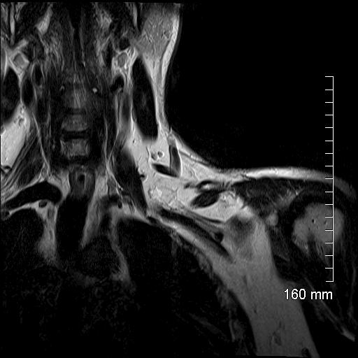
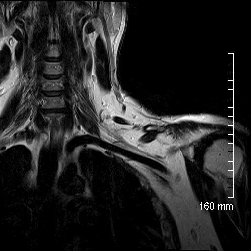
13 Branches Musculocutaneous N. Axillary N. Median N. Radial N. Ulnar N. ^ B

14 Branches

15
16 Method of choice Multi planar Exquisite soft-tissue contrast Castillo. AJR 2005, 185: S Todd et al. Top Magn Reson Imaging 2004, 15: Saifuddin. Skeletal Radiol 2003, 32: Wittenberg et al. Radiographics 2000, 20:
17 Surface coil Thin sections with no/small gap (3D) T1, T2 and STIR Contrast may be given Two imaging protocols at TOH
18 Neck coil and body array Localizer in 3 planes Sequence Time ST TR TE Cor T2 Space 5: Cor T1 2D 4: Cor T2 STIR 3: Sag T1 2D 4:

19
20 McGill/MGH The Ottawa Hospital Sagittal T1W 3/4 mm (thickness/gap), T2W 3/4 mm Coronal T1W 3/4mm and FAST STIR 3/4 mm Axial T1W 4/5 mm, T2W 3/4 mm, +/- Gadolinium enhanced: Coronal 3/4 mm,axial and sag T1W 4/5 mm with fat saturation
21 MODIFIED TECHNIQUE FOR THE STUDY OF THE BRACHIAL PLEXUS WITH MR MODIFIED TECHNIQUE : 3 plane LOCALIZER Increase number of slices in the coronal plane Parameters FSE T1 Parameters FSE T2 Matrix: 448x224 cm

22 CONVENTIONAL TECHNIQUE MODIFIED TECHNIQUE LOCALIZER


23 MODIFIED TECHNIQUE FOR THE STUDY OF THE BRACHIAL PLEXUS WITH MR MODIFIED TECHNIQUE


24 MODIFIED TECHNIQUE FOR THE STUDY OF THE BRACHIAL PLEXUS WITH MR MODIFIED TECHNIQUE: The axial oblique sequences are planned off the coronal localizer parallel to the plane of the roots, trunks and divisions of the brachial plexus.


25 MODIFIED TECHNIQUE FOR THE STUDY OF THE BRACHIAL PLEXUS WITH MR MODIFIED TECHNIQUE: The coronal sequences are planned off the axial oblique dataset following the plane of the brachial plexus.


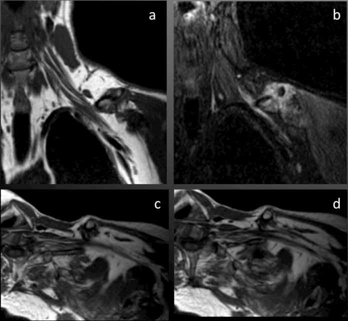
26 MODIFIED TECHNIQUE FOR THE STUDY OF THE BRACHIAL PLEXUS WITH MR MODIFIED TECHNIQUE: The sagital sequences are planned off the axial oblique images, perpendicular to the segments of the brachial plexus.


27 MODIFIED TECHNIQUE FOR THE STUDY OF THE BRACHIAL PLEXUS WITH MR CONVENTIONAL TECHNIQUE MODIFIED TECHNIQUE

28 CONVENTIONAL TECHNIQUE MODIFIED TECHNIQUE


29 CONVENTIONAL TECHNIQUE MODIFIED TECHNIQUE
30 MODIFIED TECHNIQUE FOR THE STUDY OF THE BRACHIAL PLEXUS WITH MR CONVENTIONAL TECHNIQUE MODIFIED TECHNIQUE Axial T1 : 7 min 25 sec 3 min 59 sec Axial T2: 8 min 03 sec 3 min 54 sec Coronal T1: 4 min 22 sec 4 min 36 sec Coronal T2: 4 min 44 sec 3 min 54 sec Sagital T1: 9 min 16 sec 6 min 17 sec Sagital T2: 7 min 32 sec 6 min 08 sec Total scan time: 41 min 22 sec 28 min 48 sec


31


32


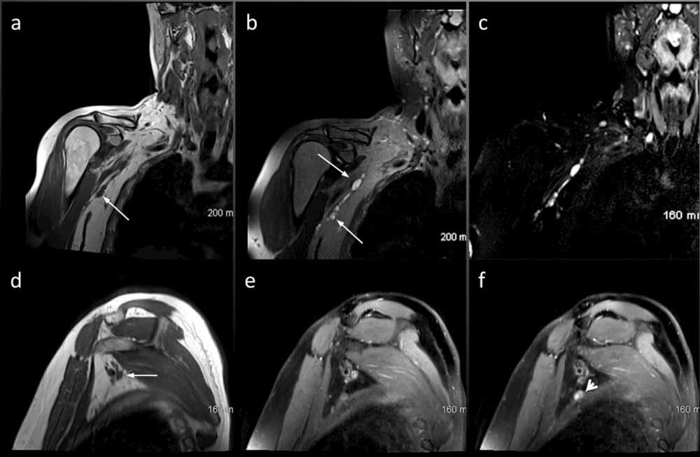
33 2005 a b c Case: 27 y/o pt with left ulnar neuropathy


34 a b c 2011 d
35

36 Pathology
37 Vague and nonspecific symptoms. Trauma: most common cause of plexopathy Tumors: 2 nd most common Post radiation Others : Inflammatory, infectious and hereditary

38 Imaging studies play an essential role in differentiating preganglionic injuries from postganglionic lesions, a differentiation that is crucial for determining the management of BPI
39 Trauma may be due to: Traction/Compression Penetrating injuries Local fractures or dislocations. What to look for? Pseudomeningoceles Clumping, thickening and signal Hematomas

40 25 y/o pt, assault
41


42 Post Traumatic Pseudo aneurysm Concentric rings of varying signal intensity due to clot that forms the walls of this pseudo aneurysm c/o Mauricio Castillo, UNC

43 MVA Stretch injury Pseudo meningoceles

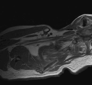
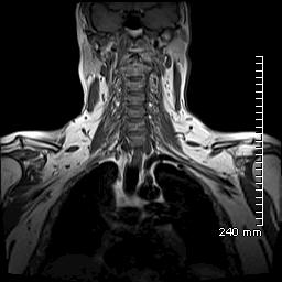
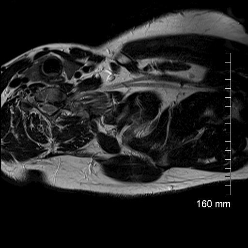
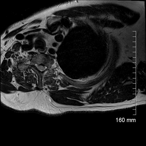
44 Stretch injury Pseudo meningocele + n root avulsion A B C
45 Primary: Schwannoma Neurofibroma Secondary: Direct extension/compression: tumors in the vicinity of the BP: lung, bones or soft tissues of the neck. Metastasis: Breast, lung.
46
47 NEUROFIBROMA
48
49 NEUROFIBROMAS NF1


50 NF1
51 10 y/o pt with neck swelling since he was 18 months c/o Manu Shroff, Sickkids Univ of Toronto.
52 Schwannomatosis - 3 rd major form of NF - Distinct from NF1 and NF2 - Noncutaneous schwannomas - Absence of vest schwannomas c/o Manu Shroff, Sickkids U of Toronto.
53

54 PANCOAST TUMOR
55

56 METS MELANOMA

57 58 y/o pt with pain in the left arm
58 NEUROLYMPHOMATOSIS: B-cell NHL
59 54 y/o pt with left brachial plexopathy A B

60

61 A B NEUROLYMPHOMATOSIS: T-cell Lymphoma


62 NEUROLYMPHOMATOSIS: T-cell Lymphoma


63
64 Post Radiation: Progressive neuropathy resulting from fibrosis and obliteration of the vasa-nervorum. Patients receiving > 60 Gy. Months years after therapy Thickenning of n. roots Low signal on both T1 and T2 Inflammatory poly neuropathy : MMN, CMT, CIDP Brachial Neuritis: viral, idiopathic, drugs, hereditary.

65 60 y/o pt with Hx of Breast Ca + Radiation

66
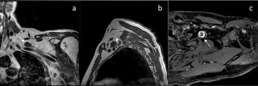
67 26 y/o pt with bilat weakness and numbness arms/legs

68 CHRONIC INFLAMMATORY DEMYELINATING POLYNEUROPATHY CIDP


69 MR imaging & nerve root thickening: Seen in ~40% children, ~60% adult CIDP patients


70 MR imaging & nerve root thickening: Nerve root thickening also noted in other diseases: CMT1A patient CIDP patient

71
72 Diffusion tensor imaging (DTI) and tractography of the brachial plexus: Feasibility and initial experience in neoplastic conditions. Vargas M et al. Neuroradiology (2010) 52: normal volunteers, 12 patients benign & malignant ( 3 Qx, 9 medical) ADC & FA maps 2 Radiologists
73 No statistically significant difference in FA and ADC values of normal fibers and fibers at the level of pathology. Tractography revealed major differences regarding fiber architecture. Benign: Malignant: Displacement Disruption/Destruction Encasement Disorganization

74 Tractography of the brachial plexus in a 37-year-old male volunteer

 suggesting an easier surgical enucleation.")
75 MIP Coronal reconstruction of the 3D STIR SPACE sequence showing a distal schwannoma of the brachial plexus. The displaced fibers of the posterior cord (white arrows) passing around the schwannoma (asterisk) suggesting an easier surgical enucleation. The findings were confirmed at surgery

76 Step-by-step reconstruction of the tractography of the brachial plexus in a 42 year-old male patient fibers within and around the benign neurogenic tumor

77 Sixty-five-year-old patient with adenocarcinoma of the lung, disorganization and interruption of nerve fibers on the tractography reconstruction image
78 Rads & Techs Drink Cold Beer MR is the imaging method of choice Different protocols: 3T vs 1.5T Advanced Imaging Techniques

79 Carlos Torres MD, FRCPC, Associate Professor of Radiology Department of Radiology, University of Ottawa
MRI of the Brachial Plexus. Christopher F. Beaulieu, MD, PhD Stanford University Medical Center
 MRI of the Brachial Plexus Christopher F. Beaulieu, MD, PhD Stanford University Medical Center Outline Anatomy MRI techniques Imaging anatomy Clinical Cases Primary tumors Metastatic disease Neuritis Trauma
G24: Shoulder and Axilla
 G24: Shoulder and Axilla Syllabus - Pg. 2 ANAT 6010- Medical Gross Anatomy David A. Morton, Ph.D. Objectives Upper limb Systemically: Bones (joints) Muscles Nerves Vessels (arteries/veins) Fascial compartments
MRI of the Brachial Plexus : A pictorial review
 MRI of the Brachial Plexus : A pictorial review Poster No.: P-0080 Congress: ESSR 2013 Type: Scientific Exhibit Authors: P. P. NAGTODE, M. Haris, C. Nel ; WFI4SL, YO/UK, Halifax/ 1 2 3 1 2 3 UK, Wakefield/UK
MR Neurography Technique & Brachial Plexus Evaluation. Avneesh Chhabra, MD
 MR Neurography Technique & Brachial Plexus Evaluation Avneesh Chhabra, MD Introduction The ventral rami of the C5-8 and T1 nerve root unite to form the brachial plexus. It is a network of nerve convergences
A simplified approach to MR imaging of the brachial plexus
 A simplified approach to MR imaging of the brachial plexus Poster No.: C-2218 Congress: ECR 2012 Type: Educational Exhibit Authors: I. Rehman, N. Uddin, F. Khosa ; Lahore/PK, Atlanta, GA/US Keywords: Trauma,
Tumors. Chapter 3. Primary neurogenic tumors. Tumors 27
 Tumors 27 Chapter 3 Tumors MR imaging of the brachial plexus is frequently requested to rule out a tumor in or near the brachial plexus, or to evaluate the extension of a known tumor in the region of the
The Role of IDEAL and DTI in Peripheral Nerve MR Imaging
 In Practice The Role of IDEAL and DTI in Peripheral Nerve MR Imaging y Darryl. Sneag, MD, Assistant Attending Radiologist, and Hollis G. Potter, MD, Chairman and The Coleman Chair, MRI Research, Department
Magnetic Resonance Imaging of Adult Traumatic Brachial Plexus Injuries
 IOSR Journal of Dental and Medical Sciences (IOSR-JDMS) e-issn: 2279-0853, p-issn: 2279-0861.Volume 16, Issue 8 Ver. XI (Aug. 2017), PP 57-62 www.iosrjournals.org Magnetic Resonance Imaging of Adult Traumatic
Neuroradiology MR Protocols
 Neuroradiology MR Protocols Brain protocols N 1: Brain MRI without contrast N 2: Pre- and post-contrast brain MRI N 3 is deleted N 4: Brain MRI without or pre-/post-contrast (seizure protocol) N 5: Pre-
Braquial plexus MRI : how to and when we do?
 Braquial plexus MRI : how to and when we do? Poster No.: C-0419 Congress: ECR 2014 Type: Educational Exhibit Authors: I. Alba de Caceres, L. Ibañez, A. Paniagua ; Madrid/ES, San Sebastián de los Reyes/ES
Assessment of the Brachial Plexus EMG Course CNSF Halifax Fraser Moore, Canadian Society of Clinical Neurophysiology McGill University
 Assessment of the Brachial Plexus EMG Course CNSF Halifax 2018 Fraser Moore, Canadian Society of Clinical Neurophysiology McGill University Angela Scott, Association of Electromyography Technologists of
Gateway to the upper limb. An area of transition between the neck and the arm.
 Gateway to the upper limb An area of transition between the neck and the arm. Pyramidal space inferior to shoulder @ junction of arm & thorax Distribution center for the neurovascular structures that serve
The brachial and sacral plexuses
 Imaging of the brachial and sacral plexus Efrat Saraf-Lavi, MD The brachial and sacral plexuses are networks of peripheral nerves responsible for innervation of the upper and lower limbs. Knowledge of
BRACHIAL PLEXUS INJURY INVESTIGATION, LOCALIZATION AND TREATMENT. Presented By : Dr.Pankaj Jain
 BRACHIAL PLEXUS INJURY INVESTIGATION, LOCALIZATION AND TREATMENT Presented By : Dr.Pankaj Jain EMBRYOLOGY l Brachial plexus (BP) is developed at 5 weeks of gestation l Afferent fibers develop from neuroblast
Brachial Plexopathy: A Review of Traumatic and Nontraumatic Causes
 Musculoskeletal Imaging Review Tharin et al. Traumatic and Nontraumatic Causes of rachial Plexopathy Musculoskeletal Imaging Review axter D. Tharin 1, 2 Jonathan. Kini 1 Gerald E. York 1 John L. Ritter
Slides of Anatomy. Spring Dr. Maher Hadidi, University of Jordan
 Slides of Anatomy Please note : These slides are Dr. Maher Hadidi s slides of spring 2016 and were edited by the Premed Academic Team to fit the slides of spring 2019. Spring 2019 Dr. Maher Hadidi, University
Management of Brachial Plexus & Peripheral Nerves Blast Injuries. First Global Conflict Medicine Congress
 Management of Brachial Plexus & Peripheral Nerves Blast Injuries Joseph BAKHACH First Global Conflict Medicine Congress Hand & Microsurgery Department American University of Beirut Medical Centre Brachial
Brachial Plexopathy in a Division I Football Player
 www.fisiokinesiterapia.biz Brachial Plexopathy in a Division I Football Player Brachial Plexus Injuries in Sport Typically a transient neurapraxia - 70% of injured players said they did not always report
Traumatic Brachial Plexus Preganglionic Injury: What to look for at MR Neurography?
 Traumatic Brachial Plexus Preganglionic Injury: What to look for at MR Neurography? Poster No.: C-1225 Congress: ECR 2017 Type: Authors: Keywords: DOI: Educational Exhibit D. Binaghi 1, M. Socolovsky 2,
G25: Brachium. ANAT Medical Gross Anatomy. David A. Morton, Ph.D.
 G25: Brachium ANAT 6010- Medical Gross Anatomy David A. Morton, Ph.D. Brachial Plexus Randy Travis Drinks Cold Beer What muscle(s) enable her to do the following exercise? What muscle(s) enable her to
Human Anatomy and Physiology I Laboratory Spinal and Peripheral Nerves and Reflexes
 Human Anatomy and Physiology I Laboratory Spinal and Peripheral Nerves and Reflexes 1 This lab involves the second section of the exercise Spinal Cord, Spinal Nerves, and the Autonomic Nervous System,
From Targeted Fascicular Biopsy of Major Nerve to Targeted Cutaneous Nerve Biopsy: Implementing Clinical Anatomy Can Catalyze a Paradigm Shift
 Clinical Anatomy 31:616 621 (2018) EDITORIAL From Targeted Fascicular Biopsy of Major Nerve to Targeted Cutaneous Nerve Biopsy: Implementing Clinical Anatomy Can Catalyze a Paradigm Shift TOMAS MAREK,
Magnetic Resonance Imaging. Basics of MRI in practice. Generation of MR signal. Generation of MR signal. Spin echo imaging. Generation of MR signal
 Magnetic Resonance Imaging Protons aligned with B0 magnetic filed Longitudinal magnetization - T1 relaxation Transverse magnetization - T2 relaxation Signal measured in the transverse plane Basics of MRI
Diffusion-weighted MR neurography for the assessment of brachial plexopathy in oncological practice
 Andreou et al. Cancer Imaging (2015) 15:6 DOI 10.1186/s40644-015-0041-5 RESEARCH ARTICLE Open Access Diffusion-weighted MR neurography for the assessment of brachial plexopathy in oncological practice
MR imaging of the brachial plexus: anatomy and pathology
 MR imaging of the brachial plexus: anatomy and pathology Poster No.: C-0265 Congress: ECR 2012 Type: Scientific Exhibit Authors: N. Bermejo; Bilbao/ES Keywords: Trauma, Neoplasia, Inflammation, Imaging
Newer MR Imaging Techniques in Traumatic Brachial Plexopathies
 Newer MR Imaging Techniques in Traumatic Brachial Plexopathies Amit Disawal 1*, Ashwini Bakde 2 1 Associate Professor, 2 Assistant Professor, Department of Radiodiagnosis, Government Medical College and
Radiation-induced Brachial Plexopathy: MR Imaging
 Radiation-induced Brachial Plexopathy 85 Chapter 5 Radiation-induced Brachial Plexopathy: MR Imaging Neurological symptoms and signs of brachial plexopathy may develop in patients who have had radiation
Brachial Plexus Injuries
 Brachial Plexus Injuries 1 / 6 2 / 6 3 / 6 Brachial Plexus Injuries A brachial plexus injury (BPI), also known as brachial plexus lesion, is an injury to the brachial plexus, the network of nerves that
Why Talk About Technique? MRI of the Knee:
 Why Talk About Technique? MRI of the Knee: Part 1 - Imaging Techniques Mark Anderson, M.D. University of Virginia Health Sciences Center Charlottesville, Virginia Always had an interest teach our fellows
ISSN X (Print) Research Article. *Corresponding author Dr. Abhinav Sahu
 Research Article. *Corresponding author Dr. Abhinav Sahu") Scholars Journal of Applied Medical Sciences (SJAMS) Sch. J. App. Med. Sci., 2015; 3(5C):1949-1953 Scholars Academic and Scientific Publisher (An International Publisher for Academic and Scientific Resources)
Scholars Journal of Applied Medical Sciences (SJAMS) Sch. J. App. Med. Sci., 2015; 3(5C):1949-1953 Scholars Academic and Scientific Publisher (An International Publisher for Academic and Scientific Resources)
Human Anatomy Biology 351
 nnnnn 1 Human Anatomy Biology 351 Exam #2 Please place your name on the back of the last page of this exam. You must answer all questions on this exam. Because statistics demonstrate that, on average,
Making sense of Nerve conduction & EMG
 Making sense of Nerve conduction & EMG Drs R Arunachalam Consultant Clinical Neurophysiologist Wessex Neurological Centre Southampton University Hospital EMG/NCS EMG machine For the assessment of patients
*Our main subject is the brachial plexus but it's important to understand the spinal cord first in order to understand the brachial plexus.
 *Our main subject is the brachial plexus but it's important to understand the spinal cord first in order to understand the brachial plexus. *Vertebral column is formed by the union of 33 sequential vertebrae
Field Strength. California clinic scrutinizes peripheral nerves role in symptomology
 Field Strength Publication for the Philips MRI Community California clinic scrutinizes peripheral s role in symptomology Oak Tree Medical Center, builds expertise in brachial/sacral plexus MRI This article
Revised Dec Spine MR Protocols
 Spine MR Protocols Sp 1: Cervical spine MRI without contrast Sp 2: Pre- and post-contrast cervical spine MRI Sp 3: Pre- and post-contrast cervical spine MRI (multiple sclerosis protocol) Sp 4: Thoracic
The Upper Limb III. The Brachial Plexus. Anatomy RHS 241 Lecture 12 Dr. Einas Al-Eisa
 The Upper Limb III The Brachial Plexus Anatomy RHS 241 Lecture 12 Dr. Einas Al-Eisa Brachial plexus Network of nerves supplying the upper limb Compression of the plexus results in motor & sensory changes
Posterior Triangle of the Neck By Prof. Dr. Muhammad Imran Qureshi
 Posterior Triangle of the Neck By Prof. Dr. Muhammad Imran Qureshi For the purpose of anatomical description the neck is sub divided into two major triangles, the Anterior and the Posterior by muscle bellies
MSK Imaging Conference. 07/22/2016 Eman Alqahtani, MD, MPH R3/PGY4 UCSD Radiology
 MSK Imaging Conference 07/22/2016 Eman Alqahtani, MD, MPH R3/PGY4 UCSD Radiology A 51 years old female with chronic thumb pain, and inability to actively flex the thumb interphalyngeal joint Possible trigger
NERVOUS SYSTEM ANATOMY
 INTRODUCTION to NERVOUS SYSTEM ANATOMY M1 - Gross and Developmental Anatomy Dr. Milton M. Sholley Professor of Anatomy and Neurobiology and Dr. Michael H. Peters Professor of Chemical and Life Science
This presentation is the intellectual property of the author. Contact them for permission to reprint and/or distribute.
 MRI of the Knee Jennifer Swart, M.D. Musculoskeletal Radiology South Texas Radiology Group Outline Coils, Patient Positioning Acquisition Parameters, Planes and Pulse Sequences Knee Arthrography Normal
NERVOUS SYSTEM ANATOMY
 NTRODUCTON to NERVOUS SYSTEM ANATOMY M1 - Gross and Developmental Anatomy Dr. Milton M. Sholley Professor of Anatomy and Neurobiology and Dr. Michael H. Peters Professor of Chemical and Life Science Engineering
This presentation is the intellectual property of the author. Contact them at for permission to reprint and/or distribute.
 MRI of the Knee Jennifer Swart, M.D. Musculoskeletal Radiology South Texas Radiology Group Financial Disclosure Dr. Jennifer Swart has no relevant financial relationships with commercial interests to disclose.
NEURO PROTOCOLS MRI NEURO PROTOCOLS (SIEMENS SCANNERS)
") Page 1 NEURO PROTOCOLS Brain Stroke Brain Brain with contrast Brain for seizures Brain for MS Brain for Pineal gland Sella FAST Scan for hydrocephalus MRA/MRV Brain MRA carotids 8 th nerve Cranial nerves
Page 1 NEURO PROTOCOLS Brain Stroke Brain Brain with contrast Brain for seizures Brain for MS Brain for Pineal gland Sella FAST Scan for hydrocephalus MRA/MRV Brain MRA carotids 8 th nerve Cranial nerves
Anatomy of the Musculoskeletal System
 Anatomy of the Musculoskeletal System Kyle E. Rarey, Ph.D. Department of Anatomy & Cell Biology and Otolaryngology University of Florida College of Medicine Outline of Presentation Vertebral Column Upper
Peripheral Nerve Injuries of the Upper Limb.
 Peripheral Nerve Injuries of the Upper Limb www.fisiokinesiterapia.biz Definitions Radiculopathy Process affecting the nerve root, most commonly by a herniated disc Weakness in muscles supplied by the
Imaging of bone metastases
 Imaging of bone metastases Antoine Feydy Service de Radiologie B Hôpital Cochin APHP Université Paris Descartes antoine.feydy@aphp.fr MEXICO 2016 INTRODUCTION Diagnostic Imaging Imaging Modalities Strengths,
Magnetic Resonance Imaging Evaluation of Nontraumatic Brachial Plexopathies
 IOSR Journal of Dental and Medical Sciences (IOSR-JDMS) e-issn: 2279-0853, p-issn: 2279-0861.Volume 16, Issue 8 Ver. XI (Aug. 2017), PP 28-35 www.iosrjournals.org Magnetic Resonance Imaging Evaluation
Anatomy of Peripheral Nerve 가톨릭대학교 재활의학과 김재민
 Anatomy of Peripheral Nerve 가톨릭대학교 재활의학과 김재민 Contents US appearance of nerves Scanning technique Peripheral nerve pathology Nerves of arm Nerves of leg US Appearance of Nerve Multiple longitudinal hypoechoic
Fractures of the shoulder girdle, elbow and fractures of the humerus. H. Sithebe 2012
 Fractures of the shoulder girdle, elbow and fractures of the humerus H. Sithebe 2012 Fractures of the Clavicle (mid-shaft). Fractures of the clavicle Fractures of the clavicle Treatment- conservative.
Brachial plexuses and axillary lymph nodes
 Brachial plexuses and axillary lymph nodes Introduction about nervous system nervous system central nervous system periphral nervous system brain spinal cord 31 pairs of spinal nerves 12 paris of cranial
Imaging the Spinal Cord & Intradural Disease
 Department of Radiology University of California San Diego Imaging the Spinal Cord & Intradural Disease John R. Hesselink, M.D. Spinal Cord Diseases Tumors Syringohydromyelia Trauma Ischemia / Infarction
Infraclavicular brachial plexus blocks have been designed
 The Supraclavicular Lateral Paravascular Approach for Brachial Plexus Regional Anesthesia: A Simulation Study Using Magnetic Resonance Imaging Øivind Klaastad, MD* and Örjan Smedby, Dr Med Sci *Department
BRACHIAL PLEXUS 11/12/2014 كيف تتكون الضفيرة FORMATION ENLARGEMENT (INTUMESCENCE) OF THE SPINAL CORD. Grey matter. Cervical intumescence - C 6 - T 2
 OF THE SPINAL CORD. Grey matter. Cervical intumescence - C 6 - T 2") BRACHIAL PLEXUS Prof. Fawzy Elnady ENLARGEMENT (INTUMESCENCE) OF THE SPINAL CORD Grey matter Cervical intumescence - C 6 - T 2 Lumbar intumescence - L 4 S 2 كيف تتكون الضفيرة FORMATION The ventral rami
BRACHIAL PLEXUS Prof. Fawzy Elnady ENLARGEMENT (INTUMESCENCE) OF THE SPINAL CORD Grey matter Cervical intumescence - C 6 - T 2 Lumbar intumescence - L 4 S 2 كيف تتكون الضفيرة FORMATION The ventral rami
Synapse Homework. Back page last question not counted. 4 pts total, each question worth 0.18pts. 26/34 students answered correctly!
 Synapse Homework Back page last question not counted 26/34 students answered correctly! 4 pts total, each question worth 0.18pts Business TASS hours extended! MWF 1-2pm, Willamette 204 T and Th 9:30-10:30am,
Richard Dobrusin DO FACOFP
 Richard Dobrusin DO FACOFP Define Thoracic Outlet Syndrome (TOS) Describe the Mechanisms of Dysfunction List Diagnostic tests for (TOS) Understand (TOS) referral patterns Discuss Treatment Options Definition:
Musculoskeletal MR Protocols
 Musculoskeletal MR Protocols Joint-based protocols MSK 1: Shoulder MRI MSK 1A: Shoulder MR arthrogram MSK 1AB: Shoulder MR arthrogram (instability protocol) MSK 2: Elbow MRI MSK 2A: Elbow MR arthrogram
imaging sequences obtained in brachial plexopathy with/without TOS MR Imaging Sequences Associated Anatomic Structures or Pathologic Conditions
 Brachial plexus imaging sequences obtained in brachial plexopathy with/without TOS MR Imaging Sequences Associated Anatomic Structures or Pathologic Conditions Sagittal TSE T2WI through cervical spine
CNS & PNS Entrapment. Disclosure - Nothing
 Peripheral Nerve Entrapments That Mimic Spinal Pathology: Evaluation And Treatment Both Medical And Surgical Michel Kliot MD Clinical Professor UCSF Department of NeuroSurgery Director Center For Evaluation
Lecture 14: The Spinal Cord
 Lecture 14: The Spinal Cord M/O Chapters 16 69. Describe the relationship(s) between the following structures: root, nerve, ramus, plexus, tract, nucleus, and ganglion. 70. Trace the path of information
Index. Note: Page numbers of article titles are in boldface type.
 Neurol Clin N Am 20 (2002) 605 617 Index Note: Page numbers of article titles are in boldface type. A ALS. See Amyotrophic lateral sclerosis (ALS) Amyotrophic lateral sclerosis (ALS) active denervation
MRI Patterns Of Shoulder Denervation: A Way To Make It Easy
 MRI Patterns Of Shoulder Denervation: A Way To Make It Easy Poster No.: C-2059 Congress: ECR 2018 Type: Educational Exhibit Authors: E. Rossetto, P. Schvartzman, V. N. Alarcon, M. E. Scherer, D. 1 2 3
MRI Patterns Of Shoulder Denervation: A Way To Make It Easy
 MRI Patterns Of Shoulder Denervation: A Way To Make It Easy Poster No.: C-2059 Congress: ECR 2018 Type: Educational Exhibit Authors: E. Rossetto, P. Schvartzman, V. N. Alarcon, M. E. Scherer, D. 1 2 3
Ultrasonography of Peripheral Nerve -upper extremity
 Ultrasonography of Peripheral Nerve -upper extremity Department of Physical Medicine and Rehabilitation Korea University Guro Hospital Korea University College of Medicine Yoon Joon Shik Normal median
Brachial plexus ultrasound in children with brachial plexus birth injury (BPBI)
") Brachial plexus ultrasound in children with brachial plexus birth injury (BPBI) Poster No.: C-0391 Congress: ECR 2015 Type: Educational Exhibit Authors: A. Doblado López, C. Bravo Bravo, M. I. Martínez
Brachial plexus ultrasound in children with brachial plexus birth injury (BPBI) Poster No.: C-0391 Congress: ECR 2015 Type: Educational Exhibit Authors: A. Doblado López, C. Bravo Bravo, M. I. Martínez
Electrophysiology of Brachial and Lumbosacral Plexopathies
 18 Electrophysiology of Brachial and Lumbosacral Plexopathies Juan A. Acosta and Elizabeth M. Raynor Summary Brachial and lumbosacral plexopathies represent a heterogeneous group of disorders including
Scapular and Deltoid Regions
 M1 Gross and Developmental Anatomy Scapular and Deltoid Regions Dr. Peters 1 Outline I. Skeleton of the Shoulder and Attachment of the Upper Extremity to Trunk II. Positions and Movements of the Scapula
region of the upper limb between the shoulder and the elbow Superiorly communicates with the axilla.
 1 region of the upper limb between the shoulder and the elbow Superiorly communicates with the axilla. Inferiorly, a number of important structures pass between arm & forearm through cubital fossa. 2 medial
4/14/2017. Disclosures. Peripheral Nerve Imaging: Lumbosacral plexus MRI. No relevant financial relationship to disclose
 Peripheral Nerve Imaging: Lumbosacral plexus MRI Disclosures No relevant financial relationship to disclose Sarah E. Stilwill M.D.- MSK Radiology Division, University of Utah Avneesh Chhabra M.D. Chief
Multiple variations involving all the terminal branches of the brachial plexus and the axillary artery a case report
 SHORT REPORT Eur J Anat, 10 (3): 61-66 (2006) Multiple variations involving all the terminal branches of the brachial plexus and the axillary artery a case report K. Ramachandran, I. Kanakasabapathy and
Chapter 14. The Nervous System. The Spinal Cord and Spinal Nerves. Lecture Presentation by Steven Bassett Southeast Community College
 Chapter 14 The Nervous System The Spinal Cord and Spinal Nerves Lecture Presentation by Steven Bassett Southeast Community College Introduction The Central Nervous System (CNS) consists of: The spinal
Lab 16: PNS: Nerves and Autonomic NS Hamilton Answers to Pre- Lab Assignments
 Lab 16: PNS: Nerves and Autonomic NS Hamilton Answers to Pre- Lab Assignments Pre-Lab Activity 1: 1. a. olfactory nerve b. optic nerve c. oculomotor nerve d. abducens nerve e. trochlear nerve f. trigeminal
Disclosures. Diffusion and Perfusion Imaging in the Head and Neck. Learning objectives ???
 Disclosures No relevant financial disclosures Diffusion and Perfusion Imaging in the Head and Neck Ashok Srinivasan, MD Associate Professor Director of Neuroradiology University of Michigan Health System
Imaging lumbosacral plexus using CT and MR: Anatomic and clinical correlations
 Imaging lumbosacral plexus using CT and MR: Anatomic and clinical correlations Poster No.: C-737 Congress: ECR 2009 Type: Educational Exhibit Topic: Neuro Authors: S. Belião, Á. Almeida; Lisbon/PT Keywords:
Neurophysiological Diagnosis of Birth Brachial Plexus Palsy. Dr Grace Ng Department of Paed PMH
 Neurophysiological Diagnosis of Birth Brachial Plexus Palsy Dr Grace Ng Department of Paed PMH Brachial Plexus Anatomy Brachial Plexus Cords Medial cord: motor and sensory conduction for median and ulnar
The grouping of nerves connecting the C4 to Th1 junctions of the spinal cord to the left and right arms.
 THE BRACHIAL The grouping of nerves connecting the C4 to Th1 junctions of the spinal cord to the left and right arms. CONTENTS Brachial plexus Brachial plexus anatomy MRI of brachial plexus Dermatome(C8-T1)
Nerve Injury. 1) Upper Lesions of the Brachial Plexus called Erb- Duchene Palsy or syndrome.
 Upper Lesions of the Brachial Plexus called Erb- Duchene Palsy or syndrome.") Nerve Injury - Every nerve goes to muscle or skin so if the nerve is injured this will cause paralysis in the muscle supplied from that nerve (paralysis means loss of function) then other muscles and other
Nerve Injury - Every nerve goes to muscle or skin so if the nerve is injured this will cause paralysis in the muscle supplied from that nerve (paralysis means loss of function) then other muscles and other
Adult Brachial Plexus Injuries: Introduction and the Role of Surgery
 Adult Brachial Plexus Injuries: Introduction and the Role of Surgery Tim Hems Scottish National Brachial Plexus Injury Service Department of Orthopaedic Surgery, Queen Elizabeth University Hospital, GLASGOW.
Upper limb Pectoral region & Axilla
 Upper limb Pectoral region & Axilla 黃敏銓 mchuang@ntu.edu.tw 1 Pectoral region Intercostal nerve Anterior branch of lateral cutaneous branch Lateral cutaneous branch Anterior cutaneous branch Anterior cutaneous
The Value of MR Neurography for Evaluating Extraspinal Neuropathic Leg Pain: A Pictorial Essay
 AJNR Am J Neuroradiol 22:786 794, April 2001 The Value of MR Neurography for Evaluating Extraspinal Neuropathic Leg Pain: A Pictorial Essay Kevin R. Moore, Jay S. Tsuruda, and Andrew T. Dailey Summary:
Josh Howard CMD Upendra Parvathaneni MBBS, FRANZCR
 Anatomic and Dosimetric Correlation in the Treatment of Advanced Larynx Cancer- When is the Brachial Plexus at Risk? Josh Howard CMD Upendra Parvathaneni MBBS, FRANZCR AAMD 39th Annual Meeting - Seattle
Peripheral Nervous Sytem: Upper Body
 Peripheral Nervous Sytem: Upper Body MSTN121 - Neurophysiology Session 10 Department of Myotherapy Cervical Plexus Accessory nerve (CN11 + C1-5) Motor: trapezius and sternocleidomastoid Greater auricular
Nerve Conduction Studies and EMG
 Nerve Conduction Studies and EMG Limitations of other methods of investigations of the neuromuscular system - Dr Rob Henderson, Neurologist Assessment of Weakness Thanks Peter Silburn PERIPHERAL NEUROPATHY
MR Neurography: Cervical Plexus and Shoulder Girdle
 MR Neurography: Cervical Plexus and Shoulder Girdle Gustav Andreisek, MD, MBA 3rd MSK MRI Meeting 2016 Date: April 23th, 2016 Time: 11:10-11:30 AM Head MSK and MR Imaging Department of Radiology University
RECENT ADVANCES IN CLINICAL MR OF ARTICULAR CARTILAGE
 In Practice RECENT ADVANCES IN CLINICAL MR OF ARTICULAR CARTILAGE By Atsuya Watanabe, MD, PhD, Director, Advanced Diagnostic Imaging Center and Associate Professor, Department of Orthopedic Surgery, Teikyo
MRI PEDIATRIC PROTOCOLS (Updated 6/19/2018)
") MRI PEDIATRIC PROTOCOLS (Updated 6/19/2018) *Please get or let us know where radiologist can review plain films. *For Texas Orthopedics and other Docs requesting only MSK section read for their pediatric
MRI PEDIATRIC PROTOCOLS (Updated 6/19/2018) *Please get or let us know where radiologist can review plain films. *For Texas Orthopedics and other Docs requesting only MSK section read for their pediatric
Region of upper limb attachment to the trunk Proximal segment of limb overlaps parts of the trunk (thorax and back) and lower lateral neck.
 and lower lateral neck.") Region of upper limb attachment to the trunk Proximal segment of limb overlaps parts of the trunk (thorax and back) and lower lateral neck. includes Pectoral Scapular Deltoid regions of the upper limb
Region of upper limb attachment to the trunk Proximal segment of limb overlaps parts of the trunk (thorax and back) and lower lateral neck. includes Pectoral Scapular Deltoid regions of the upper limb
Rad Tech 4643 MRI Torso and Extremities
 Rad Tech 4643 MRI Torso and Extremities Prostate Cancer Leiomyoma Retroverted Anteverted Ovarian Cyst Gone Wrong Fibroid (Leiomyoma) IUD Ovary Hysterectomy? What are we to see when imaging a female pelvis
Consortium of MS Centres Guidelines Revised Standardized MRI Protocol. for the Diagnosis and Follow-up of MS. David K.B.
 Consortium of MS Centres Guidelines Revised Standardized MRI Protocol for the Diagnosis and Follow-up of MS David K.B. Li MD FRCPC Indianapolis, Indiana May 27, 2015 Disclosure I have received research
Magnetic resonance imaging of ulnar nerve abscess in leprosy: a case report
 Lepr Rev (2006) 77, 381 385 CASE REPORT Magnetic resonance imaging of ulnar nerve abscess in leprosy: a case report SMRITI HARI, SUBRAMANIAN SUBRAMANIAN & RAJU SHARMA Department of Radiodiagnosis, All
Yiru Lorna Fan 1, MBChB, FRCR, Mohamad Isham Bin Othman 2, MBBS, FRCR, Niraj Dubey 2, MBBS, FRCR, Wilfred CG Peh 2, FRCP, FRCR
 Singapore Med J 2016; 57(10): 552-560 10.11622/smedj.2016166 CMEARTICLE Magnetic resonance imaging of traumatic and non-traumatic brachial plexopathies Yiru Lorna Fan 1, MBChB, FRCR, Mohamad Isham Bin
In vivo diffusion tensor imaging (DTI) of articular cartilage as a biomarker for osteoarthritis
 of articular cartilage as a biomarker for osteoarthritis") In vivo diffusion tensor imaging (DTI) of articular cartilage as a biomarker for osteoarthritis Jose G. Raya 1, Annie Horng 2, Olaf Dietrich 2, Svetlana Krasnokutsky 3, Luis S. Beltran 1, Maximilian F.
In vivo diffusion tensor imaging (DTI) of articular cartilage as a biomarker for osteoarthritis Jose G. Raya 1, Annie Horng 2, Olaf Dietrich 2, Svetlana Krasnokutsky 3, Luis S. Beltran 1, Maximilian F.
High Field MR of the Spine
 Department of Radiology University of California San Diego 3T for MR Applications Advantages High Field MR of the Spine Increased signal-to-noise Better fat suppression Increased enhancement with gadolinium
Chapter 13: The Spinal Cord and Spinal Nerves
 Chapter 13: The Spinal Cord and Spinal Nerves Spinal Cord Anatomy Protective structures: Vertebral column and the meninges protect the spinal cord and provide physical stability. a. Dura mater, b. Arachnoid,
OBJECTIVE: To obtain a fundamental knowledge of the root of the neck with respect to structure and function
 The root of the neck Jeff Dupree, Ph.D. e mail: jldupree@vcu.edu OBJECTIVE: To obtain a fundamental knowledge of the root of the neck with respect to structure and function READING ASSIGNMENT: Moore and
Imaging the Anatomy of the Brachial Plexus: Review and Self-Assessment Module*
 1 AJR Integrative Imaging LIFELONG LEARNING FOR RADIOLOGY Castillo Imaging the Brachial Plexus Neuroradiology Mauricio Castillo 1 SAM and CME available online at www.arrs.org. *To complete the self-assessment
Fig Cervical spinal nerves. Cervical enlargement C7. Dural sheath. Subarachnoid space. Thoracic. Spinal cord Vertebra (cut) spinal nerves
 spinal nerves") Fig. 13.1 C1 Cervical enlargement C7 Cervical spinal nerves Dural sheath Subarachnoid space Thoracic spinal nerves Spinal cord Vertebra (cut) Lumbar enlargement Medullary cone T12 Spinal nerve Spinal nerve
Fig. 13.1 C1 Cervical enlargement C7 Cervical spinal nerves Dural sheath Subarachnoid space Thoracic spinal nerves Spinal cord Vertebra (cut) Lumbar enlargement Medullary cone T12 Spinal nerve Spinal nerve
Human Anatomy - Problem Drill 11: The Spinal Cord and Spinal Nerves
 Human Anatomy - Problem Drill 11: The Spinal Cord and Spinal Nerves Question No. 1 of 10 Instructions: (1) Read the problem statement and answer choices carefully, (2) Work the problems on paper as needed,
Human Anatomy. Spinal Cord and Spinal Nerves
 Human Anatomy Spinal Cord and Spinal Nerves 1 The Spinal Cord Link between the brain and the body. Exhibits some functional independence from the brain. The spinal cord and spinal nerves serve two functions:
STRUCTURAL BASIS OF MEDICAL PRACTICE EXAMINATION 5. September 30, 2011
 STRUCTURAL BASIS OF MEDICAL PRACTICE EXAMINATION 5 September 30, 2011 PART l. Answer in the space provided. (12 pts) 1. Identify the structures. (2 pts) EXAM NUMBER A. Suprascapular nerve B. Axillary nerve
Case Report: Knee MR Imaging of Haemarthrosis in a Case of Haemophilia A
 Clinical > Pediatric Imaging Case Report: Knee MR Imaging of Haemarthrosis in a Case of Haemophilia A M. A. Weber, J. K. Kloth University Hospital Heidelberg, Department of Diagnostic and Interventional