BILATERAL RARE NEURO VASCULAR VARIATIONS OF UPPER LIMB A CASE REPORT
|
|
|
- Dwain Elliott
- 5 years ago
- Views:
Transcription
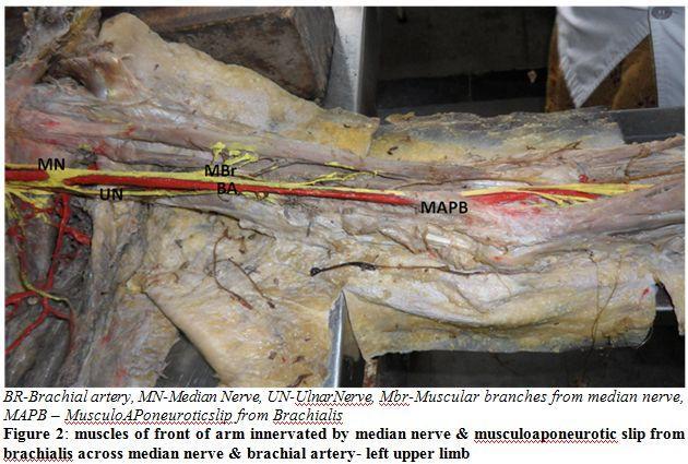
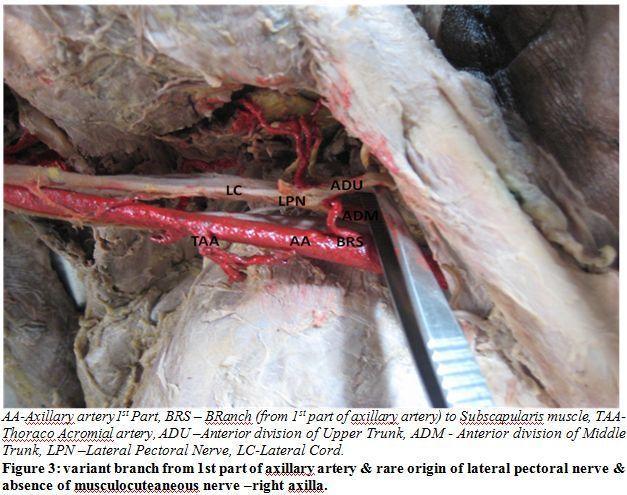
1 BILATERAL RARE NEURO VASCULAR VARIATIONS OF UPPER LIMB A CASE REPORT *N. B. S. Parimala Department of Anatomy, Dr. Pinnamaneni Siddhartha Institute of Medical Sciences & Research Foundation, Chinnaoutpalli, Gannavaram Mandal, Krishna District (AP), INDIA. * Author for Correspondence ABSTRACT Axillary artery is the direct continuation of subclavian artery at the outer border of 1 st rib. In the axilla this artery forms the central axis around which cords of brachial plexus are arranged. Variations in branches of axillary artery are not uncommon. Axillary artery continues as Brachial artery at the lower border of teres major, occasionally it may bifurcate in the middle of arm thus forming superficial radial artery. Variations in the branches of brachial plexus, particularly communications between musculocutaneous nerve and the median nerve were extensively studied owing to their surgical importance. Absence of musculocutaneous nerve is comparatively rare entity that poses difficulty in interpretation of cause of muscle weakness in injury to median nerve as it supplies the muscles of front of arm directly instead of musculocutaneous nerve. Neurovascular entrapment of median nerve and brachial artery by musculoaponeurotic slip from brachialis is a clinically important finding as it can result in compression neuropathy of median nerve and vascular compression symptoms. Key Words: Brachilias Axillary Artery, Superficial Radial Artery, Musculocutaenous Nerve, Median Nerve, INTRODUCTION The Axillary artery can be considered as the central axis of the axilla around which veins & brachial plexus are arranged. This artery is direct continuation of the subclavian artery, the change in name occurring as the vessel crosses the 1 st rib. The Axillary artery is described as having 3 parts in relation to the pectoralis minor muscle. The axillary artery usually gives rise to 6 branches. The first part of the artery gives superior thoracic artery. The second part gives lateral thoracic and thoracoacromial branches. The third part gives subscapular artery, anterior and posterior circumflex humeral arteries. It is very common to find the variations in the branching pattern. Sometimes many of the branches may originate from a common stem or arise separately (Hollinshed, 1958). Variations in the branches of 1 st part of the axillary artery is less common when compared to other 2 parts. In the present case an unusual branch was bilaterally observed from 1 st part of axillary artery closely related with anterior divisions of upper & middle trunks of Brachial plexus (Figure1 &3). The axillary artery becomes brachial as it crosses the tendon of teres major and it ends by dividing into radial and ulnar arteries in the ante cubital fossa. The named branches of brachial artery before its termination are profunda brachii, nutrient artery, superior and inferior ulnar collateral arteries. Higher bifurcation of brachial artery is a common variation reported by many authors. It may be unilateral or bilateral. In the present case it is unilateral. Anatomical variations of the radial artery are of clinical importance in creating native arteriovenous fistula for hemodialysis. Normally, the lateral cord gives its first branch to the pectoralis major muscle, named the lateral Pectoral nerve and then it divides into the musculocutaneous nerve and the lateral root of the median nerve. The musculocutaneous nerve is normally the terminal branch of lateral cord of brachial plexus and it pierces the coracobrachialis muscle. Before it pierces the coracobrachialis, it supplies the muscle. After piercing the coracobrachialis, it descends in between the biceps muscle and brachialis muscle and supplies the 194

2 above muscles and descends as lateral cutaneous nerve of forearm. Number of variations in the course and distribution of the musculocutaneous nerve have been reported.the median nerve is normally formed by two roots, the lateral root and the medial root from the corresponding cords of the brachial plexus. It descends crossing the brachial artery from lateral to medial side and descends without giving any branch in the arm. The present case reports bilateral absence of musculocutaneous nerve where median nerve by its direct branches innervates muscles of anterior compartment of arm. Injury to such median nerve results in unexpected. CASES During routine dissection of( I st M.B;B.S students batch ) a elderly male cadaver aged 65 yrs at pinnamaneni Siddhartha institute of medical sciences & research foundation Gannavaram, Krishna District, AP (INDIA),the following variations were observed. Observations with Bilaterality An unusual branch from 1 st part of axillary artery was observed associated with variation in branching pattern of lateral cord with bilateral similarity. It was noticed that an unusual branch originated from superior aspect of 1 st part of axillary artery opposite to superior thoracic artery. The course of above said artery was upwards and backwards between the anterior divisions of upper & middle trunks, descends deep to subscapularis muscle to supply the same. On both sides lateral pectoral nerve takes origin from anterior divisions of upper &middle trunks and lateral cord continues as median nerve after its formation from medial and lateral roots of corresponding cords without giving rise to musculocutaneous nerve. The median nerve gives direct muscular branches to biceps,coraco brachialis & brachialis.the nerve to brachialis continues as lateral ante brachial cutaneous nerve (Figure 1&2). 195
3 196

4 Other Unilateral Variations on Left Side In addition to above bilateral observations, 3 rd part of left axillary artery presented with common trunk for sub scapular & posterior circumflex arteries which is commonly found. The median nerve which is lateral to 3 rd part of axillary artery continues to have the same relation with brachial artery upto cubital fossa without crossing it from lateral to medial side and both brachial artery and median nerve are enclosed by musulo aponeurotic slip from infero medial aspect of brachialis muscle. At the apex of cubital fossa median nerve passes between 2 heads of pronator teres and its muscular branches cross the brachial artery to reach the muscle, further course of the nerve was normal (Figure 2). Other Unilateral Variations on Right Side Brachial artery which is the continuation of 3 rd part of axillary artery bifurcates into radial and ulnar arteries at the level of insertion of coraco brachialis. The median nerve lies lateral to 3 rd part of axillary artery and upper one third of Brachial artery after which it is crossed by radial artery from medial to lateral side, further course of the nerve was normal.the radial artery has a superficial course and its termination was normal (Figure 4).This variation is called higher bifurcation of Brachial artery. DISCUSSION De Garis (1928) reported that unnamed branch arises from superior aspect of axillary artery as its 2 nd branch and supplies upper part of sub scapularis muscle and found this vessel in 13.5% of whites and19.6%of negros. According to Bergmann et al., (1988) the first part of the axillary artery may, in rare cases, give rise to the subscapular artery or supply branch to the subscapular muscle. Any type of variation in branching pattern of first part of axillary artery were not found in study conducted by Samta gaur et al.,(2012) and in 2% cases posterior circumflex humeral artery a large trunk, arises along with subscapular artery. Clemente (1985) reported the higher origin of radial artery which is one of the common variations of upper limb arteries. The higher origin of radial artery in present case was superficial throughout its course 197
5 from its origin in the arm to palm where it continued with the superficial palmar arch. The part of the radial artery arising in the arm is brachio-radial, in the present case it is superficial throughout its course, it is known as superficial brachio-radial artery (Rodriguez-Niedenfuhr et al., 2003). The arterial anomalies in the upper limb are due to defects in embryonic development of the vascular plexus in the upper limb buds. This may be due to arrest at any stage of development of the vascular plexus showing regression, retention or reappearance and may lead to variations in the arterial origins and courses of the major upper limb vessels (Hamilton et al., 1972). The complex nature of brachial plexus makes it prone for innumerable variations of which communications between median nerve and musculocutaneous nerve were extensively studied.the present case is different because lateral cord of brachial plexus continues as lateral root of median nerve and does not give rise to any other branches. According to Kerr (1918) the lateral pectoral nerve may arise from lateral cord, from the anterior divisions of upper & middle trunks or in various ways from upper part of plexus by 1,2or 3 roots. In the present case lateral pectoral nerve arises by 2 roots from the anterior divisions of upper & middle trunks and is not reported any where in the recent studies. The variations of the musculocutaneous and median nerve may be classified into five types by Le minor. In Type V: The musculocutaneous nerve is absent. The fibers of the musculocutaneous nerve runs within the median nerve along its course. In this type the musculocutaneous nerve does not pierce the coracobrachialis muscle (Le Minor, 1990). The present observation is similar to the above said type. Absence of the musculocutaneous nerve was also reported by Mane et al., (2011). Bilecenoglu et al., (2005) described abnormal slip arising from brachialis muscle passing superficial to and causing entrapment of the nerve and the artery. According to Bilecenoglu et al., (2005) there are 7 possible compression sites for the median nerve and brachialis stands the 1 st among them. An extra Musculoaponeurotic slip from brachialis is not uncommon.the additional slip may mechanically stabilize the ulnohumeral joint, but can cause compression neuropathy of median nerve and vascular compression symptoms due to entrapment of brachial artery (Bincy et al., 2008). Conclusion Clinical implications of the above mentioned variations occurs in cases where a person with such variations suffers from injury to median nerve at the axilla or in the arm and have unexpected paralysis of the flexor muscles of the arm and hypoesthesia of the lateral surface of forearm. Prior knowledge of such variations by surgeons repairing trauma of the arm or treating tumors may be helpful to avoid accidental injury to these nerves. Variations assume significance during nerve block of infraclavicular part of the brachial plexus. Though the variations that we have mentioned here may not alter the normal functioning of the limb of the individual, it is important to keep these in mind in surgical and anaesthesiological procedures (SANNES, 2000). ACKNOWLEDGEMENT I thank my family for their support and encouragement. REFERENCES Bergman RA et al. (1988). Compendium of human anatomic variation. Munich and Baltimore Bilecenoglu B et al. (2005). Possible anatomic structures causing entrapment neuropathies of the median nerve: An anatomic study. Acta Orthopedica Belgica-Journal Bincy M et al. (2008). Median nerve and brachial artery entrapment in the abnormal brachialis muscle a case report. Neuroanatomy Clemente CD (1985). Anatomy of the Human Body. 30th Ed., Philadelphia, Lea & Febiger 829. De Garis CF and Swartley WB (1928). The axillary artery in white and negro stocks. American Journal of Anatomy
6 Hamilton WJ et al. (1972). Cardiovascular system. In: Human embryology, 4th ed. Baltimore, Williams and Wilkins Hollinshed WH (1958). Anatomy for surgeons in general surgery of upper limb, the back and limbs. A Heber Harper Book, New York Kerr AT (1918). The Brachial Plexus of Nerves. In Man:The variations in its formation and branches.american Journal of Anatomy Le Minor JM (1990). A rare variation of the median and musculocutaneous nerves in man. Archives of Anatomy, Histology and the Embryology Rodriguez-Niedenfuhr M et al. (2003). Arterial patterns of the human upper limb: update of anatomical variations and embryological development. Europian Journal of Anatomy 7(1) Samta gaur et al. (2012). A Cadaveric Study of Branching Pattern of the Axillary Artery. International Journal of Biological and Medical Research 3(1) Sannes Rey HD et al. (2000). Development of the nervous system in: Axon growth and guidance. New York, Academic Press UW Mane et al. (2011). Absence of Musculocutaneous Nerve Along With Accessory Head of Biceps Brachii. International Journal of Recent Trends in Science and Technology 1(2)
International Journal of Medical and Health Sciences
 International Journal of Medical and Health Sciences Journal Home Page: http://www.ijmhs.net ISSN: 2277-4505 Case Report An Unusual Branching Pattern of the Axillary Artery and Brachial Artery- A Case
Key Relationships in the Upper Limb
 Key Relationships in the Upper Limb This list contains some of the key relationships that will help you identify structures in the lab. They are organized by dissection assignment as defined in the syllabus.
The arm: *For images refer back to the slides
 The arm: *For images refer back to the slides Muscles of the arm: deltoid, triceps (which is located at the back of the arm), biceps and brachialis (it lies under the biceps), brachioradialis (it lies
Al-Balqa Applied University
 Al-Balqa Applied University Faculty Of Medicine *You can use this checklist as a guide to you for the lab. the items on this checklist represent the main features of the models that you have to know for
Fascial Compartments of the Upper Arm
 Fascial Compartments of the Upper Arm The upper arm is enclosed in a sheath of deep fascia and has two fascial septa: 1- Medial fascial septum (medial intermuscular septum): attached to the medial supracondylar
Multiple variations involving all the terminal branches of the brachial plexus and the axillary artery a case report
 SHORT REPORT Eur J Anat, 10 (3): 61-66 (2006) Multiple variations involving all the terminal branches of the brachial plexus and the axillary artery a case report K. Ramachandran, I. Kanakasabapathy and
UNILATERAL THIRD HEAD OF BICEPS BRACHII WITH ASSOCIATED NEUROVASCULAR VARIANTS IN BOTH THE UPPER LIMBS OF A SINGLE CADAVER ABSTRACT
 Asian Journal of Medical Science, Volume-5(2014) UNILATERAL THIRD HEAD OF BICEPS BRACHII WITH ASSOCIATED NEUROVASCULAR VARIANTS IN BOTH THE UPPER LIMBS OF A SINGLE CADAVER CASE REPORT,Vol-5 No.1 http://nepjol.info/index.php/ajms
Slides of Anatomy. Spring Dr. Maher Hadidi, University of Jordan
 Slides of Anatomy Please note : These slides are Dr. Maher Hadidi s slides of spring 2016 and were edited by the Premed Academic Team to fit the slides of spring 2019. Spring 2019 Dr. Maher Hadidi, University
The Arm and Cubital Fossa
 The Arm and Cubital Fossa Dr. Andrew Gallagher School of Anatomical Sciences University of the Witwatersrand Introduction The ARM (BRACHIUM) is the most proximal segment of the upper limb musculoskeletal
Upper limb Pectoral region & Axilla
 Upper limb Pectoral region & Axilla 黃敏銓 mchuang@ntu.edu.tw 1 Pectoral region Intercostal nerve Anterior branch of lateral cutaneous branch Lateral cutaneous branch Anterior cutaneous branch Anterior cutaneous
Gateway to the upper limb. An area of transition between the neck and the arm.
 Gateway to the upper limb An area of transition between the neck and the arm. Pyramidal space inferior to shoulder @ junction of arm & thorax Distribution center for the neurovascular structures that serve
The arterial system of upper limb begins with the
 Kathmandu University Medical Journal (2009), Vol. 7, No. 3, Issue 27 Case Note Multiple arterial anomalies in upper limb Baral P 1, Vijayabhaskar P 2, Roy S 1, Kumar S 2, Ghimire S 3, Shrestha U 3 1 Lecturer,
BRACHIAL PLEXUS. DORSAL SCAPULAR NERVE (C5) supraclavicular branch innervates rhomboids (major and minor) and levator scapulae
 supraclavicular branch innervates rhomboids (major and minor) and levator scapulae") THE BRACHIAL PLEXUS DORSAL SCAPULAR NERVE (C5) supraclavicular branch innervates rhomboids (major and minor) and levator scapulae SCHEMA OF THE BRACHIAL PLEXUS THE BRACHIAL PLEXUS PHRENIC NERVE supraclavicular
THE BRACHIAL PLEXUS DORSAL SCAPULAR NERVE (C5) supraclavicular branch innervates rhomboids (major and minor) and levator scapulae SCHEMA OF THE BRACHIAL PLEXUS THE BRACHIAL PLEXUS PHRENIC NERVE supraclavicular
Variations of median nerve and musculocutaneous nerve: Cadeveric study
 Original article: Variations of median nerve and musculocutaneous nerve: Cadeveric study 1Dr.VaishaliBondge*, 2 Dr. Ashok Khade, 3 Dr. P.H.Shingare 1Assistant Professor, Grant Medical College, Mumbai,
region of the upper limb between the shoulder and the elbow Superiorly communicates with the axilla.
 1 region of the upper limb between the shoulder and the elbow Superiorly communicates with the axilla. Inferiorly, a number of important structures pass between arm & forearm through cubital fossa. 2 medial
Upper limb Arm & Cubital region 黃敏銓
 Upper limb Arm & Cubital region 黃敏銓 1 Arm Lateral intermuscular septum Anterior (flexor) compartment: stronger Medial intermuscular septum Posterior (extensor) compartment 2 Coracobrachialis Origin: coracoid
MUSCLES. Anconeus Muscle
 LAB 7 UPPER LIMBS MUSCLES Anconeus Muscle anconeus origin: distal end of dorsal surface of humerus insertion: lateral surface of ulna from distal margin of the semilunar notch to proximal end of the olecranon
Brachial plexuses and axillary lymph nodes
 Brachial plexuses and axillary lymph nodes Introduction about nervous system nervous system central nervous system periphral nervous system brain spinal cord 31 pairs of spinal nerves 12 paris of cranial
Axilla and Brachial Region
 L 4 A B O R A T O R Y Axilla and Brachial Region BRACHIAL PLEXUS 5 Roots/Rami (ventral rami C5 T1) 3 Trunks Superior (C5, C6) Middle (C7) Inferior (C8, T1) 3 Cords Lateral Cord (Anterior Superior and Anterior
*the Arm* -the arm extends from the shoulder joint (proximal), to the elbow joint (distal) - it has one bone ; the humerus which is a long bone
, to the elbow joint (distal) - it has one bone ; the humerus which is a long bone") *the Arm* -the arm extends from the shoulder joint (proximal), to the elbow joint (distal) - it has one bone ; the humerus which is a long bone - muscles in the arm : *brachialis muscle *Biceps brachii
*the Arm* -the arm extends from the shoulder joint (proximal), to the elbow joint (distal) - it has one bone ; the humerus which is a long bone - muscles in the arm : *brachialis muscle *Biceps brachii
Nerves of the upper limb Prof. Abdulameer Al-Nuaimi. E. mail:
 Nerves of the upper limb Prof. Abdulameer Al-Nuaimi E-mail: a.al-nuaimi@sheffield.ac.uk E. mail: abdulameerh@yahoo.com Brachial plexus Median nerve After originating from the brachial plexus in the axilla,
Region of upper limb attachment to the trunk Proximal segment of limb overlaps parts of the trunk (thorax and back) and lower lateral neck.
 and lower lateral neck.") Region of upper limb attachment to the trunk Proximal segment of limb overlaps parts of the trunk (thorax and back) and lower lateral neck. includes Pectoral Scapular Deltoid regions of the upper limb
Region of upper limb attachment to the trunk Proximal segment of limb overlaps parts of the trunk (thorax and back) and lower lateral neck. includes Pectoral Scapular Deltoid regions of the upper limb
STRUCTURAL BASIS OF MEDICAL PRACTICE EXAMINATION 5 October 6, 2006
 STRUCTURAL BASIS OF MEDICAL PRACTICE EXAMINATION 5 October 6, 2006 PART l. Answer in the space provided. (8 pts) 1. Identify the structures. (2 pts) B C A. _pisiform B. _ulnar artery A C. _flexor carpi
3 Mohammad Al-Mohtasib Areej Mosleh
 3 Mohammad Al-Mohtasib Areej Mosleh ***Muscles Connecting the Upper Limb to the Vertebral Column 1.Trapezius Muscle ***The first muscle on the back is trapezius muscle, it s called so according
of the Axillary Artery
 Variation in the Origins of the Branches of the Axillary Artery DONALD F. HUELKE Department of Anatomy, The University of Michigan Medical School, Ann Arbor, Michigan Variability in the origin of certain
Bilateral Variations in the Branching Pattern of the Axillary Artery in a Single Cadaver
 Case Report Bilateral Variations in the Branching Pattern of the Axillary Artery in a Single Cadaver Dr. Purnendu Rang 1, Dr. Parijat Mukherjee 2, Dr. Aradhana Sanga 3, Dr. Arunima Nag (Ray) 4, Dr. Champak
Abnormal Pattern Of Brachial Plexus Formation: An Original Case Report. K Oluyemi, O Adesanya, D Ofusori, C Okwuonu, V Ukwenya, F Om'iniabohs, B Odion
 ISPUB.COM The Internet Journal of Neurosurgery Volume 4 Number 2 Abnormal Pattern Of Brachial Plexus Formation: An Original Case Report K Oluyemi, O Adesanya, D Ofusori, C Okwuonu, V Ukwenya, F Om'iniabohs,
Dr. Mahir Alhadidi Anatomy Lecture #9 Feb,28 th 2012
 Quick Revision: Upper arm is divided into two compartments: 1. Anterior Compartment: Contains three muscles (Biceps brachii, Coracobrachialis, Brachialis). Innervated by Musculocutaneous nerve. 2. Posterior
G24: Shoulder and Axilla
 G24: Shoulder and Axilla Syllabus - Pg. 2 ANAT 6010- Medical Gross Anatomy David A. Morton, Ph.D. Objectives Upper limb Systemically: Bones (joints) Muscles Nerves Vessels (arteries/veins) Fascial compartments
COMMUNICATIONS BETWEEN MUSCULOCUTANEOUS NERVE AND MEDIAN NERVE - A STUDY
 IJCRR Vol 05 issue 08 Section: Healthcare Category: Research Received on: 13/03/13 Revised on: 04/04/13 Accepted on: 21/04/13 COMMUNICATIONS BETWEEN MUSCULOCUTANEOUS NERVE AND MEDIAN NERVE - A STUDY Balasubramanian
Netter's Anatomy Flash Cards Section 6 List 4 th Edition
 Netter's Anatomy Flash Cards Section 6 List 4 th Edition https://www.memrise.com/course/1577581/ Section 6 Upper Limb (66 cards) Plate 6-1 Humerus and Scapula: Anterior View 1.1 Acromion 1.2 Greater tubercle
The Study of Anatomical Variations of Axillary Artery - A Case Report
 International Journal of Current Microbiology and Applied Sciences ISSN: 2319-7706 Volume 6 Number 1 (2017) pp. 639-644 Journal homepage: http://www.ijcmas.com Case Study http://dx.doi.org/10.20546/ijcmas.2017.601.077
Multiple Neurovascular... Pit Baran Chakraborty, Santanu Bhattacharya, Sumita Dutta.
 Multiple Neurovascular... Pit Baran Chakraborty, Santanu Bhattacharya, Sumita Dutta. Fig-3: Showing high formation of Median nerve. Fig-1: Showing atypical formation of cords of Brachial plexus. 1 = Upper
Upper Limb Muscles Muscles of Axilla & Arm
 Done By : Saleh Salahat Upper Limb Muscles Muscles of Axilla & Arm 1) Muscles around the axilla A- Muscles connecting the upper to thoracic wall (4) 1- pectoralis major Origin:- from the medial half of
Misc Anatomy. Upper Limb! 2. Lower Limb! 5. Venous Drainage! Head & neck! 8
 Misc Anatomy Upper Limb! 2 Arteries!... 2 Veins!... 2 Spaces!... 4 Lower Limb! 5 Arteries!... 5 Venous Drainage!... 6 Spaces!... 7 Head & neck! 8 Artery!... 8 Ultrasound View for IJ CVL!... 8 Arteries
Bifurcation Of Axillary Artery In Its 3rd Part - A Case Report
 166 J Anat. Soc. India 50(2) 166-169 (2001) Bifurcation Of Axillary Artery In Its 3rd Part - A Case Report *Patnaik, V.V.G.; **Kalsey, G.; **Singla, Rajan K. Department of Anatomy, Government Medical College,
divided by the bones ( redius and ulna ) and interosseous membrane into :
 and interosseous membrane into :") fossa Cubital Has: * floor. * roof : - Skin - superficial fasica - deep fascia ( include bicipital aponeurosis ) Structures within the roof : -cephalic and basilic veins -and between them median cubital
fossa Cubital Has: * floor. * roof : - Skin - superficial fasica - deep fascia ( include bicipital aponeurosis ) Structures within the roof : -cephalic and basilic veins -and between them median cubital
Gross Anatomy: Upper Extremity Arteries
 Gross Anatomy: Upper Extremity Arteries By: Trevor Lohman DPT Illustrated by: Dennis Breese 1 Subclavian and Axillary arteries Hardening of the heart ages people more quickly than hardening of the arteries
Neurovascular variations in upper limb
 Case report : Neurovascular variations in upper limb 1Dr. Dinendra Kumar Saha, 2 Dr. Jayeeta Burman, 3 Dr. Sudeshna Majumdar, 4 Dr. Manotosh Banerjee, 5 Dr. Sharmistha Chakraborty, 6 Dr. Sushmita Sen,
The Upper Limb III. The Brachial Plexus. Anatomy RHS 241 Lecture 12 Dr. Einas Al-Eisa
 The Upper Limb III The Brachial Plexus Anatomy RHS 241 Lecture 12 Dr. Einas Al-Eisa Brachial plexus Network of nerves supplying the upper limb Compression of the plexus results in motor & sensory changes
VENOUS DRAINAGE O US F UPPER UPPER LIM B BY dr.fahad Ullah
 VENOUS DRAINAGE OF UPPER LIMB BY dr.fahad Ullah Venous drainage of the supper limb The venous system of the upper limb drains deoxygenated blood from the arm, forearm and hand It can anatomically be divided
Nerves of Upper limb. Dr. Brijendra Singh Professor & Head Department of Anatomy AIIMS Rishikesh
 Nerves of Upper limb Dr. Brijendra Singh Professor & Head Department of Anatomy AIIMS Rishikesh 1 Objectives Origin, course & relation of median & ulnar nerves. Motor & sensory distribution Carpal tunnel
BOGOMOLETS NATIONAL MEDICAL UNIVERSITY. Department of Human Anatomy GUIDELINES. The theme of the lesson The vessels of the upper limb.
 BOGOMOLETS NATIONAL MEDICAL UNIVERSITY Department of Human Anatomy GUIDELINES Academic discipline HUMAN ANATOMY Module 2 The theme of the lesson The vessels of the upper limb. Course Faculties І Medical
The Elbow and the cubital fossa. Prof Oluwadiya Kehinde
 The Elbow and the cubital fossa Prof Oluwadiya Kehinde www.oluwadiya.com Elbow and Forearm Anatomy The elbow joint is formed by the humerus, radius, and the ulna Bony anatomy of the elbow Distal Humerus
Candidate s instructions Look at this cross-section taken at the level of C5. Answer the following questions.
 Section 1 Anatomy Chapter 1. Trachea 1 Candidate s instructions Look at this cross-section taken at the level of C5. Answer the following questions. Pretracheal fascia 1 2 5 3 4 Questions 1. Label the
Neurovascular Variations in Upper Limb
 Case report : Neurovascular Variations in Upper Limb 1Dr. Dinendra Kumar Saha, 2 Dr. Jayeeta Burman, 3 Dr. Sudeshna Majumdar, 4 Dr. Manotosh Banerjee, 5 Dr. Sharmistha Chakraborty, 6 Dr. Sushmita Sen,
Anatomy Workshop Upper Extremity David Ebaugh, PT, PhD Workshop Leader. Lab Leaders: STATION I BRACHIAL PLEXUS
 Anatomy Workshop Upper Extremity David Ebaugh, PT, PhD Workshop Leader Lab Leaders: STATION I BRACHIAL PLEXUS A. Posterior cervical triangle and axilla B. Formation of plexus 1. Ventral rami C5-T1 2. Trunks
Scapular and Deltoid Regions
 M1 Gross and Developmental Anatomy Scapular and Deltoid Regions Dr. Peters 1 Outline I. Skeleton of the Shoulder and Attachment of the Upper Extremity to Trunk II. Positions and Movements of the Scapula
*Our main subject is the brachial plexus but it's important to understand the spinal cord first in order to understand the brachial plexus.
 *Our main subject is the brachial plexus but it's important to understand the spinal cord first in order to understand the brachial plexus. *Vertebral column is formed by the union of 33 sequential vertebrae
CASE REPORT. HIGH DIVISION OF BRACHIAL ARTERY A CASE REPORT K. Smitha Elizabeth
 HIGH DIVISION OF BRACHIAL ARTERY A CASE REPORT K. Smitha Elizabeth 1. Assistant Professor. Department of Anatomy, Shri B M Patil medical College & Research Centre, Bijapur. CORRESPONDING AUTHOR K. Smitha
BRACHIAL PLEXUS 11/12/2014 كيف تتكون الضفيرة FORMATION ENLARGEMENT (INTUMESCENCE) OF THE SPINAL CORD. Grey matter. Cervical intumescence - C 6 - T 2
 OF THE SPINAL CORD. Grey matter. Cervical intumescence - C 6 - T 2") BRACHIAL PLEXUS Prof. Fawzy Elnady ENLARGEMENT (INTUMESCENCE) OF THE SPINAL CORD Grey matter Cervical intumescence - C 6 - T 2 Lumbar intumescence - L 4 S 2 كيف تتكون الضفيرة FORMATION The ventral rami
BRACHIAL PLEXUS Prof. Fawzy Elnady ENLARGEMENT (INTUMESCENCE) OF THE SPINAL CORD Grey matter Cervical intumescence - C 6 - T 2 Lumbar intumescence - L 4 S 2 كيف تتكون الضفيرة FORMATION The ventral rami
VARIANT ARTERIAL PATTERN IN THE FOREARM WITH ITS EMBRYOLOGICAL BASIS. Vaishnavi Joshi and Dr. Shaheen Sajid Rizvi
 Volume-8, Issue-3 July-Sept-2018 Coden:IJPAJX-CAS-USA, Copyrights@2018 ISSN-2231-4490 Received: 8 th June-2018 Revised: 15 th July-2018 Accepted: 16 th July-2018 DOI: 10.21276/Ijpaes http://dx.doi.org/10.21276/ijpaes
Nerve Injury. 1) Upper Lesions of the Brachial Plexus called Erb- Duchene Palsy or syndrome.
 Upper Lesions of the Brachial Plexus called Erb- Duchene Palsy or syndrome.") Nerve Injury - Every nerve goes to muscle or skin so if the nerve is injured this will cause paralysis in the muscle supplied from that nerve (paralysis means loss of function) then other muscles and other
Nerve Injury - Every nerve goes to muscle or skin so if the nerve is injured this will cause paralysis in the muscle supplied from that nerve (paralysis means loss of function) then other muscles and other
Variation in Branching Pattern of Brachial Artery
 Original Article Print ISSN: 2321-6379 Online ISSN: 2321-595X DOI: 10.17354/ijss/2017/192 Variation in Branching Pattern of Brachial Artery Amandeep Kaur 1, Anshu Sharma 2, Mahesh Sharma 3 1 Demonstrator,
Supplied in part by the musculocutaneous nerve. Forms the axis of rotation in movements of pronation and supination
 Anatomy: Upper limb (15 questions) 1. Latissimus Dorsi: Is innervated by the dorsal scapular nerve Lies above feres major muscle Medially rotates the humerus All of the above 2. Supinator muscle is: Deep
Downloaded from umj.umsu.ac.ir at 20: on Friday March 22nd
 * 1391/04/04 1392/02/01.. :... : - - : : Email: sazegargh@mums.ac.ir.( ) () () () (). (). () ... ( ) :(). :() ( ). .. :() ( ). :() ( ) () () ( ) () ().() (). ...... References: 1. Standring S. Grays Anatomy
Anatomical variations of the median nerve distribution and communication in the arm
 O R I G I N A L A R T I C L E Folia Morphol. Vol. 63, No. 3, pp. 313 318 Copyright 2004 Via Medica ISSN 0015 5659 www.fm.viamedica.pl Anatomical variations of the median nerve distribution and communication
Definition of anatomy 1 Questions 5
 Contents Chapter 1: Introduction 1 5 Definition of anatomy 1 Questions 5 Chapter 2: Skeletal System 6 74 Skeleton 6 Skeletal system 8 Bones of superior extremity 12 Articulated skeleton of hand 17 Clinical
STRUCTURAL BASIS OF MEDICAL PRACTICE EXAMINATION 5. September 30, 2011
 STRUCTURAL BASIS OF MEDICAL PRACTICE EXAMINATION 5 September 30, 2011 PART l. Answer in the space provided. (12 pts) 1. Identify the structures. (2 pts) EXAM NUMBER A. Suprascapular nerve B. Axillary nerve
Anatomy of the Musculoskeletal System
 Anatomy of the Musculoskeletal System Kyle E. Rarey, Ph.D. Department of Anatomy & Cell Biology and Otolaryngology University of Florida College of Medicine Outline of Presentation Vertebral Column Upper
213: HUMAN FUNCTIONAL ANATOMY: PRACTICAL CLASS 1: Proximal bones, plexuses and patterns
 213: HUMAN FUNCTIONAL ANATOMY: PRACTICAL CLASS 1: Proximal bones, plexuses and patterns CLAVICLE Examine an isolated clavicle and compare it with a clavicle on an articulated skeleton. Viewed from above,
Peripheral Nervous Sytem: Upper Body
 Peripheral Nervous Sytem: Upper Body MSTN121 - Neurophysiology Session 10 Department of Myotherapy Cervical Plexus Accessory nerve (CN11 + C1-5) Motor: trapezius and sternocleidomastoid Greater auricular
JMSCR Vol 3 Issue 12 Page December 2015
 www.jmscr.igmpublication.org Impact Factor 3.79 Index Copernicus Value: 5.88 ISSN (e)-2347-176x ISSN (p) 2455-0450 DOI: http://dx.doi.org/10.18535/jmscr/v3i12.41 A Cadaveric Study on Division of Brachial
This figure (of humerus) is from Dr. Maher's newest slides. -Its added here just for consideration-
 is from Dr. Maher's newest slides. -Its added here just for consideration-") This figure (of humerus) is from Dr. Maher's newest slides. -Its added here just for consideration- Slides of Anatomy Please note : These slides are Dr. Maher Hadidi s slides of spring 2016 and were edited
This figure (of humerus) is from Dr. Maher's newest slides. -Its added here just for consideration- Slides of Anatomy Please note : These slides are Dr. Maher Hadidi s slides of spring 2016 and were edited
THE SUPERFiCiAL BRACHIAL ARTERY: A CASE REPORT
 115 THE SUPERFiCiAL BRACHIAL ARTERY: A CASE REPORT J. L. PACE M.D. (MALTA), PH.D. (LOND.) Lecturer, Department of Anatomy, Royal University of Malta. and A. E. FELICE, A. J. FELICE, A. G. FELICE, E. FIORENTINO
Human Anatomy Biology 351
 1 Human Anatomy Biology 351 Upper Limb Exam Please place your name on the back of the last page of this exam. You must answer all questions on this exam. Because statistics demonstrate that, on average,
David G. Simpson, Ph.D.
 David G. Simpson, Ph.D. ARM & CUBITAL FOSSA Revised 7/08 Text References Moores 3 rd ed., p402 408, 436 439, 439 443, 478, 481 LEARNING OBJECTIVES: 1. Describe the humerus, indicating the sites of muscle
#12. Joint نبيل خوري
 #12 30 Anatomy Joint هيام الر جال 9/10/2015 نبيل خوري Salam Awn Some notes before starting : ** Not all slides are included, so I recommend having a look at the slides beside this sheet ** If you find
Research article - Human anatomy case report Abnormal branching of the axillary artery: an axillohepatic
 IJAE Vol. 121, n. 2: 172-178, 2016 ITALIAN JOURNAL OF ANATOMY AND EMBRYOLOGY Research article - Human anatomy case report Abnormal branching of the axillary artery: an axillohepatic artery Pranit N Chotai
Functional anatomy and variability of the blood vessels of the upper and lower limbs. Anastasia Bendelic Human Anatomy Departament
 Functional anatomy and variability of the blood vessels of the upper and lower limbs Anastasia Bendelic Human Anatomy Departament Plan: 1. Variations of the branching pattern of the aortic arch 2. Arterial
Clinico-AnatomicalStudy ofananomalousaxillobrachi-palmar Artery ARare ArterialDuplication
 Global Journal of Science Frontier Research Interdisciplinary Volume 13 Issue 3 Version 1.0 Year 2013 Type : Double Blind Peer Reviewed International Research Journal Publisher: Global Journals Inc. (USA)
Origin of the subscapular artery in the South African Black population
 O R I G I N L R T I C L E Folia Morphol. Vol. 73, No. 4, pp. 486 491 DOI: 10.5603/FM.2014.0073 Copyright 2014 Via Medica ISSN 0015 5659 www.fm.viamedica.pl Origin of the subscapular artery in the South
Anatomical variations of brachial plexus: anomalous branching pattern
 International Journal of Research in Medical Sciences Gopal K et al. Int J Res Med Sci. 2016 Aug;4(8):3376-3380 www.msjonline.org pissn 2320-6071 eissn 2320-6012 Research Article DOI: http://dx.doi.org/10.18203/2320-6012.ijrms20162297
Biology 323 Human Anatomy for Biology Majors Lecture 11 Dr. Stuart S. Sumida. Peripheral Circulation
 Biology 323 Human Anatomy for Biology Majors Lecture 11 Dr. Stuart S. Sumida Peripheral Circulation Structures of the Splanchnopleure: receive unpaired vessels of the abdominal aorta. Structures of the
AN UNUSUAL AXILLARY ARTERY VARIATION
 Case Report AN UNUSUAL AXILLARY ARTERY VARIATION Pallab Kumar Saha 1, Tanwi Ghosal (Sen) * 2, Parijat Mukherjee 2, Anupam Baske 2, Maitreyi Mondal 3, Sudeshna Majumder 4. ABSTRACT Background: Axillary
FUNCTIONAL ANATOMY OF SHOULDER JOINT
 FUNCTIONAL ANATOMY OF SHOULDER JOINT ARTICULATION Articulation is between: The rounded head of the Glenoid cavity humerus and The shallow, pear-shaped glenoid cavity of the scapula. 2 The articular surfaces
Cubital fossa and forearm
 Cubital fossa and forearm Cubital fossa is the triangular space in front of elbow joint. - The Cubital fossa has boundaries: apex, base, roof and floor and it has contents. The base: an imaginary horizontal
The Clavicle Right clavicle Deltoid tubercle: Conoid tubercle, conoid ligamen Impression for the
 The Clavicle Muscle Attachment Sites in the Upper Limb Pectoralis major Right clavicle Smooth superior surface of the shaft, under the platysma muscle tubercle: attachment of the deltoid Acromial facet
Muscles of the Upper Limb
 Muscles of the Upper Limb anterior surface of ribs 3 5 coracoid process Pectoralis minor pectoral nerves protracts / depresses scapula Serratus anterior Subclavius ribs 1-8 long thoracic nerve rib 1 ----------------
MCQWeek2. All arise from the common flexor origin. The posterior aspect of the medial epicondyle is the common flexor origin.
 MCQWeek2. 1. Regarding superficial muscles of anterior compartment of the forearm: All arise from the common flexor origin. The posterior aspect of the medial epicondyle is the common flexor origin. Flexor
Abduction of arm until your hand rich your head. Flexion of forearm at elbow joint. Extension of arm at elbow joint. Flexion of fingers 10.
 Num. answer 1. Medialy With the manubrium ( sternum ), and laterally with the acromion of the scapula 2. 1. Trapezius 2. Levator scapulae 3. Rhomboids 3. 1. Pectoralis major 2. Pectoralis minor 3. Latissiumus
Analysis of the Morphological Variations between Musculocutaneous Nerve and Median Nerve -A cadaveric study
 Original Research Article Analysis of the Morphological Variations between Musculocutaneous Nerve and Median Nerve -A cadaveric study Neeraj T. Master 1,*, Deepa S. Gupta 2 1 Resident & Tutor, 2 Professor
LIST OF STRUCTURES TO BE IDENTIFIED IN LAB: UPPER EXTREMITY REVIEW 2016
 LIST OF STRUCTURES TO BE IDENTIFIED IN LAB: UPPER EXTREMITY REVIEW 2016 BONES Ribs, sternum, clavicle Humerus: Head, greater tubercle, lesser tubercle, intertubercular sulcus, surgical neck, anatomical
Pectoral region. Lecture 2
 Pectoral region Lecture 2 Muscle Action Each muscle has: Origin Beginning. Insertion End. Body (belly). Law: When a muscle performs its action, its insertion, moves towards its origin. Spring 2016 Dr.
Practical 2 Worksheet
 Practical 2 Worksheet Upper Extremity BONES 1. Which end of the clavicle is on the lateral side (acromial or sternal)? 2. Describe the difference in the appearance of the acromial and sternal ends of the
ONTOGENY RECAPITULATES PHYLOGENY: COMMUNICATION BETWEEN THE MEDIAN AND THE MUSCULOCUTANEOUS NERVE: A CASE REPORT
 Case Report ONTOGENY RECAPITULATES PHYLOGENY: COMMUNICATION BETWEEN THE MEDIAN AND THE MUSCULOCUTANEOUS NERVE: A CASE REPORT Subhra Mandal * 1, Moumita Saha 2, Prabir Mandal 3, Ramprasad Saha 4. ABSTRACT
BILATERAL MULTIPLE VARIATIONS IN THE UPPER EXTREMITY OF A HUMAN CADAVER: A CASE REPORT
 Case Report BILATERAL MULTIPLE VARIATIONS IN THE UPPER EXTREMITY OF A HUMAN CADAVER: A CASE REPORT Soniya A Gupta 1, Saiprasad P Bhavsar * 2, Medha V Ambiye 3, Seema N Khambatta 4. ABSTRACT International
Dissertation submitted for
 BRACHIAL ARTERY, ITS BRANCHING PATTERN AND VARIATIONS WITH ITS CLINICAL APPLICATIONS Dissertation submitted for M.S. ANATOMY BRANCH - V DEGREE EXAMINATION THE TAMIL NADU DR.M.G.R.MEDICAL UNIVERSITY CHENNAI,
Gross Anatomy Questions That Should be Answerable After October 27, 2017
 Gross Anatomy Questions That Should be Answerable After October 27, 2017 1. The inferior angle of the scapula of a woman who was recently in an automobile accident seems to protrude making a ridge beneath
International Journal of Pharma and Bio Sciences A CADAVERIC STUDY OF THE AXILLARY ARTERY. ABSTRACT
 Research Article Allied Science International Journal of Pharma and Bio Sciences ISSN 0975-6299 A CADAVERIC STUDY OF THE AXILLARY ARTERY. HUMBERTO FERREIRA ARQUEZ* Professor of Human Morphology, Medicine
A Morphological Study on the Anatomic Variation of the Musculocutaneous Nerve with its Clinical Implication.
 Original Article ISSN (O):2395-2822; ISSN (P):2395-2814 A Morphological Study on the Anatomic Variation of the Musculocutaneous Nerve with its Clinical Implication. Ranjana S Arya 1, Suhasinee 2, Mrithunjay
Elbow Elbow Anatomy. Flexion extension. Pronation Supination. Anatomy. Anatomy. Romina Astifidis, MS., PT., CHT
 Elbow Elbow Anatomy Romina Astifidis, MS., PT., CHT Curtis National Hand Center Baltimore, MD October 6-8, 2017 Link between the arm and forearm to position the hand in space Not just a hinge Elbow = 70%
Bilateral single cord of the brachial plexus in an adult female cadaver of South Indian origin
 Case Report pissn 2093-3665 eissn 2093-3673 Bilateral single cord of the brachial plexus in an adult female cadaver of South Indian origin Uma Viswanathan, Vigneswaran Madhivadhany, Nachiket Shankar Department
AN UNUSUAL TORTUOUS BRACHIAL ARTERY AND ITS BRANCHES: HISTOLOGICAL BASIS AND ITS CLINICAL PERSPECTIVE
 Int. J. LifeSc. Bt & Pharm. Res. 2014 Ashwini C and Vasantha Kuberappa, 2014 Research Paper ISSN 2250-3137 www.ijlbpr.com Vol. 3, No. 2, April 2014 2014 IJLBPR. All Rights Reserved AN UNUSUAL TORTUOUS
Assessment of the Brachial Plexus EMG Course CNSF Halifax Fraser Moore, Canadian Society of Clinical Neurophysiology McGill University
 Assessment of the Brachial Plexus EMG Course CNSF Halifax 2018 Fraser Moore, Canadian Society of Clinical Neurophysiology McGill University Angela Scott, Association of Electromyography Technologists of
compartments of the forearm
 " forearm posterior compartment " compartments of the forearm Posterior Fascial compartment Muscles: ** The superficial group 1. Extensor carpi radialis brevis 2. Ex. digitorum 3. Ex. digiti minimi 4.
Human Anatomy and Physiology I Laboratory Spinal and Peripheral Nerves and Reflexes
 Human Anatomy and Physiology I Laboratory Spinal and Peripheral Nerves and Reflexes 1 This lab involves the second section of the exercise Spinal Cord, Spinal Nerves, and the Autonomic Nervous System,
Posterior Triangle of the Neck By Prof. Dr. Muhammad Imran Qureshi
 Posterior Triangle of the Neck By Prof. Dr. Muhammad Imran Qureshi For the purpose of anatomical description the neck is sub divided into two major triangles, the Anterior and the Posterior by muscle bellies
26/9/2016. Anatomy. 1 Nour Erekat Wejdan Amer
 26/9/2016 Anatomy st 1 Nour Erekat Wejdan Amer Notes before we start the lecture. Bring any colored Atlas with you to the lab. The main reference is clinical anatomy by regions by Richard snell the 9 th
The Muscular System. Chapter 10 Part C. PowerPoint Lecture Slides prepared by Karen Dunbar Kareiva Ivy Tech Community College
 Chapter 10 Part C The Muscular System Annie Leibovitz/Contact Press Images PowerPoint Lecture Slides prepared by Karen Dunbar Kareiva Ivy Tech Community College Table 10.9: Muscles Crossing the Shoulder