The role of mechanical stimuli induced by prenatal movements in skeletal development
|
|
|
- Gerard Griffin
- 5 years ago
- Views:
Transcription
1 The role of mechanical stimuli induced by prenatal movements in skeletal development Niamh Nowlan, PhD Department of Bioengineering, Imperial College London


2 Mechanobiology of Bone


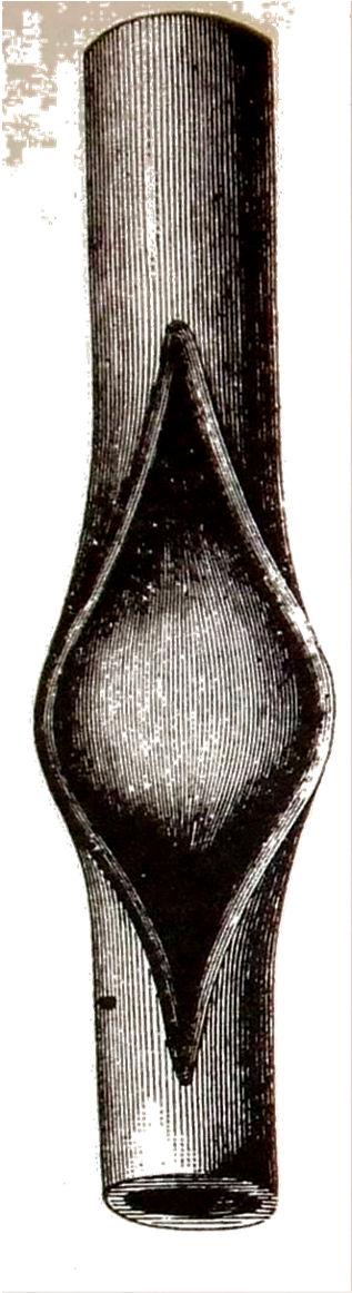
3 Mechanoregulation of Skeletogenesis Rodriguez et al., JBJS, 1988
 Roux, W., 1894. Arch. Entwicklungsmech. Org. 1: 1-42.")
4 Developmental Mechanics (Entwicklungsmechanik) the goal of developmental mechanics (is) the ascertainment of formative forces or energies (Roux, 1894) Roux, W., Arch. Entwicklungsmech. Org. 1: Wilhelm Roux ( )
5 Developmental Mechanics (Entwicklungsmechanik) the goal of developmental mechanics (is) the ascertainment of formative forces or energies (Roux, 1894) To think that heredity will build organic beings without mechanical means is a piece of unscientific mysticism (His, 1888) Wilhelm His ( ) His, W., Proc. Roy. Soc. Edinburgh 15:


6 Developmental Mechanics (Entwicklungsmechanik) Wilhelm His ( ) His, W., Proc. Roy. Soc. Edinburgh 15:


7 Genetics Existence of genes; Gregor Mendel, 1866 Location of genes on chromosomes; Thomas Hunt Morgan, 1910 Structure of DNA; Watson & Crick, 1953 Watson, JD, Crick, FHC, Nature 171, pp

8 Research Approach Rot-Nicevic et al., 2006 Control Immobilised Animal Models of Abnormal Prenatal Movement 3-D Imaging of Embryonic Bones and Joints Finite Element Analysis
9 Relevance Treatment of congenital neuromuscular diseases alleviate or minimise effects on skeletal development Inform tissue engineering of cartilage and bone What biophysical stimuli needed for bone and/or articular cartilage? What cellular or genetic factors translate biophysical stimuli to developmental change?


10 Bone Development Mammalian Avian Fell, H.B., J. Morph. & Physiol., Hall, B.K., Clin. Orthop. Relat. Res., 1987
11 Part 1: Mechanoregulation of Bone Development in a Chick Immobilisation Model System
12 Part 1: Bone Development in Immobilised Chicks Hypothesis Mechanical forces induced by embryonic muscle contractions affect the development of skeletal tissues and structures through the action of mechanosensitive genes
13 Part 1: Bone Development in Immobilised Chicks Mechanosensitive Genes Genes which are up- or down- regulated as a result of a change in the mechanical environment Provot & Schipani, Biochem. Biophys. Res. Comm., 328(3), pp
14 Part 1: Bone Development in Immobilised Chicks Methods 1. Characterise biophysical stimuli in the embryonic limb using finite element (FE) analysis 2. Characterise expression patterns of candidate mechanosensitive genes involved in bone formation - Compare with patterns of biophysical stimuli 3. Examine effect of altered mechanical environment on mechanosensitive gene expression patterns
15 Part 1: Bone Development in Immobilised Chicks ¹Sharpe et al., 2002, Science 296, pp


16 Part 1: Bone Development in Immobilised Chicks Cartilage (Alcian Blue Staining) HH30 HH32 HH34 2.4mm 4.2mm 5.9mm


17 Part 1: Bone Development in Immobilised Chicks Muscle (Anti-myosin antibody staining) HH30 HH32 HH34 1 mm

18 Part 1: Bone Development in Immobilised Chicks Tendon (Scleraxis in situ)


19 Part 1: Bone Development in Immobilised Chicks Methods: Muscle load application HH32, extension contraction Muscle CSA Force Dorsal_ mm mn Dorsal_ mm mn Dorsal_ mm mn

20 Part 1: Bone Development in Immobilised Chicks Results: Shear Strain & Fluid Vel. Octahedral Shear Strain Fluid Velocity


21 Part 1: Bone Development in Immobilised Chicks HH30
22 Part 1: Bone Development in Immobilised Chicks HH30
23 Part 1: Bone Development in Immobilised Chicks HH32
24 Part 1: Bone Development in Immobilised Chicks HH34


25 Part 1: Bone Development in Immobilised Chicks Methods: Immobilisation Decamethonium Bromide (DMB) used to prevent muscle contractions in ovo
 R")


26 CONTROL (HH36) R J IMMOBILISED (HH36) S
27 Part 1: Bone Development in Immobilised Chicks Effect on Bone Collar Formation


28 Part 1: Bone Development in Immobilised Chicks

29 Part 1: Bone Development in Immobilised Chicks Effect on Indian Hedgehog Expression

30 Part 1: Bone Development in Immobilised Chicks Results on Collagen X Expression
31 Part 1: Bone Development in Immobilised Chicks Discussion: Chick Model Correlation between Ihh, ColX expression and patterns of biophysical stimuli No correlation between FGFr2 and PTHrP ColX and Ihh expression altered by immobilisation ColX and Ihh may be key mediators in translating information from the mechanical environment to the molecular regulation of bone formation in the embryo
32 Part 2: Skeletal Development in Muscleless Limb Mice
33 Part 2: Skeletal development in muscleless mice Hypothesis Embryonic muscle contractions are critical to normal development of skeletal tissues and structures in embryonic mice
34 Part 2: Skeletal development in muscleless mice Methods Altered mechanical environment induced in muscleless limb genetically modified mice Splotch(Pax3 Sp/Sp ) No limb muscle precursor cells and therefore no skeletal muscle[1] MyoD/Myf5 knockouts Muscleless: limb muscle precursor cells migrate but do not differentiate in doubleknockout(myf5 (nlacz/nlacz) :MyoD ( / ) )[2] Reduced muscle: if one copy of Myf5 present (Myf5 (nlacz/+) : MyoD ( / ) ) amountofmuscleisreducedby35-55%[2] TheilerStage(TS)23(e14.5)mainstageofinterest Initiation of major long bone ossification sites [1] Tajbakhsh et al., Cell, 1997; [2] Rudnicki et al., Cell, 1993.
35 Part 2: Skeletal development in muscleless mice Methods Stained for cartilage and mineralised tissue with Alcian Blue & Alizarin Red Measurements of 5 rudiments: length, length of bone, & mineralised proportion Optical Projection Tomography [1] used for 3-D imaging of control and mutant limbs Histological Analysis: Weigert's Iron Hematoxylin, Fast green and Safranin- O used to stain for cartilage and bone [1] Sharpe et al., Science 296: pp

36 Results: Gross Skeletal Morphology Abnormal ossification in scapula and humerus Delayed bone formation in scapular blade but not in scapular spine Irregular pattern of bone formation in humerus Apparently normal morphology in hindlimbs reduced muscle forelimbs & hindlimbs Part 2: Skeletal development in muscleless mice


37 Part 2: Skeletal development in muscleless mice Results: Abnormal Joint Formation


38 Part 2: Skeletal development in muscleless mice Results: Bone Initiation & Progression
 ) and Reduced Muscle (Myf5 (nlacz/+) : MyoD ( / ) ) Littermates Scapula Humerus Ulna Femur Tibia")
39 Part 2: Skeletal development in muscleless mice Bone Proportion: Muscleless (Myf5 (nlacz/nlacz) : MyoD ( / ) ) and Reduced Muscle (Myf5 (nlacz/+) : MyoD ( / ) ) Littermates Scapula Humerus Ulna Femur Tibia
40 Part 2: Skeletal development in muscleless mice Conclusions: Part 1 Hypothesis partially corroborated: embryonic muscle contractions critical to normal development of some skeletal tissues& structures Skeletal rudiments differentially affected by lack of, or reduced, muscle Why the differential effects? Biochemical factors? someregionsmaybemoresensitivetothelackofdiffusiblefactors from the absent muscle bodies Biomechanical factors? different levels of mechanical forces could exist in different regions of skeleton in normal or mutant embryos
41 Part 3: Biomechanical role in the Differential Effects in Muscleless Limb Mice
42 Part 3: Biomechanical role in differential effects Hypothesis Differences in the local mechanical environment are responsible for differential effects in muscleless limb mice

43 Part 3: Biomechanical role in differential effects Methods Compare mechanosensitive gene expression patterns Compare patterns of biophysical stimuli

44 Part 3: Biomechanical role in differential effects Mechanosensitive gene analysis FGFr3 and ColX Provot & Schipani, Biochem. Biophys. Res. Comm.
45 Part 3: Biomechanical role in differential effects Methods in situ hybridisation performed on sections of femora and humeri of muscleless limb mice and normal littermate controls Myf5 nlacz/nlacz :Myod / and Pax3 Sp/Sp at (mostly) TS23 more extreme change in expression in humerus than in femur would indicate potential difference in mechanical environment between two regions


46 Part 3: Biomechanical role in differential effects Gene expression more severely affected in humerus than in femur


47 Part 3: Biomechanical role in differential effects Methods Compare mechanosensitive gene expression patterns Compare patterns of biophysical stimuli
48 Part 3: Biomechanical role in differential effects Patterns of Biophysical Stimuli Muscle Forces Limb Movements DeLaurier et al., 2008, BMC Dev Biol Normal mice only Compare humerus and femur Active movements in normal mice only Passive movements in muscleless mutants Compare forelimb and hindlimb
 2 used to obtain 3-D geometries of limbs 1. Nowlan et al., 2005, J. Biomechanics, 2. Sharpe et al.")
49 Part 3: Biomechanical role in differential effects Methods: Geometry Limbs stained for cartilage and bone using Alcian Blue and Alizarin Red 1 Optical Projection Tomography (OPT) 2 used to obtain 3-D geometries of limbs 1. Nowlan et al., 2005, J. Biomechanics, 2. Sharpe et al., 2002, Science

50 Part 3: Biomechanical role in differential effects Methods: Geometry TS22 TS23 TS24 Forelimb Hindlimb


51 Methods: Geometry Rhino and Cubit used to create meshes Part 3: Biomechanical role in differential effects

52 Part 3: Biomechanical role in differential effects Methods: Forces Muscle Forces Muscles visualised with OPT scans of myf-5 stained limbs Muscles and attachment sites identified using Mouse Limb atlas¹ Muscle forces estimated from cross sectional areas Limb Movements 10 µm displacement to distal end of limb 1µm, 50µm and 5mN also examined 1. DeLaurier et al., 2008, BMC Developmental Biology


53 Part 3: Biomechanical role in differential effects Methods: Muscle Forces TS22 TS23 TS24 Forelimb Hindlimb


54 Part 3: Biomechanical role in differential effects Methods: Boundary Conditions Muscle Forces Limb Movements


55 Part 2: Biomechanical role in differential effects Methods: Material Properties Muscle Forces Limb Movements Poroelastic material properties of murine embryonic cartilage and mineralised tissue: Tanck et al., 2004, Bone. Presumptive joint regions: Roddy et al., 2011, J. Biomechanics

56 Part 3: Biomechanical role in differential effects Results: Muscle Forces Similar patterns of biophysical stimuli between humerus and femur Low stimuli values induced by muscle forces


57 Part 3: Biomechanical role in differential effects Results: FE of Whole Limbs Higher levels of biophysical stimuli induced in hindlimb than in forelimb by 10µm displacement Stimuli magnitudes at least ten times greater than those directly induced by muscle contractions
58 Part 3: Biomechanical role in differential effects Conclusions: Part 3 Mechanosensitive gene expression patterns indicate a difference in the underlying mechanical environment between developing humerus and femur In mammalian embryos, limb movements rather than the direct application of muscle loads likely to drive mechanoregulation in normal mice In muscleless limb mice, passive movements compensate for lack of active movements, but to different degrees depending on region of the limb


59 Conclusion: Passive Movement may be Responsible for Differential Effects in Muscleless Mice Chick Mouse from Nowlan, Sharpe et al., 2010, Birth Defects Research Part C: Embryo Today

60 Future Work: The Role of Prenatal Movement in DDH Rot-Nicevic et al., 2006 Control Immobilised Animal Models of Abnormal Prenatal Movement Developmental Dysplasia of the Hip Therapeutic Passive Movement


61 Acknowledgements Collaborators James Sharpe(Centre for Genomic Regulation, Barcelona, Spain) Patrick J. Prendergast & Paula Murphy (Trinity College Dublin, Ireland) Shahragim Tajbakhsh(Pasteur Institute, Paris, France) Funding sources
Developing Bones are Differentially Affected by Compromised Skeletal Muscle Formation
 Developing Bones are Differentially Affected by Compromised Skeletal Muscle Formation Niamh C. Nowlan 1,2, Céline Bourdon 1,2, Shahragim Tajbakhsh³, Gérard Dumas³, Patrick J. Prendergast 2, Paula Murphy
A Dynamic Pattern of Mechanical Stimulation Promotes Ossification in Avian Embryonic
 A Dynamic Pattern of Mechanical Stimulation Promotes Ossification in Avian Embryonic Long Bones Niamh C. Nowlan 1, Paula Murphy 2, Patrick J. Prendergast 1 * 1. Trinity Centre for Bioengineering, School
A Dynamic Pattern of Mechanical Stimulation Promotes Ossification in Avian Embryonic
 A Dynamic Pattern of Mechanical Stimulation Promotes Ossification in Avian Embryonic Long Bones Niamh C. Nowlan 1, Paula Murphy 2, Patrick J. Prendergast 1 * 1. Trinity Centre for Bioengineering, School
Supplemental Tables and Figures. The metalloproteinase-proteoglycans ADAMTS7 and ADAMTS12 provide an innate,
 Supplemental Tables and Figures The metalloproteinase-proteoglycans ADAMTS7 and ADAMTS12 provide an innate, tendon-specific protective mechanism against heterotopic ossification Timothy Mead et al Supplemental
Mechanobiology of Embryonic Limb Development
 Mechanobiology of Embryonic Limb Development NIAMH C. NOWLAN, a PAULA MURPHY, b AND PATRICK J. PRENDERGAST a a Trinity Centre for Bioengineering, School of Engineering, Trinity College, Dublin, Ireland
Reproductive Biomechanics. Mechanobiology of Embryonic Limb Development
 October 03, 2006 Annals of the New York Academy of Sciences Special Volume on Reproductive Biomechanics Mechanobiology of Embryonic Limb Development Niamh C. Nowlan 1, Paula Murphy 2, Patrick J. Prendergast
Immobilized Chicks as a Model System for Early-Onset Developmental Dysplasia of the Hip
 Immobilized Chicks as a Model System for Early-Onset Developmental Dysplasia of the Hip Niamh C. Nowlan, 1,2 Vikesh Chandaria, 2 James Sharpe 1,3 1 EMBL-CRG Systems Biology Program, Centre for Genomic
SUPPLEMENTARY INFORMATION
 SUPPLEMENTARY INFORMATION Supplementary Figure 1. Generation of a conditional allele of the Kindlin-2 gene. (A) A restriction map of the relevant genomic region of Kindlin-2 (top), the targeting construct
The Skeletal System. Chapter 7a. Skeletal System Introduction Functions of the skeleton Framework of bones The skeleton through life
 The Skeletal System Skeletal System Introduction Functions of the skeleton Framework of bones The skeleton through life Chapter 7a Support Protection Movement Storage areas Minerals Lipids Hemopoiesis
Supplemental Data. Wnt/β-Catenin Signaling in Mesenchymal Progenitors. Controls Osteoblast and Chondrocyte
 Supplemental Data Wnt/β-Catenin Signaling in Mesenchymal Progenitors Controls Osteoblast and Chondrocyte Differentiation during Vertebrate Skeletogenesis Timothy F. Day, Xizhi Guo, Lisa Garrett-Beal, and
Chapter 2. Genetic interaction between Gli3 and Alx4 during limb and craniofacial development CH Utrecht, The Netherlands
 Genetic interaction between Gli3 and Alx4 during limb and craniofacial development Lia Panman 1, Thijs Drenth 1, Pascal te Welscher 1,3, Aimee Zuniga 1,2, Rolf Zeller 1,2 1 Department of Developmental
The Skeletal System. Chapter 8
 The Skeletal System Chapter 8 1 Introduction Movement is essential for animals. This is done by moving muscles with the skeleton. 2 Functions of the skeleton 1. Gives shape and support to the body. The
Chapter 6 & 7 The Skeleton
 Chapter 6 & 7 The Skeleton Try this Make clockwise circles with your RIGHT foot, while doing this, draw the number 6 in the air with you RIGHT hand what happens to your foot???? Bony Background Adult body
Fig. S1. Weight of embryos during development. Embryos are collected at different time points (E12.5, E14.5, E16.5 and E18.5) from matings between
 from matings between") Fig. S1. Weight of embryos during development. Embryos are collected at different time points (E12.5, E14.5, E16.5 and E18.5) from matings between Myod +/ or Myod / females and Myod +/ ;Igf2 +/ males and
Fig. S1. Weight of embryos during development. Embryos are collected at different time points (E12.5, E14.5, E16.5 and E18.5) from matings between Myod +/ or Myod / females and Myod +/ ;Igf2 +/ males and
Principles of Anatomy and Physiology
 Principles of Anatomy and Physiology 14 th Edition CHAPTER 8 The Skeletal System: The Appendicular Skeleton The Appendicular Skeleton The 126 bones of the appendicular skeleton are primarily concerned
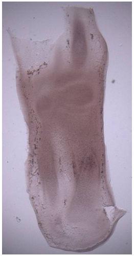
)109( COPYRIGHT 2016 BY THE ARCHIVES OF BONE AND JOINT SURGERY. A Chick Embryo in-vitro Model of Knee Morphogenesis RESEARCH ARTICLE
109( COPYRIGHT 2016 BY THE ARCHIVES OF BONE AND JOINT SURGERY. A Chick Embryo in-vitro Model of Knee Morphogenesis RESEARCH ARTICLE") )109( COPYRIGHT 2016 BY THE ARCHIVES OF BONE AND JOINT SURGERY RESEARCH ARTICLE A Chick Embryo in-vitro Model of Knee Morphogenesis Edward K. Rodriguez, MD, PhD; Jeeva Munasinghe, PhD Research performed
)109( COPYRIGHT 2016 BY THE ARCHIVES OF BONE AND JOINT SURGERY RESEARCH ARTICLE A Chick Embryo in-vitro Model of Knee Morphogenesis Edward K. Rodriguez, MD, PhD; Jeeva Munasinghe, PhD Research performed
Development and regeneration of the neonatal digit tip in mice
 Available online at www.sciencedirect.com Developmental Biology 315 (2008) 125 135 www.elsevier.com/developmentalbiology Development and regeneration of the neonatal digit tip in mice Manjong Han a, Xiaodong
Lecture 2: Skeletogenesis
 Jilin University School of Stomatology Skeletogenesis Lecture 2: Skeletogenesis Aug. 18, 2015 Yuji Mishina, Ph.D. mishina@umich.edu Student will describe Development of Bone - the general anatomy of bone
Longitudinal study to follow-up a developmental abnormality Luc De Schaepdrijver, DVM, PhD Preclinical Development & Safety Janssen R&D, Belgium
 Longitudinal study to follow-up a developmental abnormality Luc De Schaepdrijver, DVM, PhD Janssen R&D, Belgium ILSI-HESI Workshop, Washington, April 21 st, 2015 Fetal Imaging in Regulatory Developmental
9/16/2009. Fast and slow twitch fibres. Properties of Muscle Fiber Types Fast fibers Slow fibers
 Muscles, muscle fibres and myofibrils Fast and slow twitch fibres Rat hindlimb muscle ATPase staining at different ph and NADH Muscle fibre shortening velocity lengths/second Properties of Muscle Fiber
Muscles, muscle fibres and myofibrils
 Muscles, muscle fibres and myofibrils Properties of Muscle Fiber Types Fast fibers Slow fibers Characteristic IIb IIx IIa Type I V max (speed of shortening) Highest Intermediate Low Resistance to fatigue
Vertebrate Limb Patterning
 Vertebrate Limb Patterning What makes limb patterning an interesting/useful developmental system How limbs develop Key events in limb development positioning and specification initiation of outgrowth establishment
Skeletal System. Std. VIII
 Skeletal System Std. VIII The skeleton in our body serves following functions : 1. Support and shape : The skeleton provides a support or framework to all the soft parts and gives the body and its parts
Journal of Biomechanical Science and Engineering
 0123456789 Bulletin of the JSME Vol.9, No.2, 2014 Journal of Biomechanical Science and Engineering Finite element analysis of hip joint cartilage reproduced from real bone surface geometry based on 3D-CT
Finite-element study of vibration effect to fracture healing of a human tibia
 Finite-element study of vibration effect to fracture healing of a human tibia Leonid Maslov 1, Jean-Baptiste Etheve 2, Nikolay Sabaneev 3 Ivanovo State Power Engineering University, Ivanovo, Russia 1 Corresponding
L01:Name and locate the major bones within the skeletal system.
 L01:Name and locate the major bones within the skeletal system. All physical activity requires movement using bones and muscles. Name three major bones which are located in the leg. Bone 1 - Bone 2- Bone
Chapter 5 The Skeletal System
 Chapter 5 The Skeletal System The Skeletal System Parts of the skeletal system Bones (skeleton) Joints Cartilages Ligaments (bone to bone)(tendon=bone to muscle) Divided into two divisions Axial skeleton:
Anatomy. Anatomy deals with the structure of the human body, and includes a precise language on body positions and relationships between body parts.
 Anatomy deals with the structure of the human body, and includes a precise language on body positions and relationships between body parts. Proper instruction on safe and efficient exercise technique requires
Equine Skeletal System
 Equine Skeletal System EQS 110 Table of Contents Click on the different sections of the table of contents to jump through this document Functions of the Skeletal System... 3 Skeletal Strength... 3 Bone
Lecture 2. Statics & Dynamics of Rigid Bodies: Human body 30 August 2018
 Lecture 2. Statics & Dynamics of Rigid Bodies: Human body 30 August 2018 Wannapong Triampo, Ph.D. Static forces of Human Body Equilibrium and Stability Stability of bodies. Equilibrium and Stability Fulcrum
5.1 BONES: AN OVERVIEW
 Unit 5 Skeletal System 5.1 BONES: AN OVERVIEW Section Objectives Identify the major structures and functions of the skeletal system. Differentiate between the two divisions (axial and appendicular) of
Development of the mouse mandibles and clavicles in the absence of skeletal myogenesis
 Histol Histopathol (2007) 22: 51-60 http://www.hh.um.es Histology and Histopathology Cellular and Molecular Biology Development of the mouse mandibles and clavicles in the absence of skeletal myogenesis
CHAPTER 8 LECTURE OUTLINE
 CHAPTER 8 LECTURE OUTLINE I. INTRODUCTION A. The appendicular skeleton includes the bones of the upper and lower extremities and the shoulder and hip girdles. B. The appendicular skeleton functions primarily
PowerPoint Lecture Slides. Prepared by Patty Bostwick-Taylor, Florence-Darlington Technical College. The Skeletal System Pearson Education, Inc.
 PowerPoint Lecture Slides Prepared by Patty Bostwick-Taylor, Florence-Darlington Technical College CHAPTER 5 The Skeletal System 2012 Pearson Education, Inc. Title Classification of Bones and Gross Anatomy
Validation of Genetic Determinants of Skeletal Diseases. Dr Kasia Piróg (Newcastle University)
") Validation of Genetic Determinants of Skeletal Diseases Dr Kasia Piróg (Newcastle University) What is? Skeletal conditions Common Rare related to ageing and/or injury often multifactorial often difficult
Validation of Genetic Determinants of Skeletal Diseases Dr Kasia Piróg (Newcastle University) What is? Skeletal conditions Common Rare related to ageing and/or injury often multifactorial often difficult
Tetrapod Limb Development
 IBS 8102 Cell, Molecular and Developmental Biology Tetrapod Limb Development February 11, 2008 Tetrapod Limbs Merlin D. Tuttle Vicki Lockard and Paul Barry Father Alejandro Sanchez Anne Fischer Limb Patterning
The Skeletal System PART A. PowerPoint Lecture Slide Presentation by Patty Bostwick-Taylor, Florence-Darlington Technical College
 PowerPoint Lecture Slide Presentation by Patty Bostwick-Taylor, Florence-Darlington Technical College The Skeletal System 5 PART A The Skeletal System Parts of the skeletal system Bones (skeleton) Joints
The Skeletal System PART A
 5 The Skeletal System PART A PowerPoint Lecture Slide Presentation by Jerry L. Cook, Sam Houston University ESSENTIALS OF HUMAN ANATOMY & PHYSIOLOGY EIGHTH EDITION ELAINE N. MARIEB The Skeletal System
The Skeletal System ESSENTIALS OF HUMAN ANATOMY & PHYSIOLOGY PART A ELAINE N. MARIEB EIGHTH EDITION
 5 The Skeletal System PART A PowerPoint Lecture Slide Presentation by Jerry L. Cook, Sam Houston University ESSENTIALS OF HUMAN ANATOMY & PHYSIOLOGY EIGHTH EDITION ELAINE N. MARIEB The Skeletal System
OHIO STATE UNIVERSITY EXTENSION
 Cloverbud Investigators: Career Detectives October Background: Today we are going to learn about our bones and how they join together to hold up our body, all the way from our head to our toes. Did you
Classification of bones
 Classification of bones compact intramembranous axial histology development regional spongy Intra cartilaginous appendicular flat Irregular shape Sesamoid Long Short Wormian pneumatic Classification
Equine Skeletal System
 Equine Skeletal System EQS 110 Table of Contents Click on the different sections of the table of contents to jump through this document Functions of the Skeletal System... 3 Skeletal Strength... 3 Bone
What is bone? Specialized form of connective tissue: mineralized collagen matrix, therefore very rigid and strong while still retaining some degree of
 Bone What is bone? Specialized form of connective tissue: mineralized collagen matrix, therefore very rigid and strong while still retaining some degree of flexibility Other types of connective tissue:
Musculoskeletal System
 Musculoskeletal System The musculoskeletal system gives the body strength, structure, and capability of movement. Bones are the framework. Ligaments and tendons are the nails Muscles are the way we move
Human Skeletal System Glossary
 Acromegaly Apatite Acromegaly - is a condition which involves excessive growth of the jaw, hands, and feet. It results from overproduction of somatotropin in adults (after fusion of the ossification centres
The Skeletal System THE APPENDICULAR SKELETON
 The Skeletal System THE APPENDICULAR SKELETON The appendicular skeleton consists of the girdles and the skeleton of the limbs. The upper (anterior) limbs are attached to the pectoral (shoulder) girdle
Simulation of bone indentation
 Modelling in Medicine and Biology VII 113 Simulation of bone indentation S. Kasiri 1, G. Reilly 1,2 & D. Taylor 1 1 Trinity Centre for Bioengineering, Trinity College Dublin, Ireland 2 Institute of Technology
CASE OF CONGENITAL CYSTIC EYE AND ACCESSORY LIMB OF THE LOWER EYELID*
 Brit. J. Ophthal. (1966) 50, 409 CASE OF CONGENITAL CYSTIC EYE AND ACCESSORY LIMB OF THE LOWER EYELID* BY N. S. C. RICE Department ofpathology, Institute of Ophthalmology, London, AND S. P. MINWALLA AND
The Skeletal System in Action!! The Skeletal System in Action!
 Skeletal System The Skeletal System in Action!! The Skeletal System in Action! 5 Functions of the Skeletal System 1. Movement: Skeletal system provides points of attachment for muscles. Your legs and arms
UNIT 3: FORENSIC ANTHROPOLOGY
 UNIT 3: FORENSIC ANTHROPOLOGY Identifying Bones What is Forensic Anthropology? The field of study that deals with the analysis of human skeletal remains resulting from unexplained deaths Development of
Chapter 8 The Skeletal System: The Appendicular Skeleton. Copyright 2009 John Wiley & Sons, Inc.
 Chapter 8 The Skeletal System: The Appendicular Skeleton Appendicular Skeleton It includes bones of the upper and lower limbs Girdles attach the limbs to the axial skeleton The pectoral girdle consists
Four weeks of Intrauterine life
 Objective Congenital & Developmental Malformation Overview of Musculoskeletal dev. Abnormal pattern of dev. Common upper & lower ext. abnormalities READ : SPINE and more information in text book Definition
Unit 5 Skeletal System
 Unit 5 Skeletal System Nov 21 10:24 PM I. Functions A. Support: > internal framework, structure, anchors & supports soft tissue organs B. Protection: > protects vital organs C. Movement: > provides attach
Supplementary Figure 1. Expression of phospho-sik3 in normal and osteoarthritic articular cartilage in the knee. (a) Semiserial histological sections
 Semiserial histological sections") Supplementary Figure 1. Expression of phospho-sik3 in normal and osteoarthritic articular cartilage in the knee. (a) Semiserial histological sections of normal cartilage were stained with safranin O-fast
Supplementary Figure 1. Expression of phospho-sik3 in normal and osteoarthritic articular cartilage in the knee. (a) Semiserial histological sections of normal cartilage were stained with safranin O-fast
Characterising 3D Soft Tissue Features on Joint Surfaces
 Dublin Institute of Technology ARROW@DIT Conference Papers Biomedical Devices and Assistive Technology Research Group 2011-01-29 Characterising 3D Soft Tissue Features on Joint Surfaces Colm O'Kane Dublin
YEAR 9 GCSE PE Learning Programme
 YEAR 9 GCSE PE Learning Programme Half Term/Term Learning objective Learning activity Differentiation and extension Bones. Knowledge of the bones at the following locations: head/neck cranium, vertebrae
Parts of the skeletal system. Bones (skeleton) Joints Cartilages Ligaments (bone to bone)(tendon=bone to muscle)
 Joints Cartilages Ligaments (bone to bone)(tendon=bone to muscle)") The Skeletal System The Skeletal System Parts of the skeletal system Bones (skeleton) Joints Cartilages Ligaments (bone to bone)(tendon=bone to muscle) Divided into two divisions Axial skeleton Appendicular
The Skeletal System The Skeletal System Parts of the skeletal system Bones (skeleton) Joints Cartilages Ligaments (bone to bone)(tendon=bone to muscle) Divided into two divisions Axial skeleton Appendicular
Thomas A. Metzger, Stephen A. Schwaner, Jonelle M. Shudick, Anthony J. LaNeve, Glen L. Niebur. University of Notre Dame, Notre Dame, IN, USA.
 Mechanical Stimuli in Bone Marrow During Physiologic Loading Thomas A. Metzger, Stephen A. Schwaner, Jonelle M. Shudick, Anthony J. LaNeve, Glen L. Niebur. University of Notre Dame, Notre Dame, IN, USA.
Skeletal System. Prof. Dr. Malak A. Al-yawer Department of Anatomy/Embryology Section
 Skeletal System Prof. Dr. Malak A. Al-yawer Department of Anatomy/Embryology Section Learning objectives At the end of this lecture, the medical student will be able to: State the embryonic origin of skeletal
Chapter 19 Musculoskeletal
 Musculoskeletal System Chapter 19 Musculoskeletal System 1 Consists of Bones and Muscles Along with associated connective tissues Tendons, ligaments, cartilage Skeleton provides support to body and protection
October. Cloverbud Investigators: Career Detectives
 October OHIO STATE UNIVERSITY EXTENSION Cloverbud Investigators: Career Detectives Background: Today we are going to learn about our bones and how they join together to hold up our body all the way from
UNIT 5 THE SKELETAL SYSTEM
 UNIT 5 THE SKELETAL SYSTEM Nov 20 12:02 PM I. Functions A. Support: Internal framework, Structure, Anchors & Supports soft tissue/organs B. Protection: Protects vital organs C. Movement: Provide attach
QUICK ASSESSMENT: CONCEPT MAP
 FUNCTIONS OF THE SKELETAL SYSTEM 7th Grade THE SKELETAL SYSTEM Provides shape, strength, and support (3S s) Internal framework of the body Support and anchor for soft organs Protects soft internal organs
Short title: Endothelial mtorc2 controls aberrant angiogenesis
 Endothelial Rictor is crucial for midgestational development and sustained and extensive FGF2 induced neovascularization in the adult Fabio Aimi 1+, Stavroula Georgiopoulou 1+, Ina Kalus 1, Fabienne Lehner
Formation of marrow cavity and ossification in mouse limb buds grown in vitro
 /. Embryol. exp. Morph. Vol. 56, pp. 301-307, 1980 301 Printed in Great Britain Company of Biologists Limited 1980 Formation of marrow cavity and ossification in mouse limb buds grown in vitro By D. R.JOHNSON
An experimental investigation into the early development of the chick elbow joint
 /. Embryol. exp. Morph. Vol. 39,pp. 115-127, 1977 Printed in Great Britain An experimental investigation into the early development of the chick elbow joint By N. HOLDER 1 From the Department of Biology
The role of movements in the development of sutural. of chick embryos. and diarthrodial joints tested by long-term paralysis
 J. Anat. (1983), 137, 3, pp. 591-599 591 With 11 figures Printed in Great Britain The role of movements in the development of sutural and diarthrodial joints tested by long-term paralysis of chick embryos
Characteristics. Bones. Functions of the Skeleton
 Characteristics Bones The Introduction 206 bones hard, rigid bones cells (osteocyctes) are a mixture of a ground substance, collagen fibres, P, Ca highly resistant to compression and tension also somewhat
"Role of thyroid hormones in bone development and maintenance"
 "Role of thyroid hormones in bone development and maintenance" Graham R. Williams Molecular Endocrinology Group Department of Medicine & MRC Clinical Sciences Centre Imperial College London Thyroid hormones
11.2 Muscles and Movement
 11.2 Muscles and Movement 11.2.1 - State the roles of bones, ligaments, muscles, tendons and nerves in human movement Bones Act as anchors for the muscles, and levers to control the movement of muscles,
Copyright 2003 Pearson Education, Inc. publishing as Benjamin Cummings. Dr. Nabil khouri
 Dr. Nabil khouri Appendicular Skeleton The appendicular skeleton is made up of the bones of the upper and lower limbs and their girdles Two girdles: Pectoral girdles attach the upper limbs to the body
The cyclin-dependent kinase inhibitor p57 Kip2 mediates proliferative actions of PTHrP in chondrocytes
 Research article The cyclin-dependent kinase inhibitor p57 Kip2 mediates proliferative actions of PTHrP in chondrocytes Helen E. MacLean, 1 Jun Guo, 1 Melissa C. Knight, 1 Pumin Zhang, 2 David Cobrinik,
36.3 The Integumentary System The Skin. KEY CONCEPT The integumentary system has many tissues that protect the body.
 36.3 The Integumentary System The Skin KEY CONCEPT The integumentary system has many tissues that protect the body. 36.3 The Integumentary System The Skin The integument is the body system that surrounds
ARTICLE IN PRESS. Journal of Biomechanics
 Journal of Biomechanics 42 (2009) 152 157 Contents lists available at ScienceDirect Journal of Biomechanics journal homepage: www.elsevier.com/locate/jbiomech www.jbiomech.com Residual periosteum tension
Chapter 7: Skeletal System: Gross Anatomy
 Chapter 7: Skeletal System: Gross Anatomy I. General Considerations A. How many bones in an average adult skeleton? B. Anatomic features of bones are based on II. Axial Skeleton A. Skull 1. Functionally
SUPPORT, MOVEMENT AND LOCOMOTION
 SUPPORT, MOVEMENT AND LOCOMOTION Overview Bones Joints Antagonistic Muscles Forelimbs Bones Functions of the skeleton Locomotion The ability to move from place to place Support Holds the body off the ground
Ossification = Osteogenesis
 Ossification = Osteogenesis Ossification = Osteogenesis Parts of the fetal skeleton form during the first few weeks after conception By the end of the 8 th week, the skeletal pattern is formed : cartilage
SD School Anatomy Program 1: Bones QuikNotes. Student Notes
 QuikNotes The transverse plane runs from right to left and divides the body into superior (upper) and inferior (lower) sections. Student Notes The frontal plane lies vertically along the body from head
Lecture 3: Skeletogenesis and diseases
 Jilin University School of Stomatology Skeletogenesis Lecture 3: Skeletogenesis and diseases Aug. 21, 2015 Yuji Mishina, Ph.D. mishina@umich.edu Bone Development Mouse embryo, E14.5 Mouse embryo, E18.0
36 1 The Skeletal System Slide 1 of 40
 1 of 40 The Skeleton All organisms need structural support. Unicellular organisms have a cytoskeleton. Multicellular animals have either an exoskeleton (arthropods) or an endoskeleton (vertebrates). 2
Lecture 5. Skeletal and Muscular Systems. Skeletal and Muscular Systems. 1. Skeletal System Bones Cartilage Ligaments & Tendons Joints 2.
 Lecture 5 Skeletal and Muscular Systems 1 Skeletal and Muscular Systems 1. Skeletal System Bones Cartilage Ligaments & Tendons Joints 2. Muscles 2 1 Skeletal System 3 Functions of Skeleton Support of the
3D representation of the developing chick knee joint: a novel approach integrating multiple components
 J. Anat. (2009) 214, pp374 387 doi: 10.1111/j.1469-7580.2008.01040.x 3D representation of the developing chick knee joint: Blackwell Publishing Ltd a novel approach integrating multiple components Karen
BIOL2005 WORKSHEET 2008
 BIOL2005 WORKSHEET 2008 Answer all 6 questions in the space provided using additional sheets where necessary. Hand your completed answers in to the Biology office by 3 p.m. Friday 8th February. 1. Your
General osteology. General anatomy of the human skeleton. Development and classification of bones. The bone as a multifunctional organ.
 General osteology. General anatomy of the human skeleton. Development and classification of bones. The bone as a multifunctional organ. Composed by Natalia Leonidovna Svintsitskaya, Associate professor
The Skeletal System. Functions of the Skeletal System
 11/15/17 The Skeletal System The Skeletal System 1 Functions of the Skeletal System Provide support and structure Protects vital internal organs Storage site for minerals Site for blood formation Bones
Hepatogenesis I Liver development
 Hepatogenesis I Liver development HB 308 George Yeoh Room 2.59 MCS Building yeoh@cyllene.uwa.edu.au Topics Early liver development Tissue interaction - role of morphogens and cytokines Liver enriched transcription
Skeletal Considerations for Movement. Kinesiology RHS 341 Lecture 2 Dr. Einas Al-Eisa
 Skeletal Considerations for Movement Kinesiology RHS 341 Lecture 2 Dr. Einas Al-Eisa The Skeletal System Bones, cartilage, ligaments, & joints Consists of approximately 20% of total body weight Bone constitutes
Joints. Vi Michelle Austin
 Joints Vi Michelle Austin Joints Overview A joint, otherwise known as an articulation, is a point at which points connect. They are constructed to allow movement (except for skull bones) and provide mechanical
Department of Anatomy ESI-PGIMSR, Joka, Kolkata
 Teaching Schedule - 2016-2017, 1st MBBS Date and Day Time Topic Name/ Group 8-9 Orientation, Introduction to theoretical aspects of Practical SC 3.10.2016 Anatomy, Tools of Trade - Anatomical Nomenclature-
Structural Analysis of Human Body Impact
 Structural Analysis of Human Body Impact Young-Shin Lee Dept. of Mechanical Design Engineering, Chungnam National University, Korea. Young-jin Choi Graduate School, Dept. of Mechanical Design Engineering,
Development of the Axial Skeleton and Limbs. Professor Alfred Cuschieri Department of Anatomy University of Malta
 Development of the Axial Skeleton and Limbs Professor Alfred Cuschieri Department of Anatomy University of Malta During the Fourth Week the Embryo Is Segmented. Each segment consists of: a segment of neural
Fig Articular cartilage. Epiphysis. Red bone marrow Epiphyseal line. Marrow cavity. Yellow bone marrow. Periosteum. Nutrient foramen Diaphysis
 Fig. 7.1 Articular cartilage Epiphysis Red bone marrow Epiphyseal line Marrow cavity Yellow bone marrow Nutrient foramen Diaphysis Site of endosteum Compact bone Spongy bone Epiphyseal line Epiphysis Articular
Two Separated Protocols with the Most Important Comments for Skeletal Staining in Embryonic and Adulthood Period in Laboratory Animals
 May 2014, Volume 11, Number 2 Two Separated Protocols with the Most Important Comments for Skeletal Staining in Embryonic and Adulthood Period in Laboratory Animals Farzane Sadeghi * PhD in Anatomical
The Musculoskeletal system
 Level 3 BTEC Applied Science Summer Homework The Musculoskeletal system Student name:.. Tutor name: 1 Student Instructions This workbook incorporates elements of Unit 8 Learning Aim A: Understand the impact
BLUE SKY SCHOOL OF PROFESSIONAL MASSAGE AND THERAPEUTIC BODYWORK. Musculoskeletal Anatomy & Kinesiology I TERMINOLOGY, STRUCTURES, & SKELETAL OVERVIEW
 BLUE SKY SCHOOL OF PROFESSIONAL MASSAGE AND THERAPEUTIC BODYWORK Musculoskeletal Anatomy & Kinesiology I TERMINOLOGY, STRUCTURES, & SKELETAL OVERVIEW MSAK101-I Session 1 Learning Objectives: 1. Define
Challenge Question: Prediction: (Wait for directions) Evidence: (Draw, color and label how your Clay model looked when it was complete)
 Evidence: (Draw, color and label how your Clay model looked when it was complete)") Activity #12 What s happening inside? Challenge Question: Prediction: (Wait for directions) Evidence: (Draw, color and label how your Clay model looked when it was complete) Act #12 pg 1 of 3 Analysis
Activity #12 What s happening inside? Challenge Question: Prediction: (Wait for directions) Evidence: (Draw, color and label how your Clay model looked when it was complete) Act #12 pg 1 of 3 Analysis
Bones of the wrist and ankle Bones that form within tendons (e.g., patella)
") Skeletal System Review Surface Anatomy Dr. Gary Mumaugh Function of Bones Support form the framework that supports the body and cradles soft organs Protection provide a protective case for the brain, spinal
Skeletal System Review Surface Anatomy Dr. Gary Mumaugh Function of Bones Support form the framework that supports the body and cradles soft organs Protection provide a protective case for the brain, spinal
Subject Index. AXIN2, cleft defects 24, 26
 Subject Index ADAMTS, mouse mutants and palate development 37, 38 Africa, cleft lip and palate prevalence 6, 7 Alcohol dependence, pregnancy risks for cleft 25, 61 Altitude, pregnancy risks for cleft 25,
Presidency General Hospital, Calcutta
 CONGENITAL ANOMALY OF HAND: " MIRROR HAND " By M. MUKERJI, F.R.C.S. Presidency General Hospital, Calcutta Case Report.--S. B., aged 4 months, was born with eight fingers and no thumb on the left hand and
Lab Exercise #04 The Skeletal System Student Performance Objectives
 Lab Exercise #04 The Skeletal System Student Performance Objectives The material that you are required to learn in this exercise can be found in either the lecture text or the supplemental materials provided