Clinical Anatomy of the Upper Limb. Kara Mudd, MSPAS, PA-C
|
|
|
- Oliver Atkinson
- 6 years ago
- Views:
Transcription
1 Clinical Anatomy of the Upper Limb Kara Mudd, MSPAS, PA-C
2 Contents Bones and Joints Clinical Anatomy of Upper Limb Joints Clinical Anatomy of Upper Limb Muscles Clinical Anatomy of Nerve affect Upper Limb Muscles Special Diagnostic Tests
3 Bones and Joints of Upper Limb Regions Bones Joints Shoulder Girdle Clavicle Scapula Sternoclavicular Joint Acromioclavicular Joint Bones of Arm Humerus Upper End: Glenohumeral Joint Lower End: See below Bones of Forearm Bones of Wrist and Hand Radius Ulna 8 Carpal Bones 5 Metacarpal 14 Phalanges Humeroradial Joint Humeroulnar Joint Proximal Radioulnar Joint Distal Radioulnar Joint Intercarpal Joint Carpometacarpal Joint Metacarpophalangeal Joint Interphalangeal Joint
4 Upper Limb Joints
. Commonly dislocated anteriorly and inferiorly.")
5 Glenohumeral Joint Dislocation Most commonly dislocated major joint in body. Ball & Socket Large head of humerus to shallow glenoid cavity Stability depends almost entirely on the strength of surrounding muscles (Rotator Cuff). Commonly dislocated anteriorly and inferiorly. Anteriorly shoulder joint protected by subscapularis. Superiorly shoulder joint protected by supraspinatus. Posteriorly shoulder joint protected by teres minor and infraspinatus. Inferior aspect of shoulder joint completely unprotected.

6


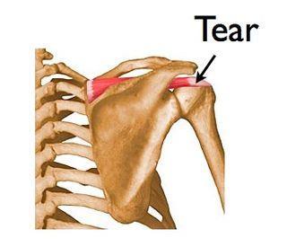
7 Glenohumeral Joint Dislocation Labral tears are common with traumatic shoulder dislocations

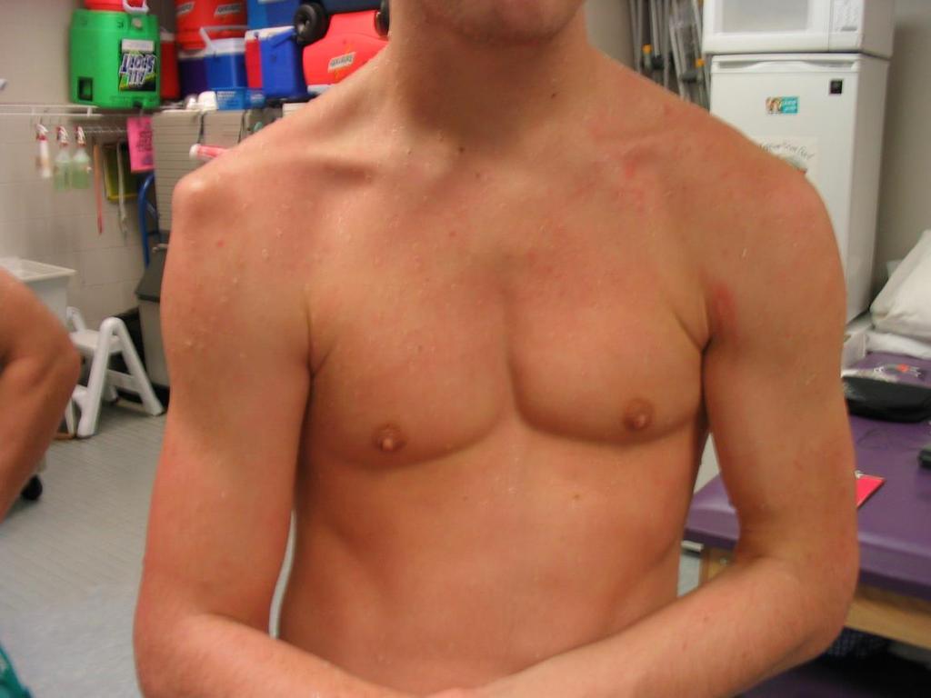
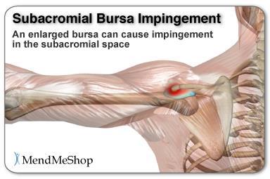
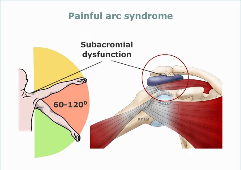
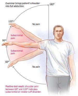
8 Subacromial Bursitis Shoulder joint in abducted position = Pain Subacromial bursa is located inferior to acromion, above supraspinatus tendon. Pain occur when abduction because supraspinatus tendon comes in contact with inferior surface of acromion, inflammed bursa slips up underneath coraco-acromial arch and gets impinged between supraspinatus tendon and acromion.
9

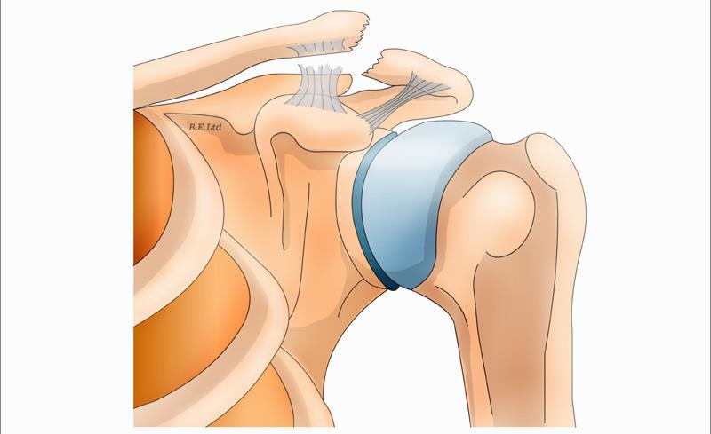
10 Injection of the Subacromial Bursa

11 Frozen Shoulder (adhesive capsulitis) Pain and uniform limitation off all movements of shoulder joint, though there is NO evidence of radiological changes in the joint. Occurs due to shrinkage and chronic inflammation of capsule of glenohumeral joint Causes: When nor moving the shoulder joint for a period of time because of: Pain Injury Chronic health condition
 2.")
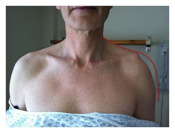
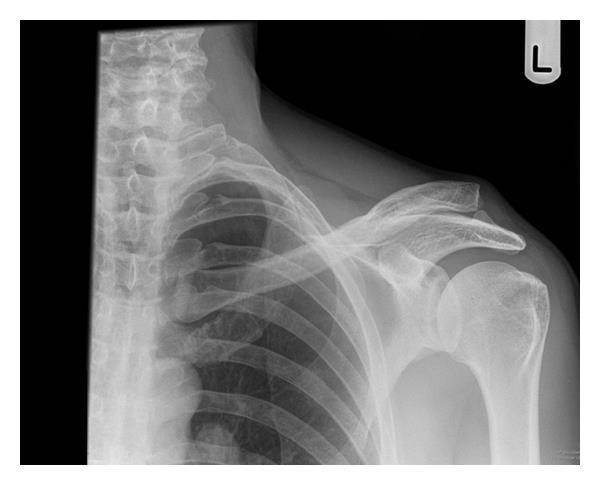
12 Shoulder Separation (Acromioclavicular Subluxation) Tearing of coracoclavicular and coracoacromial ligaments caused by downward blow on tip of shoulder. Coracoclavicular and coracoacromial joint spaces become 50% wider than in normal contralateral shoulder. Presenting features: 1. Injured arm hangs lower than normal (contralateral arm) 2. Noticeable bulge at tip of shoulder as a result of upward displacement of clavicle.
13
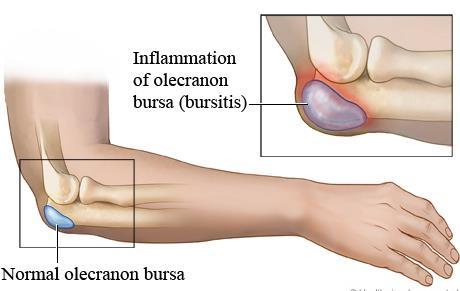
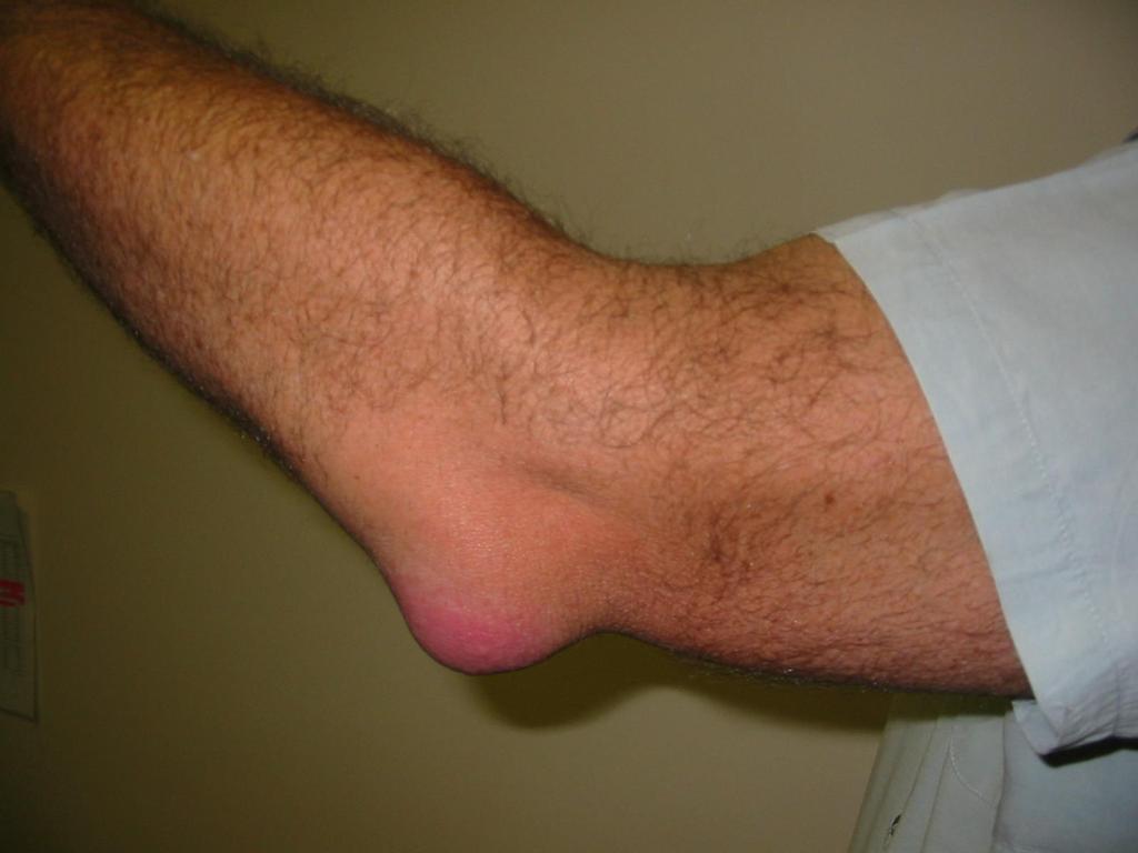
14 Olecranon Bursitis Inflammation of subcutaneous olecranon bursa (lying over the subcutaneous triangular area on dorsal surface of olecranon process of Ulna). Causes a round fluctuating painful swelling of 1 or so in circumference over olecranon. Occurs due to: 1. Repeated friction as occurs in students who read for long hours with head supported by hand and elbow resting on table. 2. Trauma during falls on elbow 3. Infection from abrasions of skin covering olecranon process.
15

16 Tennis Elbow (Lateral epicondylitis) Repeated or violent extension of the wrist with forearm pronated (i.e. movements required during backhand strokes in lawn tennis), leads to tenderness over lateral epicondyle of humerus. Possibly due to: Sprain of radial collateral ligament. Tearing of fibers of extensor carpi radialis brevis muscle. Inflammation of the bursa underneath extensor carpi radialis brevis Strain or tear of common extensor origin.

.")
17 Golfer s Elbow (Medial Epicondylitis) Repetitive use of superficial flexors of forearm, strains their common flexor origin with subsequent inflammation of medial epicondyle (medial epicondylitis). Characterised by pain on medial side of elbow.
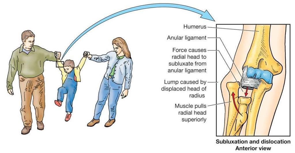
18 Radial Head Subluxation Also known as nursemaid s elbow. Vulnerable for preschool children (1-3 years old). Annular ligament is funnel-shaped in adults, but its sides are vertical in young children. (When child is suddenly lifted/pulled up when forearm is in pronated position, head or radius may slips out partially from annular ligament). Pain and limitation of supination.
19
.")

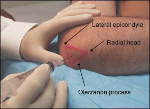
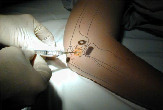
20 Aspiration of Elbow Joint Locate the head of radius and capitulum of humerus. (flex elbow to 90 degrees, pronate and supinate forearm and feel with the thumb its rotation). Insert needle in the palpable depression between proximal part of radial head and capitulum in a direction directly forwards, the joint being flexed to right angle and forearm semi pronated. Safest and most direct approach. When elbow is distended with pus, the capsule bulges to either side of triceps and hence the pus can easily and effiviently be drained.
21
22 Upper limb Muscles

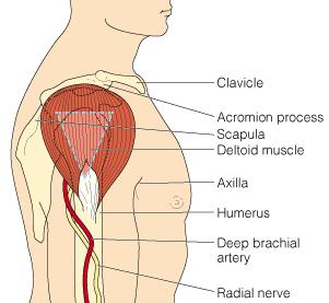
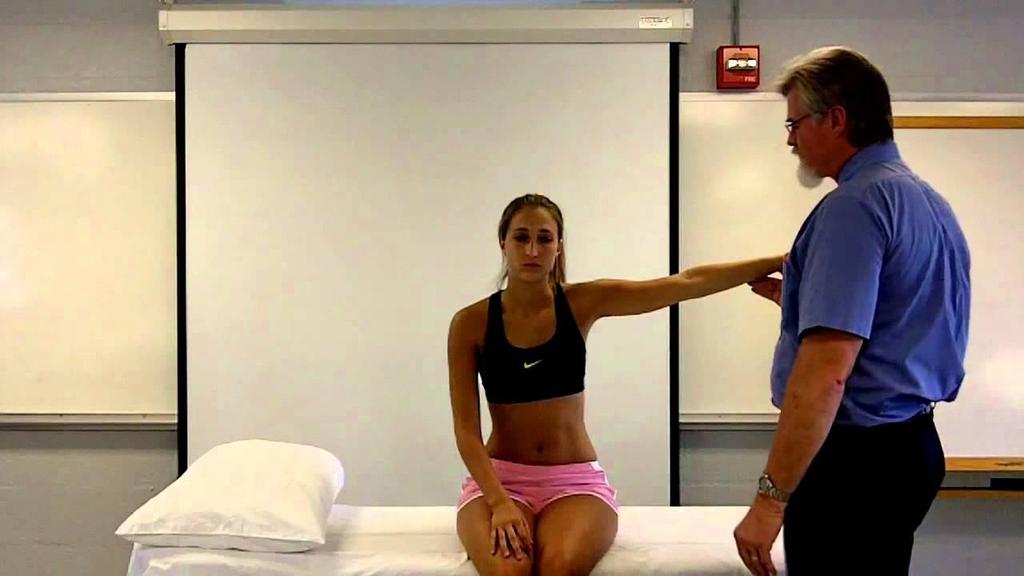
23 Preferred site for Intramuscular Injection Deltoid Muscles Well-developed in most adults and easily accessible. Injection given in the lower half to avoid injury to axillary nerve. Exact site of injection: Needle should be inserted in center of triangle. Place 4 fingers across deltoid muscle with top finger kept along acromial process, injection site is 3 fingers breadth below acromial process.
24

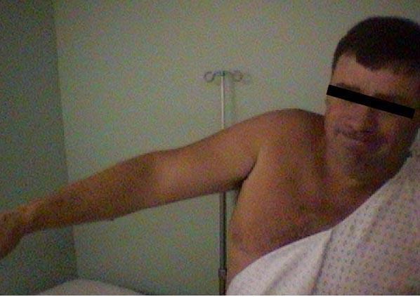
25 Ruptured Supraspinatus Active initiation of abduction is not possible. Patient gradually develop a trick of tilting his body towards injured side so that the arm swings away from the body due to gravity leading to 15 degrees initial abduction. Later deltoid and scapular rotators come into play to do the required job. Sometimes may also lift arm on the side of injury with opposite hand to cause initial abduction
26
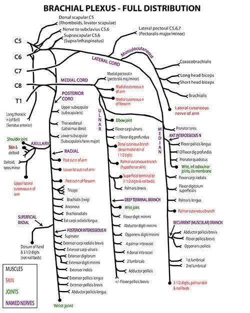
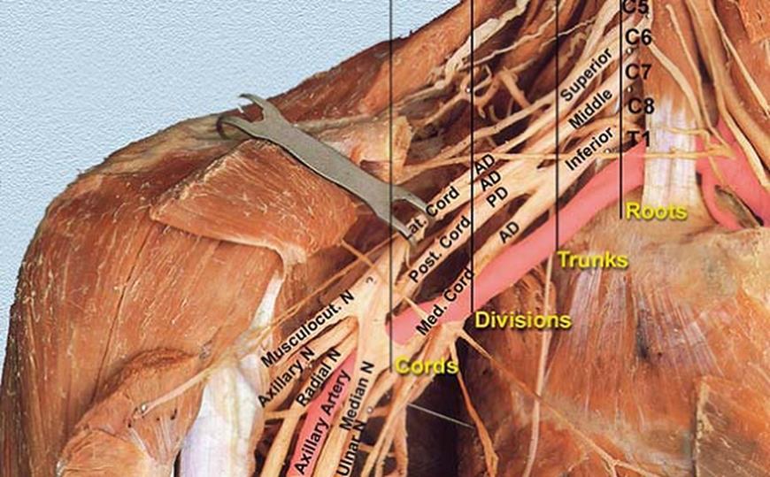
27 Upper Limb Nerves
28

29 Injury to the Long Thoracic Nerve Causes: -Surgical injury -axillary node dissection -trauma Results in paralysis of the serratus anterior and winged scapula/inability to abduct the arm

30 Paralysis of Serratus Anterior Functions of Serratus Anterior Keeps medial border of scapula in contact with chest wall. Important role in abduction of arm and elevation of arm above horizontal. Pulls scapula forward as in throwing, pushing and punching. Effects of paralysis of serratus anterior Medial border of scapula stands out from the chest wall, particularly when patient is asked to press against a wall in front of him (winged scapula). Inability to raise arm above head. Inability to carry arm forward in breast stroke swimming (Swimmer s Palsy).

31 Suprascapular Nerve Entrapment Entrapment may occur as it passes through suprascapular notch or in spinoglenoid notch. Effects: Typical dull posterior and lateral shoulder pain. Tenderness, 2.5cm lateral to midpoint of spine of scapular Weakness of shoulder abduction and external rotation due to involvement of supraspinatus and infraspinatus muscles.
.")
32 Erb-Duchenne Paralysis (Erb s Palsy) Brachial plexus injury at Erb s point (C5, C6). Maybe injured during childbirth, due to forcible downward traction of shoulder with lateral displacement of head to other side (Usually during forceps delivery). Upper limb assumes typical waiter s tip position : Shoulder adducted and medially rotated. Elbow extended Forearm pronated
33 Axillary Nerve damage Axillary nerve usually damaged by fractures of surgical neck of humerus or due to an inferior dislocation of shoulder joint. Effects: Loss or weakness of abduction of shoulder (between 15 degrees to 90 degrees) due to paralysis of Deltoid. Rounded contour/profile of shoulder is lost due to paralysis of deltoid Sensory loss over lower half of the outer aspect of shoulder regimental badge area. Paralysis of teres minor is not easily demonstrated clinically.

34 Effects of Radial Nerve Injury Severity and symptoms depend on site of lesion Most common clinical finding is wrist drop
35 Radial Nerve Injury (Axilla) Causes: Prolonged use of crutches (Crutch Paralysis) Effects: Loss of extension of elbow due to paralysis of triceps. Loss of extension of wrist due to paralysis of extensor muscles of forearm (Wrist Drop). Supination of forearm in elbow extension not possible (paralysis of supinator) Loss of sensation over: Posterior surface of lower part of arm and narrow strip over back of forearm Over lateral side of dorsum of hand and lateral 3½ fingers Loss of triceps and supinator reflexes.
36 Radial Nerve Injury (Radial Groove) Causes: Fracture of shaft of humerus Improper intramuscular injection Prolonged pressure Effects: Triceps brachii is spared (extension of elbow is possible) Other effects are similar as those of a lesion of radial nerve in axilla
37 Ulnar Nerve Usually injured at following sites: Elbow Cubital tunnel Wrist Hand Effects depending on site of lesion
 Weakness of flexion and adduction of wrist due to paralysis of flexor carpi ulnaris Ulnar Claw Hand Loss of adduction and")
38 Ulnar Nerve Injury at Elbow Easily damaged (lies in ulnar groove behind medial epicondyle of humerus) Causes: Fracture of medial epicondyle Effects: Loss of flexion of terminal phalanges of ring and little finger due to paralysis of flexor digitorum profundus (medial half) Weakness of flexion and adduction of wrist due to paralysis of flexor carpi ulnaris Ulnar Claw Hand Loss of adduction and abduction of fingers due to paralysis of Palmar (adductors) and dorsal (abductors) interossei Loss of adduction of thumb due to paralysis of adductor pollicis. Flattening of hypothenar eminence and depression of interosseous space due to atrophy of hypothenar and interosseous muscles
 Causes: Entrapment of ulnar nerve in this cubital tunnel (osseofibrous tunnel).")
39 Ulnar Nerve Injury at Cubital Tunnel (Cubital Tunnel Syndrome) Cubital Tunnel Formed by tendinous arch connecting 2 heads of flexor carpi ulnaris (arise from humerus and ulna) Causes: Entrapment of ulnar nerve in this cubital tunnel (osseofibrous tunnel). Effects same as ulnar nerve lesion in elbow
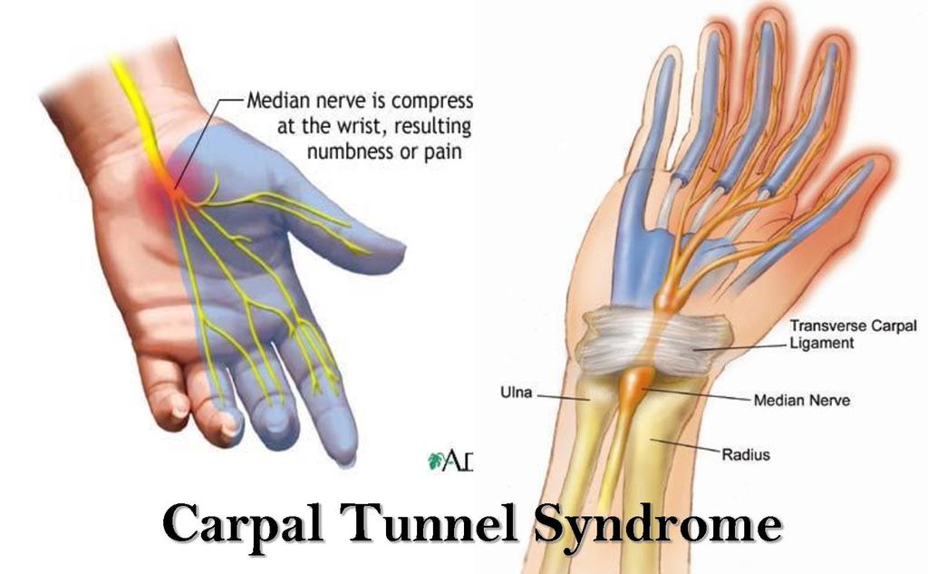
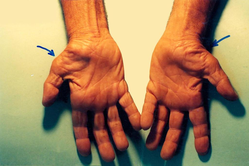
40 Median Nerve Injury usually injured at following sites: Axilla Wrist Carpal Tunnel Effects depending on site of lesion
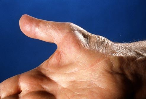
41 Median Nerve Injury at Axilla Motor Effects: Weakness of pronation of forearm due to paralysis of pronators. Deviation of wrist to Ulnar side on wrist flexion due to unopposed action of flexor carpi ulnaris. Weakness of flexion of distal phalanx of thumb and index finger. Wasting of thenar muscles due to paralysis. Loss of opposition of thumb due to paralysis of opponens pollicis. Loss of flexion and weakness of abduction of thumb. Ape-hand deformity Sensory Effects: Loss of cutaneous sensations over palmar surface of lateral 3½ digits and radial two-thirds of palm
 Myxoedema, pregnancy, hypothyroidism etc conditions resulting in fluid accumulation. Weight gain.")
42 Medial Nerve Injury at Carpal Tunnel (Carpal Tunnel Syndrome) Compression of medial nerve in carpal tunnel. More common in women than man. Causes Tendosynovitis Bony encroachment (osteoarthritis/injury) Myxoedema, pregnancy, hypothyroidism etc conditions resulting in fluid accumulation. Weight gain. Symptoms Painful paraesthesia and numbness affecting radial 3½ digits of hand. Wasting and weakness of thenar muscles. Loss of sensation or hypoaesthesia to light touch and pin prick over palmar aspect of radial 3½ digits and corresponding part of hand except skin over thenar eminence
43
44 Special Tests

45 Yergason Test To determine if biceps tendon is stable in bicipital groove. Steps: Instruct patient to fully flex elbow. Clinician grasp the flexed elbow in one hand while holding his wrist with other hand. External rotate patient s arm as he resists, at the same time pull downward his elbow. If biceps tendon is unstable in bicipital groove, it will pop out the groove and patient will experience pain; If stable, it remains secure and patient have no experience of discomfort.
46 Drop Arm Test To detects any tears in the rotator cuff. Steps: Instruct patient to fully abduct his arm. Asked patient slowly lower it to his side. Of there are tears in rotator cuff (especially supraspinatus), arm will drop to side from a position of about 90 abduction. Patient still will not be able to lower his arm smoothly and slowly no matter how many times he tries. If he is able to hold his arm in abduction, gentle tap on forearm will cause arm to fall to his side.
47
48 Apprehension Test To test for chronic shoulder dislocation or labral tear. Steps: Clinician abduct and externally rotate patient's arm to a position where it might easily dislocate If patient's shoulder is ready to dislocate, the patient will have a noticeable look of apprehension and alarm on his face and will resist further motion.
49 Elbow Stability Test To assess stability of medial and lateral collateral ligaments of elbow. Steps: Clinician cup posterior aspect of patient's elbow in one hand and hold patient's wrist with the other. The hand on the elbow will act as fulcrum, other hand will force the forearm during the test. Instruct patient to flex his elbow few degrees while forcing his forearm laterally, producing valgus stress on joint s medial side. Then do it in reverse direction. Notice if any gapping collateral ligament with the clinician hand that act as fulcrum.
50 Questions?
51 References Singh, V. (2007) Clinical & Surgical Anatomy, 2 nd edn. Elsevier; New Delhi. Snell, R.S. (2007) Clinical Anatomy: An Illustrated Review with Questions and Explanations, 3rd Edn. Lippincott Williams & Wilkins; Philadelphia. Slide share presentation by Gan Quan Fu, PT, Thieme
Nerve Injury. 1) Upper Lesions of the Brachial Plexus called Erb- Duchene Palsy or syndrome.
 Upper Lesions of the Brachial Plexus called Erb- Duchene Palsy or syndrome.") Nerve Injury - Every nerve goes to muscle or skin so if the nerve is injured this will cause paralysis in the muscle supplied from that nerve (paralysis means loss of function) then other muscles and other
Nerve Injury - Every nerve goes to muscle or skin so if the nerve is injured this will cause paralysis in the muscle supplied from that nerve (paralysis means loss of function) then other muscles and other
Connects arm to thorax 3 joints. Glenohumeral joint Acromioclavicular joint Sternoclavicular joint
 Connects arm to thorax 3 joints Glenohumeral joint Acromioclavicular joint Sternoclavicular joint Scapula Elevation Depression Protraction (abduction) Retraction (adduction) Downward Rotation Upward Rotation
Nerves of Upper limb. Dr. Brijendra Singh Professor & Head Department of Anatomy AIIMS Rishikesh
 Nerves of Upper limb Dr. Brijendra Singh Professor & Head Department of Anatomy AIIMS Rishikesh 1 Objectives Origin, course & relation of median & ulnar nerves. Motor & sensory distribution Carpal tunnel
Netter's Anatomy Flash Cards Section 6 List 4 th Edition
 Netter's Anatomy Flash Cards Section 6 List 4 th Edition https://www.memrise.com/course/1577581/ Section 6 Upper Limb (66 cards) Plate 6-1 Humerus and Scapula: Anterior View 1.1 Acromion 1.2 Greater tubercle
The Muscular System. Chapter 10 Part C. PowerPoint Lecture Slides prepared by Karen Dunbar Kareiva Ivy Tech Community College
 Chapter 10 Part C The Muscular System Annie Leibovitz/Contact Press Images PowerPoint Lecture Slides prepared by Karen Dunbar Kareiva Ivy Tech Community College Table 10.9: Muscles Crossing the Shoulder
BRACHIAL PLEXUS. DORSAL SCAPULAR NERVE (C5) supraclavicular branch innervates rhomboids (major and minor) and levator scapulae
 supraclavicular branch innervates rhomboids (major and minor) and levator scapulae") THE BRACHIAL PLEXUS DORSAL SCAPULAR NERVE (C5) supraclavicular branch innervates rhomboids (major and minor) and levator scapulae SCHEMA OF THE BRACHIAL PLEXUS THE BRACHIAL PLEXUS PHRENIC NERVE supraclavicular
THE BRACHIAL PLEXUS DORSAL SCAPULAR NERVE (C5) supraclavicular branch innervates rhomboids (major and minor) and levator scapulae SCHEMA OF THE BRACHIAL PLEXUS THE BRACHIAL PLEXUS PHRENIC NERVE supraclavicular
Gross Anatomy Questions That Should be Answerable After October 27, 2017
 Gross Anatomy Questions That Should be Answerable After October 27, 2017 1. The inferior angle of the scapula of a woman who was recently in an automobile accident seems to protrude making a ridge beneath
The Elbow and the cubital fossa. Prof Oluwadiya Kehinde
 The Elbow and the cubital fossa Prof Oluwadiya Kehinde www.oluwadiya.com Elbow and Forearm Anatomy The elbow joint is formed by the humerus, radius, and the ulna Bony anatomy of the elbow Distal Humerus
LIST OF STRUCTURES TO BE IDENTIFIED IN LAB: UPPER EXTREMITY REVIEW 2016
 LIST OF STRUCTURES TO BE IDENTIFIED IN LAB: UPPER EXTREMITY REVIEW 2016 BONES Ribs, sternum, clavicle Humerus: Head, greater tubercle, lesser tubercle, intertubercular sulcus, surgical neck, anatomical
Practical 2 Worksheet
 Practical 2 Worksheet Upper Extremity BONES 1. Which end of the clavicle is on the lateral side (acromial or sternal)? 2. Describe the difference in the appearance of the acromial and sternal ends of the
Muscles of the Upper Limb
 Muscles of the Upper Limb anterior surface of ribs 3 5 coracoid process Pectoralis minor pectoral nerves protracts / depresses scapula Serratus anterior Subclavius ribs 1-8 long thoracic nerve rib 1 ----------------
Key Relationships in the Upper Limb
 Key Relationships in the Upper Limb This list contains some of the key relationships that will help you identify structures in the lab. They are organized by dissection assignment as defined in the syllabus.
Nerves of the upper limb Prof. Abdulameer Al-Nuaimi. E. mail:
 Nerves of the upper limb Prof. Abdulameer Al-Nuaimi E-mail: a.al-nuaimi@sheffield.ac.uk E. mail: abdulameerh@yahoo.com Brachial plexus Median nerve After originating from the brachial plexus in the axilla,
Supplied in part by the musculocutaneous nerve. Forms the axis of rotation in movements of pronation and supination
 Anatomy: Upper limb (15 questions) 1. Latissimus Dorsi: Is innervated by the dorsal scapular nerve Lies above feres major muscle Medially rotates the humerus All of the above 2. Supinator muscle is: Deep
Lab Activity 11: Group II
 Lab Activity 11: Group II Muscles Martini Chapter 11 Portland Community College BI 231 Origin and Insertion Origin: The place where the fixed end attaches to a bone, cartilage, or connective tissue. Insertion:
UPPER EXTREMITY INJURIES. Recognizing common injuries to the upper extremity
 UPPER EXTREMITY INJURIES Recognizing common injuries to the upper extremity ANATOMY BONES Clavicle Scapula Spine of the scapula Acromion process Glenoid fossa/cavity Humerus Epicondyles ANATOMY BONES Ulna
Physical Examination of the Shoulder
 General setup Patient will be examined in both the seated and supine position so exam table needed 360 degree access to patient Expose neck and both shoulders (for comparison); female in gown or sports
Muscular Nomenclature and Kinesiology - One
 Chapter 16 Muscular Nomenclature and Kinesiology - One Lessons 1-3 (with lesson 4) 1 Introduction 122 major muscles covered in this chapter Chapter divided into nine lessons Kinesiology study of human
MLT Muscle(s) Patient Position Therapist position Stabilization Limb Position Picture Put biceps on slack by bending elbow.
 Patient Position Therapist position Stabilization Limb Position Picture Put biceps on slack by bending elbow.") MLT Muscle(s) Patient Position Therapist position Stabilization Limb Position Picture Put biceps on slack by bending elbow. Pectoralis Minor Supine, arm at side, elbows extended, supinated Head of Table
MLT Muscle(s) Patient Position Therapist position Stabilization Limb Position Picture Put biceps on slack by bending elbow. Pectoralis Minor Supine, arm at side, elbows extended, supinated Head of Table
Sick Call Screener Course
 Sick Call Screener Course Musculoskeletal System Upper Extremities (2.7) 2.7-2-1 Enabling Objectives 1.46 Utilize the knowledge of musculoskeletal system anatomy while assessing a patient with a musculoskeletal
Anatomy Workshop Upper Extremity David Ebaugh, PT, PhD Workshop Leader. Lab Leaders: STATION I BRACHIAL PLEXUS
 Anatomy Workshop Upper Extremity David Ebaugh, PT, PhD Workshop Leader Lab Leaders: STATION I BRACHIAL PLEXUS A. Posterior cervical triangle and axilla B. Formation of plexus 1. Ventral rami C5-T1 2. Trunks
Scapular and Deltoid Regions
 M1 Gross and Developmental Anatomy Scapular and Deltoid Regions Dr. Peters 1 Outline I. Skeleton of the Shoulder and Attachment of the Upper Extremity to Trunk II. Positions and Movements of the Scapula
Clinical examination of the wrist, thumb and hand
 Clinical examination of the wrist, thumb and hand 20 CHAPTER CONTENTS Referred pain 319 History 319 Inspection 320 Functional examination 320 The distal radioulnar joint.............. 320 The wrist.......................
Ligaments of Elbow hinge: sagittal plane so need lateral and medial ligaments
 Ligaments of Elbow hinge: sagittal plane so need lateral and medial ligaments Ulnar Collateral ligament on medial side; arising from medial epicondyle and stops excess valgus movement (lateral movement)
The Shoulder. Anatomy and Injuries PSK 4U Unit 3, Day 4
 The Shoulder Anatomy and Injuries PSK 4U Unit 3, Day 4 Shoulder Girdle Shoulder Complex is the most mobile joint in the body. Scapula Clavicle Sternum Humerus Rib cage/thorax Shoulder Girdle It also includes
FUNCTIONAL ANATOMY OF SHOULDER JOINT
 FUNCTIONAL ANATOMY OF SHOULDER JOINT ARTICULATION Articulation is between: The rounded head of the Glenoid cavity humerus and The shallow, pear-shaped glenoid cavity of the scapula. 2 The articular surfaces
ARM Brachium Musculature
 ARM Brachium Musculature Coracobrachialis coracoid process of the scapula medial shaft of the humerus at about its middle 1. flexes the humerus 2. assists to adduct the humerus Blood: muscular branches
The Elbow and Radioulnar Joints Kinesiology. Dr Cüneyt Mirzanli Istanbul Gelisim University
 The Elbow and Radioulnar Joints Kinesiology Dr Cüneyt Mirzanli Istanbul Gelisim University 1 The Elbow & Radioulnar Joints Most upper extremity movements involve the elbow & radioulnar joints. Usually
Chapter 8. The Pectoral Girdle & Upper Limb
 Chapter 8 The Pectoral Girdle & Upper Limb Pectoral Girdle pectoral girdle (shoulder girdle) supports the arm consists of two on each side of the body // clavicle (collarbone) and scapula (shoulder blade)
Anatomy of the Upper Limb
 Anatomy of the Upper Limb Figure 53: The thenar & midpalmar spaces. The synovial (tendon) sheaths of the long flexors [Figure.54] These sheaths surround the tendons of the long flexors; flexor digitorum
region of the upper limb between the shoulder and the elbow Superiorly communicates with the axilla.
 1 region of the upper limb between the shoulder and the elbow Superiorly communicates with the axilla. Inferiorly, a number of important structures pass between arm & forearm through cubital fossa. 2 medial
STRUCTURAL BASIS OF MEDICAL PRACTICE EXAMINATION 5. September 30, 2011
 STRUCTURAL BASIS OF MEDICAL PRACTICE EXAMINATION 5 September 30, 2011 PART l. Answer in the space provided. (12 pts) 1. Identify the structures. (2 pts) EXAM NUMBER A. Suprascapular nerve B. Axillary nerve
Anatomage Table Instructors Guide- Upper Limb
 The Upper Limb Anatomage Table Instructors Guide- Upper Limb Table of Contents Upper Limb 1- The Skeletal System...3 1: Clavicle...3 2: Scapula...5 3: Shoulder (Glenohumeral) and Proximal Humerus...7 4:
MUSCLES OF SHOULDER REGION
 Dr Jamila EL Medany OBJECTIVES At the end of the lecture, students should: List the name of muscles of the shoulder region. Describe the anatomy of muscles of shoulder region regarding: attachments of
SUPERIEUR ARM AND HAND
 Pectoral girdle, SUPERIEUR ARM AND HAND Danil Hammoudi.MD The pectoral girdle is the set of bones which connect the upper limb to the axial skeleton on each side. It consists of the clavicle scapula in
Main Menu. Wrist and Hand Joints click here. The Power is in Your Hands
 1 The Wrist and Hand Joints click here Main Menu K.5 http://www.handsonlineeducation.com/classes/k5/k5entry.htm[3/23/18, 1:40:40 PM] Bones 29 bones, including radius and ulna 8 carpal bones in 2 rows of
Joint G*H. Joint S*C. Joint A*C. Labrum. Humerus. Sternum. Scapula. Clavicle. Thorax. Articulation. Scapulo- Thoracic
 A*C Joint Scapulo- Thoracic Articulation Thorax Sternum Clavicle Scapula Humerus S*C Joint G*H Joint Labrum AC Ligaments SC Ligaments SC JOINT AC Coracoacromial GH GH Ligament Complex Coracoclavicular
Lecture 9: Forearm bones and muscles
 Lecture 9: Forearm bones and muscles Remember, the region between the shoulder and the elbow = brachium/arm, between elbow and wrist = antebrachium/forearm. Forearm bones : Humerus (distal ends) Radius
Pectoral girdle, SUPERIEUR ARM AND HAND. Danil Hammoudi.MD
 Pectoral girdle, SUPERIEUR ARM AND HAND Danil Hammoudi.MD The pectoral girdle is the set of bones which connect the upper limb to the axial skeleton on each side. It consists of the clavicle scapula in
Anatomy and Physiology II. Review Shoulder Girdle New Material Upper Extremities - Bones
 Anatomy and Physiology II Review Shoulder Girdle New Material Upper Extremities - Bones Anatomy and Physiology II Shoulder Girdle Review Questions From Last Lecture Can you identify the following muscles?
Upper limb Arm & Cubital region 黃敏銓
 Upper limb Arm & Cubital region 黃敏銓 1 Arm Lateral intermuscular septum Anterior (flexor) compartment: stronger Medial intermuscular septum Posterior (extensor) compartment 2 Coracobrachialis Origin: coracoid
MUSCLES. Anconeus Muscle
 LAB 7 UPPER LIMBS MUSCLES Anconeus Muscle anconeus origin: distal end of dorsal surface of humerus insertion: lateral surface of ulna from distal margin of the semilunar notch to proximal end of the olecranon
STRUCTURAL BASIS OF MEDICAL PRACTICE EXAMINATION 5 October 6, 2006
 STRUCTURAL BASIS OF MEDICAL PRACTICE EXAMINATION 5 October 6, 2006 PART l. Answer in the space provided. (8 pts) 1. Identify the structures. (2 pts) B C A. _pisiform B. _ulnar artery A C. _flexor carpi
Disclaimer. Evaluation & Treatment of Shoulder and Elbow Pain in the Adult Patient. Objectives. Anatomy
 Evaluation & Treatment of Shoulder and Elbow Pain in the Adult Patient William T. Crowe, RN-C, FNP, MSN, MBA Disclaimer! I, William T Crowe, have relevant financial relationships to be discussed, directly
REFERENCE DIAGRAMS OF UPPER LIMB MUSCLES: NAMES, LOCATIONS, ATTACHMENTS, FUNCTIONS MUSCLES CONNECTING THE UPPER LIMB TO THE AXIAL SKELETON
 REFERENCE DIAGRAMS OF UPPER LIMB MUSCLES: NAMES, LOCATIONS, ATTACHMENTS, FUNCTIONS MUSCLES CONNECTING THE UPPER LIMB TO THE AXIAL SKELETON A25LAB EXERCISES: UPPER LIMB MUSCLES Page 1 MUSCLES CONNECTING
The Biomechanics of the Human Upper Extremity. Dr Ayesha Basharat BSPT, PP.DPT. M.PHIL
 The Biomechanics of the Human Upper Extremity Dr Ayesha Basharat BSPT, PP.DPT. M.PHIL Sternoclavicular Joint Provides major axis of rotation for movement of clavicle and scapula Freely permitted frontal
Traditional Thai Acupressure Points. The anterior aspect of the body THE ANATOMICAL ATLAS
 Traditional Thai Acupressure Points The anterior aspect of the body THE ANATOMICAL ATLAS lines of the SHOULDER BLADES AND POSTERIOR ARM Scapula Line This line runs through landmarks: 1. Above the midpoint
Abduction of arm until your hand rich your head. Flexion of forearm at elbow joint. Extension of arm at elbow joint. Flexion of fingers 10.
 Num. answer 1. Medialy With the manubrium ( sternum ), and laterally with the acromion of the scapula 2. 1. Trapezius 2. Levator scapulae 3. Rhomboids 3. 1. Pectoralis major 2. Pectoralis minor 3. Latissiumus
The Shoulder Complex. Anatomy. Articulations 12/11/2017. Oak Ridge High School Conroe, Texas. Clavicle Collar Bone Scapula Shoulder Blade Humerus
 The Shoulder Complex Oak Ridge High School Conroe, Texas Anatomy Clavicle Collar Bone Scapula Shoulder Blade Humerus Articulations Sternoclavicular SC joint. Sternum and Clavicle. Acromioclavicular AC
Shoulder Joint Examination. Shoulder Joint Examination. Inspection. Inspection Palpation Movement. Look Feel Move
 Shoulder Joint Examination History Cuff Examination Instability Examination AC Joint Examination Biceps Tendon Examination Superior Labrum Examination Shoulder Joint Examination Inspection Palpation Movement
The Clavicle Right clavicle Deltoid tubercle: Conoid tubercle, conoid ligamen Impression for the
 The Clavicle Muscle Attachment Sites in the Upper Limb Pectoralis major Right clavicle Smooth superior surface of the shaft, under the platysma muscle tubercle: attachment of the deltoid Acromial facet
medial half of clavicle; Sternum; upper six costal cartilages External surfaces of ribs 3-5
 MUSCLE ORIGIN INSERTION ACTION NERVE Pectoralis Major medial half of clavicle; Sternum; upper six costal cartilages Lateral lip of intertubercular groove of horizontal adduction Medial and lateral pectoral
Human Anatomy Biology 351
 1 Human Anatomy Biology 351 Upper Limb Exam Please place your name on the back of the last page of this exam. You must answer all questions on this exam. Because statistics demonstrate that, on average,
BLUE SKY SCHOOL OF PROFESSIONAL MASSAGE AND THERAPEUTIC BODYWORK. Musculoskeletal Anatomy & Kinesiology II REVIEW
 BLUE SKY SCHOOL OF PROFESSIONAL MASSAGE AND THERAPEUTIC BODYWORK Musculoskeletal Anatomy & Kinesiology II REVIEW MSAK101-II Session 4 LEARNING OBJECTIVES: By the end of this session, the student will be
Hand and Wrist Editing file. Color Code Important Doctors Notes Notes/Extra explanation
 Hand and Wrist Editing file Color Code Important Doctors Notes Notes/Extra explanation Objectives Describe the anatomy of the deep fascia of the wrist & hand (flexor & extensor retinacula & palmar aponeurosis).
The Upper Limb II. Anatomy RHS 241 Lecture 11 Dr. Einas Al-Eisa
 The Upper Limb II Anatomy RHS 241 Lecture 11 Dr. Einas Al-Eisa Sternoclavicular joint Double joint.? Each side separated by intercalating articular disc Grasp the mid-portion of your clavicle on one side
Year 2004 Paper one: Questions supplied by Megan
 QUESTION 47 A 58yo man is noted to have a right foot drop three days following a right total hip replacement. On examination there is weakness of right ankle dorsiflexion and toe extension (grade 4/5).
Dystocia.
 Dystocia http://www.skepticalob.com/2012/10/except-for-the-nerve-damage-the-baby-was-unscathed.html Upper Lesions of the Brachial Plexus (Erb-Duchenne Palsy) excessive displacement of the head to the opposite
Shoulder joint Assessment and General View
 Shoulder joint Assessment and General View Done by; Mshari S. Alghadier BSc Physical Therapy RHPT 366 m.alghadier@sau.edu.sa http://faculty.sau.edu.sa/m.alghadier/ Functional anatomy The shoulder contains
Al-Balqa Applied University
 Al-Balqa Applied University Faculty Of Medicine *You can use this checklist as a guide to you for the lab. the items on this checklist represent the main features of the models that you have to know for
divided by the bones ( redius and ulna ) and interosseous membrane into :
 and interosseous membrane into :") fossa Cubital Has: * floor. * roof : - Skin - superficial fasica - deep fascia ( include bicipital aponeurosis ) Structures within the roof : -cephalic and basilic veins -and between them median cubital
fossa Cubital Has: * floor. * roof : - Skin - superficial fasica - deep fascia ( include bicipital aponeurosis ) Structures within the roof : -cephalic and basilic veins -and between them median cubital
Elbow, Wrist & Hand Evaluation.
 Elbow, Wrist & Hand Evaluation www.fisiokinesiterapia.biz Common Injuries to the Elbow, Wrist, Hand & Fingers Lateral epicondylitis tennis elbow Medial epicondylitis golfer s s elbow, little league elbow
SHOULDER PAIN. A Real Pain in the Neck. Michael Wolk, MD Northeastern Rehabilitation Associates October 31, 2017
 SHOULDER PAIN A Real Pain in the Neck Michael Wolk, MD Northeastern Rehabilitation Associates October 31, 2017 THE SHOULDER JOINT (S) 1. glenohumeral 2. suprahumeral 3. acromioclavicular 4. scapulocostal
Motion of Left Upper Extremity During A Right- Handed Golf Swing
 Motion of Left Upper Extremity During A Right- Handed Golf Swing Description of Movement While the movement required for a golf swing requires many muscles, joints, & ligaments throughout the body, the
Systematic Anatomy (For international students)
") Systematic Anatomy (For international students) Department of Anatomy,Fudan University Teaching contents Muscles of abdomen & upper limbs Dr.Hongqi Zhang ( 张红旗 ) Email: zhanghq58@126.com 1 Muscles of abdomen
Systematic Anatomy (For international students) Department of Anatomy,Fudan University Teaching contents Muscles of abdomen & upper limbs Dr.Hongqi Zhang ( 张红旗 ) Email: zhanghq58@126.com 1 Muscles of abdomen
Muscles of the hand Prof. Abdulameer Al-Nuaimi
 Muscles of the hand Prof. Abdulameer Al-Nuaimi a.alnuaimi@sheffield.ac.uk abdulameerh@yahoo.com Thenar Muscles Thenar muscles are three short muscles located at base of the thumb. All are innervated by
Joints of the upper limb II
 Joints of the upper limb II Prof. Abdulameer Al-Nuaimi E-mail: a.al-nuaimi@sheffield.ac.uk E. mail: abdulameerh@yahoo.com Elbow joint The elbow joint is connecting the upper arm to the forearm. It is classed
Kinesiology of The Wrist and Hand. Cuneyt Mirzanli Istanbul Gelisim University
 Kinesiology of The Wrist and Hand Cuneyt Mirzanli Istanbul Gelisim University Bones The wrist and hand contain 29 bones including the radius and ulna. There are eight carpal bones in two rows of four to
The arm: *For images refer back to the slides
 The arm: *For images refer back to the slides Muscles of the arm: deltoid, triceps (which is located at the back of the arm), biceps and brachialis (it lies under the biceps), brachioradialis (it lies
Fascial Compartments of the Upper Arm
 Fascial Compartments of the Upper Arm The upper arm is enclosed in a sheath of deep fascia and has two fascial septa: 1- Medial fascial septum (medial intermuscular septum): attached to the medial supracondylar
Axilla and Brachial Region
 L 4 A B O R A T O R Y Axilla and Brachial Region BRACHIAL PLEXUS 5 Roots/Rami (ventral rami C5 T1) 3 Trunks Superior (C5, C6) Middle (C7) Inferior (C8, T1) 3 Cords Lateral Cord (Anterior Superior and Anterior
26/9/2016. Anatomy. 1 Nour Erekat Wejdan Amer
 26/9/2016 Anatomy st 1 Nour Erekat Wejdan Amer Notes before we start the lecture. Bring any colored Atlas with you to the lab. The main reference is clinical anatomy by regions by Richard snell the 9 th
Biceps Brachii. Muscles of the Arm and Hand 4/4/2017 MR. S. KELLY
 Muscles of the Arm and Hand PSK 4U MR. S. KELLY NORTH GRENVILLE DHS Biceps Brachii Origin: scapula Insertion: radius, fascia of forearm (bicipital aponeurosis) Action: supination and elbow flexion Innervation:
Region of upper limb attachment to the trunk Proximal segment of limb overlaps parts of the trunk (thorax and back) and lower lateral neck.
 and lower lateral neck.") Region of upper limb attachment to the trunk Proximal segment of limb overlaps parts of the trunk (thorax and back) and lower lateral neck. includes Pectoral Scapular Deltoid regions of the upper limb
Region of upper limb attachment to the trunk Proximal segment of limb overlaps parts of the trunk (thorax and back) and lower lateral neck. includes Pectoral Scapular Deltoid regions of the upper limb
David G. Simpson, Ph.D.
 David G. Simpson, Ph.D. ARM & CUBITAL FOSSA Revised 7/08 Text References Moores 3 rd ed., p402 408, 436 439, 439 443, 478, 481 LEARNING OBJECTIVES: 1. Describe the humerus, indicating the sites of muscle
The Free Upper Limb. Bone of the Arm. aus: Platzer, Locomotor System (ISBN ), 2009 Georg Thieme Verlag KG
, 2009 Georg Thieme Verlag KG") : ones, Ligaments, Joints The Free The bones of the free upper limb are The humerus The radius and ulna The carpal bones The metacarpal bones The phalanges one of the Arm Humerus (A H) The humerus articulates
: ones, Ligaments, Joints The Free The bones of the free upper limb are The humerus The radius and ulna The carpal bones The metacarpal bones The phalanges one of the Arm Humerus (A H) The humerus articulates
MCQWeek2. All arise from the common flexor origin. The posterior aspect of the medial epicondyle is the common flexor origin.
 MCQWeek2. 1. Regarding superficial muscles of anterior compartment of the forearm: All arise from the common flexor origin. The posterior aspect of the medial epicondyle is the common flexor origin. Flexor
The Biomechanics of the Human Upper Extremity-The Elbow Joint C. Mirzanli Istanbul Gelisim University
 The Biomechanics of the Human Upper Extremity-The Elbow Joint C. Mirzanli Istanbul Gelisim University Structure of The Elbow Joint A simple hinge joint, actually categorized as a trochoginglymus joint
Dr. Mahir Alhadidi Anatomy Lecture #9 Feb,28 th 2012
 Quick Revision: Upper arm is divided into two compartments: 1. Anterior Compartment: Contains three muscles (Biceps brachii, Coracobrachialis, Brachialis). Innervated by Musculocutaneous nerve. 2. Posterior
Acknowledgement. Here are some flash cards all set up in a "pdf" format for you! Thanks to Laura H. (spring 08)
") Acknowledgement Here are some flash cards all set up in a "pdf" format for you! Thanks to Laura H. (spring 08) for her donation to all my anatomy students! t Here is her suggestion for making flashcards
Acknowledgement Here are some flash cards all set up in a "pdf" format for you! Thanks to Laura H. (spring 08) for her donation to all my anatomy students! t Here is her suggestion for making flashcards
Figure 27: The synovial membrane of the shoulder joint (anterior view)
") The coracoacromial ligament; is an accessory ligament that protects the superior aspect of the joint extending from the coracoid process to the acromion over the tendon of supraspinatus. The synovial membrane
The coracoacromial ligament; is an accessory ligament that protects the superior aspect of the joint extending from the coracoid process to the acromion over the tendon of supraspinatus. The synovial membrane
3 Mohammad Al-Mohtasib Areej Mosleh
 3 Mohammad Al-Mohtasib Areej Mosleh ***Muscles Connecting the Upper Limb to the Vertebral Column 1.Trapezius Muscle ***The first muscle on the back is trapezius muscle, it s called so according
THE SHORT DESCRIPTION OF THE JOINTS 1. THE UPPER LIMB (Dr. Dóra Reglődi*, version )
") THE SHORT DESCRIPTION OF THE JOINTS 1. THE UPPER LIMB (Dr. Dóra Reglődi*, version 02-2007) Shoulder girdle The shoulder girdle consists of the clavicle and scapula on both sides. The two sides are connected
THE SHORT DESCRIPTION OF THE JOINTS 1. THE UPPER LIMB (Dr. Dóra Reglődi*, version 02-2007) Shoulder girdle The shoulder girdle consists of the clavicle and scapula on both sides. The two sides are connected
compartments of the forearm
 " forearm posterior compartment " compartments of the forearm Posterior Fascial compartment Muscles: ** The superficial group 1. Extensor carpi radialis brevis 2. Ex. digitorum 3. Ex. digiti minimi 4.
Anatomical Considerations/ Pathophysiology The shoulder is the most mobile joint in the body. : Three bones:
 Introduction Musculoskeletal training is generally underrepresented in medical training and residency curriculums. There is a general deficit in musculoskeletal knowledge amongst current medical students,
Review shoulder anatomy Review the physical exam of the shoulder Discuss some common causes of acute shoulder pain Discuss some common causes of
 Review shoulder anatomy Review the physical exam of the shoulder Discuss some common causes of acute shoulder pain Discuss some common causes of chronic shoulder pain Review with some case questions Bones:
Upper Limb Muscles Muscles of Axilla & Arm
 Done By : Saleh Salahat Upper Limb Muscles Muscles of Axilla & Arm 1) Muscles around the axilla A- Muscles connecting the upper to thoracic wall (4) 1- pectoralis major Origin:- from the medial half of
THE SKELETAL SYSTEM. Focus on the Pectoral Girdle
 THE SKELETAL SYSTEM Focus on the Pectoral Girdle Appendicular Skeleton 126 bones Includes bones of the limbs (arms and legs) Pectoral girdle (shoulder) Pelvic girdle (hip) Pectoral Girdle (the shoulder)
Elbow. Chapter 2 LISTEN. Mechanism of Injury (If Applicable) Pain
 Pain") Chapter 2 Elbow LISTEN Mechanism of Injury (If Applicable) Patient usually remembers their position at the time of injury Certain mechanisms of injury result in characteristic patterns Fall on outstretched
Chapter 2 Elbow LISTEN Mechanism of Injury (If Applicable) Patient usually remembers their position at the time of injury Certain mechanisms of injury result in characteristic patterns Fall on outstretched
Anatomy of the Shoulder Girdle. Prof Oluwadiya Kehinde FMCS (Orthop)
") Anatomy of the Shoulder Girdle Prof Oluwadiya Kehinde FMCS (Orthop) www.oluwadiya.com Bony Anatomy Shoulder Complex: Sternum(manubrium) Clavicle Scapula Proximal humerus Manubrium Sterni Upper part of
Anatomy of the Shoulder Girdle Prof Oluwadiya Kehinde FMCS (Orthop) www.oluwadiya.com Bony Anatomy Shoulder Complex: Sternum(manubrium) Clavicle Scapula Proximal humerus Manubrium Sterni Upper part of
forearm posterior compartment
 Quick revision: The anterior compartment of the forearm contains of 8 muscles... -4 superficial -1 intermediate -3 deep *All supplied by median nerve except 1 and 1/2 muscle (by ulnar N.) forearm posterior
Learning Objectives. 07 Aug 12. Article E-1. At the end of this section the learner will be able to:
 Module 1: Comparative Functional Anatomy and Biomechanics Article E-1 Learning Objectives At the end of this section the learner will be able to: Describe the bones of the equine thoracic Describe the
The Arm and Cubital Fossa
 The Arm and Cubital Fossa Dr. Andrew Gallagher School of Anatomical Sciences University of the Witwatersrand Introduction The ARM (BRACHIUM) is the most proximal segment of the upper limb musculoskeletal
79b Orthopedic Massage: Technique Demo and Practice! Rotator Cuff and Carpal Tunnel!
 79b Orthopedic Massage: Technique Demo and Practice! Rotator Cuff and Carpal Tunnel! 79b Orthopedic Massage: Technique Demo and Practice! Rotator Cuff and Carpal Tunnel! Class Outline" 5 minutes" "Attendance,
SMALL GROUP SESSION 16 January 8 th or 10 th Shoulder pain case/ Touch workshop/ Upper and Lower Extremity Examination
 SMALL GROUP SESSION 16 January 8 th or 10 th Shoulder pain case/ Touch workshop/ Upper and Lower Extremity Examination Suggested Readings: Opatrny L. The Healing Touch. Ann Int Med 2002; 137:1003. http://www.annals.org/cgi/reprint/137/12/1003.pdf
Hands PA; Obl. Lat.; Norgaard s Thumb AP; Lat. PA. PA; Lat.: Obls.; Elongated PA with ulnar deviation
 Projections Region Basic projections Additional / Modified projections Upper Limbs Hands PA; Obl. Lat.; Norgaard s Thumb ; Lat. PA Fingers PA; Lat. Wrist PA; Lat. Obls. Scaphoid Lunate Trapezium Triquetral
CHAPTER 6: THE UPPER EXTREMITY: THE ELBOW, FOREARM, WRIST, AND HAND
 CHAPTER 6: THE UPPER EXTREMITY: THE ELBOW, FOREARM, WRIST, AND HAND KINESIOLOGY Scientific Basis of Human Motion, 12 th edition Hamilton, Weimar & Luttgens Presentation Created by TK Koesterer, Ph.D.,
Chapter 6 The Elbow and Radioulnar Joints
 The Elbow & Radioulnar Chapter 6 The Elbow and Radioulnar Manual of Structural Kinesiology R.T. Floyd, EdD, ATC, CSCS Most upper extremity movements involve the elbow & radioulnar joints Usually grouped
Sports Medicine Unit 16 Elbow
 Sports Medicine Unit 16 Elbow I. Bones a. b. c. II. What movements does the elbow perform? a. Flexion b. c. Pronation d. III. Muscles in motion a. FLEXION (supinated) i Brachialis (pronated) ii (neutral)